R E S E A R C H A R T I C L E
Open Access
TNF
α
expressed on the surface of
microparticles modulates endothelial cell
fate in rheumatoid arthritis
Cristiana Barbati
1*, Marta Vomero
1, Tania Colasanti
1, Marco Diociaiuti
2, Fulvia Ceccarelli
1, Sara Ferrigno
1,
Annacarla Finucci
1, Francesca Miranda
1, Lucia Novelli
1, Carlo Perricone
1, Francesca Romana Spinelli
1,
Simona Truglia
1, Fabrizio Conti
1, Guido Valesini
1and Cristiano Alessandri
1Abstract
Background:Rheumatoid arthritis (RA) is associated with a high prevalence of atherosclerosis. Recently increased levels of microparticles (MPs) have been reported in patients with RA. MPs could represent a link between autoimmunity and endothelial dysfunction by expressing tumor necrosis factor alpha (TNFα), a key cytokine involved in the pathogenesis of RA, altering endothelial apoptosis and autophagy. The aim of this study was to investigate TNFαexpression on MPs and its relationship with endothelial cell fate.
Methods:MPs were purified from peripheral blood from 20 healthy controls (HC) and from 20 patients with
RA, before (time (T)0) and after (T4) 4-month treatment with etanercept (ETA). Surface expression of TNFα was performed by flow cytometry analysis. EA.hy926 cells, an immortalized endothelial cell line, were treated with RA-MPs purified at T0 and at T4 and also, with RA-MPs in vitro treated with ETA. Apoptosis and autophagy were then evaluated.
Results: RA-MPs purified at T0 expressed TNFα on their surface and this expression significantly decreased at T4. Moreover, at T0 RA-MPs, significantly increased both apoptosis and autophagy levels on endothelial cells, in a dose-dependent manner. RA-MPs did not significantly change these parameters after 4 months of in vivo treatment with ETA.
Conclusions: Our data demonstrate that MPs isolated from patients with RA exert a pathological effect on
endothelial cells by TNFα expressed on their surface. In vivo and in vitro treatment with ETA modulates this effect, suggesting anti-TNF therapy protects against endothelial damage in patients with RA.
Keywords: Rheumatoid arthritis, Microparticles, Autophagy, Endothelial cells
Background
Microparticles (MPs) are small membrane vesicles (0.1– 1.0μm) released by many cell types under physiological and pathological conditions. In the past these particles were considered as inert cell debris, but recently many studies have demonstrated they could be involved in intercellular communication. Generation and scattering of MPs occurs during different biological processes, in-cluding apoptosis and cellular activation [1–3]. Due to
their formation, MPs have an array of surface markers derived from their parental cell that can be used to as-sess their origin. Thus, MPs can transfer biological mes-sages from parental to target cells by direct interaction with the ligands expressed on the surface of target cells and activate cascade signaling; or they can transfer pro-teins, messenger RNA (mRNA), micro RNA (miRNA), and bioactive lipids by fusion or internalization with tar-get cells [4].Thus, MPs are able to modulate various biological phenomena such as cell proliferation, angio-genesis, immune response, and coagulation [5,6].
Increased levels of MPs have been reported in various pathological conditions including infections, malignancies, * Correspondence:cristiana.barbati1@gmail.com
1Arthritis Center, Department of Internal Medicine and Medical Specialties,
Sapienza University of Rome, Rome, Italy
Full list of author information is available at the end of the article
and autoimmune diseases, such as rheumatoid arthritis (RA) [7]. RA is an autoimmune systemic inflammatory disease characterized by chronic synovial inflammation, resulting in cartilage and bone damage with accelerated atherosclerosis and increased mortality [8]. Tumor necro-sis factor alpha (TNFα) is the main cytokine involved in the pathogenesis of RA and many studies agree on the pro-atherogenic effect of TNFα in patients with RA. TNF-inhibitors are effective treatments for joint inflam-mation in RA; however, very little is known about their ef-fect on atherosclerosis and endothelial dysfunction, which occur in this disease. Previous studies have shown that TNF-inhibitors can improve endothelial function and de-crease cardiovascular events in responder patients, highlight-ing the pro-atherogenic effect of TNFαin RA [9,10].
According to the literature, MPs could also have a role in endothelial dysfunction, contributing to atheroscler-osis in patients with RA [11]. Moreover, an imbalance between apoptosis and autophagy mechanisms seems to be involved in endothelial dysfunction. Apoptosis is pro-grammed cell death and many studies suggest the in-volvement of endothelial apoptosis in the atherosclerosis process [12]. Autophagy is a reparative process by which cytoplasmic components are sequestered in double-membrane vesicles and degraded on fusion with lyso-somal compartments. It has been shown that basal autophagy is essential to proper vascular function [13].
Taking into account these considerations, the aim of this study was to analyze MP subsets in patients with RA and their contribution to endothelial dysfunction, with special focus on apoptosis and autophagy pathways, and to investigate if biological therapy could modulate these effects.
Materials and methods
Patients and controls
We enrolled 20 patients with RA fulfilling the 2010 American College of Rheumatology RA criteria [14], re-cruited from the Rheumatology Unit of Sapienza Univer-sity of Rome at time zero (T0) and after 4 months (T4) of therapy with the TNF-inhibitor etanercept (ETA). The main clinical and laboratory variables assessed were the erythrocyte sedimentation rate (ESR), C-reactive protein (CRP), rheumatoid factor (RF) and anti-cyclic citrulli-nated peptide (anti-CCP) titer, tender and swollen joint count, patient’s assessment of pain, patient’s assessment of disease activity, physician’s global assessment of dis-ease activity, and the health assessment questionnaire (HAQ). The European League Against Rheumatism (EULAR) response to therapy was also recorded before and after treatment. All patients had a history of failed treatment with at least one disease-modifying antirheu-matic drug (DMARD). Patients were allowed to continue DMARDs, steroids, and non-steroidal anti-inflammatory
drugs at a stable dose for at least 4 weeks before and during ETA treatment. Patients received ETA at a dose of 50 mg given by subcutaneous injection (sc) weekly. Methotrexate (MTX) was given at a dose of 10–20 mg weekly. In addition to MTX, hydroxychloroquine (200– 400 mg daily) and steroids (maximum daily dose 10 mg of oral prednisone or equivalent) were also permitted.
Simultaneously, 20 healthy controls (HC) within a similar age range and gender as the patients were re-cruited. Also 10 patients with RA who had only been treated with traditional DMARDs were enrolled.
The protocol of study was approved by the Ethics Committee of Sapienza University of Rome (Protocol number 109/18), and informed written consent was ob-tained from all patients prior to enrollment.
Isolation of MPs
A fasting blood sample was obtained from patients with RA, at T0 and T4, and from HC by venipuncture in 5-ml tubes containing citrate, which were centrifuged two times at 2500 g for 15 min at room temperature, to obtain platelet-poor plasma (PPP), rich in MPs. The resulting plasma was divided into five aliquots and stored at−80 °C until analysis [15,16].
Energy-filtered transmission electron microscopy (EF-TEM)
For electron microscopy analysis MPs were centrifuged at 14000 rpm for 1 h at 4 °C and pellets were fixed. The samples were observed using a Zeiss EM902 transmis-sion electron microscope, operating at 80 kV and equipped with an“in column”electron energy filter. The filter was set to collect only elastic electrons (ΔE 1/4 0), with the result of enhancing image contrast and reso-lution, thanks to the elimination of inelastic electrons in the image formation (reduction of the chromatic aberra-tion). The sample was stained with 2% (w/v) phospho-tungstic acid (PTA) in buffered aqueous solution at pH 7.3 (NaOH), previously filtered by polycarbonate 0.2-μm filters to eliminate impurities. Images were ac-quired with a digital charge-coupled device camera, model PROSCAN HSC2 (1 K × 1 K pixels), thermostated by a Peltier unit. The analysis was performed using a digital analyzer SIS 3.0. The overall resolution is in the range of 2 nm [16].
Assessment of MP subsets and surface expression of TNFαby flow cytometry
Antibodies anti-CD41a–(Percp), anti-CD45–(APC) anti-CD31–(PE) were added to MP samples to identify specific MP subsets: platelet MPs (PMP (CD41a+ CD31+)), leukocyte MPs (LMP (CD45+)) and endothelial MPs (EMP (CD41a− CD31+)) [17]. To evaluate the sur-face expression of TNFα, MPs were incubated with anti-bodies anti-TNFα–(fluorescein isothiocyanate (FITC)). MPs were also labeled using FITC-conjugated Annexin-V (AAnnexin-V). Fluorescent-conjugated isotype mAbs were used as controls. Incubation was performed at room temperature for 30 min in AV buffer and then MP sus-pensions were transferred into count tubes that were im-mediately processed by flow cytometry. MP number was calculated as described [18,19]. All antibodies were pur-chased from BD Biosciences, San Josè, CA, USA.
RA-MP incubation with ETA
In vitro treatment of MPs was performed with ETA. Briefly, 2*106MPs/ml RA-MPs purified at T0 were cul-tured in Dulbecco’s modified Eagle’s medium (DMEM) containing 10% fetal bovine serum (FBS), and treated with ETA at different concentrations (1, 3, 5, and 10μg/ ml) for 30 min, 2 h and 4 h. Surface expression of TNFα was evaluated at the end of each time period.
In vitro culture of EA.hy926 cells
The in vitro effects of plasma-isolated MPs on the endo-thelium were evaluated using human umbilical vein cell line EA.hy926. Cells were cultured in DMEM containing 10% FBS, 50 IU/ml penicillin, 50μg/ml streptomycin and 2 mM l-glutamine at 37 °C under an atmosphere of 5% CO2. To this end, MP suspensions purified from pa-tients with RA at T0 and T4 and from HC were pre-pared. Briefly, MPs were centrifuged at 14000 rpm for 1 h at room temperature and resuspended in DMEM 10% FBS to obtain the desired concentration. Cells were cul-tured with MP suspensions at different concentrations (0.5, 2 and 8*106MPs/ml) for 6 h, 16 h, 24 h, 48 h, and 72 h. Cells were also treated with RA-MPs pre-incubated with ETA. Where indicated, cells were treated in the presence of lysosomal inhibitors E64d and pepstatin A (PepA) (both at 10μg/ml; Sigma-Aldrich) for 2 h before the end of the culture. For inhibition of autophagy, cells were treated with 10 mM 3-methyladenine (3MA), which pharmacologically blocks this catabolic process (Sigma-Aldrich). Apoptosis and autophagy were evalu-ated at the end of experiments.
Evaluation of EA.hy926 cell apoptosis
After in vitro treatment with MPs, EA.hy926 cell apop-tosis was analyzed by flow cytometry using a FITC-conjugated Annexin V and propidium iodide apoptosis detection kit [20] according to the manufac-turer’s instructions (MBL).
Analysis of autophagy and TNF pathway by western blot
For analysis of autophagy EA.hy926 lysates (30μg) were loaded on SDS-PAGE and expression levels of the autopha-gic markers LC3-II and P62 were analyzed by western blot [21]. Rabbit anti-human p65 antibody was used for NF-kB-p65 protein detection,. All antibodies were pur-chased from Cell Signaling Technology, Beverly, MA, USA.
Statistical analysis
Normal distribution of variables was assessed using the Kolmogorov-Smirnov test. Statistical analysis was performed using the program GraphPad Prism Version 6 (GraphPad Software, San Diego, CA, USA). The Mann–Whitney un-paired test or Student’sttest was used to compare quantita-tive variables in different groups. Statistical correlation was examined using Spearman’s rank correlation coefficient. Values ofp< 0.05 were considered statistically significant.
Results
Patients and controls
The demographic, serological and clinical characteristics of patients with RA and of HC are summarized in Table1.
Electron microscopy
Transmission electron microscopy of PPP confirms the purity of the samples used to conduct all our in vitro ex-periments. The images show vesicles that are heteroge-neous in terms of shape and density, with the majority in the range of between 0.2μm and 1μm. Those sizes represent the typical size of MPs that differ from exo-somes and apoptotic bodies in size, composition and mechanism of formation (Fig.1).
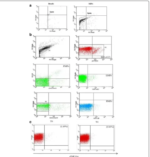
Evaluation of MP subsets and surface expression of TNFα
The number of MPs in serum from patients with RA and HC was quantified by flow cytometry analysis. The strat-egy used to identify MP morphology, MP subsets and TNFαexpression is shown in Fig.2a, b, and c.
patients with RA compared to HC (p = 0.0009). After 4 months of in vivo treatment with ETA the percentage of sTNFαwas significantly decreased with respect to baseline (p = 0.0002) (Fig. 3e). Instead, there was no significant change in the percentage of sTNFα-MPs after therapy in patients treated only with traditional DMARDs (data not shown).This result corroborated our hypothesis on the capacity of ETA to bind TNFαcarried on the surface of the MPs, as explained in the next paragraph. Moreover, as shown in Additional file1, in relation to the expression of sTNF on MP subsets, we observed that EMPs expressed a greater percentage of TNFαthan PMPs. LMPs expressed a high percentage of TNFαbut this was not significantly different to the percentage in the other subsets.
Finally, to compare the percentage of TNFαexpressed on the surface of the MPs and the serum TNFα, we also performed ELISA of serum TNF on the same patients in whom we evaluated the surface expression of TNFα on MPs, as shown in Additional file1.
Surface expression of TNFαon MPs after incubation with ETA
We demonstrated for the first time the expression of TNF on the surface of human MPs. On the basis of this result
we decided to incubate MPs with ETA at different con-centrations in order to demonstrate that the drug was able to bind and inhibit surface MP-TNFα expression. ETA, a fully soluble, human dimeric fusion protein, functions as a TNF inhibitor by competitively binding to TNF and pre-venting its activation of the inflammatory cascade. ETA is a soluble form of the p75 receptor that inhibits TNFα, by blocking its interaction with cell-surface TNF receptors and is different from adalimumab and infliximab, which are monoclonal antibodies to TNF. Moreover, its peculiar-ity with respect to the other TNFαinhibitors, is that be-sides recognizing soluble TNFα, it also recognizes the membrane-related portion.
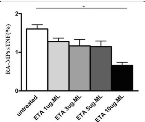
[image:4.595.305.539.85.319.2]For this reason, we hypothesized that ETA, in addition to the soluble portion, recognizes and binds the portion transported on the membrane of the MPs, preventing the link with its receptor and then the inflammatory cas-cade. Results obtained confirmed our hypothesis, in fact, in vitro experiments showed that the percentage of sTNFαexpressed on RA-MPs significantly decreased, in a dose-dependent manner, after incubation with ETA at each timepoint of the experiment. In particular, the major effect was at 10μg/ml of ETA at (Fig.4).
Table 1Clinical, demographic, serological, and therapeutic characteristics of patients with RA and HC
Patients with RA (n= 20)
HC (n= 20)
Demographic parameters
Female/male 19/1 20/0
Age, years (median (IQR)) 58.5 (50–67) 58 (50–65)
Disease duration, years (mean ± SD), 5.3 ± 5.6
Disease activity
DAS28 (mean ± SD) 4.1 ± 1.7
TJ,n(mean ± SD) 6.16 ± 5.9
SJ,n(mean ± SD) 2.79 ± 2.7
CDAI (mean ± SD) 18.8 ± 10.7
HAQ (mean ± SD) 1.15 ± 0.8
Laboratory parameters N (%)
RF+ 15 (75)
ACPA+ 15(75)
ESR, mm/h (mean ± SD) 16.5 ± 10.3
CRP, mg/dl (mean ± SD) 0.7 ± 0.73
Therapy
Concurrent csDMARDs (n(%)) 20 (100)
ETA (n(%)) 20 (100)
RArheumatoid arthritis,HChealthy controls,SDstandard deviation,DAS28 Disease Activity Score in 28 joints,TJtender joints,SJswollen joints,CDAI Clinical Disease Activity Index,HAQHealth Assessment Questionnaire,RF rheumatoid factor,ACPAanti-citrullinated peptide antibodies,ESRerythrocyte sedimentation rate,CRPC-reactive protein,csDMARDsconventional synthetic disease-modifying antirheumatic drugs
Fig. 1 Transmission electron microscopy (TEM) of microparticles
(MPs). Micrographs show the negative staining of a typical sample observed by the TEM.aWhite particles of different sizes ranging from a few nanometers to 1 mm were present. As expected, all particles appeared as white structures in the dark background, due to phosphotungstic acid (PTA) negative staining. They were round-shaped and sometimes superimposed. This observation suggests that the smallest nanoparticles and MPs coexist in our sample, because it is well-known that MP size ranges from 100 to 1000 nm.
[image:4.595.56.294.110.384.2]The percentage of sTNFαcorrelates with clinical parameters of patients with RA
The purpose of our study was to test for correlation be-tween MPs and endothelial damage. However, observing a decrease in the surface expression of MP-TNFα and a concomitant improvement in the parameters of disease activity in patients after therapy with ETA, we decided to
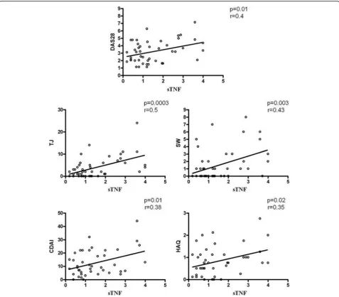
test correlation between them. When we compared the sTNFα of RA-MPs and clinical parameters, such as the Disease Activity Score in 28 joints (DAS28), tender joints (TJ), swollen joints (SW), CDAI and HAQ, identified sig-nificant correlation between these parameters as shown in Fig.5. These results lead us to suppose there is a link be-tween sTNFαon MPs and the status of patients’disease.
[image:5.595.61.542.86.588.2]sTNFαof RA-MPs mediates in vitro induction of apoptosis and autophagy in EA.hy926 cells
TNFα is the main cytokine involved in RA pathogenesis and has a pathogenic effect, with a direct effect on endo-thelial cells. Earlier studies have shown that TNFα inhibi-tors are able to improve endothelial function and decrease cardiovascular events in those patients who respond clin-ically [9,10]. After demonstrating the expression of TNFα on the surface of MPs we decided to investigate whether MPs induce apoptosis and autophagy on endothelial cells
and whether TNFα carried on their surface was respon-sible for this effect. Our in vitro results on EA.hy926 cells showed that at baseline (T0), serum RA-MPs significantly increased both apoptosis and autophagy levels compared to untreated cells, in a dose-dependent manner (p =0.005 andp =0.02,respectively)(Fig.6a–d).
After 4 months of in vivo treatment with ETA, RA-MPs did not significantly change these parameters (p > 0.05 versus untreated cells for both parameters) (Fig. 6e, f ). These effects were comparable to those
Fig. 3Plasma levels of microparticles (MPs) in patients with rheumatoid arthritis (RA) at time zero (baseline (T0)) and after 4 months of treatment
[image:6.595.59.541.86.517.2]obtained with the same number of MPs from HC (Fig. 6e, f ). Interestingly, in the same experiment de-scribed above, treatment with the 3MA restored a situ-ation comparable to that at T0, with an increase in endothelial apoptosis after autophagy block in EA.hy926 cells treated with MPs at T4, asserting the protective role of autophagy (Fig. 6e, f ). The experiments reported above showed the results at 16 h, but a cellular response, comparable to that obtained at 16 h, had begun by 6 h when the cells were incubated with RA-MPs, (data not shown). However, at 24 h, 48 h, and 72 h the cellular re-sponse in terms of apoptosis and autophagy gave rise to confused and uninterpretable results, probably because, after some hours, other surface molecules started a cel-lular response, or the MPs fused themselves with the cells or were internalized, and so the molecular content of MPs may have altered the initial response (data not shown).
Moreover, in vitro studies showed that RA-MPs treated with ETA were no longer able to significantly modulate apoptosis and autophagy in EA.hy926 cells. In particular, when MPs were treated with ETA at 10μg/ ml, both endothelial apoptosis and autophagy reached levels comparable to those of untreated cells (p = 0.01 and p = 0.02, respectively versus cells treated with RA-MPs at T0), to confirm possible involvement of TNFαcarried on MPs in the induction of apoptosis and autophagy (Fig.6g, h).
Finally, the result of western blot for NF-kB showed a significant decrease in p65 protein when endothelial cells were incubated with RA-MPs purified at T0 and
treated with ETA at 10μg/ml (p = 0.02 versus cells treated with RA-MPs at T0). This result confirms the hypothesis that MPs carry TNFαon their surface, which activates TNF-signaling pathways by interaction with its surface receptors (Fig.7).
Discussion
In our study, for the first time we demonstrated TNFα expression on the surface of MPs from patients with RA [22]. Higher circulating levels of TNFα are present in serum from patients with RA, and TNFαitself is able to directly damage endothelial function triggering NF-κB activation and accumulation of reactive oxygen species (ROS) [23].
Some evidence indicates a beneficial effect of TNF in-hibitors on vascular wall physiology, increasing the pos-sibility that TNF blockade may improve endothelial function, with consequent improvement of arterial stiff-ness and reduced progression of subclinical atheroscler-osis [24, 25]. However, clinical studies conducted to investigate the effect of anti-TNF therapy on endothelial function in patients with RA have had contradictory re-sults, generating controversy on this subject.
Based on our results and these considerations, we hy-pothesized that TNFαcarried on the surface of RA-MPs could participate in the atherosclerotic process in pa-tients with RA. Impaired endothelial cell function is a hallmark of atherosclerosis. The endothelial dysfunction is directly associated with the development of athero-sclerosis, and it is present during all stages of the dis-ease. Moreover, alterations in endothelial function precede morphological atherosclerotic changes [26]. In-jury to the endothelium is believed to be a preliminary event in most vascular diseases [27]. Furthermore, those alterations could include an imbalance between apop-tosis and autophagy pathways.
Endothelial apoptosis has been associated with multiple cardiovascular events that cause vascular wall damage and atherosclerotic plaque, and consequently it promotes vas-cular injury [28]. MPs have been shown to initiate cell death when applied to cultured primary rat brain micro-vascular endothelial cells (RBMVECs) and a variety of dif-ferent mechanisms have been identified [29].
Previous studies showed that MPs derived from hu-man umbilical vein endothelial cells [30] and platelets [31] contain caspase 3, and Schneider et al. [32] have suggested that circulating MPs induce apoptosis by the transfer of caspases into target cells. In relation to this, Abid Hussein et al. [30] suggest that MPs could be phagocytosed by cells, causing the release of caspase 3 within the cell itself and initiating apoptosis. In this study, treatment of MPs with a caspase 3 inhibitor sig-nificantly improved cell survival, indicating that the MPs impart pro-apoptotic caspase 3 signals.
[image:7.595.58.290.87.283.2]Moreover, many studies showed that basal levels of au-tophagy are protective for the endothelium, continuously stimulated by mechanical and physical stress [33]. During injury, endothelial cell autophagy may occur to protect the cells from damage, while the failure of autophagy re-sults in endothelial cell apoptosis, leading to the break-down of the integrity of endothelium to induce the local lipid deposition into atheroma, plaque instability, and even acute coronary occlusion and death [34–37]. Nevertheless, the mechanisms that control the autophagy of endothelial cells are still limited. Moreover, autophagy has been shown to play an important role in endothelial cells to prevent development of atherosclerosis.
From the discovery of MPs up to now there has been no scientific evidence of the involvement of MPs in the regula-tion of autophagy pathway. Thus, in light of the numerous reported data, we can assume that MPs could be involved in alteration of apoptosis and autophagy, which together could contribute to the formation and progression of ath-erosclerotic plaque [38]. On the basis of this observation, after investigating surface expression of TNFα on MPs purified from patients with RA, we evaluated if MPs were able to modulate endothelial apoptosis and autophagy in vitro, and if TNFαcarried on the surface of MPs could be involved in these cellular processes. In this regard Schock and coworkers identified three independent pro-apoptotic
Fig. 5Correlation between the percentage of surface (s)TNFαexpression on rheumatoid arthritis (RA)-microparticles (MPs) and clinical parameters
[image:8.595.59.537.87.505.2]signals induced by MPs on RBMVECs cells: caspase 3 and activation of TNF-related apoptosis-inducing ligand (TRAIL) and TNF receptors. To support our last point on the induction of apoptosis by sTNF-MPs on EC, this study showed that treatment with a neutralizing antibody to
TRAIL and TNF receptor blocker did improve cell survival. Genetic analysis confirmed that Tumor necrosis factor re-ceptor 1 and 2(TNFR1, TNFR2) and TRAIL receptor 4 are present in the cells. TRAIL may activate the extrinsic apop-totic pathway, but in some circumstances may activate
Fig. 6Induction of apoptosis and autophagy in EA.hy926 cells by microparticles (MPs) isolated from patients with rheumatoid arthritis (RA) after in
[image:9.595.61.537.84.517.2]pro-survival or proliferative pathways [29]. TNF is synthe-sized as a type-2 transmembrane protein and cleaved by the tumor necrosis factor-alpha-converting enzyme (TACE) ADAM17 to release soluble TNF, which may then activate TNF receptors. The authors showed that treating MPs with the TACE inhibitor (TAPI-0) prior to applying MPs to cul-tured cells provides a significant improvement in cell sur-vival. This result indicates that TACE is present and active in MP membranes. Given the small size of MPs it is uncer-tain whether they are able to shed soluble TNF for an ex-tended period of time.
According to these reported data, our results confirm that MPs from RA at T0 significantly increased apop-tosis in endothelial cells. Interestingly, MPs purified from RA at T4, after in vivo treatment with ETA, and RA-MPs after in vitro treatment with ETA, were no lon-ger able to significantly modify this parameter.
We obtained the same results for autophagy; indeed MPs obtained from RA at T0 significantly increased
autophagy in endothelial cells and MPs purified from RA after in vivo treatment with ETA, and RA-MPs after in vitro treatment with ETA, were no longer able to sig-nificantly modify the autophagy pathway. The experi-ments with the 3MA, which is able to block autophagy and increase apoptosis of endothelial cells treated with RA-MPs, led us to suppose that MPs, if they induce cell death on one side, on the other side they induce cells to upregulate autophagy as a protective mechanism, at least at an early stage.
Furthermore, we demonstrated that RA-MPs induced an increase in NF-kB expression in endothelial cells, and that RA-MPs in vitro treated with ETA did not. These conclusive results support the concept, already proposed in other studies [39,40], that the TNF transported on the surface of MPs interacts with TNF receptors on endothe-lial cells and activates the TNF-signaling cascade, includ-ing apoptosis and autophagy pathways.
Moreover, we observed significant correlation between DAS28 and TNFαexpression on the surface of RA-MPs. This result supports other clinical observations that demonstrated the efficacy and safety of ETA in patients with active RA.
Finally, the interpretation of the ELISA results, i.e. that serum TNF does not change or increase after therapy, as demonstrated in other studies [41, 42], induced us to hypothesize the possibility of using surface expression of TNF on MPs as a possible therapy response marker. Ob-viously, to confirm this statement we need a far greater cohort of patients.
Conclusions
Our results suggest that MPs isolated from patients with RA could exert their pathological effect on endothelial cells by TNFα expressed on their surface. In vivo treat-ment with ETA attenuates this effect, probably binding and blocking the TNF carried on the surface of MPs, as confirmed by our in vitro studies. To conclude, these re-sults show a new pathway of endothelial damage mediated by MPs and confirm the protective effect of anti-TNF therapy against endothelial damage in patients with RA.
Certainly, we cannot assume that sTNF on MPs is the only architect of a cellular response; we hypothesize that it acts in synergy with multiple factors, some of which are already widely discussed in the literature, like serum TNF.
Additional file
Additional file 1:Supplementary material and figures. (PDF 424 kb)
Abbreviations
3MA:3-Methyladenine; AV: Annexin-V; anti-CCP: Anti-cyclic citrullinated peptide; APC: Allophycocyanin; CDAI: Clinical Disease Activity Index; CRP: C-reactive protein; DAS28: Disease Activity Score in 28 joints; DMARD: Disease-modifying antirheumatic drug; DMEM: Dulbecco’s modified Eagle’s medium;
Fig. 7Activation of the TNFαpathway by microparticles (MPs).aWestern blot analysis of NF-kB levels in EA.hy926 cells cultured with MPs purified from patients with rheumatoid arthritis (RA) at time zero (baseline (T0) and with MPs from patients with RA at T0 in vitro-treated with etanercept (ETA). Blot shown is representative of three independent experiments.b
[image:10.595.56.292.87.414.2]EA.hy926: Human umbilical vein cell line; EF-TEM: Energy-filtered transmission electron microscopy; ELISA: Enzyme-linked immunosorbent assay;
EMPs: Endothelial-derived microparticles; ESR: Erythrocyte sedimentation rate; ETA: Etanercept; FBS: Fetal bovine serum; FITC: Fluorescein isothiocyanate; FSC: Forward scatter; HAQ: Health Assessment Questionnaire; HC: Healthy controls; LMPs: Leukocyte-derived microparticles; mAbs: Monoclonal antibodies; MPs: Microparticles; PE: Phycoerythrin; PepA: Pepstatin A; PerCP: Peridin chlorophyll protein; PMPs: Platelet-derived microparticles; PTA: Phosphotungstic acid; RA: Rheumatoid arthritis; SJ: Swollen joints; SSC: Side scatter; STNFα: TNFαexpressed on the surface of microparticles; T0: Time zero (before treatment); T4: Four months after treatment; TJ: Tender joints; TNFα: Tumor necrosis factor alpha
Acknowledgements
None.
Funding
None.
Availability of data and materials
Datasets analyzed during the current study are available from the corresponding author on reasonable request.
Disclosures
None
Authors’contributions
CB and CA collected the clinical data, examined and interpreted the data, performed statistical analyses, and wrote and revised the manuscript. FCe, AF, FM, FRS, and ST recruited patients. CB, TC, and MV carried out the laboratory experiments. CP and LN revised the manuscript. AF and SF collected the clinical data. MD performed EF-TEM experiments. CA, FCo, and GV designed, coordinated and supervised the study, and wrote and revised the manuscript. All authors read and approved the final manuscript.
Authors’information
Cristiana Barbati, Marta Vomero, Tania Colasanti, Marco Diociaiuti: PhD. Fulvia Ceccarelli, Francesca Miranda, Carlo Perricone, Francesca Romana Spinelli, Simona Truglia: MD, PhD. Annacarla Finucci, Lucia Novelli: MD. Fabrizio Conti, Guido Valesini and Cristiano Alessandri: MD, PhD, professor. Sara Ferrigno: medical student
Ethics approval and consent to participate
The study protocol was approved by the Ethics Committee of Sapienza University of Rome (Protocol number 109/18), and informed written consent was obtained from all patients prior to enrollment.
Competing interests
The authors declare that they have no competing interests.
Publisher’s Note
Springer Nature remains neutral with regard to jurisdictional claims in published maps and institutional affiliations.
Author details 1
Arthritis Center, Department of Internal Medicine and Medical Specialties, Sapienza University of Rome, Rome, Italy.2Technology and health
Department, Istituto Superiore di Sanità, Rome, Italy.
Received: 26 July 2018 Accepted: 13 November 2018
References
1. Raposo G, Stoorvogel W. Extracellular vesicles: exosomes, microvesicles, and friends. J Cell Biol. 2013;200:373–83.
2. Piccin A, Murphy WG, Smith OP. Circulating microparticles: pathophysiology and clinical implications. Blood Rev. 2007;21:157–71.
3. Morel O, Toti F, Hugel B, Freyssinet J-M. Cellular microparticles: a disseminated storage pool of bioactive vascular effectors. Curr Opin Hematol. 2004;11:156–64.
4. Loyer X, Vion AC, Tedgui A, Boulanger CM. Microvesicles as cell-cell messengers in cardiovascular diseases. Circ Res. 2014;114:345–53. 5. Connor DE, Exner T, Ma DD, Joseph JE. Detection of the procoagulant
activity of microparticle-associated phosphatidylserine using XACT. Blood Coagul Fibrinolysis. 2009;20:558–64.
6. Morel O, Toti F, Freyssinet JM. Markers of thrombotic disease: procoagulant microparticles. Ann Pharm Fr. 2007;65:75–84.
7. Boilard E, Nigrovic PA, Larabee K, Watts GF, Coblyn JS, Weinblatt ME. Platelets amplify inflammation in arthritis via collagen-dependent microparticle production. Science. 2010;327:580–3.
8. Aviña-Zubieta JA, Choi HK, Sadatsafavi M, Etminan M, Esdaile JM, Lacaille D. Risk of cardiovascular mortality in patients with rheumatoid arthritis: a meta-analysis of observational studies. Arthritis Rheum. 2008;59:1690–7. 9. Hürlimann D, Forster A, Noll G, Enseleit F, Chenevard R, Distler O, et al.
Anti-tumor necrosis factor-a treatment improves endothelial function in patients with rheumatoid arthritis. Circulation. 2002;106:2184–7.
10. Irace C, Mancuso G, Fiaschi E, Madia A, Sesti G, Gnasso A. Effect of antiTNF-a therapy on arterial diameter and wall shear stress and HDL cholesterol. Atherosclerosis. 2004;177:113–8.
11. Pamuk GE, Vural O, Turgut B, Demir M, Pamuk ON, Cakir N. Increased platelet activation markers in rheumatoid arthritis: are they related with subclinical atherosclerosis? Platelets. 2008;19:146–54.
12. Fukuo K, Nakahashi T, Nomura S, Hata S, Suhara T, Shimizu M, et al. Possible participation of Fas-mediated apoptosis in the mechanism of
atherosclerosis. Gerontology. 1997;1:35–42.
13. Salabei JK, Hill BG. Implications of autophagy for vascular smooth muscle cell function and plasticity. Free Radic Biol Med. 2013;65:693–703. 14. Shanthini K, Barbara LG, Susan M, Elizabeth WK, Karen HC. Comparison of
the 1987 American College of Rheumatology and the 2010 American College of Rheumatology/European League against Rheumatism Criteria for Classification of Rheumatoid Arthritis in the Nurses’Health Study Cohorts. Rheumatol Int. 2014;34:407–11.
15. Biró É, Nieuwland R, Sturk A. Measuring circulating cell‐derived microparticles. J Thromb Haemost. 2004;2:1843–54.
16. Jy W, Horstman LL, Jimenez JJ, Ahn YS. Measuring circulating cell-derived microparticles. J Thromb Haemost. 2004;2:1842–51.
17. Diociaiuti M. Electron energy loss spectroscopy microanalysis and imaging in the transmission electron microscope: example of biological applications. J Electron Spectrosc Relat Phenom. 2005;143:189–203.
18. Ayers L, Kohler M, Harrison P, Sargent I, Dragovic R, Schaap M, et al. Measurement of circulating cell-derived microparticles by flow cytometry: sources of variability within the assay. Thromb Res. 2011;127:370–7.
19. Niccolai E, Squatrito D, Emmi G, Silvestri E, Emmi L, Ciucciarelli L, et al. A new cytofluorimetric approach to evaluate the circulating microparticles in subjects with antiphospholipid antibodies. Thromb Res. 2015;136:1252–8. 20. Alessandri C, Barbati C, Vacirca D, Piscopo P, Confaloni A, Sanchez M, et al. T
lymphocytes from patients with systemic lupus erythematosus are resistant to induction of autophagy. FASEB J. 2012;26:4722–32.
21. Barbati C, Alessandri C, Vomero M, Vona R, Colasanti T, Vacirca D, et al. Autoantibodies specific to D4GDI modulate Rho GTPase mediated cytoskeleton remodeling and induce autophagy in T lymphocytes. J Autoimmun. 2015;58:78–89.
22. Beyer C, Pisetsky DS. The role of microparticles in the pathogenesis of rheumatic diseases. Nat Rev Rheumatol. 2010;6:21–9.
23. Gao X, Belmadani S, Picchi A, Xu X, Potter BJ, Tewari-Singh N, et al. Tumor necrosis factor-αinduces endothelial dysfunction in Lepr(db) mice. Circulation. 2007;115:245–54.
24. Libby P. Inflammation in atherosclerosis. Nature. 2002;420:868–74. 25. Brezinski EA, Follansbee MR, Armstrong EJ, Armstrong AW. Endothelial
dysfunction and the effects of TNF inhibitors on the endothelium in psoriasis and psoriatic arthritis: a systematic review. Curr Pharm Des. 2014; 20:513–28 Review.
26. Tam LS, Kitas GD, Gonzalez-Gay MA. Can suppression of inflammation by anti-TNF prevent progression of subclinical atherosclerosis in inflammatory arthritis? Rheumatology. 2014;53:1108–19.
27. Bonetti PO, Lerman LO, Lerman A. Endothelial dysfunction: a marker of atherosclerotic risk. Arterioscler Thromb Vasc Biol. 2003;23:168–75. 28. Vita JA. Endothelial function. Circulation. 2011;124:e906–12.
30. Schock SC, Edrissi H, Burger D, Cadonic R, Hakim A, Thompson C. Microparticles generated during chronic cerebral ischemia deliver proapoptotic signals to cultured endothelial cells. Biochem Biophys Res Commun. 2014;450:912–7. 31. Abid Hussein MN, Nieuwland R, Hau CM, Evers LM, Meesters EW, Sturk A.
Cell-derived microparticles contain caspase 3 in vitro and in vivo. J Thromb Haemost. 2005;3:888–96.
32. Böing AN, Hau CM, Sturk A, Nieuwland R. Platelet microparticles contain active caspase 3. Platelets. 2008;19:96–103.
33. Schneider J, Chromik AM, Uhl W, Mügge A, Bulut D. Apoptosis in esophagus and pancreas carcinoma cells induced by circulating microparticles is related to phosphatidyl serine and microparticle-associated caspases. Med Oncol. 2012;29:962–9.
34. Torisu K, Singh KK, Torisu T, Lovren F, Liu J, Pan Y. Intact endothelial autophagy is required to maintain vascular lipid homeostasis. Aging Cell. 2016;15:187–91.
35. Ding Z, Liu S, Wang X, Dai Y, Khaidakov M, Romeo F, et al. Lox-1, oxidant stress, mtDNA damage, autophagy, and immune response in atherosclerosis. Can J Physiol Pharmacol. 2014;92:524–30.
36. Martinet W, De Meyer GR. Autophagy in atherosclerosis. Curr Atheroscler Rep. 2008;10:216–23.
37. Wang L, Li H, Zhang J, Lu W, Zhao J, Su L, et al. Phosphatidylethanolamine binding protein 1 in vacular endothelial cell autophagy and atherosclerosis. J Physiol. 2013;591:5005–15.
38. Ding Z, Liu S, Wang X, Khaidakov M, Dai Y, Mehta JL. Oxidant stress in mitochondrial DNA damage, autophagy and inflammation in atherosclerosis. Sci Rep. 2013;3:1077.
39. Mesri M, Altieri DC. Endothelial cell activation by leukocyte microparticles. J Immunol. 1998;161:4382–7.
40. Distler JH, Huber LC, Gay S, Distler O, Pisetsky DS. Microparticles as mediators of cellular cross-talk in inflammatory disease. Autoimmunity. 2006;39:683–90.
41. Kotyla P, Jankiewicz-Ziobro K, Owczarek A, Kucharz EJ. Etanercept increases tumor necrosis factor-alpha level but not sFas level in patients with rheumatoid arthritis. Isr Med Assoc J. 2015;17:14–8.