S T U D Y P R O T O C O L
Open Access
Effects of osteopathic treatment on
pulmonary function and chronic thoracic
pain after coronary artery bypass graft
surgery (OstinCaRe): study protocol for a
randomised controlled trial
Gert Roncada
1,2Abstract
Background:Coronary artery bypass graft surgery (CABG) is an effective and widespread coronary revascularisation technique, nevertheless there are a number of long-term postoperative complications from which patients can suffer. One year after CABG surgery pulmonary function is decreased by 12% and 30% of the patients suffer from chronic thoracic pain. To date and to our knowledge there are no effective treatments for these conditions. The aim of the present clinical trial is to explore the effectiveness of osteopathic treatment on these conditions. Methods:The study is designed as a randomised controlled trial with two parallel groups. Group A will receive a standard cardiac rehabilitation programme during 12 weeks and group B will receive the same standard cardiac rehabilitation programme supplemented with four osteopathic treatments (OT). OT will be performed at week 4, 5, 8 and 12 after surgery. Three hundred and eight patients (Group A:n= 154, Group B:n= 154) will be enrolled from the cardiothoracic surgery department of the Jessa Hospital Hasselt. Blinding will be assured for the staff of the cardiac rehabilitation centre and outcome assessors. Primary outcome measure will be the mean difference in change from baseline in slow vital capacity (SVC) at 12 weeks after surgery between groups. Secondary outcome measures will be the change from baseline in quality of life, pain, thoracic stiffness and maximal aerobic capacity at 12 weeks after surgery. A follow-up is planned 52 weeks after surgery for SVC, quality of life, pain and thoracic stiffness. Intention to treat analysis will be executed.
Discussion:The OstinCare study has been designed to explore the potential long-term added value of osteopathic
treatment in the management of decreased pulmonary function, chronic thoracic pain and diminished thoracic mobility after CABG surgery.
Trial registration:The protocol has been retrospectively registered on ClinicalTrials.gov (NCT01714791).
Keywords:Osteopathic treatment, Coronary artery bypass graft surgery, Slow vital capacity, Pulmonary function, Chronic thoracic pain
Correspondence:[email protected]
1Jessa Hospital, Heart Centre Hasselt, Stadsomvaart 11, 3500 Hasselt, Belgium 2Commission for Osteopathic Research, Practice and Promotion, Mechelen,
Belgium
Background
Approximately 640,000 coronary artery bypass graft (CABG) surgery procedures are performed in Europe and the United States each year to restore or optimize myocardial perfusion in coronary artery dis-ease [1, 2]. Worldwide there are approximately over two million open-heart surgeries per year [3]. In this surgical intervention, venous and/or arterial grafts are used to bypass the coronary occlusion or stenosis. Be-cause cardioplegia (induction of temporary cardiac ar-rest) is routinely used, the patient’s circulation is coupled to a cardiopulmonary bypass and access to the heart is most often achieved by median sternot-omy. After CABG surgery, a hospital stay of 1 to 2 weeks is generally required [4, 5].
Although CABG surgery is an effective coronary revascularisation technique, there are a number of postoperative complications from which patients can suffer. For example, a decrease in pulmonary function is a frequently observed complication after CABG surgery. During the first week after CABG surgery vital capacity (VC) decreases by 30–60% [6–9] and even up to 1 year this remains reduced by 12% [10, 11]. Reduced VC has a negative effect on exercise tolerance (Vo2max) [12] and therefore it is important to optimize pulmonary function after CABG surgery. No method of postoperative therapy has been distinguished in treating or preventing these long-term changes [6].
In addition, Ragnarsdòttir et al. [8] found a diminished mobility of the left hemithorax at 3 months after CABG surgery. This decreased thoracic mobility was still present 12 months after surgery [11]. Thoracic mobility and vital capacity were affected more when the left in-ternal thoracic artery (LITA)-retractor was used and re-duced thoracic mobility is related to a diminished pulmonary function [10].
Chronic pain, which is defined as pain without ap-parent biological value that has persisted beyond the normal tissue healing time, which usually takes 3 months [13] after CABG surgery. Chronic pain after CABG surgery is described in several studies reaching a time period from 3 to 28 months [14–20]. Kehlet et al. [14] reported a pain prevalence of 30–50%, from which 5–10% suffer from severe disabling pain after more than 6 months after surgery. Numerous other studies report a pain prevalence of 28–56% from 3 to 28 months after CABG surgery [15–20]. Many theories for its cause have been proposed in litera-ture, but the aetiology is still not clear and no ther-apy or technique has been shown to reduce chronic pain after CABG surgery [17, 21]. In literature this syndrome is described as chronic chest pain [19], chronic thoracic pain [18], chronic post-sternotomy pain [14–20].
Chronic pain after CABG surgery is a major clinical problem, which is distressing and reduces the quality of life of patients [14].
As a result, many patients undergoing CABG surgery suffer from decreased pulmonary function, reduced thor-acic mobility and/or chronic thorthor-acic pain. These anom-alies have significant clinical repercussion and may have an effect on the patients’quality of life [14].
According to current clinical guidelines, exercise intervention should be initiated early after CABG sur-gery [22]. According to these recommendations, exer-cise training should be individually tailored according to the clinical condition, baseline exercise capacity and ventricular function. Upper-body training can begin when the sternal wound is stable. The general applicable exercise training in cardiac rehabilitation consists of walking, jogging, cycling, swimming, row-ing, stair climbrow-ing, elliptical trainers and aerobic dan-cing, at low to moderate exercise intensity, for 3–5 days/week. Programmes should last up to 12 weeks for outpatient settings. However in this trajectory, pulmonary function, thoracic pain and thoracic mobil-ity are not specifically targeted.
In fact, to our knowledge there are no effective treat-ments to treat the latter conditions or effective prevent-ive interventions. Osteopathic treatment (OT) has been used to treat and manage pain symptoms. Several arti-cles have been published addressing acute and chronic pain in different medical conditions [23–25]. However, no trials have been conducted to test the effect of OT on chronic pain after CABG surgery.
Methods/Design Aim of the study
The aim of this randomised controlled trial is to exam-ine whether OT could lead to a better treatment of chronic thoracic pain, decreased pulmonary function and/or decreased thoracic mobility.
We hypothesized that OT reduces the decrease in SVC, reduces chronic thoracic pain, reduces thoracic stiffness and improves the quality of life in patients at 12 weeks and 52 weeks after CABG surgery.
Design
Participants
Subjects admitted to the hospital for elective CABG with median sternotomy will be eligible for this study. Partici-pant recruitment began in January 2010 and is expected to finish in December 2017. Subjects with diagnosed chronic obstructive pulmonary disease, diagnosed neuro-logic disease that prevents participation in the cardiac rehabilitation programme, diagnosed nephrological dis-ease that requires haemodialysis, prior thoracic surgery, surgery in the epigastric, right or left hypochondriac re-gion will be excluded. Subjects will also be excluded if the subject has a prolonged stay (>5 days) in the inten-sive care unit. All CABG surgery procedures will be per-formed by the same surgical team. Subjects may not receive any other manual treatment on the spine and/or thorax during the study.
Randomisation and masking
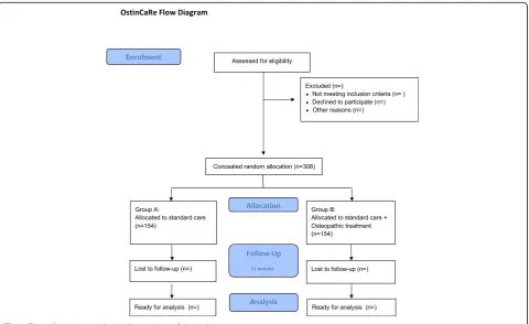
Patients will be randomly assigned in a 1:1 ratio to either group A or group B (Fig. 1). A blocked allocation sched-ule will be used. Randomisation will be performed by means of opaque, sealed envelopes. A physical therapist of the cardiac rehabilitation centre will perform and store the randomisation. The personnel of the cardiac rehabilitation centre performing the outcome measure-ments in this study are unaware of patient’s allocation. Osteopathic treatments will not be performed in the
presence of the personnel of the cardiac rehabilitation centre. The treatments will be performed in another lo-cation to assure that the personnel of the cardiac re-habilitation centre remains blinded to patient’s allocation. Only the treating osteopaths will be aware of the patient’s allocation. The enrolment and procedures are visualised in Fig. 2 according to the Standard Proto-col Items: Recommendations for Interventional Trials (SPIRIT) guidelines [26, 27].
Intervention
Cardiac rehabilitation programme
The cardiac rehabilitation programme is a multidisciplin-ary programme in line with the current guidelines. Com-ponents of the multidisciplinary programme include patient assessment, physical activity counselling, exercise training, diet/nutritional counselling, weight control man-agement, lipid manman-agement, blood pressure monitoring, smoking cessation, and psychosocial management [22].
The outpatient exercise-based cardiac rehabilitation programme includes endurance training. No strength training exercises are executed. According to cardiac rehabilitation literature, all patients exercise under close supervision 3 days per week for a total duration of 3 months [28], and because this frequency is easily attainable for most patients. Exercise training intensity is determined by baseline Vo2peak assessment [29].
[image:3.595.59.540.426.720.2]Patients exercise at a heart rate corresponding to 65% of baseline Vo2peak. Each exercise training session takes 45 min. Exercise time is apportioned as follows: 42% on the cycle ergometer, 33% on the treadmill and 25% on the arm-cranking device [30].
Osteopathic procedure
The protocol used for OT and osteopathic examin-ation (OE) incorporates a number of osteopathic techniques and will be performed by five registered osteopaths with a minimum experience of 5 years. The protocol used is based on the work of Dickey [31] and supplemented with the findings of other au-thors [32–35]. The most common findings found in literature are:
Decrease in costal mobility [8,10,11,36]
Decrease in pulmonary function [6,8,10]
Decreased thoracic mobility [8,10,11,31]
Dysfunction of the abdominal diaphragm [31,37–41]
The nomenclature, indications and contraindications for the OE and OT are based on the work of Nicholas and Nicholas [32], Chila [42], on the professional com-petence profile of an osteopath [43–45] and the bench-marks of the World Health Organisation [46].
The OE protocol is a set of standardised test for a first evaluation of the patient. The findings are noted on a predefined Microsoft excel-file by the Osteopath.
Inspection: in standing, seated and supine: observe the patient in posterior, anterior and lateral view to develop the most complete understanding of the patients makeup before performing the remainder of the OE ([32] p. 3–14).
Position test of the thoracic spine (seated) ([42] p.561)
Intersegmental motion testing:
thoracic seated ([32] p. 42–49)
cervical supine ([32] p. 60–65)
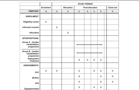
costal supine ([32] p. 52–59 Fig. 2Study content for the schedule of enrolment, interventions, and assessments. -t2: preoperative, -t1: 9
th
[image:4.595.60.539.85.431.2]Costal motion testing supine [32] (p.53–56)
Evaluation of the abdominal diaphragm (seated and supine) ([42] p. 567)
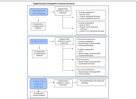
The OT consists of a standardised treatment protocol (Table 1) and a supplementary treatment protocol of the dysfunctions found during the examination. In order to be reproducible, the different treatment possibilities are discussed and presented (Fig. 3). The OT will take 30– 45 min. The examination room is air-conditioned and has a constant temperature of 21–22 °C. The OT will be performed at 4 weeks postoperative (t1), 5 weeks postop-erative (t2), 9 weeks postoperative (t3) and at 12 weeks postoperative (t4). OT can be considered as safe, and major adverse events are very rare [47, 48]. In case ad-verse events should occur, they will be recorded and dis-cussed in the final paper.
Outcome measures
Primary outcome measure will be the mean difference in change from baseline in SVC at 12 weeks after surgery between the two groups.
Secondary outcome measures will include:
Change from baseline in SVC at 52 weeks
Change from baseline in MacNew QLQ at 12 and 52 weeks after surgery
Change in pain from baseline on Visual Analogue Scale (VAS) at 12 and 52 weeks after surgery
Change in thoracic stiffness from baseline on VAS at 12 and 52 weeks after surgery
Change from baseline in maximal aerobic capacity (VO2max) at 12 weeks after surgery
Spirometry
The Pocket-Spiro USB100 (Medical Electronic Construc-tion & Logistic nv, Belgium) will be used for measuring pulmonary function. All patients are asked to perform a slow vital capacity (SVC) test, consisting of three mea-surements. For VC the best of the three measurements is used and for IVC the average of three measurements is used [49]. The instrument is calibrated prior to the test. When an OT is planned, SVC will be measured at least 2 days after OT, because the study wants to meas-ure long-term effects (effects of the entire intervention), instead of any short-term effect (within the first hours after OT). SVC will be measured preoperative (-t2), 9th postoperative day (-t1), 4 weeks after surgery (t1), 8 weeks after surgery (t3), 12 weeks after surgery (t4) and at 12 months after surgery (t5).
Ergospirometry
[image:5.595.57.539.433.734.2]All patients will perform a maximal cardiopulmonary ex-ercise test on a cycle ergometer [50]. All the exex-ercise
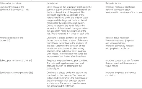
Table 1Standard treatment protocol OstinCaRe study
Osteopathic technique Description Rationale for use
Doming/stretching of the abdominal diaphragm [31–33]
Direct release of the respiratory diaphragm: the patient is supine and the osteopath stands on the homolateral side of the patient. The osteopath places the cubital side of the heterolateral hand under the anterior costal margin and the fingers of the homolateral hand under the posterior costal margin. During inspiration, the hands follow the expansion of the ribs and during expiration, the osteopath holds the expansion of the ribs. This is repeated 3–4 times on each side.
Improves motion of diaphragm Releases connective tissue
tension within structures of the thorax
Myofascial release of the thorax [33]
One hand is placed posterior on one hemi thorax, the other hand anterior of the same hemi thorax (according to the anatomy of the ribs). Determine the direction of free movement with passive motion testing. Maintain either indirect or direct position until release. The osteopath stimulates the expression of the fasciae after the release.
Releases tissue restriction Promotes improved lymphatic and venous drainage Improves pulmonary function and lymphatic circulation
Suboccipital inhibition [31,33,34] Fingertips are placed on occipital condyles. The osteopath applies an outward and cephalad traction to decompress the occipital joint
Improves parasympathetic function Releases restricted tissues around vagus nerves
Equilibration anterior-posterior [35] One hand is placed under the sacrum and one hand on the sternum. The osteopath follows and synchronizes the expression of the primary respiration between sacrum and sternum. The same is done between the occiput and the sternum.
tests will be performed at the same time of day (between 8.30 and 11.30 am). The test will be performed at 4 weeks after surgery (t1), 9 weeks after surgery (t3) and at 12 weeks after surgery (t4).
During the exercise test, an electronically braked e-Bike (Acertys) is used. The cycling frequency is set at 70 cycles/min. In addition, exercise tests will be prema-turely ended when myocardial ischemia and/or severe ventricular arrhythmias would occur and the subject will be excluded from the study. Both the starting and incre-mental cycling resistance will be set between 10 and 40 W and increased every minute to volitional fatigue.
Pulmonary gas exchange analysis will be performed by using cardiopulmonary ergospirometry device (Jaeger MasterScreen CPX). Before every test, a gas and volume calibration will be executed. During the test, environ-mental temperature is kept stable (19–21 °C). Oxygen uptake, expiratory volume and respiratory exchange ratio are collected breath-by-breath and averaged every 10 s. Using a 12-lead ECG device, heart rate is monitored and averaged every 10 s. In addition, maximal cycling resist-ance and total test duration are reported. By V-slope method, ventilatory threshold is calculated. The criteria for defining a maximal cardiopulmonary exercise test are an achieved heart rate > 85% of the maximal
predicted heart rate, and/or a Respiratory gas Exchange Ratio (RER) > 1,09 [51].
MacNew quality of life questionnaire (QLQ) and Visual analogue scale (VAS)
The Flemish version of the MacNew QLQ and the VAS for pain and thoracic stiffness are delivered to the pa-tients by a blinded person of the cardiac rehabilitation centre at enrolment (t0), 12 weeks after surgery (t4) and at 12 months after surgery (t5). The Flemish version of the MacNew QLQ demonstrates good psychometric properties and is recommended as a specific instrument for assessing and evaluate health-related quality of life in Flemish-speaking patients [52].
Data management
[image:6.595.60.537.87.396.2]be maintained in storage for a period of 3 years after completion of the study. During this time, access to the completely encrypted dataset can be obtained on indi-vidual demand. A back-up of all data will be performed every week on a USB-stick and on the hospitals’ backup server. Because the known minimal risk of this study a data monitoring committee is not needed [53]. Import-ant protocol modifications during this study will be communicated to the trial registry and the journal of publication.
Sample size
A priori sample size calculation is based on pulmonary function (by GPower 3.1). The hypothesis for the Ostin-CaRe study is that the addition of OT towards endur-ance exercise training increases inspiratory vital capacity (IVC) by 12% during the follow-up of CABG patients. Power analysis is based on a 12% decrease in IVC at 12 weeks after surgery [11]. As a result, an increase of approximately 12% of the IVC is expected at 12 weeks after CABG. Sample size is computed considering an ef-fect size of 0.50, a statistical power of 0.80 and an alpha level of 0.05. The power analysis outcome defines that 128 subjects per group are needed. Based on unpub-lished data of the cardiac rehabilitation centre a dropout rate of 20% is to be expected. Therefore, 154 subjects per group are needed. Participant recruitment began in January 2010 and is expected to finish in December 2017. Interim analysis is planned when 154 (50%) pa-tients are included. The study can be stopped before reaching sample size if there is a significant change in surgery technique, which could compromise reproduci-bility and comparareproduci-bility throughout the study. The au-thor makes the final decision to terminate the trial.
Statistical analysis
Data will be analysed by a statistician blinded to group allocations, using the Statistical Package for the Social Sciences (SPSS) v. 22.0 (IBM). First, descriptive statistics will be executed, with calculation of means and standard deviations, and analysis of data distribution (by Shapiro-Wilk test) and evaluation of outliers. In case of normal data distribution, one-way ANOVA with repeated mea-sures will be executed to analyse and compare changes in parameters between groups (with Bonferroni correc-tions for multiple comparisons). Relacorrec-tions between pa-rameters will be examined by Pearson correlations. In case of non-normal data distribution, absolute changes in parameters will be compared between groups by Mann Whitney U-tests (with Bonferroni corrections for multiple comparisons). Relations between parameters will be examined by Spearman correlations. Statistical significance is set atp< 0.05, two-tailed. Observed statis-tical power will be calculated by use of GPower v. 3.1.
Intention to treat analysis will be executed. Missing data will be handled using the last observation carried for-ward imputation technique. Dropouts and withdrawals from the study will be recorded through the intervention and follow-up periods. When differences in baseline phenotype are present, these differences will be taken into account during analysis of treatment effect between groups, by regarding them as co-variates.
Discussion
Although CABG surgery is an effective coronary revascularisation technique, there are a number of postoperative complications, such as diminished pul-monary function and chronic thoracic pain, from which patients are prone to suffer from [10, 11, 14–20]. To our knowledge, there are no effective treatments to treat these conditions or effective preventive interventions [6, 17, 21]. The OstinCaRe study has been designed to ex-plore the potential long-term added value of OT in the management of decreased pulmonary function, chronic thoracic pain and diminished thoracic mobility after CABG surgery. The present study is the first study to examine long-term effects of OT after CABG surgery using rigorous procedures and gold standard methods for clinical trials. Previous studies studied the short-term ef-fects of OT after CABG surgery. One study has proven that OT has immediate, beneficial haemodynamic effects after CABG surgery when administered while the patient is sedated [34]. Another study mentioned beneficial, though statistically insignificant, effect of OT on length of stay and recovery of bowel function of CABG surgical patients [54].
The expected outcomes from the present study will be increased pulmonary function, reduction in thoracic pain and increased thoracic mobility. The study has the potential to deliver the first valuable complement in cardiac rehabilitation programmes to address these problems.
Abbreviations
CABG:Coronary artery bypass graft; HVLA: High velocity low amplitude; IVC: Inspiratory vital capacity; LITA: Left internal thoracic artery; MET: Muscle energy technique; OE: Osteopathic examination; OT: Osteopathic treatment; QLQ: Quality of life questionnaire; RER: Respiratory gas exchange ratio; SVC: Slow vital capacity; VAS: Visual analogue scale; VC: Vital capacity
Acknowledgements
The author sincerely thanks the staff of the cardiac rehabilitation centre of the Jessa Hospital for their well-appreciated contribution.
Funding
This research receives no specific grant from any funding agency in the public, commercial or not-for-profit sectors.
Availability of data and material
Authors’contributions
GR conceived the idea and wrote the draft as well as the final paper.
Authors’information
Researcher at the cardiac rehabilitation centre, Jessa Hospital, Stadsomvaart 11, 3500 Hasselt, Belgium. Board member of the Commission for Osteopathic Research, Practice and Promotion vzw (CORPPvzw).
Competing interests
The author declares that he/she has no competing interests.
Consent for publication
Not applicable.
Ethics approval and consent to participate
This study was approved by the local medical ethical committee (number 09.07/cardio09.01, Jessa Hospital, Hasselt, Belgium). Written informed consents will be obtained from all subjects by the treating physical therapist of the subjects. The results of this study will be reported in accordance with the Consolidated Standards of Reporting Trials (CONSORT)
recommendations. The results of this study will be disseminated in a peer-reviewed journal and presented at international congresses.
Received: 2 June 2016 Accepted: 19 November 2016
References
1. Lafortune G, Balestat G, Durand A. Comparing activities and performance of the hospital sector in Europe: how many surgical procedures performed as inpatient and day cases? Paris: Organisation for economic co-operation and development; 2012.
2. Services USDohah, Prevention CFDCA, Statistics NCFH. National Hospital Discharge Survey 2010. Maryland; 2012. ftp://ftp.cdc.gov/pub/Health_ Statistics/NCHS/Dataset_Documentation/NHDS/NHDS_2010_ Documentation.pdf.
3. Pezzella AT. Global aspects of cardiothoracic surgery with focus on developing countries. Asian Cardiovasc Thorac Ann. 2010;18(3):299–310. 4. Hansen D, Linsen L, Verboven K, Hendrikx M, Rummens JL, van Erum M, Eijnde BO, Dendale P. Magnitude of muscle wasting early after on-pump coronary artery bypass graft surgery and exploration of etiology. Exp Physiol. 2015.
5. Roncada G, Dendale P, Linsen L, Hendrikx M, Hansen D. Reduction in pulmonary function after CABG surgery is related to postoperative inflammation and hypercortisolemia. Int J Clin Exp Med. 2015;8(7):10938–46. 6. Westerdahl E, Lindmark B, Bryngelsson I, Tenling A. Pulmonary function
4 months after coronary artery bypass graft surgery. Respir Med. 2003; 97(4):317–22.
7. Baumgarten MC, Garcia GK, Frantzeski MH, Giacomazzi CM, Lagni VB, Dias AS, Monteiro MB. Pain and pulmonary function in patients submitted to heart surgery via sternotomy. Revista brasileira de cirurgia cardiovascular : orgao oficial da Sociedade Brasileira de Cirurgia Cardiovascular. 2009;24(4): 497–505.
8. Ragnarsdottir M, KristjAnsdottir A, Ingvarsdottir I, Hannesson P, Torfason B, Cahalin L. Short-term changes in pulmonary function and respiratory movements after cardiac surgery via median sternotomy. Scand Cardiovasc J. 2004;38(1):46–52.
9. Morsch KT, Leguisamo CP, Camargo MD, Coronel CC, Mattos W, Ortiz LD, Lima GG. Ventilatory profile of patients undergoing CABG surgery. Revista brasileira de cirurgia cardiovascular : orgao oficial da Sociedade Brasileira de Cirurgia Cardiovascular. 2009;24(2):180–7.
10. Kristjansdottir A, Ragnarsdottir M, Hannesson P, Beck HJ, Torfason B. Chest wall motion and pulmonary function are more diminished following cardiac surgery when the internal mammary artery retractor is used. Scand Cardiovasc J. 2004;38(6):369–74.
11. Kristjansdottir A, Ragnarsdottir M, Hannesson P, Beck HJ, Torfason B. Respiratory movements are altered three months and one year following cardiac surgery. Scand Cardiovasc J. 2004;38(2):98–103.
12. Fisher LR, Cawley MI, Holgate ST. Relation between chest expansion, pulmonary function, and exercise tolerance in patients with ankylosing spondylitis. Ann Rheum Dis. 1990;49(11):921–5.
13. Harstall C, Ospina M. How prevalent is chronic pain? International Association for the Study of Pain. 2003;11(2):1–4.
14. Kehlet H, Jensen TS, Woolf CJ. Persistent postsurgical pain: risk factors and prevention. Lancet. 2006;367(9522):1618–25.
15. Kalso E, Mennander S, Tasmuth T, Nilsson E. Chronic post-sternotomy pain. Acta Anaesthesiol Scand. 2001;45(8):935–9.
16. Meyerson J, Thelin S, Gordh T, Karlsten R. The incidence of chronic post-sternotomy pain after cardiac surgery–a prospective study. Acta Anaesthesiol Scand. 2001;45(8):940–4.
17. van Leersum NJ, van Leersum RL, Verwey HF, Klautz RJ. Pain symptoms accompanying chronic poststernotomy pain: a pilot study. Pain Med. 2010;11(11):1628–34.
18. van Gulik L, Janssen LI, Ahlers SJ, Bruins P, Driessen AH, van Boven WJ, van Dongen EP, Knibbe CA. Risk factors for chronic thoracic pain after cardiac surgery via sternotomy. Eur J Cardiothorac Surg. 2011;40(6): 1309–13.
19. Bruce J, Drury N, Poobalan AS, Jeffrey RR, Smith WC, Chambers WA. The prevalence of chronic chest and leg pain following cardiac surgery: a historical cohort study. Pain. 2003;104(1-2):265–73.
20. Eisenberg E, Pultorak Y, Pud D, Bar-El Y. Prevalence and characteristics of post coronary artery bypass graft surgery pain (PCP). Pain. 2001;92(1-2):11–7. 21. Alston RP, Pechon P. Dysaesthesia associated with sternotomy for heart
surgery. Br J Anaesth. 2005;95(2):153–8.
22. European Association of Cardiovascular P, Rehabilitation Committee for Science G, Eacpr, Corra U, Piepoli MF, Carre F, Heuschmann P, Hoffmann U, Verschuren M, Halcox J, et al. Secondary prevention through cardiac rehabilitation: physical activity counselling and exercise training: key components of the position paper from the Cardiac Rehabilitation Section of the European Association of Cardiovascular Prevention and
Rehabilitation. Eur Heart J. 2010;31(16):1967–74.
23. Tozzi P, Bongiorno D, Vitturini C. Low back pain and kidney mobility: local osteopathic fascial manipulation decreases pain perception and improves renal mobility. J Bodyw Mov Ther. 2012;16(3):381–91.
24. Licciardone JC. Systematic review and meta-analysis conclusions relating to osteopathic manipulative treatment for low back pain remain valid and well accepted. J Bodyw Mov Ther. 2013;17(1):2–4.
25. Schwerla F, Kaiser AK, Gietz R, Kastner R. Osteopathic treatment of patients with long-term sequelae of whiplash injury: effect on neck pain disability and quality of life. J Altern Complement Med. 2013;19(6):543–9. 26. Chan AW, Tetzlaff JM, Altman DG, Laupacis A, Gotzsche PC, Krleza-Jeric K,
Hrobjartsson A, Mann H, Dickersin K, Berlin JA, et al. SPIRIT 2013 statement: defining standard protocol items for clinical trials. Ann Intern Med. 2013; 158(3):200–7.
27. Hoffmann TC, Glasziou PP, Boutron I, Milne R, Perera R, Moher D, Altman DG, Barbour V, Macdonald H, Johnston M, et al. Better reporting of interventions: template for intervention description and replication (TIDieR) checklist and guide. BMJ. 2014;348:g1687.
28. Hansen D, Dendale P, Berger J, Meeusen R. Rehabilitation in cardiac patients:what do we know about training modalities? Sports Med. 2005; 35(12):1063–84.
29. Hansen D, Dendale P, Berger J, Meeusen R. Low agreement of ventilatory threshold between training modes in cardiac patients. Eur J Appl Physiol. 2007;101(5):547–54.
30. Hansen D, Dendale P, Raskin A, Schoonis A, Berger J, Vlassak I, Meeusen R. Long-term effect of rehabilitation in coronary artery disease patients: randomized clinical trial of the impact of exercise volume. Clin Rehabil. 2010;24(4):319–27.
31. Dickey J. Postoperative osteopathic manipulative management of median sternotomy patients. Journal of the American Osteopathic Association. 1989;89(10):1309.
32. Nicholas AS, Nicholas EA. Atlas of Osteopathic Techniques. 2nd ed. Philadelphia: Lippincot Williams & Wilkins; 2012.
33. Noll DR, Degenhardt BF, Fossum C, Hensel K. Clinical and research protocol for osteopathic manipulative treatment of elderly patients with pneumonia. J Am Osteopath Assoc. 2008;108(9):508–16.
34. O-Yurvati AH, Carnes MS, Clearfield MB, Stoll ST, McConathy WJ. Hemodynamic effects of osteopathic manipulative treatment immediately after coronary artery bypass graft surgery. Journal of the American Osteopathic Association. 2005;105(10):475–81.
36. Locke TJ, Griffiths TL, Mould H, Gibson GJ. Rib cage mechanics after median sternotomy. Thorax. 1990;45(6):465–8.
37. Canbaz S, Turgut N, Halici U, Balci K, Ege T, Duran E. Electrophysiological evaluation of phrenic nerve injury during cardiac surgery–a prospective, controlled, clinical study. BMC Surg. 2004;4:2–-6.
38. Deng Y, Byth K, Paterson HS. Phrenic nerve injury associated with high free right internal mammary artery harvesting. Ann Thorac Surg. 2003; 76(2):459–63.
39. Katz MG, Katz R, Schachner A, Cohen AJ. Phrenic nerve injury after coronary artery bypass grafting: will it go away? Ann Thorac Surg. 1998;65(1):32–5. 40. Laub GW, Muralidharan S, Chen C, Perritt A, Adkins M, Pollock S, Bailey B, McGrath LB. Phrenic nerve injury. A prospective study. Chest. 1991;100(2): 376–9.
41. Tripp HF, Bolton JW. Phrenic nerve injury following cardiac surgery: a review. J Card Surg. 1998;13(3):218–23.
42. Chila AG, editor. Foundations of Osteopathic Medicine. 3rd ed. Philadelphia: Lippincot Williams & Wilkins; 2011.
43. van Dun PLS. Beroepscompetentieprofiel Osteopathie. 1st ed. Brussel: Groepering Nationaal en Representatief van de Professionele Osteopaten vzw. (GNRPO vzw.); 2010. p. 59.
44. Kouwenberg T, van Well T, van Wolde H, Jansen T.
Beroepscompetentieprofiel Osteopathie. Hilversum: Nederlandse vereniging voor osteopathie en het Nederlands register voor osteopathie; 2009. p. 114. 45. Roggen L. De Osteopaat: Beroepscompetentieprofiel. 1st ed. Beveren:
Register voor de osteopaten van België (ROB); 2011. p. 32. 46. WHO. Benchmarks for Training in Osteopathy. Geneva: World Health
Organizaton; 2010. p. 36.
47. Carnes D, Mars TS, Mullinger B, Froud R, Underwood M. Adverse events and manual therapy: a systematic review. Man Ther. 2010;15(4):355–63. 48. Vogel S. Adverse events and treatment reactions in osteopathy.
International Journal of Osteopathic Medicine. 2010;13(3):83–4.
49. Miller MR, Hankinson J, Brusasco V, Burgos F, Casaburi R, Coates A, Crapo R, Enright P, van der Grinten CPM, Gustafsson P, et al. Standardisation of spirometry. Eur Respir J. 2005;26(2):319–38.
50. Fletcher GF, Balady GJ, Amsterdam EA, Chaitman B, Eckel R, Fleg J, Froelicher VF, Leon AS, Pina IL, Rodney R, et al. Exercise standards for testing and training: a statement for healthcare professionals from the American Heart Association. Circulation. 2001;104(14):1694–740. 51. Balady GJ, Arena R, Sietsema K, Myers J, Coke L, Fletcher GF, Forman D,
Franklin B, Guazzi M, Gulati M, et al. Clinician’s guide to cardiopulmonary exercise testing in adults: a scientific statement from the American Heart Association. Circulation. 2010;122(2):191–225.
52. Vandereyt F, Dendale P, Vanhees L, Roosen J, Hofer S, Oldridge N. Psychometric properties of the Flemish version of the MacNew heart disease health-related quality of life questionnaire. Acta Cardiol. 2012; 67(1):31–9.
53. Sydes MR, Altman DG, Babiker AB, Parmar MK, Spiegelhalter DJ, Group D. Reported use of data monitoring committees in the main published reports of randomized controlled trials: a cross-sectional study. Clin Trials. 2004;1(1): 48–59.
54. Wieting JM, Beal C, Roth GL, Gorbis S, Dillard L, Gilliland D, Rowan J. The effect of osteopathic manipulative treatment on postoperative medical and functional recovery of coronary artery bypass graft patients. J Am Osteopath Assoc. 2013;113(5):384–93.
• We accept pre-submission inquiries
• Our selector tool helps you to find the most relevant journal • We provide round the clock customer support
• Convenient online submission • Thorough peer review
• Inclusion in PubMed and all major indexing services • Maximum visibility for your research
Submit your manuscript at www.biomedcentral.com/submit