Georgia State University
ScholarWorks @ Georgia State University
Biology Theses Department of Biology
5-9-2015
The Survival and Recovery of ϕ6 Virus from
Fomites
Richard L. Bearden II
Follow this and additional works at:https://scholarworks.gsu.edu/biology_theses
This Thesis is brought to you for free and open access by the Department of Biology at ScholarWorks @ Georgia State University. It has been accepted for inclusion in Biology Theses by an authorized administrator of ScholarWorks @ Georgia State University. For more information, please contact [email protected].
Recommended Citation
by
RICHARD L. BEARDEN II
Under the Direction of Lisa Casanova, PhD
ABSTRACT
Viral transmission from the environment can occur via fomites, but there is uncertainty
about which factors most affect viral persistence on fomites. Children are a population highly
susceptible to viral infection, and sharing common fomites like toys may spread infection. The
objective of this research was to assess the survival of enveloped viruses on the surfaces of
children’s toys, using bacteriophage ϕ6 as a surrogate for enveloped human viruses. The survival
of infectious ϕ6 virions was observed over a 24 hour period at 22°C and relative humidities of
40% & 60%. On the surface of children’s toys, ϕ6 was better able to persist at 60% RH (log10
reduction< 2 log10) over a 24 hour period than it was at 40% RH (log10 reduction> 6 log10). If ϕ6
virus persists on toy material for up to 24 hours, then viral transmission via shared fomites is
certainly significant.
THE SURVIVAL AND RECOVERY OF ϕ6 VIRUS FROM FOMITES
by
RICHARD L. BEARDEN II
A Thesis Submitted in Partial Fulfillment of the Requirements for the Degree of
Master of Science
in the College of Arts and Sciences
Georgia State University
Copyright by Richard Louis Bearden II
THE SURVIVAL AND RECOVERY OF ϕ6 VIRUS FROM FOMITES
by
RICHARD L. BEARDEN II
Committee Chair: Lisa Casanova
Committee: Sidney Crow
Robert Simmons
Electronic Version Approved:
Office of Graduate Studies
College of Arts and Sciences
Georgia State University
DEDICATION
I would like to dedicate this body of work to my family. I’m forever grateful for their
v
ACKNOWLEDGEMENTS
I would like to thank Dr. Casanova for her mentorship and eagerness to help me achieve
my goals throughout my graduate studies. Without her wisdom, guidance, and support this
project would not have been possible. I would also like to thank Dr. Crow and Dr. Simmons for
taking the time to be a part of the committee. Their knowledge and expertise have been
invaluable throughout this process. Last, but certainly not least, I would like to thank Dr. Luo for
providing me with the skills to use the data to model virus survival beyond the parameters used
for the experiments. All of you were essential to this project and I am eternally grateful for your
TABLE OF CONTENTS
ACKNOWLEDGEMENTS ... v
LIST OF FIGURES ... vii
LIST OF TABLES ... viii
LIST OF ABBREVIATIONS ... ix
1 INTRODUCTION ... 1
2 Research Design & Methods ... 4
2.1 Propagation of Virus Stock ... 4
2.2 Suspension Media Experiments ... 4
2.3 Core Survival Experiments ... 5
3 RESULTS ... 7
3.1 Suspension Media Experiments ... 7
3.2 Core Survival Experiments ... 8
3.3 Linear Regression Analysis Fitting the Weibull Model ... 10
4 Discussion ... 12
vii
LIST OF FIGURES
Figure 1. ϕ6 Survival in 1X PBS and DIW. Survival of ϕ 6 virus suspended in either
deionized water or 1X PBS solution for t=2 hours at 22°C, 60%RH. Gray column, survival in
DIW; white column, survival in 1X PBS. Bars, 95% confidence interval. ... 7
Figure 2. ϕ6 Survival at 22°C over a 24 hour period. Circles, 60%RH. Squares, 40%RH.
Bars, 95% confidence intervals. ... 8
Figure 3. ϕ6 Survival at 22°C, 60%RH. Circles, second aliquots of virus stock. Triangles,
first aliquots of virus stock. Bars, 95% confidence intervals. ... 9
Figure 4. Linear Regression Analysis Fitting the Weibull Model. (a) Log transformations
of survival of the second aliquots of virus stock at 40 & 60% RH. Squares, log transformations
of inactivation of second aliquots of virus stock at 40% RH; circles, log transformations of
inactivation at 60% RH. (b) Log transformations of inactivation of the first and second aliquots
of virus stock at 60% RH. Triangles, log transformations of inactivation of the first aliquots of
LIST OF TABLES
Table 1. Linear Regression Analysis of the Weibull Function. Linear model fitted with
ln(log10 NT/N0) as the response variable and ln(t), time, as the predictor to get estimations of n =
ix
LIST OF ABBREVIATIONS
°C ϕ6 BE DIW Hr mL µL PBS PFU RH rpm TA TSA TSB Degrees Celsius Bacteriophage phi 6 1.5% Beef Extract Deionized Water Hour
Milliliter Microliter
Phosphate Buffer Solution Plaque Forming Units Relative Humidity Rotations Per Minute Top Agar
1 INTRODUCTION
Viral particles consisting of an outer layer of lipids in addition to their protein capsid are
called enveloped viruses. Orthomyxoviruses such as influenza viruses H1N1and H5N1 as well as
Coronoavirus SARS-CoV and other coronaviruses (CoV) are examples of very pathogenic
strains of enveloped viruses [13]. These viruses are responsible for causing many respiratory
tract infections that often result in fatality in humans. The WHO estimates that seasonal
influenza epidemics alone will result in about 3-5 million cases of severe illness, and about
250,000 to 500,000 deaths globally [21]. Respiratory viruses can be transmitted from person to
person through a variety of modes. For instance, influenza can be transmitted via contaminated
fomites or inanimate objects, droplets from infected persons, and persistent droplet nuclei
suspended in aerosols [5]. The efficiency of various modes of transmission depends partly on the
survival of the virus in the environment before it interacts with its next host, and the efficiency
with which enveloped viruses spread from one host to the next depends partly on which mode of
transmission leaves viruses most vulnerable to inactivation [10]. In environments where a large
number of people interact with shared surfaces, there could be continuous contamination of those
surfaces with virus and subsequent spread of the virus throughout that population [5]. If viral
infection stemming from interaction with contaminated fomites is a major source of viral
persistence and spread within a population, then it is extremely important to understand which
type of environmental conditions are conducive to a virus’s survival on shared surfaces and the
risks associated with those surfaces [5]. This knowledge could better equip agencies to foster
indoor environmental standards specifically targeted at the inactivation of enveloped viruses
using a variety of strategies – controlling/maintaining relative humidity, maintaining
2
protocols [13]. Such standards could prove effective at preventing illness from viruses, especially
for vulnerable populations such as children.
It has generally been accepted that enveloped viruses are more sensitive to environmental
conditions and have limited survival outside of their host when compared to non-enveloped
viruses. Many studies have concluded that humidity is an extremely important factor impacting
virus survival but there is a large amount of variation in the findings of these studies [13]. Some
studies suggest that as humidity increases, viral inactivation increases [2, 8, 9]. Others suggest
that lower humidity levels increase viral inactivation [2, 6]. Currently there is no consensus as to
a defined minimum or maximum relative humidity that reduces a virus’ survival or ability to
infect a new host [13]. Another, often missing, component in some of the existing literature is the
significance of the amount of time the virus is exposed to various environmental conditions and
how time impacts survival under these conditions [13]. There are also studies that assess virus
survival on household surfaces [5]. Virus survival on fomites or surfaces is dependent upon
several factors, e.g. the fomite or surface characteristics (porosity, chemical residue, etc.), the
matrix surrounding the virus, and the environmental conditions.
If viruses can survive on a fomite for a period of time, than it is probable that viral
transmission through direct contact with the contaminated fomite can occur [5]. Virus survival
and recovery from children’s toys are an exceptional choice because toys are often communal
objects. In daycare facilities, schools, or even in homes with multiple children close in age, toys
are often shared and circulated from child to child. Children also represent a susceptible
population to viral infection. Understanding the factors influencing the persistence of viruses on
fomites under varying simulated indoor environments could have a profound impact on how we
cases of disease, particularly amongst susceptible populations, and how we evaluate the risk
posed by contaminated objects that are shared, like children’s toys. Therefore, the objective of
this experiment is to assess how relative humidity, temperature, and time influence survival of an
4
2 Research Design & Methods
2.1 Propagation of Virus Stock
Bacteriophage ϕ6 was propagated in Pseudomonas syringae (host) using the soft agar
preparation method [3]. 30mL of host bacterial culture was grown for 24 hours with shaking at
100rpm at room temperature (22°C). 2mL of ϕ6 virus stock was added and incubated with
shaking for an additional 24 hours. 0.5mL of this virus culture and 0.5mL of host culture were
added to 30mL of soft agar (0.7% agar), dispensed into tryptic soy agar (TSA) plates, and
incubated at room temperature for 24 hours. The top layer was then harvested, pooled and
centrifuged (5900g, 30 minutes at 4°C), and stored as stock in tryptic soy broth (TSB) with 20%
glycerol at -80°C.
2.2 Suspension Media Experiments
Host was prepared by adding a 1.5mL volume of host to 150mL of TSB and incubating
with shaking at 22°C for 24 hours. 100µL of ϕ6 virus stock (stored at -80C in 20% glycerol) was
diluted into 900µL of deionized water (DIW) and 900µL of 1X PBS (8.0g NaCl2, 0.2g KCl,
0.12g KH2PO4, 0.91g Na2HPO4 /liter of DIW). 10µL of virus suspended in DIW (target
concentration ~107 plaque-forming units (PFU)) was added to 6 toy coupons (UV sterilized 2cm x 2cm pieces of a child’s toy). 10µL of virus suspended in 1X PBS was added to an additional 6
toy coupons. 3 coupons from each group of 6 were immediately placed in tubes containing 5mL
of 1.5% beef extract (BE) (~7.5 pH) using sterile forceps and placed on a shaker (220rpm) at
22°C for 20 minutes. These coupons represented the concentration of virus at t=0hr in both DIW
and 1X PBS. Samples were serially diluted in TSB and assayed using the double agar layer
method. Plates were incubated at room temperature ~22°C for 24 hours. The remaining 3
and temperature environments were created using sealed glass tanks containing saturated salt
solutions (40% - magnesium chloride, 60% - magnesium nitrate). After 2 hours, each coupon
was placed into a tube containing 5mL of 1.5% beef extract and placed on the shaker (220rpm) at
22°C for 20 minutes. These coupons represent infectious virus concentration at t=2hr in DIW
and 1X PBS. After 24 hours incubation, the number of plaques on each plate were counted to
quantify the number of infectious viruses remaining at after 2 hours in DIW and 1X PBS and the
log reduction of viruses was calculated log10 (Nt/N0), where Nt is the number of viruses at time t
and N0 is the number of viruses at t=0.
2.3 Core Survival Experiments
The core survival experiments were carried out in a similar fashion as the suspension
media experiments. 100µL of ϕ6 virus stock was diluted into 900µL of DIW. 10µL of virus
suspended in DIW (target concentration ~107 plaque-forming units (PFU)) was then added 12
coupons. 3 coupons were immediately added to tubes containing 5mL of 1.5% BE (~7.5 pH)
using sterile forceps and placed on a shaker at (220rpm) for 20 minutes. These coupons
represented the concentration of virus at t=0hr. Samples were serially diluted in TSB and assayed
using the double agar layer method. Plates were allowed to incubate at room temperature ~22°C
for 24 hours. The remaining BE eluent was stored at -80°C.The 9 remaining coupons were
grouped into groups of 3 and were placed in either the 40% or 60% humidity chamber at ~22°C.
Every 2 hours, 1 group of coupons was removed from the humidity chamber and the above
procedure was repeated. These experiments were completed at 6-hour intervals (t=2hr, t=4hr,
and t=6hr) up to 24 hours for both relative humidity levels at ~22°C. After 24 hours of
incubation, the number of plaques on each plate were counted to quantify the number of
6
log10 (Nt/N0). When ϕ6 virus could not be recovered from the plaque assay for the 7-dilution
titer, the remaining volume of viral eluent (recovered virus in 1.5% BE) was divided into equal
volumes and plated onto TSA plates with 1mL of host and 5mL of TA. The data was analyzed
using Excel 2011 (Mac) and GraphPad Prism 5 (GraphPad). The data was fitted to the Weibull
model by completing log transformations of the parameter log NT/N0 and plotting them against
the log transformation of time, t, for survival at each relative humidity [1,7]. Linear regression
analysis was conducted to determine the slope of the inactivation line and to predict the shape of
3 RESULTS
3.1 Suspension Media Experiments
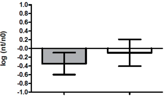
To determine whether the choice of suspension media (PBS vs. deionized water) affected
[image:18.612.163.430.186.351.2]virus survival, virus survival in PBS was compared to deionized water (Figure 1).
Figure 1. ϕ6 Survival in 1X PBS and DIW. Survival of ϕ 6 virus suspended in either deionized water or 1X PBS solution for t=2 hours at 22°C, 60%RH. Gray column, survival in DIW; white column, survival in 1X PBS. Bars, 95% confidence interval.
A t test was used to compare virus survival (log NT/N0) in deionized water (n=9) as a
suspension media to 1X PBS (n=6). Experiments were conducted at 22°C in a controlled
chamber at 60% relative humidity. There was not a statistically significant difference in virus
inactivation at t=2 hours between the two types of suspension media (p=0.1564). PBS was used
8
3.2 Core Survival Experiments
[image:19.612.158.434.114.302.2]
Figure 2. ϕ6 Survival at 22°C over a 24 hour period. Circles, 60%RH. Squares, 40%RH. Bars, 95% confidence intervals.
Over a 24 hour period, there was a ~2 log10 reduction (99% inactivation) in the number of
infectious viruses recovered at 60% RH (Fig.2). At t=8 hours post application to the coupon, the
number of infectious viral particles declines by ~1 log10 and then remains somewhat stable
(between 1-2 log10 reduction) up to 24 hours at 60%RH.
At 40%RH, there was a more rapid decline (~3 log10 reduction, 99.9% inactivation) in the
number of infectious viral particles recovered after t=2 hours post application of the viruses to
the coupon, and the level of the inactivation was considerably greater at 40%RH (Fig.2). At t=8
to 10 hours post-application most trials were below the detection limit for the double agar layer
Figure 3. ϕ6 Survival at 22°C, 60%RH. Circles, second aliquots of virus stock. Triangles, first aliquots of virus stock. Bars, 95% confidence intervals.
In initial survival experiments conducted at 60% RH, two different pools of virus stock
were used that were propagated at different times using identical methods from the same initial
seed stock. The survival experiments using the first set of aliquots of virus stock (Fig.3), yielded
a ~2.5 log10 reduction (99% inactivation) in the number of infectious viruses recovered from the
coupons after t=4 hours of exposure, reaching ~5 log10 reduction (99.999% inactivation) after
t=12 hours. However, completing the double agar layer plaque assay under the same conditions
with aliquots of virus stock created at a later time revealed dissimilar results. Survival
experiments using the second set of aliquots never reached >2 log10 reduction (99% inactivation)
in the number of infectious viruses recovered from the coupons even after t=24 hours of
exposure. It is important to note that the 40% RH survival experiments were also carried out
10
3.3 Linear Regression Analysis Fitting the Weibull Model
[image:21.612.141.402.93.452.2]
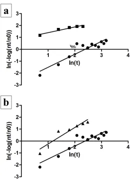
Figure 4. Linear Regression Analysis Fitting the Weibull Model. (a) Log
transformations of survival of the second aliquots of virus stock at 40 & 60% RH. Squares, log transformations of inactivation of second aliquots of virus stock at 40% RH; circles, log transformations of inactivation at 60% RH. (b) Log
transformations of inactivation of the first and second aliquots of virus stock at 60% RH. Triangles, log transformations of inactivation of the first aliquots of virus stock; circles, log transformations of inactivation of the second aliquots of virus stock.
After fitting the data to the Weibull model, a linear regression analysis was conducted for
each set point of relative humidity. The log transformations of average inactivation (ln(-log10
(NT/N0))) (response variable) at each time point were plotted against the log transformation of
the time (ln(t)) at which survival was being evaluated. For the second set of aliquots of virus
However, the slopes of the survival plot for both sets of aliquots of virus stock at 60% RH were
not significantly different (p=0.3002).
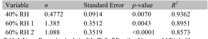
Variable n Standard Error p-value R2
40% RH 0.4772 0.0914 0.0070 0.9362
60% RH 1 1.385 0.3512 0.0043 0.8951
[image:22.612.122.488.127.197.2]60% RH 2 1.088 0.3519 <0.0001 0.8573
Table 1. Linear Regression Analysis of the Weibull Function. Linear model fitted with ln(log10 NT/N0) as the response variable and ln(t), time, as the predictor to get
estimations of n = slope of the log time.
Modeling this experimental data with the Weibull model and comparing the slopes of the
inactivation plots at each set point of relative humidity, allows for extrapolation of what the
actual shape of the survival curve may be. Linear regression analysis of this model reveals that
the model fits the expectation of the data. At 40% RH, the slope (n) of the survival plot = 0.4772.
When n <1, the rate of inactivation will decrease over time due to significant inactivation at early
time points. At 60% RH, n >1, indicating that the inactivation of ϕ6 will increase over time at
12
4 Discussion
These experiments show that ϕ6 virus is able to persist at 22°C and 60% RH for up to 24
hours, and potentially longer. The rate of inactivation appears to be slower at 60% RH than at
40% over a 24 hour period. The rate of inactivation of infectious virions is much more
pronounced at 40% RH, with virus reaching undetectable levels 8 hours post application (>6
log10 reduction, ~99.9999% inactivation). However, at 60% RH, the amount of infectious virus
recovered from the toy coupons declines most around t=8 hours post application, and then
appears to remain relatively stable up to t=24 hours (~1-2 log10 reduction, ~90-99% inactivation).
This data suggests that ϕ6 is better able to resist inactivation at 22°C and 60% RH on the surface
of the toy coupon. This is contrary to some of the existing literature centered on the survival of
enveloped viruses, where virus survival appears to be more stable at lower relative humidities [2,
9, 10].
There is 99.9999% inactivation of ϕ6 within the first 10 hours of exposure to 40% RH. It
is expected that the rate of inactivation will decrease over time due to the vast majority of virions
becoming inactivated early on in their exposure to this relative humidity. The plot of survival at
40% RH supports this prediction. In comparing n at both 40 and 60% RH, the opposite is true for
the rate of inactivation at 60% RH. Although inactivation of the second aliquots of ϕ6 was no
greater than 99% inactivation after 24 hours of exposure to the simulated environment, the slope
calculated from the linear regression analysis suggests that the rate of inactivation will increase
over time. However, the plot of survival at 60% relative humidity for the second aliquots of virus
stock does not necessarily support this. Additional experiments carried out beyond 24 hours at
Toys were chosen as a fomite for these experiments because they have been shown to be
significant sources of viral and bacterial contamination in healthcare settings and nurseries [7,
11, 16]. Due to their age, children are often much more susceptible to viral and bacterial
infections. They often lack the immunity provided by previous exposure or vaccination,
especially when very young [16]. They also exhibit behavior conducive to contracting infections,
such as putting foreign objects in their mouths or failure to wash their hands. In healthcare and
nursery environments, toys are communal objects and often shared between children [11]. As a
result, they are likely significant sources of the spread of infection throughout populations in
those environments. It has been previously shown that respiratory syncytial virus, an enveloped
virus, can survive on the surface of toys for up to 6 hours [11]. At 22°C and 60%RH, ϕ6
inactivation was between 90-99% 8 hours post application to the surface and never reached
greater than 99% inactivation over a 24 hour period. Although ϕ6 virus is non-pathogenic to
humans, its survival on the surface of the toy coupon highlights the necessity of examining how
enveloped viruses become inactivated in indoor environments and the importance of fostering
effective decontamination protocols for communal objects, like toys, that children interact with
in these environments.
Virus survival and persistence on surfaces is influenced by several factors, whether it be
the viral species, the surface the virus is applied to, the temperature and humidity of the
environment, or the media the virus is suspended in. It is somewhat difficult to compare the
findings of this paper with those of the existing literature because each set of experiments differs
by 1 or more of these factors. For instance, Casanova and Waka completed survival experiments
using ϕ6 virus and identical research methods [3]. The only major differences between the
14
used (1X PBS) and the surface to which the virus was applied; coupons of N95 respirator
material. ϕ6 virus survival at 22°C appeared to be much more stable at 40%RH (<2 log10
reduction, 90-99% inactivation) on N95 material then at 60% RH (>3 log10 reduction, 99.9%
inactivation) over a 24 hour time period [3]. For the survival experiments carried out for this
paper at 22°C and 40% RH, >3 log10 reduction (>99.9% inactivation) of ϕ6 was achieved after 2
hours post application of the virus to the coupon. At 60% RH, the level of inactivation (~5 log10
reduction, 99.999% inactivation) achieved at t=12hr using the first set of aliquots seemed to be
somewhat comparable to the inactivation achieved by Casanova and Waka (~4 log10 reduction,
99.99% inactivation). However, the second set of aliquots did not reach >2 log10 reduction (99%
inactivation) until 24 hours after application of the virus to the coupon. That degree of ϕ6
inactivation is reached by 6 hours post application by Casanova and Waka. It is also important to
note that virus stock propagated at different times from the same seed stock appear to have
varying rates of inactivation (n=1.385, n=1.088) when exposed to identical environmental
conditions (22°C, 60% RH). The survival plots for the first set of aliquots of viral stock exhibit a
significantly greater rate of inactivation than the survival plots of the second set of aliquots at
60%RH. At t=2 hours post application, both populations have similar rates of inactivation, but at
t=4 hours post application, the inactivation diverges for the two populations of virus. Although
the rate of inactivation for the first set of aliquots at 60% RH is higher than that of the second set,
the slopes from the linear regression analysis were not significantly different (p=0.3002).
In a recent paper, filoviruses ZEBOV (Zaire Ebola virus) and MARV (Lake Victoria
Marburg virus) were used for long-term survival experiments. Neither virus could be recovered
from glass, plastic, or metal when applied to the surface at ~22°C, 55±5% RH suspended in
plastic at 4°C and 55±5% RH, and remained detectable 14 days post application [15]. A
juxtaposition of this discovery with the results of the experiments using ϕ6 within this paper
reveals a dichotomy. Although ϕ6 and filoviruses share structural similarities (envelope and
nucleocapsid), they do not have the same resistance to similar environmental conditions. This is
also true for viruses of the same species. In another paper, the survival of 2 strains of coronavirus
in suspension and dried onto various surfaces to estimate the risk of viral transmission on items
found in hospital settings [17]. They found that when HCV-229E and HCV-OC43 were dried
onto surfaces, infectious HCV-229E was still detectable up to 6 hours post drying on sterile
sponges (4% recovery) and aluminum (8% recovery) whereas, HCV-OC43 was undetectable
(0% recovery) on any surface after 3 hours post drying on those surfaces.
One interesting explanation for the variation in survival of viruses is what happens at the
air-water interface (AWI) when viruses are suspended in water on surfaces. The theory is that the
proteins of virions are strongly attracted to the AWI, especially when suspended in liquids of
high ionic strength [19, 20]. These proteins are often embedded in the envelope of the virion.
When these proteins interact with the AWI, they cause the loss of the envelope due to
hydrophobic interactions and rearrangement of the viral capsid. The rearrangement of the capsid,
which is a highly organized structure in many viruses, would lead to the release of the viral
genome and subsequent inactivation of the virus [12]. Other experiments have conclusively
shown that viruses meeting the AIW become inactivated but it is not the sole reason viruses
become inactivated [19, 20]. If multiple virions within a suspension become attracted to the AWI
and form an aggregate of virions, it is possible that those virions at the AWI will become
inactivated but will also form a barrier, blocking additional virions from reaching the AWI [12,
16
liquid volume would decrease in the suspension due to evaporation, and the viruses will become
larger aggregates over time (more concentrated). This aggregation could have a protective effect
for viruses at the interior of the viral suspension [12]. However, at 40% RH, perhaps the benefits
of aggregation are outweighed by the increased rate of the disappearance of water, leading to
desiccation.
Although virally contaminated environmental surfaces can contribute to the transmission
of virus from one person to another, it is unclear how significant this mode of transmission is.
This uncertainty stems from the fact that virus survival appears to be quite dynamic, in that it is
impacted by a number of factors including but not limited to temperature, humidity, surface
material, and the intrinsic properties of the virus. The impact of one particular environmental
factor is hard to estimate because each species and/or strain of virus appears to respond to similar
environmental conditions in a unique way. More investigation is needed to better understand the
survival kinetics of viruses under various environmental conditions and the factors that confound
REFERENCES
1. Berger, R. W., & Lawrence, K. (1974). Estimating Weibull Parameters by Linear and Nonlinear Regression. Technometrics, 16(4), 617-619.
doi:10.1080/00401706.1974.10489245
2. Casanova, L., et al. Effects of air temperature and relative humidity on coronavirus
survival on surfaces. Applied and Environmental Microbiology, 76 (9):712–2717.
3. Casanova, L., & Waka, B. Survival of a Surrogate Virus on N95 Respirator Material. Infection Control and Hospital Epidemiology, 34, 1334-1335.
4. Enstone J. Influenza and related infection control issues. In: Van-Tam J, Sellwood C, eds.
Introduction to pandemic influenza. Wallingford: CAB International, 57–72.
5. Greatorex JS, Digard P, Curran MD, Moynihan R, Wensley H, et al. (2011) Survival of
Influenza A(H1N1) on Materials Found in Households: Implications for Infection Control. PLoS ONE 6(11): e27932. doi:10.1371/journal.pone.0027932
6. Harper, G.J. Airborne Micro-organisms: Survival Tests with Four Viruses.
Microbiological Research Establishment. Journal of Hygiene, 59, 479-486.
7. Ibfelt, T., E.H. Engelund, A.C. Schultz, and L.P. Andersen. "Effect of cleaning and
disinfection of toys on infectious diseases and micro-organisms in daycare nurseries." Journal of Hospital Infection 89 (2015): 109-115. Print.
8. Kim, S. J., Jiyeon, S., Lee, J. E., & Ko, G. (2012). Temperature and Humidity Influences on Inactivation Kinetics of Enteric Viruses on Surfaces. Environmental Science & Technology, 46, 13303-13310.
9. Koep, T., et al. Predictors of indoor absolute humidity and estimated effects on influenza virus survival in grade schools. BMC Infectious Diseases, 13, 1471-2334.
10.Kohn, M., & Shaman, J. Absolute humidity modulates influenza survival, transmission, and seasonality. Proceedings of the National Academy of Sciences, 106, 3243-3248.
11.Little, Kathryn, and Stanley Cutcliffe. "The safe use of children’s toys within the
healthcare setting." Nursing Times 102.38 (2006): 34-37. Print.
18
13.Memarzadeh, F. Literature Review of the Effect of Temperature and Humidity on
Viruses. ASHRAE, 12, 1049-1060.
14.Myatt, T., et al. Modeling the airborne survival of influenza virus in a residential setting:
the impacts of home humidification. Environmental Health, 9, 1-7.
15.Piercy, T. J., Smither, S. J., Steward, J. A., Eastaugh, L., & Lever, M. S. (2010). The survival of filoviruses in liquids, on solid substrates and in a dynamic aerosol: Survival of filoviruses. Journal of Applied Microbiology. doi:10.1111/j.1365-2672.2010.04778.x
16.Posfay-Barbe, Klara M., Danielle M. Zerr, and Didier Pittet. "Infection Control in Paediatrics." Lancet Infectious Diseases 8 (2008): 19-31. Print.
17.Sizun, J., Yu, M. W., & Talbot, P. J. (2000). Survival of human coronaviruses 229E and OC43 in suspension and after drying on surfaces: a possible source of hospital-acquired infections. Journal of Hospital Infection. doi:10.1053/jhin.2000.0795
18.Thomas, Y. et al. Survival of influenza virus on human fingers. Clinical Microbiology
and Infection, 20, O58-O64.
19.Thompson, S. S., & Yates, M. V. (1999). Bacteriophage Inactivation at the Air-Water-Solid Interface in Dynamic Batch Systems. Journal of Hospital Infection.
20.Thompson, S. S., Flury, M., Yates, M. V., & Jury, W. A. (1998). Role of the
Air-Water-Solid Interface in Bacteriophage Sorption Experiments. Journal of Applied Microbiology.
21.World Health Organization. (2014, March). WHO | Influenza (Seasonal). Retrieved from