R E S E A R C H A R T I C L E
Open Access
Antiangiogenesis and antioxidant activity of
ethanol extracts of
Pithecellobium jiringa
Nahdzatul Syima Muslim
1, Zeyad D Nassar
1,2, Abdalrahim FA Aisha
1,3, Armaghan Shafaei
3, Norshirin Idris
1,
Amin Malik Shah Abdul Majid
1*and Zhari Ismail
3Abstract
Background:Angiogenesis plays a critical role in embryonic development and various physiological processes. However, excessive angiogenesis is associated with several pathological conditions including cancer.Pithecellobium jiringa(Jack) Prain is a traditional medicinal plant from the family Leguminosae. It is native to the Southeast Asia, where it has been used traditionally for treatment of various ailments such as hypertension and diabetes. The present work is aimed to study antioxidant and antiangiogenesis activities ofP. jiringaethanol extracts.
Methods:P. jiringafruit rinds were extracted with ethanol and 50% ethanol. The antioxidant property was analysed using, 1,1-diphenyl-2-picryl-hydrazyl free radical scavenging assay. Phytochemical analysis was performed using thin layer chromatography and colorimetric methods. Then, cell growth inhibition was studied against a panel of human cell lines by MTT test.In vitroinhibition of angiogenesis was studied by the following assays: isolated rat aortic rings cell viability, colony formation, endothelial cell migration, endothelial tube formation on matrigel, and expression of vascular endothelial growth factor by endothelial cells.In vivoantiangiogenesis effect was studied by utilising fertilised chick embryos assay. The results were statistically analysed by analysis of variance.
Results:Ethanolic and 50% hydro-ethanolic extracts showed relatively high concentration of total phenolics associated with potent antioxidant activity. The rat aortic rings study conducted showed potent inhibition of the microvessels outgrowth with IC50s5.27 ± 0.81μg/ml (ethanolic) and 4.45 ± 0.63μg/ml (50% hydro-ethanolic). Both extracts arrested the growth of human endothelial cells via down-regulation of VEGF expression, leading to inhibition of other angiogenesis cascades including migration of endothelial cells, and formation of capillary network on matrigel matrix. The extracts also inhibited the neovascularisation of chick embryo chorioallantoic membrane.
Conclusions:P. jiringaextracts inhibit angiogenesis by blocking the VEGF expression thus inhibiting endothelial cells proliferation, migration and differentiation most likely due to presence of the antioxidant phenolics.
Keywords:Pithecellobium jiringa, Antiangiogenesis, Antioxidant, Phytochemical analysis
Background
Angiogenesis is the formation of new blood vessels sprouting from existing vascularisation [1]. The process plays essential role in embryonic development, wound healing and reproductive functions. In early 1970s, it was noted that solid tumours appear to be highly vascu-larised [2]. It was then established that all solid tumours are dependent on the angiogenesis in order for the
tumour to grow larger and metastasize [1,2]. Endothelial cells in tumour bed tend to be more susceptible to cyto-toxic agents due to their high proliferation rate. In addition, endothelial cells, on the contrary to cancerous cells, are genetically stable as they do not undergo muta-tions and hence more sensitive to apoptotic effects of the cytotoxic agents. Thus, these features of endothelial cells make them a compelling target for antiangiogenesis treatment [3]. Consequently, cytotoxic agents pose as candidates as antiangiogenic agents on top of their po-tent activity in causing death of cancerous cells. Pacli-taxel, an anti-cancer drug has been shown to inhibit * Correspondence:aminmalikshah@gmail.com
1EMAN Testing & Research Laboratory, Department of Pharmacology, School of Pharmaceutical Sciences, Universiti Sains Malaysia, Minden, Penang 11800, Malaysia
Full list of author information is available at the end of the article
proliferation of endothelial cells as well as the cells’ mi-gration and invasiveness dose-dependently both in vitro andin vivo[3].
Extensive studies have been conducted to assess the role of oxidative stress and hence the use of antioxidants in the prevention of many diseases such as cancer, in-flammation, and atherosclerosis [4-6]. In the initial stage of these diseases, oxidative stress plays a major role in damaging essential components of cells [4]. To date, there have been extensive studies on natural product compounds and extracts that showed potent antiangio-genic activity, in conjunction to having good antioxidant activities [7-10].
Pithecellobium jiringa (Jack) Prain is a traditional me-dicinal plant native to the Southeast Asia which belongs to the family Leguminosae [11]. Traditionally, a drink mixture of pounded whole fruit with ginger is taken daily to eliminate bladder stones [12]. The plant has been traditionally used in treating hypertension and dia-betes, the fruit rinds are also used for flavouring and as fragrance in manufacturing traditional soaps, shampoos and detergents. The matured leaves of the plant are burnt to ashes as a cure for itchy parts of the body [13,14]. Though there is a widespread traditional use of the whole fruit as anti-diabetes agent,P. jiringahas not been studied extensively [14]. This study has been undertaken in order to investigate the antioxidant and antiangiogenic activity of Pithecellobium jiringa ethano-lic and 50% hydro-ethanoethano-lic extracts.
Methods Materials
Analytical grade solvents were purchased from Avan-tor Performance Materials (Petaling Jaya, Selangor, Malaysia). Earles’ salt (M199) medium, trypsin, Dul-becco’s modified eagle medium (DMEM), minimum essential medium (MEM), fibrinogen and foetal bovine serum were obtained from Bio-Diagnostics (Petaling Jaya, Selangor, Malaysia). Aprotinin, L-glutamine, thrombin, sodium chloride, fungizone, gentamycin, 6-aminocaproic acid, suramin, dimethyl sulfoxide (DMSO), crystal violet, phosphate buffered saline (PBS), 3-(4,5-dimethylthiazol-2-yl-2,5-diphenyl) tetrazolium bromide (MTT), para-formaldehyde, agarose, potassium acetate, aluminum chloride, 1,1-diphenyl-2-picryl-hydrazyl (DPPH), quer-cetin, gallic acid, betulinic acid, Folin-Ciocalteau re-agent, sodium carbonate, anisaldehyde rere-agent, natural product-polyethylene glycol 4000’ reagent (NP-PEG) and Dragendorff reagent were from Sigma-Aldrich (Subang Jaya, Selangor, Malaysia). Human umbilical vein endothelial cells (HUVEC), hormone-dependent breast carcinoma cells (MCF 7), human hepatocarci-noma cells (Hep G2) and normal colonic fibroblasts (CCD-18Co) were purchased from ATCC (Manassas,
Virginia). Endothelial cell medium (ECM) was obtained from Team Medical Scientific (Shah Alam, Selangor, Malaysia). Matrigel matrix was purchased from Bio-Diagnostics (Petaling Jaya, Selangor, Malaysia), and VEGF-165 ELISA kit was obtained from Chemtron Bio-technology Sdn Bhd (Kuala Lumpur, Malaysia). All other chemicals used in the study were of analytical grade.
Plant material and extraction
The plant material was authenticated by the Herbarium of School of Biological Sciences, Universiti Sains Malay-sia, where a voucher specimen was deposited (Reference number of 11242). The fruit rinds were separated from the seeds and oven-dried at 40°C and powdered using a milling machine (Retsch GmbH, Germany). Fine pow-dered material (30 g) was macerated separately in 500 ml ethanol (EtOH) and 50% ethanol (EW) overnight at 40°C with intermittent shaking. After cooling, extracts were filtered using Whatman filter paper No.1 (What-man, England), concentrated at 50°C under vacuum using a rotary evaporator (RE121 Buchi, Switzerland), and freeze-dried using freeze-drying system (Labconco, USA).
Experimental animals
The thoracic aortas were excised from 8–12 weeks old male Sprague Dawley rats obtained from the Animal House Facility, Universiti Sains Malaysia. The rats were kept in well ventilated cages and allowed to free access of tap water and normal laboratory diet. All experimen-tal procedures were executed following the Animal Ethics Guidelines of Universiti Sains Malaysia (USM/ Animal Ethics Approval/2011/(66)(302)).
Rat aortic ring assay
using an inverted light microscope (Olympus) at 4× mag-nification. The blood vessels outgrowth of an explant was quantified by using Leica QWin computerised imaging software [16].
Cell viability assay
Inhibition of cell growth was evaluated by the MTT test. The cells were treated with extracts as well as 1% DMSO as a negative control for 48 h. Cell viability was then assessed following a previously described method [17]. The absorbance of the treated cells was then taken by using a microtitre plate reader (Tecan, Austria) at 570 nm. The results are presented as average percentage of cell viability to that of the negative control.
Colony formation assay
The colony formation assay was conducted following a previously described method [18]. Single cell suspension of HUVEC cells was seeded at 500 cells/ml in a 6-well plate and incubated for 24 h to allow attachment. Subse-quently, culture medium containing various concentra-tions of the test extracts was added; betulinic acid at 20 μg/ml was used as a positive control and 1% DMSO served as a negative control. After a 48 h treatment, the medium was discarded, the cells were washed by PBS, and a fresh medium was added. On the sixth day of in-cubation, the cells were fixed with 4% (v/v) paraformal-dehyde and subsequently stained with 0.2% (w/v) crystal violet solution in PBS. Colonies consisting of more than 50 cells were counted by using a stereomicroscope, and the results are reported as a mean percentage of survival fraction ± SD (n=2).
Cell migration assay
Cell migration was performed following previously described procedures [19]. In brief, HUVEC cells were seeded in a 6-well plate until a confluent monolayer was formed. Subsequently, a wound was created using a ster-ile 200 μl micropipette tip. The detached cells were removed by washing with PBS and the plates were then treated with the test extracts at 0.5 μg/ml. The wounds were photographed after 12 and 18 h, and the width of the wound was measured using Leica QWin imaging system; 10 fields per well were taken and a minimum of 30 readings per field were measured. The results are pre-sented as a mean percentage of migration inhibition in comparison to control ± SD (n=3).
Tube formation assay on matrigel matrix
The matrigel matrix (5 mg/ml) was obtained by diluting the stock at 1:1 with serum-free medium, before 300 μl of the solution was transferred into each well of a 48-well plate. The matrix was allowed to solidify at 37°C and 5% CO2 for 45 min. The HUVEC cells were then
trypsinized and seeded (3 × 10⁴cells/well) in 100μl fresh medium containing various concentrations of the test extracts. After 6 h incubation, the tubular structures were visualised and photographed under an inverted light microscope at 4× magnification (Olympus, Japan). Quantitative assessment of the tube formation inhibition was carried out by using the ImageJ Software by measur-ing the area of the tubular structures [20]. The results are presented as a mean percentage inhibition ± SD (n=3).
VEGF concentration in HUVEC cells lysates
The VEGF concentration was determined by using a human VEGF-165 ELISA kit (Raybio, USA) following the manufacturer’s protocols. HUVEC cells were treated with the test extracts at 2.5 μg/ml for 24 h before the cell lysates were prepared using cell lysis buffer provided with the kit. A calibration curve of VEGF standard was prepared at the same time. Subsequently, the concentra-tion of VEGF in cell lysates was quantified using the log-log regression equation of the standard calibration curve (y = 0.0099x0.6137, R2 = 0.996). The experiment was repeated two times in triplicates.
In vivochorioallantoic membrane assay
Thein vivo antiangiogenic effect of the test extracts was investigated by CAM assay as described previously [21]. Fertilized chicken eggs of 5-days old were obtained from a local hatchery. Albumin (5 ml) was withdrawn and the eggs were incubated horizontally to allow the CAM de-tachment of the shell. The extracts were dissolved in ethanol and prepared as agarose discs of 1.2% at extracts concentration of 25 and 50μg/disc. Discs contained the vehicle only (ethanol) were used as negative control. A small window was made on the shell through which the discs were applied to the CAM. The window was closed back and sealed with a sterile surgical tape and the eggs were incubated for another 24 h. The images of each treated CAM were captured under dissecting micro-scope. The blood vessels in the disc application site were counted to calculate the percentage inhibition [22].
DPPH scavenging activity
200 μM DPPH solution. Reduced optical density indi-cated higher free radical scavenging activity. The free radical scavenging activity of the extracts and gallic acid was determined as follows:
Percentage of free radical scavenging activity = [1− (AS−AB)/AC−AB] × 100, where:
AS: absorbance of sample/standard AB: absorbance of blank
AC: absorbance of control
Phytochemical screening
P. jiringafruit rinds extracts were screened for presence of various classes of chemical constituents as previously described [24]. High performance thin layer chromatog-raphy (HPTLC) (Camag, Switzerland) was conducted using a mobile phase system of ethyl acetate-formic acid-acetic acid-water (100:11:11:26). The developed TLC plates were then sprayed with various spraying reagents as the following; anisaldehyde reagent for terpe-noids, natural product reagent for flavoterpe-noids, Dragen-dorff reagent for alkaloids and Folin-Ciocalteu reagent for phenolic compounds. After spraying, the plates were heated at 100°C for 5 min, and were visualised at 366 nm. The presence of the compounds was confirmed by the presence of bands after spraying with respective reagents. All tests were repeated thrice.
Estimation of total phenolics content
The total phenolics content was determined using a col-orimetric assay [25]. Test extracts (100μl at 1 mg/ml in methanol) was added to 750μl Folin-Ciocalteau reagent (diluted 1:10 with deionised water) and incubated for 5 min in the dark at RT. Then, 750μl of 60 g/l sodium bi-carbonate solution was added and further incubated in the dark for 90 min at 30°C. The absorbance was read at 725 nm. Gallic acid was used as the standard reference (50–1600 μg/ml). The results are expressed as mg gallic acid equivalents per gram of the extract.
Total flavonoids content
The total flavonoids content was determined using as previously described [26]. Quercetin (3.91 - 250 μg/ml in methanol) was used as a standard reference. The standard and the extracts solutions (500 μl) were mixed with 0.1 ml of 10% (w/v) aluminum chloride, 0.1 ml of 1M potassium acetate, 1.5 ml methanol and 2.8 ml water. As for the blank, both potassium acetate and aluminum chloride were not added and their volume was replaced by water. The reaction mixture was incu-bated for 30 min at RT and the absorbance was taken at 415 nm.
Statistical analysis
Statistical analysis was carried out using SPSS software version 16.0 (SPSS, Chicago, Illinois). All values are shown as a mean ± SD. Comparisons among multiple groups were done via analysis of variance (ANOVA),
P < 0.05 was considered significant.
Results
Rat aortic ring assay
The antiangiogenic effect of EtOH and EW extracts was first investigated using the rat aortic ring model. Figure 1A show the microvessels outgrowth from the untreated aortic rings. Figures 1B, 1C and 1D show re-duction in the microvessels outgrowth by both extracts (10 μg/ml) as well as Suramin (100μg/ml) as a positive control. IC50s were calculated from the dose response curves (Figure 1E (i) and (ii)) and were found to be 5.27 ± 0.81 μg/ml and 4.45 ± 0.63 μg/ml for EtOH and EW extracts, respectively. Suramin, as a positive control, showed almost 100% inhibition of the microvessels out-growth at 100μg/ml.
Effect of EtOH and EW extracts on HUVEC cells proliferation
Cytotoxic effect of both extracts was assessed by MTT test on HUVEC cells, revealing a selective cytotoxicity towards the endothelial cells with IC500.60 ± 0.15μg/ml (EtOH extract) and 3.01 ± 0.05μg/ml (EW extract). The extracts’cytotoxicity was also tested on MCF 7 (breast cancer cell line), Hep G2 (liver cancer cell line), and CCD-18Co (normal colonic fibroblasts). However, the extracts showed lower growth inhibition towards these cell lines. Table 1 depicts the calculated half maximal in-hibitory concentration (IC50s) on all tested cell lines.
Inhibition of colony formation
In order to figure out whether the extracts exhibit cyto-toxic or cytostatic effect on HUVEC cells, the colony formation assay was performed. The results indicate that both extracts are cytostatic as shown by the percentage of survival cells [27]. The plating efficiency (PE) was 13.7 ± 1.00%. The IC50s were 2.32 ± 0.28 μg/ml (EtOH extract) and 6.24 ± 0.54 μg/ml (EW extract). Figure 2 describes the survival percentages of HUVEC cells after treatment with the test extracts and their colonies after treatment.
Cell migration
factors across a horizontal surface. In the present study, the results demonstrated effective reduction in the mi-gratory capacity of the extracts-treated cells as compared to their untreated counterparts (Figure 3). In EW extract-treated cells at 0.5μg/ml, the percentage of wound closure at 12 and 18 h was 48.02 ± 1.94% (P= 0.002) and 72.90 ± 0.9%, respectively. At the same concentration of the EtOH extract, the percentage of wound closure was 41.13 ± 4.38% and 49.50 ± 1.39% at 12 and 18 h, respectively.
Tube formation
Under normal culture conditions endothelial cells cul-tured on a matrigel matrix form tube-like structures within 6 h. Treatment of HUVEC cells with EtOH and EW extracts has reduced the formation of the tube-like structures in a dose dependent manner (Figure 4). The extracts showed IC50s of 3.62 ± 0.83 μg/ml (EtOH) and 2.19 ± 0.19 μg/ml (EW). The positive control (suramin) inhibited endothelial tube formation by 97 ± 0.72% at 100μg/ml.
Inhibition of VEGF expression
The inhibitory effect of the extracts on VEGF expression was determined by quantifying VEGF 165 concentration in lysates of endothelial cells. The results showed that both extracts caused significant inhibition of VEGF ex-pression in HUVEC cells. The VEGF concentration in cell lysates in cells treated with EtOH extract at 2.5μg/ ml (57.77 ± 4.65 pg/ml) (P = 0.004) and EW extract (69.06 ± 3.71 pg/ml) (P< 0.007) was significantly lower than that measured in untreated cells (253.36 ± 8.19 pg/ ml) (P = 0.01). Hence, the present results propose for down-regulation of VEGF expression as one of the mechanism of actions involved.
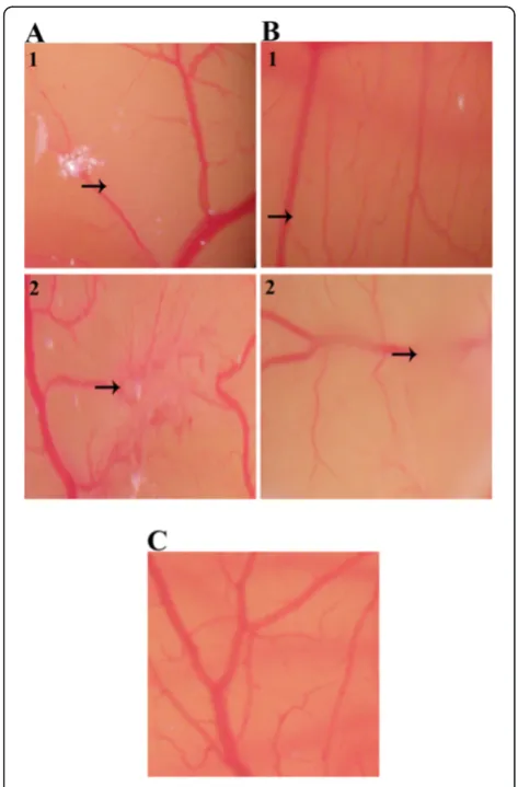
In vivoinhibition of CAM neovascularisation
[image:5.595.56.291.84.697.2]Inhibition of normal vascularisation in chick embryo was observed by the treatment of the CAM with 25 and
Table 1 IC50values (μg/ml) of EtOH and EW extracts on
different human cell lines
Extract HUVEC MCF 7 Hep G2 CCD-18Co
EtOH 0.60 ± 0.15 28.66 ± 1.86 40.63 ± 0.63 4.24 ± 0.15 EW 3.01 ± 0.05 101.34 ± 1.51 38.58 ± 0.66 11.50 ± 1.02
[image:5.595.303.539.691.732.2]50 μg of EtOH and EW extracts. Figure 5C represents normal vascularisation in the untreated CAM which consisted of primary, secondary and tertiary microves-sels. In comparison, the CAM treated with 25 and 50μg of EtOH and EW extracts displayed distorted vascular-isation as well as perturbation on existing vasculatures (Figure 5A1 and 5A2; Figure 5B1 and 5B2). The percent-age nhibition in EtOH extract treated CAMs was 32.75 ± 5.59% (25μg) (P= 0.01) and 62.56 ± 4.20% (50μg) while that in EW extract treatment was 51.30 ± 6.50% (25μg) and 71.11 ± 3.08% (50μg).
DPPH scavenging activity
The results showed that the extracts possess potent scavenging capacity of the stable free radical DPPH. EW
extract was more effective free radical scavenging agent with IC50 18.48 ± 1.60 μg/ml than the EtOH extract which showed an IC50of 33.52 ± 2.05μg/ml.
Phytochemical analysis
The preliminary phytochemical analysis revealed the presence of phenolics, flavonoids, terpenoids and alka-loids in both extracts. The TLC plates which were sprayed with the natural product reagent and enhanced with 5% ethanolic PEG 4000’showed presence of differ-ent flavonoid compounds. Appearance of blue bands indicates the presence of phenolic acids; yellow-orange bands indicate the presence of flavonols and yellow-green bands indicate flavones.
Spraying with Folin-Ciocalteu reagent and enhancing with 20% sodium carbonate solution has marked the presence of phenolic compounds in all extracts, indi-cated by appearance of dark blue bands. Spraying the TLC plates with anisaldehyde reagent indicate the pres-ence terpenoids in both extracts, as indicated by the deep purple bands, however with higher band intensity in EtOH extract than in EW extract. Presence of alka-loids was also noted in both extracts, as shown by the presence of brown bands upon spraying with Dragen-dorff reagent. From these results, it can be seen that P.
jiringa extracts contain abundant number of phyto-constituents which may contribute to various pharmaco-logical activities.
Total phenolics and total flavonoids
The EtOH and EW extracts were subjected to a quanti-tative analysis of total phenolics and flavonoids. In gen-eral, EW and EtOH extracts has showed relatively similar amount of total phenolics and flavonoids. EtOH extract contained 143.95 ± 0.22 mg/g total phenolics and 2.21 ± 0.17 mg/g of total flavonoids while EW ex-tract contained 146.22 ± 1.17 mg/g total phenolics and 2.84 ± 0.83 mg/g total flavonoids.
Discussion
Plants contain tremendous amount of phytochemical constituents such as phenolics and flavonoids com-pounds, these have a great potential in promoting and maintaining a good health [28]. Antioxidants from plant origin have always been tagged with possibilities in treat-ing and lowertreat-ing the risk of various diseases such as in-flammation and cancer [4]. Antioxidants have the ability to scavenge reactive oxygen species that may cause damage to DNA, proteins and lipids. In addition, antioxidants may suppress cancer cells through affecting cyclooxygenase-2 enzyme or inhibiting oncogene expression [cyclooxygenase-28].
[image:6.595.57.292.88.439.2]Phytochemical analysis of both EtOH and EW extracts of P. jiringafruit rinds revealed the presence of various secondary metabolites including phenolics, flavonoids, Figure 2Effect ofP. jiringafruit rinds extracts on clonogenicity
terpenoids and alkaloids. In support of the present finding, previous GC-MS-TOF analysis ofP. jiringaseeds extracts prepared by carbon dioxide supercritical extraction has shown the presence of flavonoids, terpenoids, alkaloids, vitamin E, allyl sulphur and some fatty acids [13,14].
EW extract showed higher potency in scavenging the DPPH free radicals than EtOH extract. Significant rela-tionship between elevated antioxidant activities with high amount of total phenolics content has been exten-sively discussed [29-31]. Generally, the antioxidant activ-ity of phenolics is greatly contributed by their structures, stressing on the presence of hydrogen-donating hydroxyl groups, and those with more hydroxyl groups possess greater antioxidant capacity [29]. Several studies have shown anti-mutagenic and anti-carcinogenesis effects of phenolic acids such as chlorogenic and caffeic acids due to their high antioxidant effect [31]. Previously, methanolic extract of P. jiringa seeds was found to inhibit Epstein-Barr Virus (EBV) activation in Raji
cells, a model of anti-tumour screening, indicating the potential anti-cancer effect of P. jiringa [32]. The present study found relatively high concentration of total phenolics in both extracts and potent antioxidant activity which suggests a possible chemopreventive effect.
[image:7.595.60.539.88.460.2]removal of the extracts. Hence, their antiangiogenic ef-fect may be explained due to the cytostatic efef-fect on HUVEC cells particularly at low concentration of the extracts.
Collectively, the anti-proliferation and rat aortic ring assay results indicate that the antiangiogenic effect of the extracts is due to the selective inhibition of the growth of the endothelial cells. During degradation of the vascular basement membrane and extracellular matrix, activated endothelial cells migrate into the peri-vascular space, differentiate and form capillary network [33]. The significant inhibition of endothelial cells migra-tion at low extract concentramigra-tion (0.5μg/ml) and the in-hibition differentiation of endothelial cells on matrigel
indicate these steps in the angiogenesis cascade as pos-sible targets of theP. jiringaactive principles.
[image:8.595.60.538.86.500.2]growth and metastasis [34], hence P. jiringa extracts may provide a new source of VEGF inhibitors as anti-tumour candidates.
In order to confirm that the antiangiogenic effect ofP.
jiringa extracts is reproduced in vivo, CAM assay was conducted. Treatment of the CAMs with either the EtOH or EW extracts changed the vascularisation pat-tern; both extracts inhibited the new blood vessels for-mation in the treated CAMs as well as distortion of existing vasculature. This result further supports the antiangiogenic activity ofP. jiringa.
Conclusions
The present study reports for the first time the inhib-ition of angiogenesis by P. jiringa extracts by blocking the VEGF expression leading to inhibition of endothelial cell proliferation, migration, and differentiation into a functional capillary network on matrigel matrix. This plant may provide a new source of antiangiogenesis
agents which can be considered as potential candidates in the treatment of angiogenesis related diseases such as cancer, psoriasis, rheumatoid arthritis and diabetic retinopathy.
Competing interests
The authors declare that they have no competing interests.
Authors’contributions
NSM conducted the plant extraction, cell culture work, rat aortic ring assay, antioxidant assays, performed the statistical analysis and wrote the manuscript. ZDN conducted the tube formation assay, participated in designing the experimental details and critically revising the paper. AFAA conducted thein vivoCAM assay and critically revised the paper. AS conducted the thin layer chromatography and interpreted the results and revised the work. NI conducted the rat aortic ring assay and cell culture work. AMSAM participated in designing and interpreting the work, and wrote and revised the paper. ZI participated in designing and interpreting the phytochemistry work. All authors read, edited and approved the final manuscript.
Acknowledgements
NSM, ZDN and AFAA would like to acknowledge Universiti Sains Malaysia for financial support given under the USM Fellowship Scheme. The authors would also like to thank Mr. Shanmugan A/P Vellosamy for his invaluable assistance in identifying the plant.
Author details
1
EMAN Testing & Research Laboratory, Department of Pharmacology, School of Pharmaceutical Sciences, Universiti Sains Malaysia, Minden, Penang 11800, Malaysia.2School of Pharmacy, University of Queensland, Brisbane, QLD, Australia.3Department of Pharmaceutical Chemistry, School of
Pharmaceutical Sciences, Universiti Sains Malaysia, Minden, Penang 11800, Malaysia.
Received: 16 April 2012 Accepted: 31 October 2012 Published: 5 November 2012
References
1. Folkman J:What is the evidence that tumors are angiogenesis dependent?J Natl Cancer Inst1990,82(1):4.
2. Folkman J:Tumor Angiogenesis: Therapeutic Implications.N Engl J Med
1971,285(21):1182–1186.
3. Folkman J:Angiogenesis and apoptosis.Semin Cancer Biol2003,
13(2):159–167.
4. Fernández-Pachón M, Villano D, García-Parrilla M, Troncoso A:Antioxidant activity of wines and relation with their polyphenolic composition. Anal Chim Acta2004,513(1):113–118.
5. Hc-C C, Lo Y-J, Lu F-J:Xanthine Oxidase Inhibitors from the Leaves of Alsophila Spinulosa (HOOK) Tryon.J Enzyme Inhib Med Chem1994,
8(1):61–71.
6. Cos P, Ying L, Calomme M, Hu JP, Cimanga K, Van Poel B, Pieters L, Vlietinck AJ, Berghe DV:Structure-Activity Relationship and Classification of Flavonoids as Inhibitors of Xanthine Oxidase and Superoxide Scavengers.J Nat Prod1998,61(1):71–76.
7. Cao Y, Cao R:Angiogenesis inhibited by drinking tea.Nature1999,
398(6726):381–381.
8. Lamy S, Blanchette M, Michaud-Levesque J, Lafleur R, Durocher Y, Moghrabi A, Barrette S, Gingras D, Béliveau R:Delphinidin, a dietary anthocyanidin, inhibits vascular endothelial growth factor receptor-2 phosphorylation. Carcinogenesis2006,27(5):989.
9. Sartippour MR, Shao ZM, Heber D, Beatty P, Zhang L, Liu C, Ellis L, Liu W, Go VL, Brooks MN:Green tea inhibits vascular endothelial growth factor (VEGF) induction in human breast cancer cells.J Nutr2002,132(8):2307. 10. Lamy S, Gingras D, Béliveau R:Green tea catechins inhibit vascular
endothelial growth factor receptor phosphorylation.Cancer Res2002,
62(2):381.
11. Barceloux DG:Djenkol Bean [Archidendron jiringa(Jack) I. C. Nielsen]. In
[image:9.595.55.292.87.447.2]Medical Toxicology of Natural Substances: Foods, Fungi, Medicinal Herbs, Toxic Figure 5Effects ofP jiringafruit rind extracts on
Plants, and Venomous Animals. Hoboken, New Jersey: John Wiley & Sons, Inc; 2008:59–61.
12. Zakaria M, Ali Mohd M:Traditional Malay Medicinal Plants. Kuala Lumpur: Institut Terjemahan Negara Malaysia Berhad; 2010.
13. Mohd Azizi CY, Nik Norulaini NA, Wahyu BS, Mohd Omar AK:Supercritical carbon dioxide extraction of constituents of pithecellobium jiringan seeds and their identification using time of flight gas spectrometry.; 2006.
14. Norulaini NANIK, Zaidul ISM, Azizi CYM, Zhari I, Noramin MN, Sahena F, Omar AKM:Supercritical carbon dioxide fractionation of pithecellobium jiringan jack seed compositions using fast gas chromatography time of flight mass spectrometry.J Food Process Eng2010,34(5):1746–1758. 15. Brown KJ, Maynes SF, Bezos A, Maguire DJ, Ford MD, Parish CR:A novel
in vitro assay for human angiogenesis.Lab Investig1996,75(4):539–555. 16. Nicosia RF, Lin YJ, Hazelton D, Qian X:Endogenous regulation of
angiogenesis in the rat aorta model. Role of vascular endothelial growth factor.Am J Pathol1997,151(5):1379. doi:5.
17. Mosmann T:Rapid colorimetric assay for cellular growth and survival: Application to proliferation and cytotoxicity assays.J Immunol Methods
1983,65(1–2):55–63.
18. Franken NAP, Rodermond HM, Stap J, Haveman J, van Bree C:Clonogenic assay of cells in vitro.Nat Protocols2006,1(5):2315–2319.
19. Liang CC, Park AY, Guan JL:In vitro scratch assay: a convenient and inexpensive method for analysis of cell migration in vitro.Nat Protoc
2007,2(2):329–333.
20. Bandyopadhyay A, YONG Z, Malik SN, Kreisberg J, Brattain MG, Sprague EA, JIAN L, Lopez-Casillas F, Sun LZ:Extracellular domain of TGFÎ2type III receptor inhibits angiogenesis and tumor growth in human cancer cells. Oncogene2002,21(22):3541–3551.
21. West DC, Thompson WD, Sells PG, Burbridge MF:Angiogenesis assays using chick chorioallantoic membrane.Methods Mol Med2001,
46:107–130.
22. Nassar ZD, Aisha AFA, Ahamed MBK, Ismail Z, Abu-Salah KM, Alrokayan SA, Majid AMSA:Antiangiogenic properties of Koetjapic acid, a natural triterpene isolated from Sandoricum koetjaoe Merr.Cancer Cell International2011,11(1):12.
23. Sharma OP, Bhat TK:DPPH antioxidant assay revisited.Food Chem2009,
113(4):1202–1205.
24. Trease G, Evans W:Pharmacognosy, 12* edition.English Language Book Society/Bailliere Tindall1983,1.
25. Lizcano LJ, Bakkali F, Begoña Ruiz-Larrea M, Ignacio Ruiz-Sanz J:Antioxidant activity and polyphenol content of aqueous extracts from Colombian Amazonian plants with medicinal use.Food Chem2010,119(4):1566–1570. 26. Kosalec I, Bakmaz M, Pepeljnjak S, Vladimir-Knezevic S:Quantitative analysis of the flavonoids in raw propolis from northern Croatia.Acta Pharm2004,
54(1):65–72.
27. Houghton P, Howes MJ, Lee C, Steventon G:Uses and abuses of in vitro tests in ethnopharmacology: visualizing an elephant.J Ethnopharmacol
2007,110(3):391–400.
28. Olsson ME, Gustavsson KE, Andersson S, Nilsson Ã, Duan RD:Inhibition of cancer cell proliferation in vitro by fruit and berry extracts and correlations with antioxidant levels.J Agric Food Chem2004,
52(24):7264–7271.
29. Cai Y, Luo Q, Sun M, Corke H:Antioxidant activity and phenolic compounds of 112 traditional Chinese medicinal plants associated with anticancer.Life Sci2004,74(17):2157–2184.
30. Yang CS, Landau JM, Huang MT, Newmark HL:Inhibition of carcinogenesis by dietary polyphenolic compounds.Annu Rev Nutr2001,21(1):381–406. 31. Tapiero H, Tew K, Nguyen Ba G, Mathe G:Polyphenols: do they play a role
in the prevention of human pathologies?Biomed Pharmacother2002,
56(4):200–207.
32. Murakami A, Ali AM, Mat-Salleh K, Koshimizu K, Ohigashi H:Screening for the in vitro anti-tumor-promoting activities of edible plants from Malaysia.Biosci Biotechnol Biochem2000,64(1):9–16.
33. Eccles SA, Court W, Patterson L, Sanderson S:In Vitro Assays for Endothelial Cell Functions Related to Angiogenesis: Proliferation, Motility, Tubular Differentiation, and Proteolysis. InMethods in Molecular Biology, Angiogenesis Protocols. 467th edition. Edited by Martin S, Murray C. Clifton, New Jersey: Humana Press; 2009:159–181.
34. Kondo T, Ohta T, Igura K, Hara Y, Kaji K:Tea catechins inhibit angiogenesis in vitro, measured by human endothelial cell growth, migration and tube formation, through inhibition of VEGF receptor binding.Cancer Lett
2002,180(2):139–144.
doi:10.1186/1472-6882-12-210
Cite this article as:Muslimet al.:Antiangiogenesis and antioxidant activity of ethanol extracts ofPithecellobium jiringa.BMC Complementary and Alternative Medicine201212:210.
Submit your next manuscript to BioMed Central and take full advantage of:
• Convenient online submission
• Thorough peer review
• No space constraints or color figure charges
• Immediate publication on acceptance
• Inclusion in PubMed, CAS, Scopus and Google Scholar
• Research which is freely available for redistribution