ISSN Online: 2333-9721 ISSN Print: 2333-9705
Effects of Extract and Phenol Glycoside from
Rose Petals on the Amylin Fibrils
Karlen Hovnanyan1, Svetlana Sharoyan2, Alvard Antonyan2, Narek Hovnanyan1, Sona Mardanyan2
1Institute of Molecular Biology of Armenian National Academy of Sciences, Yerevan, Armenia 2Institute of Biochemistry of Armenian National Academy of Sciences, Yerevan, Armenia
Abstract
Peptide amyloidoses are considered as causes of a variety of pathologies (Alz-heimer’s disease, Parkinson’s disease, type 2 diabetes, etc.). In the present work, the results of the transmission and scanning electron microscopy (TEM and SEM) were used to study the effects of the ethanol extract of rose petals and phenol glycoside fraction, isolated from it, on the fibrillation of the amy-loid polypeptide amylin (AIAPP), which is toxic for islet cells. At TEM and SEM visualization of amylin fibrils, the size, form-factor, distribution by di-mension and by polymorphism degree were taken into account. The nature of conformational diversity of aggregates of varying degree was analyzed. The analyses showed simultaneous presence of various structural forms: protofi-brils, mature fibrils and ribbon-like forms. In case of plant preparations, a) in-clusions of their particles caused increase of fibril dimention; b) amorphous bundles without clear configuration of the structure appeared, etc. These ob-servations are in concordance with the earlier observed ability of these plant preparations to hinder the amylin fibrillation and to protect the islet cells from the toxicity of aggregated amylin. The findings of the present work demonstrate TEM and SEM as reasonable approaches in seeking effective an-tiamyloidogenic agents.
Subject Areas
Biochemistry
Keywords
Electron Microscopy, Amylin, Amyloidosis, Ethanol Extract of Rose Petals, Phenol Glycoside from Rose Petals
How to cite this paper: Hovnanyan, K., Sharoyan, S., Antonyan, A., Hovnanyan, N. and Mardanyan, S. (2017) Effects of Extract and Phenol Glycoside from Rose Petals on the Amylin Fibrils. Open Access Library Journal, 4: e3343.
http://dx.doi.org/10.4236/oalib.1103343
Received: December 30, 2016 Accepted: January 17, 2017 Published: January 20, 2017
Copyright © 2017 by authors and Open Access Library Inc.
This work is licensed under the Creative Commons Attribution International License (CC BY 4.0).
1. Introduction
Misfolding of proteins and peptides leads to the formation of strictly ordered amyloid aggregates [1][2]. Amyloid fibrils are similar in morphology and struc-ture, regardless of the amino acid composition and sequence of the forming pep-tides [3][4]. The amyloid state of a peptide is more stable thermodynamically, and its native state is the metastable state [5]. Peptide amyloidosis is considered as a cause of a variety of pathologies [6]. Misfolding of peptides, self-assembly into insoluble amyloid fibrillar structure, and formation of strictly ordered ac-cumulations underlie many amyloid-related diseases (the amyloid proteins are indicated in brackets): Alzheimer’s disease (amyloid β-peptides, tau protein); Parkinson’s disease (α-synuclein); type 2 diabetes (amylin); dialysis amyloidosis (β2-microglobulin); cataract (crystallin); lysozyme-systemic amyloidosis (lyso-zyme); etc. [6][7][8][9].
Currently, there are no approved therapeutic agents against formation of fi-brillar assemblies. The high cost and side effects of synthetic drugs, as well as non-complete recovery of patients due to their usage, induced researches on the development of approaches based on the use of natural products. Natural com-pounds with a broad spectrum of biological activities are considered as agents reducing the risk of many diseases [10] [11] [12] [13] [14], and can serve as sources for the development of new therapeutic drugs [15].
Amylin, a 37 amino acid containing amyloid polypeptide, is a major secretory product of pancreatic β-cells [16][17]. It is a regulatory peptide inhibiting the secretion of glucagon and insulin in the islet β-cells and functioning in some distant organs also (for instance, in brain). Amyloid deposits of amylin were found in the islet β-cells in type 2 diabetes (T2D) in humans. The aggregated amylin plays a significant role in the loss of β-cells in T2D and in pancreatic is-lets transplanted into individuals with T1D [18].
It had been shown that such natural plant compounds as flavonoids (myrice-tin, querce(myrice-tin, kaempferol, etc.), polyphenols (rosmarinic acid, curcumin, etc.) [19][20][21] are capable of inhibiting the amyloidosis of amylin. Epigallocate-chingallate, a polyphenol from green tea, inhibited the in vitro amyloidogenesis of amylin and destabilized its preformed aggregates [22]. Obviously, the cyto-toxicity of amylin against β-cells can be reduced by the plant preparations, pos-sessing such properties.
Earlier, using the transmission electron microscopy, fluorescent microscopy and Thioflavin-T staining, we demonstrated in vitro the ability of the purified bovine kidney dipeptidyl peptidase IV to hinder the aggregation/fibrillation of Aβ(1-40) and Aβ(1-42) peptides and to disaggregate their preformed fibrils [23].
finding was in strong correlation with the protection of β-cells. Then the reverse of preformed amylin aggregates in the presence of several plant preparations was shown, and the promotion of viability of β-cells has been registered with DNA- comet analysis and trypan blue exclusion test [25].
The present work studies (with the use of transmission and scanning electron microscopies) the effects of ethanol extract from rose petals, and the isolated from it phenol glycoside fraction on amylin fibrils.
2. Materials and Methods
2.1. General
Thioflavin T (ThT) was purchased from Sigma Ltd, USA; G-25 and LH-20 Se-phadex—from Pharmacia Biotech, Uppsala, Sweden, and amylin—from ‘‘Ge-neCust’’ (Luxembourg). All the other chemicals were of the highest purity.
Spectral measurements were performed on spectrophotometer Specord M-40 UV-VIS (Germany) and spectrofluorometer Perkin-Elmer MPF-44A (USA), using quartz cuvettes with light path 0.5 and 1 cm at 25˚C in thermostatic cu-vette holders.
2.2. Preparation of Specimens for Electron Microscopy
On the copper grids coated with formvar the suspension of the preparate was applied dropwise, in 1 minute the liquid was removed and the specimens were negatively stained with 1.0% phosphorous tungsten solution, pH 7.2 or with 2% aqueous uranyl acetate. They were registered in the transmission electron mi-croscope (TEM) JEM-1400PLUS TUNGSTEN, operating at accelerating voltage of 80 kV, and in the scanning electron microscope (SEM) JSM-5410 of the com-pany JEOL, operating at accelerating voltage from 0.5 to 30 kV. Both the micro-scopes were provided with the image recording digital system.
2.3. Plant Material
The rose petals (Rosa damascena) purchased from Phytotherapeutic Center “Artemisia” (Armenia) were dried in the shade. The dried material was grinded and 10% (w/v) extract was prepared in 70% (v/v) ethanol (72 hours at ambient temperature). The extract was filtered through a sterile cheese cloth, dried by evaporation at 37˚C and stored at −18˚C until using [26].
To obtain the fractions, 1 - 3 mg of the extract was dissolved in 2 ml of 70% ethanol and subjected to sequential gel filtrations on LH-20 and G-25 Sephadex columns, as described earlier [26] [24]. The constituents of the extract and the isolated fractions were characterized by optical absorbance in UV-Vis region, identified by qualitative chemical analysis and thin layer chromatography [26].
2.4. Peptide Preparation
The molar concentration of the peptide was evaluated using the molar extinction coefficient of tyrosine at 276 nm, 1.39 mM−1∙cm−1. The fibrils of amylin were formed at incubating for 7 days at 37˚C the protein at concentration of 125 μM in 20 mM HEPES buffer, pH 7.2, containing 0.02% Na azide (w/v). The fibriliza-tion state of peptide was evaluated by ThT staining as described elsewhere [23] [27] and measuring the fluorescence intensity at λex = 430 nm and λem = 485 nm. To evaluate the effects on the amylin fibrilation of the ethanol extracts of rose petals and the isolated from it phenol glycoside fraction, the identical solutions of the peptide were incubated in their absence and presence, in the conditions, noted above.
3. Results
At electron-microscopic visualization and identification of amylin fibrils, the size, form-factor, distribution by dimension and by polymorphism degree were accounted. The nature of the accumulation of fibrils as separate particles or as aggregations of varying degrees was assessed.
The TEM images of the suspension of 125 μM amylin in 20 mM HEPES buf-fer, pH 7.2, incubated for 7 days at 37◦C are shown in Figure 1. Preliminarily, the fibrillation of the peptide was manifested by ThT staining and measuring the fluo-rescence intensity (see Materials and Methods). Figure 1 shows polymorphism of amylin fibrillation, expressed both in the length of the fibrils (from 100 nm to 31.4 μm), and in the diameter (from 7 nm to 100 nm). Besides, the presence of different forms of oligomerization, specifically, of protofibrils (Figure 1(a)) and mature fibrils (Figure 1(b)), also suggest polymorphism. In the aggregated fi-brils, the transformation into ribbon-like formations with the diameter in the 20 - 60 nm range (Figure 1(c)) is observed. Figure 1(d) demonstrates that rib-bon-like formations are observed concurrently with aggregated fibrils.
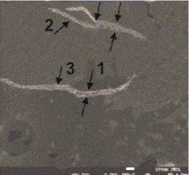
As we noted in the Introduction, the amelioration of amylin fibrillation state by the ethanol extracts from rose petals (Rosa damascena) and several other plants, as well as by isolated eleven fractions (phenol glycosides among them) was shown earlier [24]. Therefore, we compared the above described properties of amylin fibrils with those, formed during the peptide incubation in the pres-ence of 0.02% ethanol extract of rose petals and phenol glycoside fraction from this extract. Their SEM images are shown in Figure 2 and Figure 3, respectively.
The analysis with SEM technique evidenced increased polymorphism of fibrils at incubation of amylin in the presence of the ethanol extract from rose petals or phenol glycoside fraction isolated from it. In the presence of the extract (Figure 2), the diameter of the fibrils increased up to 50 - 60 nm (Figure 2, arrow 1) and the longitudinal breakthrough appeared (Figure 2, arrow 2). The formed clear-ances were occupied with extract particles with dimensions up to 8 nm (Figure 2, arrow 3).
This pattern is similar to that for amylin fibrils interacting with enzyme cata-lase, described in the literature [28].
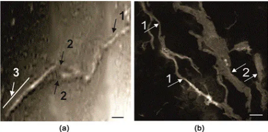
fibrillation extent for the phenol glycoside fraction is more pronounced than that for the extract. In Figure 3(a), the increased diameter of fibril is shown by arrow 1, the longitudinal breakthrough of the fibrils are shown by arrows 2. Un-
Figure 1. The TEM images of the suspension of amylin, aggregated for 7 days incubation
in 20 mM HEPES buffer, pH 7.2, at 37˚C. (a) protofibril forms; (b) mature fibrils (shown by arrows); (c) the protofibrils are indicated by arrow 1, mature form fibrils transformed into ribbon-like formations—by arrow 3; (d) the mature fibrils are indicated by arrow 1, the ribbon-like formations—by arrow 2. Scale bars: 100 nm.
Figure 2. The SEM picture of the suspension of amylin, aggregated in the presence of
[image:5.595.281.468.500.673.2]Figure 3. The SEM pictures of the suspension of amylin, aggregated in the presence of 0.02% of phenol glycoside fraction from the ethanol extract of rose petals (the conditions as in the Legend of Figure 1). (a) arrow 1 indicates the amylin fibrils with the diameter of 50 - 60 nm, arrows 2—the longitudinal breakthrough forms of the fibrils, arrow 3—the spiralized part of the fibrils; (b) the amorphous bundles of fibrils without clear configura-tion are seen. Scale bars: 100 nm.
der the impact of phenol glycoside, spiralization of amylin fibrils was also ob-served (Figure 3(a), arrow 3). In this case, concurrently with the fibrillar struc-tures, the amorphous bundles of fibrils without clear configuration were ob-served (Figure 3(b)).
4. Discussion
The transmission and scanning electron microscopy analysis of conformational changes at amylin oligomerization manifested simultaneous presence of various structural forms: protofibrils, mature fibrils and their ribbon-like formations. In case of amylin incubation in the presence of plant preparations, the inclusion of their components increased the fibril dimensions. In addition to the fibril struc-tures, the amorphous bundles are observed without clear configuration of their structure.
These observations can be considered as manifestation of the loosening of the fibril structure in the presence of the ethanol extract and phenol glycoside frac-tion, which can induce the decomposition of fibrils. Indeed, in our previous re-searches significantly low IC50 values have been estimated for the extract and phenol glycoside from rose petals (1.45 ± 0.26 and 0.15 ± 0.05 μg/ml, respective-ly) in protecting islet beta cells from killing by fibriled amylin [24]. These para-meters were in strong correlation with the ability of the preparations to inhibit the peptide fibrillation and disaggregate its preformed fibrils [25].
The presented observations confirm the TEM and SEM analyses as reasonable approaches to the evaluation of the effectiveness at search of anti-amyloidogenic agents.
Acknowledgements
Conflict of Interests
The authors declare no conflict of interests with respect to the present paper.
References
[1] Zerovnik, E. (2002) Amyloid Fibril Formation. Proposed Mechanisms and relev-ance to Conformational Disease. European Journal of Biochemistry, 269, 3362- 3371. https://doi.org/10.1046/j.1432-1033.2002.03024.x
[2] Greenwald, J. and Riek, R. (2010) Biology of Amyloid: Structure, Function, and Regulation. Structure, 18, 1244-1260. https://doi.org/10.1016/j.str.2010.08.009 [3] Chiti, F. and Dobson, C.M. (2006) Protein Misfolding, Functional Amyloid, and
Human Disease. Annual Review of Biochemistry, 75, 333-366. https://doi.org/10.1146/annurev.biochem.75.101304.123901
[4] Chiti, F. and Dobson, C.M. (2009) Amyloid Formation by Globular Proteins under Native Conditions. Nature Chemical Biology, 5, 15-22.
https://doi.org/10.1038/nchembio.131
[5] Baldwin, A.J., Knowles, T.P.J., Tartaglia, G.G., Fizpatrick, A.W,, Devlin, G.L., Shammas, S.L., Waudby, C.A., et al. (2011) Metastability of Native Proteins and the Phenomenon of Amyloid Formation. Journal of the American Chemical Society, 133, 14160-14163. https://doi.org/10.1021/ja2017703
[6] Sipe, J.D., Benson, M.D., Buxbaum, J.N., Ikeda, S., Merlini, G., Saraiva, M.J.M. and Westermark, P. (2016) Amyloid Fibril Proteins and Amyloidosis: Chemical Identi-fication and Clinical ClassiIdenti-fication International Society of Amyloidosis 2016 No-menclature Guidelines. Amyloid, 23, 209-213.
https://doi.org/10.1080/13506129.2016.1257986
[7] Invernizzi, G., Papaleo, E., Sabate, R. and Ventura, S. (2012) Protein Aggregation: Mechanisms and Functional Consequences.The International Journal of Biochemi-stry & Cell Biology, 44, 1541-1554. https://doi.org/10.1016/j.biocel.2012.05.023 [8] Pastore, A. and Temussi, P. (2012) Protein Aggregation and Misfolding: Good or
Evil? Journal of Physics:Condensed Matter, 24, 1-9. https://doi.org/10.1088/0953-8984/24/24/244101
[9] Stefani, M. (2004) Protein Misfolding and Aggregation: New Examples in Medicine and Biology of the Dark Side of the Protein World. Biochimica et Biophysica Acta, 1739, 5-25. https://doi.org/10.1016/j.bbadis.2004.08.004
[10] Clardy, J. and Walsh, C. (2004) Lessons from Natural Molecules. Nature, 432, 829- 837. https://doi.org/10.1038/nature03194
[11] Carocho, M. and Ferreira, I.C.F.R. (2013) A Review on Antioxidants, Pro-Oxidants and Related Controversy: Natural and Synthetic Compounds, Screening and Analy-sis Methodologies and Future Perspectives. Food and Chemical Toxicology, 51, 15- 25. https://doi.org/10.1016/j.fct.2012.09.021
[12] Hawkes, C.A., Ng, V. and McLaurin, J.A. (2009) Small Molecule Inhibitors of a
β-Aggregation and Neurotoxicity. Drug Development Research,70, 111-124. https://doi.org/10.1002/ddr.20290
[13] Joynera, P.M. and Cichewicz, R.H. (2011) Bringing Natural Products into the Fold- Exploring the Therapeutic Lead Potential of Secondary Metabolites for the Treat-ment of Protein-Misfolding Related Neurodegenerative Diseases. Natural Product Reports,28, 26-47.
638. https://doi.org/10.1016/j.bbadis.2011.10.006
[15] Porat, Y., Abramowitz, A. and Gazit, E. (2006) Inhibition of Amyloid Fibril Forma-tion by Polyphenols: Structural Similarity and Aromatic InteracForma-tions as a Common Inhibition Mechanism. Chemical Biology & Drug Design,67, 27-37.
https://doi.org/10.1111/j.1747-0285.2005.00318.x
[16] Westermark, P., Wernstedt, C., Wilander, E., Hayden, D.W., O’Brien, T.D. and Johnson, K.H. (1987) Amyloid Fibrils in Human Insulinoma and Islets of Langer-hans of the Diabetic Cat Are Derived from a Neuropeptide-Like Protein also Present in Normal Islet Cells. Proceedings of the National Academy of Sciences,84, 3881-3885. https://doi.org/10.1073/pnas.84.11.3881
[17] Cooper, G.J.S., Leighton, B., Dimitriadis, G.D., Parry-Billings, M., Kowalchuk, J.M., Howland, K., Rothbard, J.B., et al. (1988) Amylin Found in Amyloid Deposits in Human Type 2 Diabetes Mellitus May Be a Hormone That Regulates Glycogen Me-tabolism in Skeletal Muscle. Proceedings of the National Academy of Sciences, 85, 7763-7767. https://doi.org/10.1073/pnas.85.20.7763
[18] Westermark, P., Andersson, A. and Westermark, G.T. (2011) Islet Amyloid Poly-peptide, Islet Amyloid, and Diabetes Mellitus. Physiological Reviews,91, 795-826. https://doi.org/10.1152/physrev.00042.2009
[19] Aarabi, M.-H. and Mirhashemi, S.M. (2012) The Role of Two Natural Flavonoids on Human Amylin Aggregation. African Journal of Pharmacy and Pharmacology, 6, 2374-2379. https://doi.org/10.5897/AJPP12.616
[20] Meng, F., Abedini, A., Plesner, A., Verchere, C.B. and Raleigh, D.P. (2010) The Fla-vanol (−)-Epigallocatechin 3-Gallate Inhibits Amyloid Formation by Islet Amyloid Polypeptide, Disaggregates Amyloid Fibrils, and Protects Cultured Cells against IAPP-Induced Toxicity. Biochemistry, 49, 8127-8133.
https://doi.org/10.1021/bi100939a
[21] Mirhashemi, S.M. and Aarabi, M.-H. (2012) Effect of Two Herbal Polyphenol Compounds on Human Amylin Amyloid Formation and Destabilization. Journal of Medicinal Plants Research, 6, 3207-3212.
[22] Mirhashemi, S.M. and Aarabi, M.-H. (2012) To Evaluate Likely Antiamyloidogenic Property of Ferulic Acid and Baicalein against Human Islet Amyloid Polypeptide Aggregation, in Vitro Study. African Journal of Pharmacy and Pharmacology, 6, 671-676.
[23] Sharoyan, S., Antonyan, A., Mardanyan, S., Harutyunyan, H., Movsisyan, N., Hov-nanyan, N. and HovHov-nanyan, K. (2013) Interaction of Dipeptidyl Peptidase IV with Amyloid Peptides. Neurochemistry International, 62, 1048-1054.
https://doi.org/10.1016/j.neuint.2013.03.017
[24] Sharoyan, S.G., Antonyan, A.A., Harutyunyan, H.A. and Mardanyan, S.S. (2015) Inhibition of Amylin Fibril Formation and Protection of Islet β-Cells by Medicinal Plants. International Journal of Pharmacognosy, 2, 234-241.
[25] Sharoyan, S.G., Antonyan, A.A., Harutyunyan, H.A. and Mardanyan, S.S. (2015) Medicinal Plants Support the Amylin-Suppressed Viability of Islet β-Cells. Interna-tional Journal of Pharmacognosy, 2, 448-453.
[26] Antonyan, A., Sharoyan, S., Harutyunyan, H., Movsisyan, N., Sargisova, Y.G., Ste-panyan, H. and Mardanyan, S. (2014) Cytotoxicity of Some Edible Plants toward Ehrlich Ascites Carcinoma Cells. Research Journal of Medicinal Plants, 8, 20-31. https://doi.org/10.3923/rjmp.2014.20.31
[28] Milton, G.N. and Harris, J.R. (2010) Human Islet Amyloid Polypeptide Fibril Bind-ing to Catalase. The Scientific World Journal, 10, 879-893.
https://doi.org/10.1100/tsw.2010.73
Submit or recommend next manuscript to OALib Journal and we will pro-vide best service for you:
Publication frequency: Monthly
9 subject areas of science, technology and medicine Fair and rigorous peer-review system
Fast publication process
Article promotion in various social networking sites (LinkedIn, Facebook, Twitter, etc.)
Maximum dissemination of your research work