www.impactjournals.com/oncotarget/ Oncotarget, Vol. 5, No. 21
Specific genomic and transcriptomic aberrations in tumors
induced by partial hepatectomy of a chronically inflamed murine
liver
Ezra Ella1, Denise Heim2, Evgeniy Stoyanov1, Rona Harari-Steinfeld1, Israel Steinfeld3, Orit Pappo4, Temima Schnitzer Perlman1, Natalie Nachmansson5, Ludmila Rivkin1, Devorah Olam1, Rinat Abramovitch1,5, Henning Wege2, Eithan Galun1 and Daniel Goldenberg1
1 The Goldyne Savad Institute of Gene Therapy, Hadassah-Hebrew University Medical Center, Jerusalem, Israel 2 Department of Gastroenterology and Hepatology, University Medical Center Hamburg-Eppendorf, Hamburg, Germany 3 Computer Science Department, Technion-Israel Institute of Technology, Haifa, Israel
4 Department of Pathology, Hadassah-Hebrew University Medical Center, Jerusalem, Israel
5 Magnetic Resonance Imaging/Magnetic Resonance Spectroscopy Laboratory, Human Biology Research Center, Hadassah-Hebrew University Medical Center, Jerusalem, Israel
Correspondence to: Daniel Goldenberg, email: [email protected]
Keywords: HCC, liver regeneration, chronic hepatitis, genomic instability, Crem
Received: August 24, 2014 Accepted: September 24, 2014 Published: September 25, 2014
This is an open-access article distributed under the terms of the Creative Commons Attribution License, which permits unrestricted use, distribution, and reproduction in any medium, provided the original author and source are credited.
ABSTRACT
INTRODUCTION
Tumor resection and ablation are currently the main treatment options for an early-stage hepatocellular carcinoma (HCC), while tumor recurrence is the main complication following these treatments [1].
Chronic inflammation precedes the majority of HCC cases and significantly contributes to genetic
instability [2]. To explore the molecular mechanisms of hepatocarcinogenesis following resection of a chronically
inflamed liver in a small animal model, we performed
partial hepatectomy (PHx) in the Mdr2-KO mice, a
well-characterized model of inflammation-mediated HCC development [3, 4]. Previously, we demonstrated accelerated hepatocarcinogenesis following 35% PHx of either 3-month-old or 9-month-old Mdr2-KO males [5].
We found genomic instability in PHx-induced tumors
of the hepatectomized mutants and activation of DNA damage response genes in the non-tumor liver of these mice, probably caused by the regenerative proliferative stress [5]. Now, we used 70% PHx of both male and female Mdr2-KO mice at the age of either 3 or 6 months
in order to explore the effects of gender and operation time
on accelerated HCC development in this model. In order to explore the specific molecular pathways responsible for the accelerated tumor development, we compared genomic landscapes and gene expression profiles of the PHx-induced and spontaneous HCC tumors developed in this model. We demonstrate that 70% PHx of the chronically inflamed liver induces a specific pattern of chromosomal amplifications in the early HCC tumors and specific patterns of gene expression both in tumors and in the non-tumor liver which are different from those in
the non-hepatectomized Mdr2-KO mice. Furthermore, we demonstrate that the gene Crem, which is frequently
amplified and over-expressed in the Mdr2-KO HCC, is also frequently over-expressed, although rarely amplified,
in human HCC, and stimulates proliferation of human HCC cells.
RESULTS
Strain, but not gender, determines the tumor-promoting effect of PHx on HCC development in the Mdr2-KO mice
In order to delineate the roles of gender and time of PHx in the development of HCC in the chronically inflamed liver, we performed 70% PHx in male and female
Mdr2-KO/FVB mice at either three or six months of age;
sham operations served as controls (Supplemental Figure 1). PHx significantly increased liver tumor incidence in
females operated at the age of three months, and in males
operated at the age of six months (Figure 1A,E). For both
genders together, the tumor-promoting effect of PHx
was statistically significant only when the operation was
performed at the age of three months (Figure 1C; Fisher
test p=0.002), while, it was only marginally significant
when the operation was performed at the age of six months
(Figure 1F; Fisher test p=0.067). Tumor load and size were profoundly increased in post-PHx mice: the average tumor load per mouse for both genders together was significantly
higher in the post-PHx compared to the sham group at
both operation ages, and a proportion of mice having tumors with a diameter larger than 3 mm was two-fold higher in the post-PHx group (Supplemental Table 1A). For both genders, tumors with a diameter larger than 7 mm (for males - larger than 5 mm) were observed only in the PHx-treated mice (Supplemental Table 1B). Remarkably, frequency, size, morphology and developmental stage of the post-PHx liver tumors in Mdr2-KO/FVB mice at the
age of nine months, were similar to those of spontaneous
tumors observed in the untreated mutants at the age of 13 months (females) or 14 months (males).
To determine the effect of genetic background on the development of HCC in the chronically inflamed liver following PHx, we performed 70% PHx in male Mdr2-KO/B6 mice at six months of age and sacrificed these animals for tumor score at 14 months of age. Surprisingly, there was no significant difference in liver tumor development between the PHx-treated and control groups (Supplemental Figure 2). Both PHx-operated and sham-operated Mdr2-KO/B6 mice at 14 months of age produced
a similarly low number of tumors and nodules, compared to the PHx-operated Mdr2-KO/FVB mice at the age of
nine months (Figure 1E). Thus, 70% PHx significantly increased liver tumor incidence, load and size in Mdr2-KO mice operated at early ages; however, this tumor-promoting effect was specific for the FVB, but not for the B6 genetic background.
Comparison of cell proliferation and DNA damage markers in non-tumor and tumor liver tissues between post-PHx and untreated Mdr2-KO/FVB mice
To explore the mechanisms of the accelerated HCC
development by PHx, we compared liver tumors and their matched non-tumor liver tissues between 9-month-old hepatectomized and 13-14-month-old untreated
Mdr2-KO mice. From each experimental group, we selected six well differentiated HCC tumors similar in size range
and morphology between groups. All tumors were well protruding from the liver and contained at least 90%
of tumor cells on histological sections. We compared
cell proliferation and DNA damage markers in tumors and their adjacent non-tumor (surrounding) as well as distant non-tumor (non-tumor) liver tissues between
Figure 1: Accelerated HCC development in the Mdr2-KO/FVB mice following 70% PHx. Either PHx or sham surgery was performed at the age of either three (A-C) or six (D-F) months; all mice were sacrificed at the age of nine months. Tumor incidence in female (A) or male (B) mice, or females and males together (C) operated at the age of three months (females: PHx n=17, sham n=16; males: PHx n=18, sham n=22). Tumor incidence in female (D) or male (E) mice, or females and males together (F) operated at the age of six months (females: PHx n=25, sham n=10; males: PHx n=20, sham n=11).
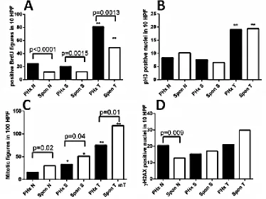
Figure 2: Quantification of proliferation and DNA damage markers in the liver of hepatectomized and untreated
[image:3.612.111.485.367.646.2]for all three tissue types was significantly higher in the
post-PHx compared to the untreated (spontaneous) group
(Figure 2A, Supplemental Figure 3A-D). However, there was no difference in the level of phosphorylated histone 3, a marker of the mitotic M phase, between groups (Figure 2B, Supplemental Figure 3E-H), while there were decreased numbers of mitotic figures in the
post-PHx compared to the untreated group in all tissue types
(Figure 2C, Supplemental Figure 3I-L). Immunostaining revealed no difference in nuclear p21 level between non-tumor liver tissues of experimental groups (not shown). TUNEL assay revealed similar and very low levels of positive cells in both post-PHx and untreated non-tumor liver (not shown), while the level of the phosphorylated histone 2AX, a marker of either DNA damage, or stalked replication forks, was significantly higher in the post-PHx non-tumor liver (Figure 2D, Supplemental Figure 3M-P). These results are consistent with the increased DNA repair process in the non-tumor and tumor liver tissues,
and stalked replication forks in the non-tumor liver tissue
of hepatectomized compared to the untreated Mdr2-KO/ FVB mice.
Specific genomic amplifications in PHx-induced liver tumors
To understand the molecular mechanisms of the
accelerated tumor development in post-PHx mice, we
compared, using aCGH, genomic aberrations in six tumors from each of the experimental groups described
above (Supplemental Figure 4; aCGH maps of four
among six post-PHx tumors were published by us
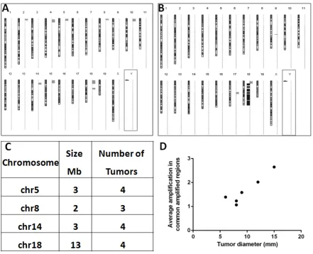
previously [5]). Among PHx-induced tumors, 5 of 6 had major chromosomal aberrations, and all of them were amplifications affecting multiple chromosomes (Figure 3A). Most amplifications were located near the
[image:4.612.86.527.292.650.2]acrocentric centromeres of murine chromosomes. Four
different chromosomal regions (size 2 to 13 Mb) were amplified (1.3 to 12.3 fold) each in at least three tumors (Figure 3C). The frequency of amplifications of common amplified regions correlated with tumor size (R2= 0.8527; Figure 3D). All six spontaneous tumors developed by
untreated Mdr2-KO mice had chromosomal aberrations,
including both deletions (4/6 tumors) and amplifications (5/6 tumors). Amplifications in spontaneous tumors affected significantly less chromosomes and were not
located preferentially at chromosomal edges. PHx-induced and spontaneous tumors shared the same frequent
amplification only at chromosome 18 (at least 3 samples in each group; Figure 3A,B; Supplemental Table 2).
Bioinformatic comparison of the genomic data of
post-PHx tumors from Mdr2-KO mice with 76 tumors from three different HCC mouse models (Supplemental Figure 5), and with 155 human HCCs (compared by synteny; Supplemental Figure 6) revealed only rare overlaps of genome amplification patterns between the PHx-induced tumors and other murine or human HCCs. Nevertheless,
analysis of the published literature demonstrated that many human chromosomal regions syntenic to the four
murine frequently amplified regions were amplified in some human HCC tumors (Supplemental Table 3).
Specific patterns of gene expression in the PHx-induced tumors
Genome-scale gene expression analysis of the
PHx-induced tumors and their distant non-tumor liver tissues using PCA (Supplemental Figure 7) and hierarchical clustering (Supplemental Figure 8) revealed that most
non-tumor samples clustered together. Two female tumors produced a separate cluster, while four male tumors did
not; age at PHx and the number and level of amplifications
did not affect clustering. The distance between the matched tumor and non-tumor samples in both of these analyses directly correlated with the total number of
aberrantly expressed genes in the tumor (Supplemental Table 4).
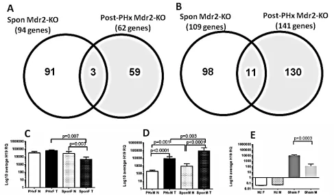
Comparison of the genome-scale gene expression
profiles of these six post-PHx tumors with those of spontaneous tumors of the 16-month-old Mdr2-KO mice which were analyzed previously [4] revealed surprisingly few common down-regulated (Figure 4A) and up-regulated (Figure 4B) genes in both tested datasets. We
compared both datasets also for enrichment in four cancer-associated gene expression signatures: up-regulation of
[image:5.612.66.553.359.643.2]HCC-specific oncogenes, or chromosomal instability
(CIN) genes, or E2F1 targets, and down-regulation of HCC-specific tumor suppressors (Table 1) [6, 7]. At least 50% of post-PHx tumors were significantly enriched in Oncogenic, CIN, and E2F1 gene expression signatures,
while in spontaneous tumors only solitary tumors were
significantly enriched in either Oncogenic or E2F1
signature. For both groups, only solitary tumors were
significantly enriched in the tumor suppressor signature. The microarray data demonstrated a very high expression of the H19 transcript in the non-tumor liver of hepatectomized females. The H19 transcript is known to be frequently highly expressed in HCC [8]. We confirmed
this result and explored the effect of gender and PHx on
H19 expression using qRT-PCR of tumor and non-tumor RNA samples from untreated (13-14-month-old) and post-PHx (9-month-old) livers from Mdr2-KO males
and females. There was a prominent gender disparity in
the H19 expression in tumor versus non-tumor liver: in
females, it was similar in the post-PHx tumors, while it
significantly decreased in spontaneous tumors (Figure 4C), whereas in males, it was significantly increased in both spontaneous and post-PHx groups (Figure 4D). Altogether,
gene expression data indicate that hepatocarcinogenesis
differed significantly between the post-PHx and
spontaneous tumors of Mdr2-KO/FVB mice.
Correlation between gene amplification and expression in the PHx-induced tumors
There was a significant correlation between genomic amplification and elevation of gene expression in all amplified regions of the PHx-induced tumors (Supplemental Table 5). Furthermore, while the observed average ratio between the up-regulated and down-regulated genes in all PHx-induced tumors was about 50%, in the common amplified regions, 95% of differentially
expressed genes were up-regulated. Correlation between
[image:6.612.61.550.301.638.2]the amplification and expression levels was confirmed by
semi-qRT-PCR for 22 genes in common amplified regions (Supplemental Table 6). The best correlation with gene expression was found for genomic amplifications over 2.5-fold (Supplemental Figure 9), similar to what was described previously for breast cancer tumors [9].
Pathway analysis of genes in the common amplified regions of the post-PHx tumors
The common amplified regions of the post-PHx tumors contained 122 genes; 32 among them were known in the literature as having a role in cancer. Analysis of signaling pathways for these 122 genes by GO, revealed a significant prevalence of the Wnt signaling pathway (Supplemental Table 7). However, immunostaining for beta-catenin revealed only rare scattered positive hepatocyte nuclei and variable cytoplasmic staining in both post-PHx and spontaneous tumors (Supplemental
Figure 10). The only post-PHx tumor sample which did not contain amplifications (PHx5M) had a typical gene expression signature of the activated Wnt signaling (Supplemental Table 8), but not beta-catenin-positive
nuclear immunostaining (not shown).
Gene signatures of the early HCC development/ recurrence in tumor and non-tumor liver tissues of the post-PHx Mdr2-KO/FVB mice
To explore the relevance of this model (PHx of the chronically inflamed murine liver) to the process of HCC development/recurrence in patients, we analyzed the significance of the known appropriate gene expression
signatures in the tumor and non-tumor samples of the
[image:7.612.68.549.85.407.2]post-PHx Mdr2-KO/FVB mice. Recently, it was shown that in tumors removed by curative surgery, up-regulation of the Racgap1 together with several other genes of its Table 1: Gene expression signatures in the PHx-induced and spontaneous HCC tumors of Mdr2-KO/FVB mice. The PHx-induced tumors as compared to spontaneous tumors were enriched with the “Oncogene”, “CIN” and “E2F1”, but not with “Tumor suppressors”, signatures.
Tumor ID Tumor suppressors Oncogenes CIN signature genes E2F1 signature genes Fold change
thresholda: ≤ -1.5 ≥ 1.5 ≥ 1.8 ≥ 1.8
Mdr2-KO mice following PHx 9-month-old
PHx2F 0.59 0.0216 1.06E-07 2.89E-09
PHx4M 1 0.0498 4.53E-12 8.13E-08
PHx3M 0.77 0.11 1 0.09
PHx1M 1 0.46 0.15 1
PHx5M 0.63 0.32 0.0058 0.06
PHx6F 0.0384 0.0269 8.95E-09 8.07E-07
Mdr2-KO mice untreated 16-month-old
49-T1 0.82 1 1 1
84-T1 0.0270 1 0.09 3.51E-02
93-T1 1 5.61E-05 0.17 0.65
93-T2 0.11 1 1 0.19
96-T1 0.19 1 1 0.73
96-T2 0.82 0.58 1 0.54
aFold change thresholds in tumors vs. matched non-tumorous tissue.
Gene lists for analysis of expression signatures of oncogenes, tumor suppressors and E2F1 targets have been described by us previously [7]; the 25-gene CIN signature – as described in [6].
Statistical significance (p value) of each gene signature was determined by Fisher test using R-software. Bold numbers indicate p-value < 0.05.
interactome is associated with the early HCC recurrence
[10]. Here we show that five of the six post-PHx tumors
had a similar co-upregulation pattern for most of
Racgap1-associated genes (Figure 5A), while none of the spontaneous tumors from 16-month-old Mdr2-KO/ FVB mice had this expression signature (Figure 5B). A prominent prognostic expression signature of 186 genes wherein the over-expression of its specific subsets was
correlated with either poor or good prognosis, has been
confirmed recently as having a prognostic significance for HCC development based on the expression profile of the early-stage cirrhotic liver samples [11]. We next compared the expression of the top 40 genes which were over-expressed in the “poor prognosis” signature between non-tumor liver tissues of the post-PHx males obtained in
the current study, and the age-matched untreated
Mdr2-KO/FVB and control liver samples that were studied by us previously [5]. Among the tested 40 genes, 31 were
presented in both microarrays, and 22 of them were up-regulated in the post-PHx compared to the untreated
Mdr2-KO livers (Figure 5C). Expression of the most of 16 housekeeping genes, which were used for normalization of expression levels in both comparative datasets, was similar between experimental groups (Figure 5D). Overall, the human “poor prognosis” HCC signatures were significantly more prominent in the post-PHx compared to the untreated Mdr2-KO/FVB liver indicating that
PHx of these mice increases the expression of the HCC
development/recurrency markers in both tumor and non-tumor liver tissues even several months following the
operation.
Crem is a new candidate HCC oncogene which is frequently amplified and over-expressed following PHx
The most common amplified region among both
post-PHx and spontaneous tumors was the one on chromosome 18, containing 22 unique genes: it was
amplified in 4/6 post PHx tumors and in 3/6 spontaneous tumors (Figure 3C, Supplemental Table 2). We focused
on the gene Crem encoded by this region due to its
involvement in cancer [12, 13], including HCC metastasis [14], LR following PHx [15], and circadian regulation [16]. Using semi-qRT-PCR of the matched tumor and non-tumor samples, we confirmed that Crem was
over-expressed (FC>=1.8) in all tumor samples where it
was amplified (FC >= 1.4) (Supplemental Table 6, Supplemental Figure 11). The correlation between Crem genomic amplification and transcript expression was significantly higher in post-PHx compared to spontaneous tumors (Supp Figure 11A,B).
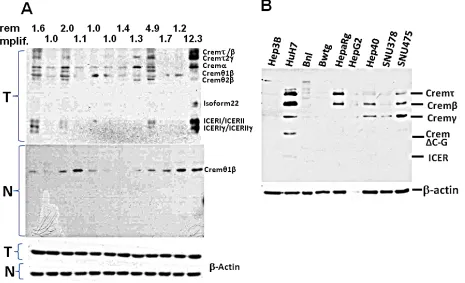
[image:8.612.77.542.390.673.2]Crem has multiple splicing isoforms (Supplemental
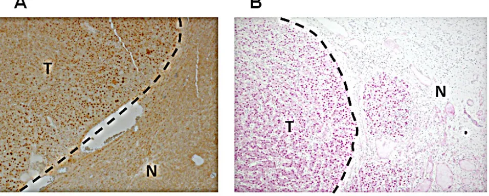
Table 9). Using immunoblotting, we revealed that more Crem protein isoforms and at higher levels were expressed in tumors than in matched non-tumor liver tissues, reaching the highest levels in tumors having genomic Crem amplifications (FC>1.5; Figure 6A). Immunostaining of paraffin-embedded murine liver tissues revealed strong Crem staining in hepatocyte nuclei of tumors containing Crem amplifications compared to the surrounding non-tumor tissues (Figure 7A). We have tested CREM protein expression in 13 human HCC samples of various etiologies, and have found CREM-positive nuclei in 11 HCCs (85%), but not in the non-tumor cirrhotic liver (Table 2, Figure 7B).
Several Crem isoforms were detected by immunoblotting in five out of nine tested human and
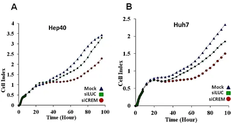
mouse HCC cell lines (Figure 6B). Human lines Huh7 and Hep40 were selected for studying the role of CREM in cancer cell proliferation by knockdown experiments using CREM-specific siRNA. Immunoblotting revealed that the selected CREM-specific siRNAs efficiently decreased protein levels of the most CREM isoforms, especially ICER (Supp Figure 11D). The xCELLigence proliferation assay demonstrated a significant decrease of the cell
proliferation rate following transient transfection of both
Huh7 and Hep40 cell lines with CREM-specific siRNA, suggesting that CREM is important for the proliferation of these HCC cells (Figure 8). Importantly, no significant effect of this transfection on cell size was observed (Supp Figure 11E). These results demonstrate that gene Crem, frequently amplified in both post-PHx and spontaneous
[image:9.612.68.551.59.247.2]Figure 7: Increased Crem protein expression in the nuclei of HCC tumor cells. Representative images of IHC staining of the Crem protein in murine (A) and human (B) HCC (T) and adjacent non-tumor liver (N). Magnification x100.
Table 2: Characteristics of the HCC patients and their tumors.
Patient CREM1 Age Gender Etiology Cirrhosis2 Grading T-Status
1 Negative 71 Male Hepatitis C Yes 2 1
2 Positive 56 Male Crypotgenic Yes 1 2
3 Negative 60 Male Alcohol Yes 2 2
4 Positive 75 Male Cryptogenic No 2 1
5 Positive 62 Male Hepatitis C Yes 2 3
6 Positive 60 Male Hepatitis C, alcohol Yes 2 3b
7 Positive 55 Female Hepatitis C Yes 2 3a
8 Positive 67 Male NASH No 3 3a
9 Positive 65 Male Hepatitis C Yes 1 1
10 Positive 58 Female Cryptogenic No 2 2
11 Positive 30 Female Adenoma No 2 3a
12 Positive 76 Male Hemochormatosis No 2 2
13 Positive 67 Male Hepatitis B No 2 1
1Results of the IHC staining of tumor samples for CREM. Positive CREM-staining in the tumor was more intense at the borders.
[image:9.612.74.552.496.688.2]tumors of the Mdr2-KO mice, is expressed in hepatocyte
nuclei in correlation with its genomic amplification in murine HCC tumors; human CREM is expressed in the tumor cell nuclei of the majority of evaluated HCCs, and is required for the efficient proliferation of human HCC
cell lines in vitro.
DISCUSSION
Strain specificity of the PHx-induced tumors in the Mdr2-KO liver suggests a direct correlation between the degree of chronic liver inflammation and a number of tumor cell precursors at a young age
We have revealed strain-specificity of the effect of
PHx on tumor acceleration in Mdr2-KO males operated
at the age of six months: in the FVB/N strain, the
tumor-promoting effect of PHx could be already detected at the
age of nine months, whereas in the B6 strain, no effect of PHx on HCC development could be observed even at the age of 14 months. This result correlates with our previous findings of a reduced inflammation at the early age and retarded tumor development at the adult age in the Mdr2-KO/B6 compared to the Mdr2-KO/FVB strain [17]. Importantly, 70% PHx of B6 males does
accelerate tumor appearance in chemically-induced
hepatocarcinogenesis [18]. In our previous work we
suggested that genomic unstable hepatocytes generated
during chronic inflammation are the tumor-initiating cells
in this model, which escape cell cycle arrest and re-enter
cell cycle due to PHx-induced regenerative proliferative stress [5]. We suggest that in the chemically-induced carcinogenesis and in the highly inflamed Mdr2-KO/FVB
strain, such potentially tumorigenic cells appear early in
the liver, and thus PHx accelerates tumor development. In the Mdr2-KO/B6 males, chronic liver inflammation decreases significantly after two months of age, probably blocking the formation of the tumor-initiating cells at
least till the age of six months; thus, PHx in these mice
at this age did not have a tumor-promoting effect. This
probably explains also the gender difference in the Mdr2-KO/FVB model: the tumor-promoting effect of PHx in
females was significant already at the age of three months, while in males – mainly at six months. The Mdr2-KO/ FVB females are more inflamed at the early ages than
males (our unpublished data) and thus, probably generate
[image:10.612.67.539.422.674.2]the tumor-initiating cells in the liver earlier. Interestingly, inflammation-mediated HCC develops in aged Mdr2-KO/FVB females earlier than in males (see “Results”), whereas spontaneous HCC develops in the parental FVB/ NJ strain in old males only [19].
Potential mechanisms of the observed genome amplifications
Recently, it was demonstrated that HCCs from
Mdr2-KO/FVB mice are similar to pediatric HCCs of similar etiology in terms of low frequency of point
mutations and high frequency of gene amplifications [20]. In contrast to our findings, the authors could detect only
a negligible number of deletions in spontaneous tumors from Mdr2-KO/FVB mice. This discrepancy could arise from a different methodology: we used the direct
aCGH method, whereas Iannelli et al. used a non-direct
method for the detection of chromosomal aberrations
in mice [20]. The existence of deletions in spontaneous
tumors of Mdr2-KO/FVB mice was also reported by us
previously [4]. A striking difference in the patterns of
chromosomal aberrations between PHx-induced and spontaneous tumors that we found here, raises a question
on the potential mechanism of PHx-induced specific genomic amplifications without deletions (a probability to observe six tumors without deletions by chance here is 0.0014). The recent finding of genomic amplifications
of the Kras oncogene in the early lung tumors appearing
in the FVB/N, but not in the mixed genetic background [21], indicates that this “amplification pattern” may be also strain-specific. However, gene amplification
by nonhomologous subtelomeric exchanges has been
documented even for the WT murine species [22], and centromeres are known hotspots of mitotic recombination [23]. Replication stress is a prominent pathway to genomic instability [24]; loss of DNA replication control
may cause inappropriate re-replication resulting in gene
amplification [25]. Our analysis of the cell proliferation and DNA damage markers (Figure 2 and Supplemental Figure 3) indicates that an increased replication stress and DNA repair in the post-PHx liver of Mdr2-KO/FVB mice could be responsible for the increased CIN in the PHx-induced tumors, in agreement with our previous findings in mice [5]. The aCGH method that we used provides only a relative quantification of different genomic regions, and not locations of the amplified genes: whether they are
extra-, intra- or inter-chromosomal.
Functional analysis of the genes that were frequently amplified in PHx-induced tumors
Determination of causative cancer genes in chromosomal aberrations is complicated by the known examples of cooperation between both co-amplified oncogenes [26] and co-deleted tumor suppressors [27]. PHx-induced tumors had a significantly higher proportion of common up-regulated genes (69.5%) compared to
spontaneous tumors of untreated Mdr2-KO/FVB mice
(53.7%), whereas the total number of common aberrantly
expressed genes in each group was identical (according to
the parameters described in Figure 4). These up-regulated genes included HCC-specific oncogenes (they are listed in our recent work [7]), E2F1 and CIN signature genes [6]. Remarkably, CIN and E2F1 genes significantly overlap, as most CIN genes are E2F1 targets [7]. Although GO analysis of the 122 genes from the common amplified regions revealed a prevalence of the Wnt signaling pathway, IHC demonstrated that all PHx-induced tumors either did not have, or had only a rare and fragmentary β-catenin-positive nuclear staining.
Other gene expression signatures that characterize the PHx-induced tumors
Prediction of tumor recurrence which jeopardizes survival of about 70% of HCC patients within 5 years
following tumor resection or ablation is of high clinical
importance [28]. Several gene expression signatures of the resected tumor and/or non-tumor liver tissue that may predict early HCC development/recurrence either alone [10, 11], or in combination with clinical and pathological data [28] were recently developed. Our finding that both tumor-specific [10] and non-tumor-specific [11] “poor prognosis” signatures were significantly more prominent in the liver of hepatectomized compared to untreated Mdr2-KO/FVB mice indicates that these mice may serve as a model for human HCC development/recurrence.
Validation of H19 expression on an expanded set of liver samples by qRT-PCR revealed different expression
patterns between PHx-induced and spontaneous tumors for Mdr2-KO/FVB females, but not for males (Figure
4C-E). The H19 expression is known to be frequently
up-regulated in cancer including HCC [8], acting in
different models either as a tumor suppressor [29], or as an oncogene [30]; its role in hepatocarcinogenesis is still debated. Our findings of a prominent gender disparity in the relative tumor/non-tumor H19 expression in both naïve
and hepatectomized Mdr2-KO/FVB mice and of a high
H19 expression in the non-tumor liver tissue of Mdr2-KO/ FVB females may indicate that H19 has different functions
in hepatocarcinogenesis of each gender.
Crem is a new candidate HCC oncogene
In our search for candidate cancer-causing genes in frequently amplified regions, we focused on the gene Crem because it was amplified in 4/6 post PHx tumors and in 3/6 spontaneous tumors, and also due to its involvement in cancer [12, 13], including HCC metastasis [14], LR following PHx [15], circadian regulation [16, 31] and cytokine production [32]. Remarkably, murine liver tumors containing Crem amplifications demonstrated high nuclear Crem expression (Figure 7A) and more expressed
Crem protein isoforms compared to the matched
datasets, CREM was infrequently up-regulated in tumors
(although frequently up-regulated in cirrhosis); similarly,
CREM DNA was infrequently amplified (Supplemental Table 3, 10p11.21 region). Nevertheless, IHC demonstrated CREM-positive nuclear staining in 11/13
tested human HCC tumors, but not in the surrounding
non-tumor liver, independently on the patient’s age and gender
and on HCC etiology (Table 2), probably indicating a
transcript level-independent mechanism of CREM up-regulation. CREM knock-down experiments in vitro
revealed its pro-proliferation role in two human HCC cell lines, thus demonstrating that CREM is a new candidate
HCC oncogene.
In summary, we demonstrated that PHx of the chronically inflamed murine liver accelerated HCC development by a specific pathway characterized by amplification of chromosomal regions located near the
acrocentric centromeres of murine chromosomes and by
expression of specific tumor-promoting genes, including those of the “poor prognosis” HCC signatures. We also identified Crem as a new candidate HCC oncogene frequently amplified and up-regulated in this murine HCC model and frequently over-expressed in human HCC.
MATERIALS AND METHODS
Full details are available in the Supplemental
Material
Mice
All animals received human care. All animal
experiments were performed according to national
regulations and guidelines of the Institutional Animal Welfare Committee (NIH approval number OPRR-A01-5011). The FVB.129P2-Abcb4tm1Bor (Mdr2-KO/FVB) and the C57Bl/6 Mdr2-KO (Mdr2-KO/B6) mice were described previously [17]. The 70% PHx or sham surgery was performed as described [5].
RNA analysis
Total RNA was isolated from frozen liver tissues and subjected to gene expression profiling using GeneChip Mouse Gene 1.0 ST Array (Affymetrix, Santa Clara, CA) as described [4]. Data were analyzed by GCRMA preprocessing algorithm using the Partek software (ProSoftware, Partek, St Louis, MO, USA). For validation of gene expression by RT–PCR, reverse transcription of total RNA was performed using the qScriptTM cDNA Synthesis Kit (#95047) and Perfecta Sybr Green Fast Mix ROX (#95073) (both from Quanta BioSciences Inc., Gaithersburg, MD, USA). The quantitative PCR assay was performed on an AB 7900 HT fast real-time PCR system (Applied Biosystems, Foster City, CA, USA) or CFX384
TM Real-Time System with C1000 Touch Thermal Cycle (BioRad, Hercules, CA, USA). The semi-quantitative RT-PCR and the primers used for RT-RT-PCR are described in the Supplemental Materials and Methods.
DNA analysis
Total DNA was isolated from frozen tumors and their matched non-tumor liver tissues as described [4] and subjected to array-based comparative genomic
hybridization (aCGH) analysis using Mouse Genome
CGH Microarrays 4x44K (Agilent, Santa Clara, CA). Data
for both genome and transcriptome microarray analyses
can be accessed from the GEO-NCBI database repository (GSE61427).
Protein analysis
Protein detection by immunoblotting and immunohistochemial staining was performed as described
in the Supplemental Materials and Methods.
Statistical analysis and calculation software
All the in vitro experiments were performed in triplicates, excluding the cell proliferation rate assay
using a xCELLigence system, which was performed in
duplicates; in the in vivo experiments, at least 3 mice per group were used. All parameters were evaluated with
the two-tailed t-test. Statistical evaluation of differential
expression between the experimental groups was
performed using the ANOVA test. A p-value of 0.05 or less was considered significant. t-test, correlation test, and
f-test were performed using the GraphPad Prism version 6.04 for Windows (GraphPad Software, La Jolla California USA, www.graphpad.com). Stained hepatocytes IHC sections were counted by the NIS-Elements Microscope Imaging Software (Nikon Instruments Inc.1300 Walt Whitman Road, Melville, NY, U.S.A). The results of protein immunoblotting and semi-quantitative RT-PCR were calculated using ImageJ (Image processing and analysis in Java , NIH, U.S.A). To evaluate the effect
of PHx on tumor incidence, the Fisher exact test was
calculated online (http://www.quantpsy.org/fisher/fisher. htm). The Pearson’s correlation coefficient (Supplemental Table 5) was calculated for all 1,256 genes with matching copy number and expression profiles. The distribution of correlation values was compared to a randomized background distribution and was found to be significantly enriched with positive correlation values (p<E-33). The randomized background distribution was generated by repeatedly (100 times) shuffling the samples in the
expression dataset only and recalculating the non-sample
two distributions was estimated using the Kolmogorov-Smirnov test.
List of Abbreviations
Abcb4 (Mdr2), adenosine triphosphate–binding cassette b4 (multi-drug resistance 2); aCGH, array-based comparative genomic hybridization; B6, C57Black/6 strain; BrdU, bromodeoxyuridine; CIN, Chromosomal Instability; Crem, cAMP responsive element modulator; FC, Fold Change; FVB, Friend virus B-type/N strain; GO, Gene Ontology; GCRMA, GeneChip robust multiarray analysis; HCC, hepatocellular carcinoma; ICER, Inducible CAMP Early Repressor; IHC, immunohistochemistry; LR, liver regeneration; KO, knockout; mRNA, messenger RNA; PHx, Partial hepatectomy; PCA, Principal Component Analysis; qPCR, quantitative RT-PCR; RT-PCR, reverse transcription polymerase chain reaction; TUNEL, Terminal deoxynucleotidyl transferase dUTP Nick End Labeling; Wnt, Wingless-Type MMTV Integration Site Family; WT, wild type.
ACKNOWLEDGMENTS
We thank Prof. Mehmet Ozturk for providing us with HCC cell lines Hep40, SNU378, SNU475, Dr. Ido Weiss for his help with FACS analysis, and Mery Clausen
for assistance in manuscript preparation. This study was
supported by: Kamea Scientific Foundation of the Israeli Government (D.G.); Deutsche Forschungsgemeinschaft (DFG) SFB841 project C3 and I-CORE ISF center of excellence and the ISF grant (E.G.); Deutsche Forschungsgemeinschaft, Collaborative Research Center 841 project C5 (H.W. and D.H.).
Conflict of interests
Nothing to disclose.
Editorial note
This paper has been accepted based in part on
peer-review conducted by another journal and the authors’ response and revisions as well as expedited peer-review
in Oncotarget.
REFERENCES
1. Forner A, Llovet JM and Bruix J. Hepatocellular carcinoma. Lancet. 2012; 379(9822):1245-1255.
2. Colotta F, Allavena P, Sica A, Garlanda C and Mantovani A. Cancer-related inflammation, the seventh hallmark of cancer: links to genetic instability. Carcinogenesis. 2009; 30(7):1073-1081.
3. Mauad TH, van Nieuwkerk CM, Dingemans KP, Smit JJ, Schinkel AH, Notenboom RG, van den Bergh Weerman MA, Verkruisen RP, Groen AK, Oude Elferink RPJ, van der Valk MA, Borst P and Offerhaus GJA. Mice with
homozygous disruption of the mdr2 P-glycoprotein gene.
A novel animal model for studies of nonsuppurative inflammatory cholangitis and hepatocarcinogenesis. Am J Pathol. 1994; 145(5):1237-1245.
4. Katzenellenbogen M, Mizrahi L, Pappo O, Klopstock N, Olam D, Jacob-Hirsch J, Amariglio N, Rechavi G, Domany E, Galun E and Goldenberg D. Molecular mechanisms of liver carcinogenesis in the Mdr2-knockout mice. Mol Cancer Res. 2007; 5(11):1159-1170.
5. Barash H, Gross ER, Edrei Y, Ella E, Israel A, Cohen I, Corchia N, Ben-Moshe T, Pappo O, Pikarsky E, Goldenberg D, Shiloh Y, Galun E, et al. Accelerated carcinogenesis following liver regeneration is associated with chronic inflammation-induced double-strand DNA breaks. Proc Natl Acad Sci U S A. 2010; 107(5):2207-2212.
6. Carter SL, Eklund AC, Kohane IS, Harris LN and Szallasi Z. A signature of chromosomal instability inferred from gene expression profiles predicts clinical outcome in multiple human cancers. Nat Genet. 2006; 38(9):1043-1048.
7. Condiotti R, Goldenberg D, Giladi H, Schnitzer-Perlman T, Waddington SN, Buckley SM, Heim D, Cheung W, Themis M, Coutelle C, Simerzin A, Osejindu E, Wege H, et al. Transduction of Fetal Mice With a Feline Lentiviral Vector Induces Liver Tumors Which Exhibit an E2F Activation Signature. Mol Ther. 2013.
8. Gabory A, Jammes H and Dandolo L. The H19 locus: role of an imprinted non-coding RNA in growth and development. Bioessays. 2010; 32(6):473-480.
9. Hyman E, Kauraniemi P, Hautaniemi S, Wolf M, Mousses S, Rozenblum E, Ringner M, Sauter G, Monni O, Elkahloun A, Kallioniemi OP and Kallioniemi A. Impact of DNA amplification on gene expression patterns in breast cancer. Cancer Res. 2002; 62(21):6240-6245.
10. Wang SM, Ooi LL and Hui KM. Upregulation of Rac GTPase-activating protein 1 is significantly associated with
the early recurrence of human hepatocellular carcinoma.
Clin Cancer Res. 2011; 17(18):6040-6051.
11. Hoshida Y, Villanueva A, Sangiovanni A, Sole M, Hur C, Andersson KL, Chung RT, Gould J, Kojima K, Gupta S, Taylor B, Crenshaw A, Gabriel S, et al. Prognostic gene
expression signature for patients with hepatitis C-related
early-stage cirrhosis. Gastroenterology. 2013; 144(5):1024-1030.
12. Passon N, Puppin C, Lavarone E, Bregant E, Franzoni A, Hershman J, Fenton M, D’Agostino M, Durante C, Russo D, Filetti S and Damante G. CREM inhibits promoter activity of NIS gene in thyroid cancer cells. Thyroid. 2012. 13. Pigazzi M, Manara E, Baron E and Basso G. ICER
14. Budhu A, Forgues M, Ye QH, Jia HL, He P, Zanetti KA, Kammula US, Chen Y, Qin LX, Tang ZY and Wang XW. Prediction of venous metastases, recurrence, and prognosis
in hepatocellular carcinoma based on a unique immune
response signature of the liver microenvironment. Cancer Cell. 2006; 10(2):99-111.
15. Servillo G, Della Fazia MA and Sassone-Corsi P. Coupling cAMP signaling to transcription in the liver: pivotal role of CREB and CREM. Exp Cell Res. 2002; 275(2):143-154. 16. Zmrzljak UP, Korencic A, Kosir R, Golicnik M,
Sassone-Corsi P and Rozman D. Inducible cAMP early repressor
regulates the Period 1 gene of the hepatic and adrenal
clocks. J Biol Chem. 2013; 288(15):10318-10327.
17. Potikha T, Stoyanov E, Pappo O, Frolov A, Mizrahi L, Olam D, Shnitzer-Perlman T, Weiss I, Barashi N, Peled A, Sass G, Tiegs G, Poirier F, et al. Interstrain differences in chronic hepatitis and tumor development in a murine model of inflammation-mediated hepatocarcinogenesis. Hepatology. 2013; 58(1):192-204.
18. Hanigan MH, Winkler ML and Drinkwater NR. Partial
hepatectomy is a promoter of hepatocarcinogenesis in
C57BL/6J male mice but not in C3H/HeJ male mice. Carcinogenesis. 1990; 11(4):589-594.
19. Mahler JF, Stokes W, Mann PC, Takaoka M and Maronpot RR. Spontaneous lesions in aging FVB/N mice. Toxicol Pathol. 1996; 24(6):710-716.
20. Iannelli F, Collino A, Sinha S, Radaelli E, Nicoli P, D’Antiga L, Sonzogni A, Faivre J, Annick Buendia M, Sturm E, Thompson RJ, Knisely AS, Natoli G, et al. Massive gene amplification drives paediatric hepatocellular carcinoma caused by bile salt export pump deficiency. Nat Commun. 2014; 5:3850.
21. To MD, Quigley DA, Mao JH, Del Rosario R, Hsu J, Hodgson G, Jacks T and Balmain A. Progressive genomic instability in the FVB/Kras(LA2) mouse model of lung cancer. Mol Cancer Res. 2011; 9(10):1339-1345.
22. Brannan CI, Disteche CM, Park LS, Copeland NG and Jenkins NA. Autosomal telomere exchange results in the rapid amplification and dispersion of Csf2ra genes in wild-derived mice. Mamm Genome. 2001; 12(12):882-886. 23. Jaco I, Canela A, Vera E and Blasco MA. Centromere
mitotic recombination in mammalian cells. J Cell Biol. 2008; 181(6):885-892.
24. Burrell RA, McGranahan N, Bartek J and Swanton C. The
causes and consequences of genetic heterogeneity in cancer
evolution. Nature. 2013; 501(7467):338-345.
25. Green BM, Finn KJ and Li JJ. Loss of DNA replication control is a potent inducer of gene amplification. Science. 2010; 329(5994):943-946.
26. Huang G and Singh B. Coamplification and cooperation: toward identifying biologically relevant oncogenes. Clin Cancer Res. 2013; 19(20):5549-5551.
27. Xue W, Kitzing T, Roessler S, Zuber J, Krasnitz A, Schultz N, Revill K, Weissmueller S, Rappaport AR, Simon J,
Zhang J, Luo W, Hicks J, et al. A cluster of cooperating
tumor-suppressor gene candidates in chromosomal
deletions. Proc Natl Acad Sci U S A. 2012; 109(21):8212-8217.
28. Villanueva A, Hoshida Y, Battiston C, Tovar V, Sia D, Alsinet C, Cornella H, Liberzon A, Kobayashi M, Kumada H, Thung SN, Bruix J, Newell P, et al. Combining clinical,
pathology, and gene expression data to predict recurrence
of hepatocellular carcinoma. Gastroenterology. 2011; 140(5):1501-1512 e1502.
29. Yoshimizu T, Miroglio A, Ripoche MA, Gabory A, Vernucci M, Riccio A, Colnot S, Godard C, Terris B, Jammes H and Dandolo L. The H19 locus acts in vivo
as a tumor suppressor. Proc Natl Acad Sci U S A. 2008; 105(34):12417-12422.
30. Matouk IJ, DeGroot N, Mezan S, Ayesh S, Abu-lail R, Hochberg A and Galun E. The H19 non-coding RNA is essential for human tumor growth. PLoS One. 2007; 2(9):e845.
31. Acimovic J, Fink M, Pompon D, Bjorkhem I, Hirayama J, Sassone-Corsi P, Golicnik M and Rozman D. CREM modulates the circadian expression of CYP51, HMGCR and cholesterogenesis in the liver. Biochem Biophys Res Commun. 2008; 376(1):206-210.