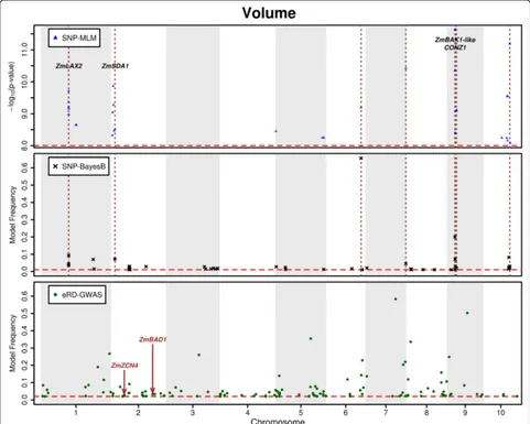
Substantial contribution of genetic variation in the expression of transcription factors to phenotypic variation revealed by eRD GWAS
Full text
Figure


Related documents
Figure 5: Photomicrographs of the wound area of the tongue of Non-diabetic subgroups (N1A, N1B, N1C, N2A, N2B, N2C, N3A, N3B, and N3C) and diabetic subgroups (D1A, D1B, D1C, D2A,
The secondary effectiveness end point of technical success was defined as ‘30-day freedom’ from: type I/III endoleak; loss of device integrity; graft infection, occlusion (defined
Treatment of acquired bilateral nevus of Ota-like macules (Hori's nevus) using a combination of scanned carbon dioxide laser followed by Q-switched ruby laser. Kunachak
In this work, the 3D structure of the genome of a human Jurkat cell is characterized through a graph theoretic approach, by using spectral clustering methods, of the Hi-C
The savings generate saving points and the loans consume saving points but, as we have more savings than loans, the members as a whole have a positive saving points balance and,
(2011) performed a detailed examination of frontal lobe morphology in children ages 8 –12 years, reporting reduced gray matter volume in the left lateral premotor cortex in girls