c
o
m
m
e
n
t
re
v
ie
w
s
re
ports
deposited research
interactions
information
refereed research
Minireview
The function and regulation of vasa-like genes in germ-cell
development
Erez Raz
Address: Department for Developmental Biology, Institute for Biology I, Freiburg University, 79104 Freiburg, Germany. E-mail: [email protected]
Abstract
The vasagene, essential for germ-cell development, was originally identified in Drosophila, and has since been found in other invertebrates and vertebrates. Analysis of these vasa homologs has revealed a highly conserved role for Vasa protein among different organisms, as well as some important differences in its regulation.
Published: 1 September 2000
GenomeBiology2000, 1(3):reviews1017.1–1017.6
The electronic version of this article is the complete one and can be found online at http://genomebiology.com/2000/1/3/reviews/1017 © GenomeBiology.com (Print ISSN 1465-6906; Online ISSN 1465-6914)
Germ-cell development in vertebrates and
invertebrates
In sexually reproducing organisms, primordial germ cells (PGCs) give rise to gametes that are responsible for the development of a new organism in the next generation. These cells must remain totipotent - able to differentiate into each and every cell type of all the different organs. In many organisms, maintenance of totipotency is achieved by the specification of germ cells early in embryogenesis: a small group of cells is set aside to follow a unique pathway of dif-ferentiation into gametes (reviewed in [1-3]).
Information on the specification of PGCs has been gained from detailed microscopical analysis, embryological experi-ments (for example transplantation of cells or cytoplasm) and gene identification through genetic screens for maternal-effect mutations in Drosophila and Caenorhabditis elegans
(reviewed in [1-4]). The main conclusion from these two inver-tebrate model organisms is that asymmetrical localization of cytoplasmic determinants - the germ plasm - is responsible for the early specification of the germline lineage. The importance of localized cytoplasmic determinants for germ-cell develop-ment has been most clearly shown in Drosophila. Here, for example, cytoplasmic germ plasm determinants concentrated at the posterior pole of the embryo in pole plasm can direct cells towards a germ-cell fate when transplanted to an ectopic
location. In mutants in which the formation of this morpho-logically characteristic cytoplasm is disrupted, germ-cell for-mation is impaired (reviewed in [5]). The pole plasm is characterized by the presence of the polar granules, electron-dense structures not delimited by a membrane that contain many RNAs and proteins and that are associated with mito-chondria. The distribution of the pole plasm correlates with the site of PGC formation. On the basis of their unique mor-phology, germ plasm components have also been identified in other organisms such as C. elegans (where they are termed P granules), Xenopus laevis (germinal granules), chick and zebrafish [1-3,6-9].
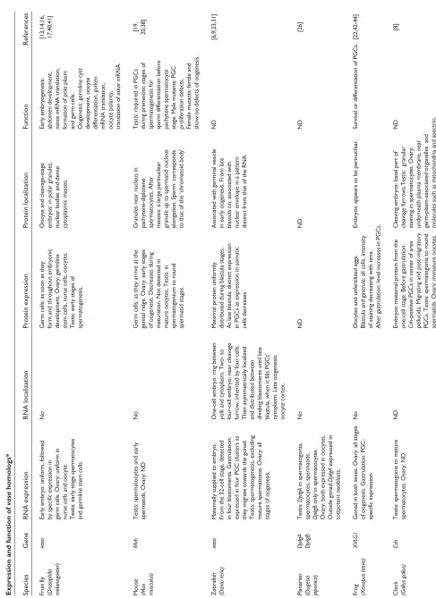
Table 1 Expression and function of
vasa
homologs*
Species
Gene
RNA expression
RNA localization
Protein expression
Protein localization
Function
References
Fruit fly
vasa
Early embryo: uniform, followed
No
Germ cells: as soon as they
Oocyte and cleavage-stage
Early embryogenesis:
[13,14,16,
(
Drosophila
by specific expression in
form and throughout embryonic
embryos: in polar granules,
abdomen development,
17,40,41]
melanogaster
)
germ cells. Ovary: uniform in
development. Ovary: germline
nuclear bodies and dense
nanos
mRNA translation,
nurse cells and oocyte.
stem cells, nurse cells, oocytes.
cytoplasmic masses.
formation of pole plasm
Testis: early stage spermatocytes
Testis: early stages of
and germ cells.
and germline stem cells.
spermatogenesis.
Oogenesis: germline cyst development, oocyte differentiation,
gurken
mRNA translation, oocyte polarity, translation of
oskar
mRNA.
Mouse
Mvh
Testis: spermatocytes and early
No
Germ cells: as they arrive at the
Granules near nucleus in
Testis: required in PGCs
[19,
(Mus
spermatids. Ovary: ND
genital ridge. Ovary: early stages
pachytene-diplotene
during premeiotic stages of
20,38]
musculus)
of oogenesis. Decreases during
spermatocytes. After
spermatogenesis for
maturation. Not detected in
meiosis: a large perinuclear
sperm differentiation before
mature oocytes. Testis: in
granule up to spermatid nucleus
pachytene spermatocyte
spermatogonium to round
elongation. Sperm: corresponds
stage. Male mutants: PGC
spermatid stages.
to that of the ‘chromatoid body’.
proliferation defects. Female mutants: fertile and show no defects of oogenesis.
Zebrafish
vasa
Maternally supplied to embryo.
One-cell embryo: ring between
Maternal protein uniformly
Associated with germinal vesicle
ND
[6,9
,23,31]
(Danio rerio)
From the 32-cell stage, detected
yolk and cytoplasm. Two- to
distributed during blastula stages.
in early oogenesis. From late
in four blastomeres. Gastrulation:
four-cell embryo: near cleavage
At late blastula, distinct expression
blastula on, associate
d with
expressed in four PGC clusters as
furrow, inherited by four cells.
in PGCs as expression in somatic
nuclear envelope in a patte
rn
they migrate towards the gonad.
Then asymmetrically localized
cells decreases.
distinct from that of the RNA
Testis: spermatogenesis, excluding
and distributed between
mature spermatozoa. Ovary: all
dividing blastomeres until late
stages of oogenesis.
blastula, when it fills PGCs’ cytoplasm. Late oogenesis: oocyte cortex.
Planarian
DjvlgA
Testis:
DjvlgA
in spermatogonia,
No
ND
ND
ND
[26]
(
Dugesia DjvlgB
spermatocytes, spermatids.
japonica
)
DjvlgB
only in spermatocytes.
Ovary: both expressed in oocytes. Outside gonad
DjvlgA
expressed in
totipotent neoblasts.
Frog
XVLG1
Gonad in both sexes. Ovary: all stages
No
Oocytes and unfertilized eggs.
Embryos: appears to be perinuclear.
Survival or differe
ntiation of PGCs.
[22,42-44]
(Xenopus laevis)
of oogenesis. Gastrulation:
PGC-Blastula and gastrula: all cells, intensity
specific expression.
of staining decreasing with time. After gastrulation: level increases in PGCs.
Chick
Cvh
Testis: spermatogonia to mature
ND
Embryos: maternal protein from the
Cleaving embryos: basal part of
ND
[8]
(Gallus gallus)
spermatocytes. Ovary: ND
one-cell stage. Before gastrulation:
cleavage furrows.Testis: granular
Cvh
-positive PGCs in center of area
staining in spermatocytes. Ovary:
pellucida. Migrating and post-migratory
underneath plasma membrane, near
PGCs. Testis: spermatogonia to round
germ-plasm-associated organelles and
spermatids. Ovary: immature oocytes.
comment
reviews
reports
deposited research
interactions
information
refereed research
showed that, at these stages, these cells are not yet committed to the germline, and when grafted into distal positions they can develop into somatic tissues [1,2]. Consistent with the notion of germ-cell induction through cell-cell interactions, distal cells that would develop as somatic cells can develop into germ cells when grafted into the region where germ cells normally form [1,2]. Indeed, formation of the founding popu-lation of PGCs in the mouse was shown to depend on the function of at least one extracellular factor - bone mor-phogentic protein 4 (Bmp4) [3].
Some of the methodologies used in the invertebrate models cannot be applied to vertebrates. In particular, the maternal-effect screens that were instrumental in analyzing the speci-fication of PGCs in invertebrates are not practicable in the frog, chick or mouse. Fortunately, such screens can be carried out in the zebrafish [11], although in this system too, it would be very difficult to achieve saturation for all mater-nal-effect mutations involved in PGC specification using classical forward genetic analysis.
Homologs of
vasa
in invertebrates and
vertebrates
The vasa gene was originally identified in Drosophila as a maternal-effect gene required for the formation of the abdominal segments and for germ-cell specification [12]. The Vasa protein can be detected in the germline cells of
Drosophila throughout their development and in early
embryos it is specifically localized to polar granules, which are located where the germ cells are specified. Drosophila
embryos that inherit mutant maternal vasa RNA and protein fail to form germ cells, and females carrying null mutations in
vasa display a range of defects in oogenesis. The vasagene
encodes an ATP-dependent RNA helicase of the DEAD-box family and is required for promoting translation of at least two known mRNAs, nanosand gurken[13-19].
Following the isolation of the Drosophila vasagene, vasa-like DEAD-box RNA helicase genes that are expressed in germ cells were identified in many species, including mouse, rat, frog, zebrafish, medaka (Oryzias latipes), trout, planarian, chick, ascidian, nematode, silkworm, human and the flour beetle (Table 1; [8,20-30], and R. Schröder and D. Tautz, per-sonal communication). The distribution of vasa RNA or protein was determined during different stages of develop-ment, thus providing information on the possible function of
vasaduring germ-cell development in these species. The vasa
loss-of-function phenotype in the fly, the mouse and the nematode provided direct evidence for the role of vasain the development of germ cells in these organisms. And in cases where the origin and precise route of germ-cell migration towards the gonad were unknown (for example in fish, chick and ascidians), it proved possible, using vasaas a molecular marker, to trace back the migration path and establish the position in which these cells originate [8,23,27,31].
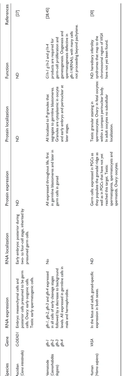
Table 1 (continued) Species
Gene
RNA expression
RNA localization
Protein expression
Protein localization
Function
References
Ascidian
Ci-DEAD1
Embryo: mesenchymal cells and
Early embryos: posterior during
ND
ND
ND
[27]
(
Ciona intestinalis
)
posterior cells presumed to be germ
two- to four-cell stage, inherited by
cells. Ovary: early oogonic cells.
presumed germ cells.
Testis: early spermatogenic cells.
Nematode
glh-1 glh-1
,
glh-2, glh-3
and
glh-4
expressed
No
All expressed throughout life, first
All localized to P-granules that
G1h-1 g1h-2
and
g1h-4
[28,45]
(
Caenorhabditis glh-2
in all cells of early cleavage stages
in germline blastomeres and later in
segregate to germline blastomeres.
products are requ
ired for
elegans
)
glh-3
followed by a decrease to background
germ cells in gonad
Granules are cytoplasmic in oocyte
germ-cell proliferation and
glh-4
levels. All expressed in germline cells in
and early embryo and perinuclear at
gametogenesis. Oogenesis and
male and hermaphrodite.
later stages.
spermatogenesis defective in glh-1/4
(RNAi), with many cells
not proceeding beyond pachytene.
Human
VASA
In the fetus and adult, gonad-specific
ND
Germ cells: expressed in PGCs as
Testis: granular staining in
ND: hereditary infertili
ty [30]
(
Homo sapiens
)
expression in both sexes.
they populate the gonadal ridge as
spermatocytes. Ovary: in fetal oocytes:
syndromes that map to the
well as in PGCs that have not yet
within a compact perinuclear body.
chromosomal region of
VASA
reached this target. Testis:
In adult oocytes no subcellular
have not yet been found.
spermatogonia, spermatocysts and
localization.
spermatids. Ovary: oocytes.
*
Not all the known
vasa
homologs are listed in the table. Homologs for which little functional information is available have been omitted unless they s
[image:3.609.63.278.82.740.2]Regulation of expression and subcellular
localization of
vasa-
like gene products
As can be seen from Table 1, vasaRNA is expressed in the germ cells of many organisms. But translational control, post-translational control and subcellular localization of the Vasa protein, as well as interaction with other proteins, appear to play a major part in controlling Vasa function. For example,
vasa RNA is uniformly distributed in early Drosophila
embryos but, consistent with its function, the protein is found localized to the posterior pole, where it is associated with the polar granules in the germ plasm. Similar discrepancies between RNA and protein expression, in which the Vasa protein is found in a restricted number of cells relative to the RNA and is localized to specific subcellular structures, have also been described in Xenopusand the nematode.
A recent thorough analysis of the distribution of the vasa
gene products in zebrafish revealed a unique and surprising difference between the localization of RNA and that of the protein [9]. The RNA is localized through a microtubule-dependent process in a novel pattern to the first and second cleavage planes of the early zebrafish embryo [23,32]. Knaut
et al. [9] also showed that the vasaRNA aggregates reside
within an electron-dense matrix similar to structures associ-ated with germ plasm in other organisms. The localization of
vasaRNA to the germ plasm allowed them to follow pre-cisely the distribution of the germ plasm to the cells of the early embryo [9]. Until late blastula stages, the four cells that contain germ plasm divide asymmetrically, so that only one of the blastomeres resulting from each division inherits the germ plasm labeled by vasa RNA; these will become the future PGCs. Towards the end of the blastula stage, the RNA fills the cytoplasm of the vasa-positive cells and is inherited by both daughter cells after cell division. The distribution of the Vasa protein at these early stages of development dif-fered from that of the RNA in a striking and unexpected manner. When the vasaRNA is asymmetrically segregating to the future PGCs, the Vasa protein is uniformly distributed in the cytoplasm of all blastomeres. This initial pattern of protein expression changes at late blastula stages, when stronger protein expression is detected in the germ cells, which now divide symmetrically. At this time, the distribu-tion of the protein within the cells becomes perinuclear.
Given that Vasa protein is implicated in establishing func-tional germ plasm and in the specification of germ cells in other organisms, the findings in zebrafish seem paradoxical.
In Drosophila, for example, loss-of-function mutations in
vasa that affect the formation of the pole plasm were shown
to affect either the biochemical activity of the protein or its localization [19]. In zebrafish, on the other hand, the vasa
RNA, rather than the protein, is initially localized to the germ plasm. There are several possible explanations for this paradox. One is that in zebrafish it is the vasa RNA and not the protein that is important for the early determination of germ plasm. It is important to note, however, that in
zebrafish, vasaRNA expressed ectopically during embryoge-nesis is unable by itself to alter the number or position of the PGCs [31]. Furthermore, cloning of the medaka vasa
homolog and analysis of its expression pattern showed that in this fish, which like the zebrafish is also a teleost, the RNA is uniformly expressed until gastrulation, when germ-cell-specific expression is observed [24].
Another possible explanation of the paradox is that although most of the Vasa protein is uniformly distributed in all cells, it is active only in the germ cells, as a result of cell-specific post-translational modifications. Vasa activity can indeed be affected by post-translational modification, as described during oogenesis in Drosophila[33]. Finally, there could be a higher concentration of Vasa protein in germ cells as a result of translation of the maternally localized RNA. In this sce-nario, localization of vasaRNA, and presumably other RNAs, important for germ-cell determination, generates high levels of these proteins in some cells, thereby inducing the zygotic PGC differentiation path. An interesting recent discovery was recently made by Schröder and Tautz, who cloned a vasa-like gene from the short germ band beetle, whose embryogenesis represents a more ancestral form of embryogenesis in insects.
Tribolium castaneum (R. Schröder and D. Tautz, personal
communication). They found that the distribution of vasa
RNA in this insect is more reminiscent of that in zebrafish rather than the long germ band Drosophila. After early uniform distribution of the RNA, the Tribolium vasa RNA appears to be located exclusively at the posterior of the early embryo, where the germ cells presumably form.
The function of
vasa
in germ-cell development
The function of the vasa gene can be inferred from its expression pattern in different organisms and from pheno-typic analysis of animals lacking a functional gene. With the exception of the mouse (and probably other mammals), the
vasagene product is expressed in or localized to the PGCs very early in development, consistent with the idea that its activity is required for specification of this cell lineage. Inter-estingly, in planarians, where a vasa homolog is also expressed in the soma, the somatic cells that express the gene were identified as neoblasts - a totipotent cell type that functions in regeneration. The function of vasacould there-fore be described as important for preserving totipotency. One mechanism for preserving totipotency is to inhibit expression of genes that would lead to somatic differentia-tion [34]. An indirect role for vasa in transcriptional inhibi-tion is suggested by the finding that one of the few known targets of Vasa, nanos, can repress gene expression in the
Drosophila germline [15,35,36].
arrive at the gonad, and expression is induced by interaction between the germ cells and the somatic cells of the develop-ing gonad [37]. The expression of vasaat this stage in the mouse, as well as in all the other organisms described above, is likely to reflect a requirement for the gene product for dif-ferentiation of the germ cells into gametes. Indeed, loss of
vasa function in the mouse affects differentiation of the male germ cells, resulting in male sterility (no other pheno-type is observed in the knockout mice) [38]. Similarly, a late function of vasaduring gametogenesis has been described in the nematode and the fly [16,17,28,33]. The first mechanistic evidence coupling progression in gamete differentiation and
vasafunction is the demonstration that a meiotic checkpoint during oogenesis in Drosophila appears to control the activ-ity of the Vasa protein [33].
The search for new factors involved in germ-cell
development
To gain a more comprehensive understanding of germline development in different species, one would obviously seek to identify most or all of the components relevant to the process and determine the functional relationships between them. Genome sequencing and the availability of expressed sequence tag (EST) libraries now allow us to identify homologs through database screens of different organisms. Isolation of new genes essential for germ-cell development is more demanding. Classical genetic analyses in invertebrates and in zebrafish are likely to identify new genes and proteins. This approach will fail, however, where there is functional redundancy among genes or when a zygotic requirement for the gene complicates the analysis of its function as a maternal factor. By using DNA microarray chips and tissue-specific probes, on the other hand, one could identify genes that are specifically expressed in the germ cells. The yeast two-hybrid system can be used to reveal new proteins that physically interact with previously identified gene products (see [19]). In addition, screening cDNA libraries by in situhybridization can identify new genes even if they are expressed in other cell types as well as in germ cells. This approach has led to identi-fication of many homologs of known genes in the zebrafish, as well as of new genes and proteins that are expressed in the germline (C. Thisse, B. Thisse and E.R., unpublished observa-tions). Functional analysis of zebrafish genes isolated by such reverse genetics approaches can be carried out using mor-pholino antisense oligonucleotides [39], which inhibit trans-lation of their specific mRNAs in this organism (S. Ekker, personal communication).
The approaches described above are most useful in model organisms for which investment in genomic resources has provided the necessary tools. Nevertheless, other species such as planarian, flour beetle, silkworm and medaka display interesting parallels as well as important differences in the way the vasa gene is expressed and regulated. Contin-uing and expanding the work in different model systems is
likely to contribute to our understanding of the molecular mechanisms of specification and differentiation of the germ cells across the animal kingdom.
Acknowledgements
I thank Randy Cassada, Holger Knaut, Michal Reichman and Gilbert Wei-dinger for comments on the manuscript. The work in my lab is supported by grants from the Deutsche Forschungsgemeinschaft (DFG).
References
1. Saffman E, Lasko P: Germline development in vertebrates and invertebrates.Cell Mol Life Sci1999, 55:1141-1163.
2. Wylie C: Germ cells.Cell1999, 96:165-174.
3. Wylie C: Germ cells.Curr Opin Genet Dev2000, 10:410-413. 4. Williamson A, Lehmann R: Germ cell development in
Drosophila. Annu Rev Cell Dev Biol1996, 12:365-391.
5. Rongo C, Lehmann R: Regulated synthesis, transport and assembly of the Drosophilagerm plasm.Trends Genet1996, 12: 102-109.
6. Braat A, Zandbergen T, van de Water S, Goos H, Zivkovic D: Char-acterization of zebrafish primordial germ cells: morphology and early distribution of vasa RNA.Dev Dyn1999, 216:153-167. 7. Pitt J, Schisa J, Priess J: P granules in the germ cells of
Caenorhabditis elegansadults are associated with clusters of
nuclear pores and contain RNA.Dev Biol2000, 219:315-333. 8. Tsunekawa N, Naito M, Sakai Y, Nishida T, Noce T: Isolation of
chicken vasa homolog gene and tracing the origin of pri-mordial germ cells.Development2000, 127:2741-2750.
9. Knaut H, Pelegri F, Bohmann K, Schwarz H, Nusslein-Volhard C: Zebrafish vasa RNA but not its protein is a component of the germ plasm and segregates asymmetrically before germline specification.J Cell Biol2000, 149:875-888.
10. Zernicka-Goetz M: Fertile offspring derived from mammalian eggs lacking either animal or vegetal poles.Development1998, 125:4803-4808.
11. Pelegri F, Schulte-Merker S: A gynogenesis-based screen for maternal-effect genes in the zebrafish, Danio rerio.Methods
Cell Biol1999, 60:1-20.
12. Schupbach T, Wieschaus E: Maternal-effect mutations altering the anterior-posterior pattern of the Drosophila embryo.
Roux’s Arch Dev. Biol1986, 195:302-317.
13. Hay B, Jan L, Jan Y: A protein component of Drosophilapolar granules is encoded by vasa and has extensive sequence similarity to ATP-dependent helicases.Cell1988, 55:577-587. 14. Lasko P, Ashburner M: The product of the Drosophila gene
vasais very similar to eukaryotic initiation factor-4A.Nature 1988, 335:611-617.
15. Gavis E, Lunsford L, Bergsten S, Lehmann R: A conserved 90 nucleotide element mediates translational repression of nanos RNA.Development1996, 122:2791-2800.
16. Styhler S, Nakamura A, Swan A, Suter B, Lasko P: vasa is required for GURKEN accumulation in the oocyte, and is involved in oocyte differentiation and germline cyst development.
Devel-opment1998, 125:1569-1578.
17. Tomancak P, Guichet A, Zavorszky P, Ephrussi A: Oocyte polarity depends on regulation of gurken by Vasa.Development1998, 125:1723-1732.
18. Liang L, Diehl-Jones W, Lasko P: Localization of vasa protein to the Drosophilapole plasm is independent of its RNA-binding and helicase activities.Development1994, 120:1201-1211. 19. Carrera P, Johnstone O, Nakamura A, Casanova J, Jackle H, Lasko P:
VASA mediates translation through interaction with a Drosophila yIF2 homolog.Mol Cell2000, 5:181-187.
20. Fujiwara Y, Komiya T, Kawabata H, Sato M, Fujimoto H, Furusawa M, Noce T: Isolation of a DEAD-family protein gene that encodes a murine homolog of Drosophila vasaand its spe-cific expression in germ cell lineage. Proc Natl Acad Sci USA 1994, 91:12258-12262.
21. Komiya T, Tanigawa Y: Cloning of a gene of the DEAD box protein family which is specifically expressed in germ cells in rats.Biochem Biophys Res Commun1995, 207:405-410.
comment
reviews
reports
deposited research
interactions
information
22. Komiya T, Itoh K, Ikenishi K, Furusawa M: Isolation and charac-terization of a novel gene of the DEAD box protein family which is specifically expressed in germ cells of Xenopus
laevis.Dev Biol1994, 162:354-563.
23. Yoon C, Kawakami K, Hopkins N: Zebrafish vasa homologue RNA is localized to the cleavage planes of 2- and 4-cell-stage embryos and is expressed in the primordial germ cells.
Development1997, 124:3157-3165.
24. Shinomiya A, Tanaka M, Kobayashi T, Nagahama Y, Hamaguchi S:
The vasa-like gene, olvas, identifies the migration path of
primordial germ cells during embryonic body formation stage in the medaka, Oryzias latipes. Dev Growth Differ2000, 42: 317-326.
25. Yoshizaki G, Sakatani S, Tominaga H, Takeuchi T: Cloning and characterization of a vasa-like gene in rainbow trout and its expression in the germ cell lineage.Mol Reprod Dev 2000, 55: 364-371
26. Shibata N, Umesono Y, Orii H, Sakurai T, Watanabe K, Agata K: Expression of vasa(vas)-related genes in germline cells and totipotent somatic stem cells of planarians. Dev Biol 1999, 206:73-87.
27. Fujimura M, Takamura K: Characterization of an ascidian DEAD-box gene, Ci-DEAD1: specific expression in the germ cells and its mRNA localization in the posterior-most blas-tomeres in early embryos.Dev Genes Evol2000, 210:64-72. 28. Kuznicki K, Smith P, Leung-Chiu W, Estevez A, Scott H, KL B:
Com-binatorial RNA interference indicates GLH-4 can compen-sate for GLH-1; these two P granule components are critical for fertility in C. elegans. Development2000, 127: 2907-2916.
29. Nakao H: Isolation and characterization of a Bombyx vasa-like protein. Dev Genes Evol1999, 209:312-316.
30. Castrillon DH, Quade BJ, Wang TY, Quigley C, Crum CP: The human VASA gene is specifically expressed in the germ cell lineage.Proc Natl Acad Sci USA 2000, in press.
31. Weidinger G, Wolke U, Koprunner M, Klinger M, Raz E: Identifica-tion of tissues and patterning events required for distinct steps in early migration of zebrafish primordial germ cells.
Development1999, 126:5295-5307.
32. Pelegri F, Knaut H, Maischein H, Schulte-Merker S, Nusslein-Volhard C: A mutation in the zebrafish maternal-effect gene nebel affects furrow formation and vasaRNA localization.Curr Biol 1999, 9:1431-1440.
33. Ghabrial A, Schupbach T: Activation of a meiotic checkpoint regulates translation of Gurken during Drosophila
oogene-sis.Nat Cell Biol1999, 1:354-357.
34. Seydoux G, Strome S: Launching the germline in Caenorhabdi-tis elegans: regulation of gene expression in early germ cells.
Development1999, 126:3275-3283.
35. Deshpande G, Calhoun G, Yanowitz J, Schedl P: Novel functions of nanos in downregulating mitosis and transcription during the development of the Drosophilagermline. Cell 1999, 99: 271-281.
36. Parisi M, Lin H: Translational repression: a duet of Nanos and Pumilio.Curr Biol2000, 10:R81-R83.
37. Toyooka Y, Tsunekawa N, Takahashi Y, Matsui Y, Satoh M, Noce T: Expression and intracellular localization of mouse vasa-homologue protein during germ cell development.Mech Dev 2000, 93:139-149.
38. Tanaka S, Toyooka Y, Akasu R, Katoh-Fukui Y, Nakahara Y, Suzuki R, Yokoyama M, Noce T: The mouse homolog of Drosophila Vasa is required for the development of male germ cells.
Genes Dev2000, 14:841-853.
39. Summerton J, Weller D: Morpholino antisense oligomers: design, preparation, and properties.Antisense Nucleic Acid Drug Dev1997, 7:187-195.
40. Hay B, Ackerman L, Barbel S, Jan L, Jan Y: Identification of a com-ponent of Drosophilapolar granules. Development1988, 103: 625-640.
41. Markussen F, Michon A, Breitwieser W, Ephrussi A: Translational control of oskar generates short OSK, the isoform that induces pole plasma assembly. Development 1995, 121: 3723-3732.
42. Ikenishi K, Tanaka T, Komiya T: Spatio-temporal distribution of the protein of the Xenopus vasahomologue (Xenopus vasa-like gene 1, XVLG1) in embryos. Dev Growth Differ1996, 38: 527-535.
43. Ikenishi K, Tanaka T: Involvement of the protein of Xenopus vasahomolog (Xenopus vasa-like gene 1, XVLG1) in the dif-ferentiation of primordial germ cells. Dev Growth Differ1997, 39:625-633.
44. Ikenishi K, Tanaka T: Spatio-temporal expression of Xenopus vasa homolog, XVLG1, in oocytes and embryos: the pres-ence of XVLG1 RNA in somatic cells as well as germline cells.Dev Growth Differ2000, 42:95-103.
45. Gruidl M, Smith P, Kuznicki K, McCrone J, Kirchner J, Roussell D, et al.: Multiple potential germ-line helicases are components of the germ-line-specific P granules of Caenorhabditis elegans.