www.impactjournals.com/oncotarget/
Oncotarget, May, Vol.2, No 5
c-JUN prevents methylation of p16
INK4a(and Cdk6): the villain
turned bodyguard
Karoline Kollmann
1, Gerwin Heller
2, Veronika Sexl
11 Institute of Pharmacology and Toxicology, University of Veterinary Medicine, Vienna, Austria
2 Clinical Division of Oncology, Department of Medicine I, Comprehensive Cancer, Medical University of Vienna (MUV), Vienna, Austria
Correspondence to: Karoline Kollmann, email: Karoline.Kollmann@vetmeduni.ac.at
Keywords: AP-1, CDK6, p16, leukemia
Received: May 27, 2011, Accepted: May 28, 2011, Published: May 28, 2011
Copyright: © Kollmann et al. This is an open-access article distributed under the terms of the Creative Commons Attribution License, which permits unrestricted use, distribution, and reproduction in any medium, provided the original author and source are credited.
AbstrAct:
A novel way by which the AP-1 factor c-JUN interferes with tumorigenesis has recently been elucidated [1]. In a model of murine leukemia, c-JUN prevents the epigenetic silencing of the cell cycle kinase CDK6. In the absence of c-JUN, CDK6 is down-regulated and the 5’region of the gene is methylated. Down-regulation
of CDK6 results in significantly delayed leukemia formation. Here we show that
c-JUN is also involved in protecting the promoter region of the tumor suppressor p16INK4a, which is consistently methylated over time in c-JUN deficient cells. In cells
expressing c-JUN, p16INK4a promoter methylation is a less frequent event. Our study
unravels a novel mechanism by which the AP-1 factor c-JUN acts as a “bodyguard”, and preventing methylation of a distinct set of genes after oncogenic transformation.
the AP-1 trAnscriPtion fActor
fAmily – tumor suPPressors And
Promoters
AP-1 (Activator Protein-1) transcription factors are
basic leucine-zipper (bZIP) proteins [2-5]. They act as
dimeric transcription factors composed of members of the
JUN family (c-JUN, JUNB, JUND), forming homo- or
heterodimers with members of the FOS, ATF (activating
transcription factor) and MAF (musculoaponeurotic
fibrosarcoma) protein family [6-7]. The expression and
physiological role of the diverse AP-1 complexes is cell
type- and even differentiation state-dependent [8-10].
AP-1 complexes modulate transcription of target genes by
binding to their TRE or CRE consensus elements
[3],[11-13],[8, 12, 14]. Consequently AP-1 dimers are involved
in the regulation of various biological processes such as
proliferation, differentiation and apoptosis [3, 15]. It is
therefore not surprising that AP-1 factors are involved in
cellular transformation and tumorigenesis [3, 16-17].
c-JUN was first discovered as the homologue of the
viral oncoprotein v-JUN, which induces avian sarcoma
[18-19]. Early evidence revealed a cooperation of c-JUN
with oncogenic RAS in cellular transformation [4, 20-21].
On the other hand, some AP-1 family members suppress
and block tumor development [4, 22-23]. This task has
been previously been attributed to JUNB, inactivation of
which provokes the development of myeloproliferative
disorders [24].
Despite the similarity in their primary structure
and DNA binding specificity, c-JUN and JUNB differ
in transcriptional capacity. Both may act either as a
transcriptional activator or as a repressor depending on
the promoter context and the heterodimerization partner
[4, 25-31]. In some tissues c-JUN and JUNB even exert
antagonistic functions in biological processes such as cell
proliferation [4, 26-29, 31]. Thus AP-1 is able to modulate
opposing functions, such as promoting or suppressing
tumor development.
c-Jun - AccelerAting
tumorigenesis by driving
trAnscriPtion
Since the initial discovery of v-JUN, solid evidence
has accumulated linking AP-1 members and in particular
c-JUN to tumor development. Using mouse models,
c-JUN has been implicated in tumor formation in both
skin and liver [32-36]. Inhibiting c-JUN activity in basal
figure 1:
p16
INK4amrnA levels are down-regulated in
c-Jun
Δ/Δp185
BCR-ABL-transformed cell lines.
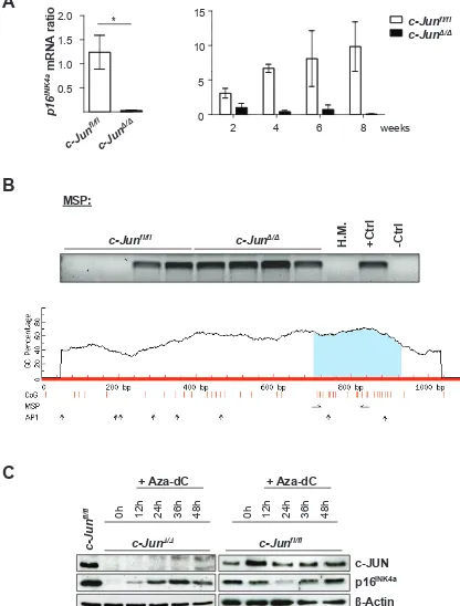
A) p16INK4a mRNA levelsof c-JunΔ/Δ and c-Junfl/fl cells 2, 4, 6 and 8 weeks after p185BCR-ABL transformation were analyzed by q-PCR. The fold change compared to c-JunΔ/Δ 2 weeks p16INK4a mRNA level is shown. Results were normalized by comparison to their Gapdh mRNA expression. B) upper
panel: Methylation-specific PCR analysis of p16INK4a in stable c-Junfl/fland c-JunΔ/Δ cell lines as detected by MSP analysis. A visible PCR
product indicates the presence of methylated alleles. Abbreviations: H.M., bone marrow of a healthy mouse; +Ctrl (control for methylated
samples); -Ctrl (control for unmethylated samples). Lower panel: Graphical overview of the CpG island associated with p16INK4a (Cdkn2a)
(ENSMUSG00000044303). The following genomic region is shown: NCBIM37:4:88927717:88928797:-1. Vertical bars (orange) indicate the location of CpG dinucleotides, horizontal arrows indicate MSP primer binding sites and vertical arrows indicate AP1 transcription factor binding sites predicted using the transcription factor binding profile database JASPAR (http://jaspar.genereg.net/). C) Immunoblot for c-JUN and p16INK4a of c-JunΔ/Δ and c-Junfl/fl cells after 12, 24, 36 and 48 hours of Aza-dC treatment. β-Actin served as loading control.
One representative set of data is depicted.
MSP:
Figure 1
A
c-Jun
fl/fl
c-Jun
Δ/Δ0h 12h 24h 36h 48h
+ Aza-dC
β
-Actin
p16
INK4ac-JUN
c-Jun
fl/fl0h 12h 24h 36h 48h
+ Aza-dC
B
p16
INK4
a
mRNA ratio
0.5 1.0 1.5 2.0c-J
un
fl/fl
c-J
un
Δ/Δ *C
0 5 10 15
c-Jun
fl/flc-Jun
Δ/Δ2 4 6 8 weeks
H.M.
+Ctrl
-Ctrl
c-Jun
fl/flc-Jun
Δ/ΔPOL II
C-JUN X Y
transcription
POL II X
Y DNMT
DNMT DNMT
DNMT DNMT
DNMT
formation because of the lack of expression of the AP-1
target genes [37]. In hepatocellular carcinoma, gene
deletion of
c-Jun
after tumor onset leads to a significant
reduction in tumor size. This effect has been explained
as resulting from increased apoptosis induced by c-JUN
dependent suppression of the pro-apoptotic gene p53 [36,
38]. Upon loss of c-JUN, p53 mediated apoptosis kicks in
and leads to a significant reduction of tumor burden [36].
Similarly, murine embryonic fibroblasts (MEFs) lacking
c-JUN show severe cell cycle abnormalities with a block
in the G1-phase of the cell cycle [39]. These fibroblasts
demonstrate increased expression of the pro-apoptotic
gene p53 and its target gene, the cell cycle inhibitor p21CIP
[38]. As in hepatocytes, c-JUN negatively regulates the
transcription of p53 in MEFs by direct binding to the
promoter region of p53. The concomitant deletion of
p53
rescues the apoptotic and proliferative disadvantage [38,
40].
Besides c-JUN’s “survival” function in interfering
with p53 induced apoptosis, c-JUN has been shown
to drive cell proliferation. One defined “hallmark” of
tumor formation is a deregulated cell cycle and c-JUN’s
ability to effect this represents a further way in which
the protein contributes to tumorigenesis. A direct
pro-proliferative function of c-JUN is mediated through the
regulation of CyclinD1 [38-39, 41]. c-JUN is able to bind
to the CyclinD1 promoter and induces transcription in a
phosphorylation-dependent manner, as the exchanges
of critical serine residues to alanine (
Jun
AA/AA) decrease
CyclinD1 transcription [30]. Both CyclinD1 and c-JUN
are overexpressed in a broad range of human cancer and
both molecules are considered proto-oncogenes [10,
42-45]. Another cell-cycle regulator under the control of AP-1
members is the cell cycle inhibitor p16
INK4a. Several AP-1
binding sites within the
p16
INK4apromoter enable AP-1 to
control expression of the gene. JUNB has been defined as
a positive regulator and induces p16
INK4aexpression [27,
46].
A novel mechAnism for c-Jun
An additional cell cycle component regulated by
c-JUN is the G1-cell cycle kinase CDK6. CDK6 is most
closely related to CDK4 and is required to allow cells
to progress through the G1-phase of the cell cycle after
binding to D-type Cyclins [47-50]. CDK6 and CDK4
generally have overlapping and redundant functions but
CDK6, unlike CDK4, has been recently recognized to be
a regulator of differentiation [51-54].
When we analyzed pro B-cells transformed by the
oncoprotein BCR-ABL we found a peculiar regulation
of CDK6 downstream of c-JUN [1]. Over time
BCR-ABL-transformed cells lacking c-JUN had lower levels of
CDK6 protein as well as mRNA. The decline in protein
expression was independent of c-JUN phosphorylation
as CDK6 was stably expressed in BCR-ABL
+Jun
AA/ AAcells, in which the mutant JUN protein can no longer
be phosphorylated. Interestingly, the reduction of CDK6
was accompanied by methylation of the 5’region of
Cdk6
and could be reversed by treatment of the cells with the
DNA methyltransferase (DNMT) inhibitor
5-aza-2’-deoxycytidine (Aza-dC). Two AP-1 binding sites are
found in the vicinity of the methylated region. This led
us to suggest that c-JUN exerts a “bodyguard” function,
being required to prevent the
Cdk6
promoter from being
silenced. Only in the absence of c-JUN can methylation
of the 5’region of
Cdk6
take place and CDK6 expression
be silenced. This effect is restricted to transformed cells
and induced by the oncogenic event, as primary
non-transformed lymphoid cells display unaltered CDK6
expression levels. It is attractive to speculate that c-Jun
may also shape age-related DNA methylation by acting
as a “bodyguard” to protect distinct DNA regions during
aging [55-57].
Altering CDK6 expression has major consequences
for leukemia and lymphoma development. Using
Cdk6
-/-animals we detected a significant delay in tumor
formation. Thus, it is safe to conclude that by preventing
the silencing of CDK6 c-JUN accelerates and promotes
leukemogenesis. In the same murine model of
BCR-ABL
+leukemia, JUNB exerts an antagonistic role. In
accordance with its role as tumor suppressor, the enforced
expression of JUNB suppresses tumor formation and the
lack of JUNB lead to a highly aggressive and rapidly
progressing disease. Again we found a role for CDK6; the
lack of JUNB was accompanied by high CDK6 expression
levels. How precisely JUNB interferes with the regulation
of CDK6 remains to be determined [46].
CDK6 is not the only cell cycle regulator under the
control of c-JUN. We found a consistent downregulation
of the tumor suppressor and cell cycle inhibitor p16
INK4ain
c-JUN deficient BCR-ABL+ cells [1]. The levels of both
the protein and the mRNA (Figure 1A) of p16
INK4adecline
with time after BCR-ABL-transformation, as verified by
micro-array experiments [1].
As shown for
Cdk6
, we found promoter methylation
of
p16
INK4ain all stable BCR-ABL+
c-Jun
Δ/Δcell lines. In
the case of wild type, only half the cell lines displayed
methylated CpG islands (Figure 1B). Treatment of the
cells with Aza-dC reverted the reduction of p16
INK4ain
cells lacking c-JUN (Figure 1C). Interestingly, no changes
could be induced by treating wild type cells with this
agent, indicating that additional regulatory mechanisms
had shut off p16
INK4ain the tumor cell lines. As is the case
for
Cdk6
, the promoter region of the
p16
INK4agene contains
a number of AP1-binding sites, which is consistent with
our model, that c-JUN is required to protect the
p16
INK4apromoter from being silenced by methylation.
conclusion
c-JUN is a common regulator of cell cycle
components that has been described to have a direct
role in regulating the transcription of p53 and CyclinD1.
Based on our studies we postulate a novel mechanism
for how c-JUN accelerates leukemogenesis and regulates
genes required for cell cycle progression in tumor cells.
We propose that binding of c-JUN to the promoter region
exerts a protective function and prevents methylation and
silencing of the genes. This function of c-JUN has recently
been demonstrated for the cell cycle kinase CDK6. In the
absence of c-JUN, CDK6 is downregulated accompanied
by methylation of the 5’region. We now show that the
same mechanism occurs at the promoter region of the
cell cycle inhibitor and tumor suppressor
p16
INK4a. Our
results indicate a novel mechanism by which AP-1 factors
modulate tumor formation.
Acknowledgments
This work was supported by the Vienna Science and
Technology Fund (WWTF, project number LS07-019)
and by the Austrian Science Foundation (grants P19723).
references
1. Kollmann K, Heller G, Ott RG, Scheicher R, Zebedin-Brandl E, Schneckenleithner C, Simma O, Warsch W, Eckelhart E, Hoelbl A, Bilban M, Zochbauer-Muller S, Malumbres M, Sexl V. c-JUN promotes BCR-ABL-induced lymphoid leukemia by inhibiting methylation of the 5’ region of Cdk6. Blood. 2011; 117:4065-4075. 2. Angel P, Szabowski A, Schorpp-Kistner M. Function
and regulation of AP-1 subunits in skin physiology and pathology. Oncogene. 2001; 20:2413-2423.
3. Angel P, Karin M. The role of Jun, Fos and the AP-1 complex in cell-proliferation and transformation. Biochim Biophys Acta. 1991; 1072:129-157.
4. Eferl R, Wagner EF. AP-1: a double-edged sword in tumorigenesis. Nat Rev Cancer. 2003; 3:859-868.
5. Jochum W, Passegue E, Wagner EF. AP-1 in mouse development and tumorigenesis. Oncogene. 2001; 20:2401-2412.
6. Chinenov Y, Kerppola TK. Close encounters of many kinds: Fos-Jun interactions that mediate transcription regulatory specificity. Oncogene. 2001; 20:2438-2452. 7. Hai T, Curran T. Cross-family dimerization of transcription
factors Fos/Jun and ATF/CREB alters DNA binding specificity. Proc Natl Acad Sci U S A. 1991; 88:3720-3724. 8. Karin M, Liu Z, Zandi E. AP-1 function and regulation.
Curr Opin Cell Biol. 1997; 9:240-246.
9. Schorpp-Kistner M, Wang ZQ, Angel P, Wagner EF. JunB is essential for mammalian placentation. EMBO J. 1999; 18:934-948.
10. Eferl R, Sibilia M, Hilberg F, Fuchsbichler A, Kufferath I, Guertl B, Zenz R, Wagner EF, Zatloukal K. Functions of c-Jun in liver and heart development. J Cell Biol. 1999; 145:1049-1061.
11. Angel P, Imagawa M, Chiu R, Stein B, Imbra RJ, Rahmsdorf HJ, Jonat C, Herrlich P, Karin M. Phorbol ester-inducible genes contain a common cis element recognized by a TPA-modulated trans-acting factor. Cell. 1987; 49:729-739. 12. Halazonetis TD, Georgopoulos K, Greenberg ME, Leder
P. c-Jun dimerizes with itself and with c-Fos, forming complexes of different DNA binding affinities. Cell. 1988; 55:917-924.
AP-1 interacts with TPA-inducible enhancer elements. Cell. 1987; 49:741-752.
15. Shaulian E, Karin M. AP-1 as a regulator of cell life and death. Nat Cell Biol. 2002; 4:E131-136.
16. Suzuki T, Hashimoto Y, Okuno H, Sato H, Nishina H, Iba H. High-level expression of human c-jun gene causes cellular transformation of chicken embryo fibroblasts. Jpn J Cancer Res. 1991; 82:58-64.
17. Raitano AB, Halpern JR, Hambuch TM, Sawyers CL. The Bcr-Abl leukemia oncogene activates Jun kinase and requires Jun for transformation. Proc Natl Acad Sci U S A. 1995; 92:11746-11750.
18. Bos TJ, Bohmann D, Tsuchie H, Tjian R, Vogt PK. v-jun encodes a nuclear protein with enhancer binding properties of AP-1. Cell. 1988; 52:705-712.
19. Maki Y, Bos TJ, Davis C, Starbuck M, Vogt PK. Avian sarcoma virus 17 carries the jun oncogene. Proc Natl Acad Sci U S A. 1987; 84:2848-2852.
20. Johnson R, Spiegelman B, Hanahan D, Wisdom R. Cellular transformation and malignancy induced by ras require c-jun. Mol Cell Biol. 1996; 16:4504-4511.
21. Smeal T, Binetruy B, Mercola DA, Birrer M, Karin M. Oncogenic and transcriptional cooperation with Ha-Ras requires phosphorylation of c-Jun on serines 63 and 73. Nature. 1991; 354:494-496.
22. Yang MY, Liu TC, Chang JG, Lin PM, Lin SF. JunB gene expression is inactivated by methylation in chronic myeloid leukemia. Blood. 2003; 101:3205-3211.
23. Shaulian E, Karin M. AP-1 in cell proliferation and survival. Oncogene. 2001; 20:2390-2400.
24. Passegue E, Wagner EF, Weissman IL. JunB deficiency leads to a myeloproliferative disorder arising from hematopoietic stem cells. Cell. 2004; 119:431-443.
25. Szabowski A, Maas-Szabowski N, Andrecht S, Kolbus A, Schorpp-Kistner M, Fusenig NE, Angel P. c-Jun and JunB antagonistically control cytokine-regulated mesenchymal-epidermal interaction in skin. Cell. 2000; 103:745-755. 26. Behrens A, Sibilia M, Wagner EF. Amino-terminal
phosphorylation of c-Jun regulates stress-induced apoptosis and cellular proliferation. Nat Genet. 1999; 21:326-329. 27. Passegue E, Wagner EF. JunB suppresses cell proliferation
by transcriptional activation of p16(INK4a) expression. EMBO J. 2000; 19:2969-2979.
28. Szremska AP, Kenner L, Weisz E, Ott RG, Passegue E, Artwohl M, Freissmuth M, Stoxreiter R, Theussl HC, Parzer SB, Moriggl R, Wagner EF, Sexl V. JunB inhibits proliferation and transformation in B-lymphoid cells. Blood. 2003; 102:4159-4165.
29. Andrecht S, Kolbus A, Hartenstein B, Angel P, Schorpp-Kistner M. Cell cycle promoting activity of JunB through cyclin A activation. J Biol Chem. 2002; 277:35961-35968. 30. Bakiri L, Lallemand D, Bossy-Wetzel E, Yaniv M.
Cell cycle-dependent variations in c-Jun and JunB phosphorylation: a role in the control of cyclin D1
expression. EMBO J. 2000; 19:2056-2068.
31. Chiu R, Angel P, Karin M. Jun-B differs in its biological properties from, and is a negative regulator of, c-Jun. Cell. 1989; 59:979-986.
32. Young MR, Farrell L, Lambert P, Awasthi P, Colburn NH. Protection against human papillomavirus type 16-E7 oncogene-induced tumorigenesis by in vivo expression of dominant-negative c-jun. Mol Carcinog. 2002; 34:72-77. 33. Dhar A, Hu J, Reeves R, Resar LM, Colburn NH.
Dominant-negative c-Jun (TAM67) target genes: HMGA1 is required for tumor promoter-induced transformation. Oncogene. 2004; 23:4466-4476.
34. Cooper SJ, MacGowan J, Ranger-Moore J, Young MR, Colburn NH, Bowden GT. Expression of dominant negative c-jun inhibits ultraviolet B-induced squamous cell carcinoma number and size in an SKH-1 hairless mouse model. Mol Cancer Res. 2003; 1:848-854.
35. Thompson EJ, MacGowan J, Young MR, Colburn N, Bowden GT. A dominant negative c-jun specifically blocks okadaic acid-induced skin tumor promotion. Cancer Res. 2002; 62:3044-3047.
36. Eferl R, Ricci R, Kenner L, Zenz R, David JP, Rath M, Wagner EF. Liver tumor development. c-Jun antagonizes the proapoptotic activity of p53. Cell. 2003; 112:181-192. 37. Young MR, Li JJ, Rincon M, Flavell RA, Sathyanarayana
BK, Hunziker R, Colburn N. Transgenic mice demonstrate AP-1 (activator protein-1) transactivation is required for tumor promotion. Proc Natl Acad Sci U S A. 1999; 96:9827-9832.
38. Schreiber M, Kolbus A, Piu F, Szabowski A, Mohle-Steinlein U, Tian J, Karin M, Angel P, Wagner EF. Control of cell cycle progression by c-Jun is p53 dependent. Genes Dev. 1999; 13:607-619.
39. Wisdom R, Johnson RS, Moore C. c-Jun regulates cell cycle progression and apoptosis by distinct mechanisms. EMBO J. 1999; 18:188-197.
40. Shaulian E, Schreiber M, Piu F, Beeche M, Wagner EF, Karin M. The mammalian UV response: c-Jun induction is required for exit from p53-imposed growth arrest. Cell. 2000; 103:897-907.
41. Jiang W, Kahn SM, Zhou P, Zhang YJ, Cacace AM, Infante AS, Doi S, Santella RM, Weinstein IB. Overexpression of cyclin D1 in rat fibroblasts causes abnormalities in growth control, cell cycle progression and gene expression. Oncogene. 1993; 8:3447-3457.
42. Maeno K, Masuda A, Yanagisawa K, Konishi H, Osada H, Saito T, Ueda R, Takahashi T. Altered regulation of c-jun and its involvement in anchorage-independent growth of human lung cancers. Oncogene. 2006; 25:271-277. 43. Liu JH, Yen CC, Lin YC, Gau JP, Yang MH, Chao TC,
Hsiao LT, Wang WS, Tsai YC, Chen PM. Overexpression of cyclin D1 in accelerated-phase chronic myeloid leukemia. Leuk Lymphoma. 2004; 45:2419-2425.
PJ. c-Jun activation is associated with proliferation and angiogenesis in invasive breast cancer. Hum Pathol. 2006; 37:668-674.
45. Arykok AT, Onal BU, Han U. Expressions of cyclin D1, p53, bcl-2, and bax in infiltrative ductal carcinoma of the breast: correlations with clinicopathologic characteristics. Breast J. 2006; 12:391-392.
46. Ott RG, Simma O, Kollmann K, Weisz E, Zebedin EM, Schorpp-Kistner M, Heller G, Zochbauer S, Wagner EF, Freissmuth M, Sexl V. JunB is a gatekeeper for B-lymphoid leukemia. Oncogene. 2007; 26:4863-4871.
47. Sherr CJ. Mammalian G1 cyclins and cell cycle progression. Proc Assoc Am Physicians. 1995; 107:181-186.
48. Malumbres M, Barbacid M. Mammalian cyclin-dependent kinases. Trends Biochem Sci. 2005; 30:630-641.
49. Lundberg AS, Weinberg RA. Control of the cell cycle and apoptosis. Eur J Cancer. 1999; 35:1886-1894.
50. Malumbres M, Sotillo R, Santamaria D, Galan J, Cerezo A, Ortega S, Dubus P, Barbacid M. Mammalian cells cycle without the D-type cyclin-dependent kinases Cdk4 and Cdk6. Cell. 2004; 118:493-504.
51. Matushansky I, Radparvar F, Skoultchi AI. CDK6 blocks differentiation: coupling cell proliferation to the block to differentiation in leukemic cells. Oncogene. 2003; 22:4143-4149.
52. Ericson KK, Krull D, Slomiany P, Grossel MJ. Expression of cyclin-dependent kinase 6, but not cyclin-dependent kinase 4, alters morphology of cultured mouse astrocytes. Mol Cancer Res. 2003; 1:654-664.
53. Ogasawara T, Kawaguchi H, Jinno S, Hoshi K, Itaka K, Takato T, Nakamura K, Okayama H. Bone morphogenetic protein 2-induced osteoblast differentiation requires Smad-mediated down-regulation of Cdk6. Mol Cell Biol. 2004; 24:6560-6568.
54. Ogasawara T, Katagiri M, Yamamoto A, Hoshi K, Takato T, Nakamura K, Tanaka S, Okayama H, Kawaguchi H. Osteoclast differentiation by RANKL requires NF-kappaB-mediated downregulation of cyclin-dependent kinase 6 (Cdk6). J Bone Miner Res. 2004; 19:1128-1136.
55. Murgatroyd C, Wu Y, Bockmuhl Y, Spengler D. The Janus face of DNA methylation in aging. Aging (Albany NY). 2010; 2:107-110.
56. Agrawal A, Tay J, Yang GE, Agrawal S, Gupta S. Age-associated epigenetic modifications in human DNA increase its immunogenicity. Aging (Albany NY). 2010; 2:93-100.