The First Experiences of Robotic Single-Site Cholecystectomy
in Asia: A Potential Way to Expand Minimally-Invasive
Single-Site Surgery?
Sung Hwan Lee, Myung Jae Jung, Ho Kyoung Hwang, Chang Moo Kang, and Woo Jung Lee
Department of Surgery, Yonsei University College of Medicine, Pancreaticobiliary Cancer Clinic,Institute of Gastroenterology, Severance Hospital, Seoul, Korea.
Received: February 11, 2014 Revised: May 8, 2014 Accepted: May 16, 2014
Corresponding author: Dr. Chang Moo Kang, Department of Surgery,
Yonsei University College of Medicine, 50-1 Yonsei-ro, Seodaemun-gu, Seoul 120-752, Korea.
Tel: 82-2-2228-2100, Fax: 82-2-313-8289 E-mail: cmkang@yuhs.ac
∙ The authors have no financial conflicts of interest.
© Copyright:
Yonsei University College of Medicine 2015
This is an Open Access article distributed under the terms of the Creative Commons Attribution Non-Commercial License (http://creativecommons.org/ licenses/by-nc/3.0) which permits unrestricted non-commercial use, distribution, and reproduction in any medium, provided the original work is properly cited.
Purpose: Herein, we firstly present the robotic single-site cholecystectomy (RSSC) as performed in Asia and evaluate whether it could overcome the limitations of con-ventional laparoscopic single-site cholecystectomy. Materials and Methods: From October 2013 to November 2013, RSSC for benign gallbladder (GB) disease was firstly performed consecutively in five patients. We evaluated these early experiences of RSSC and compared factors including clinicopathologic factors and operative out-comes with our initial cases of single-fulcrum laparoscopic cholecystectomy (SFLC). Results: Four female patients and one male patient underwent RSSC. Neither open conversion nor bile duct injury or bile spillage was noted during surgery. In compari-sons with SFLC, patient-related factors in terms of age, sex, Body Mass Index, diag-nosis, and American Society of Anesthesiologist score showed no significant differ-ences between two groups. There were no significant differdiffer-ences in the operative outcomes regarding intraoperative blood loss, bile spillage during operation, postop-erative pain scale values, postoppostop-erative complications, and hospital stay between the two groups (p<0.05). Actual dissection time (p=0.003) and total operation time (p=0.001) were significantly longer in RSSC than in SFLC. There were no drain in-sertion or open conversion cases in either group. Conclusion: RSSC provides a com-fortable environment and improved ergonomics to laparoscopic single-site cholecys-tectomy; however, this technique needs to be modified to allow for more effective intracorporeal movement. As experience and technical innovations continue, RSSC will soon be alternative procedure for well-selected benign GB disease.
Key Words: Robotic, single-site, single port, cholecystectomy
INTRODUCTION
Robotic single-site surgical system
The da Vinci Single-Site™ Instrumentation and the da Vin-ci® SiTM System (Intuitive Surgical®, Sunnyvale, CA, USA) was adopted for RSSC. A single-site port was used for RSSC that included five lumens consisting of an 8.5-mm endoscope, a 5-mm or 10-mm accessory port, a curved can-nula, and an insufflation adaptor. Additionally, a curved 5-mm instrument cannula designed for optimizing triangu-lation toward the operative field and several 5-mm semi-rigid instruments were employed for the operative proce-dure (Fig. 1).
Surgical procedure
The robot single-site surgical system was applied to RSSC with a specialized single port for robotic surgery and a curved cannula with flexible instruments. A vertical 2-cm transumbilical skin incision was made, and the fascia layer was opened in same direction. After the single port consist-ing of a pliable silicone architecture was inserted into the fascial opening using Kelly forceps and an Army retractor, a pneumoperitoneum was created by carbon dioxide gas in-flation. A camera was inserted to explore the peritoneal cav-ity and localize the fundus of the gallbladder after inserting the 8.5-mm camera port. The patient table was rotated to align with the orientation of the umbilicus to the fundus of the gallbladder (main axis). The patient-side cart of the ro-botic surgical system was moved to the patient table along the main axis. The robotic arm was docked to the camera port, curved cannula, and accessory trocar, according to pre-determined sequences.7 Flexible robotic instruments were inserted through the curved cannula, and the robotic system provided a switching motion between right-hand and left-hand orientations to improve the surgeon’s ergonomics. The assistant surgeon performed gallbladder traction toward the lateral and upward direction to expose Calot’s triangle. After dissecting around the GB neck and cystic duct, the cystic duct and cystic artery were ligated securely by intracorpo-real tie ligation and a Hem-o-lok clip and then divided with robotic scissors. Compared to laparoscopic single-port sur-gery, the robotic single-port surgical system provided a sta-ble environment to perform intracorporeal tie ligation, de-spite the absence of EndoWrist movement. Additionally, the robotic single-port surgical system has a special feature, called Intraoperative FireflyTM Fluorescence Imaging. This feature allows the surgeon to visualize the biliary system during the operation by intravenous injection of ICG before the operation and a NIR light for real-time fluorescent chol-goal of providing patients with minimally invasive cosmetic
surgery, stimulated by several pioneering gastrointestinal en-doscopists who tried to perform surgical procedures using endoscopic routes; this technique is known as natural orifice transluminal endoscopic surgery.6 However, several technical difficulties such as inter-instrumental cloudiness from laparo-scopic movements within the limited space (single port) re-quire specialized laparoscopic instruments for improving sur-geons’ ergonomics and the effectiveness of dissection. Therefore, while it is true that single-port LC is regarded as feasible and safe, it is not very popular.
A robotic surgical system has been introduced to over-come the limitations of conventional laparoscopic surgery, and its clinical application is currently available for single-port surgery.7,8 The most outstanding features of the robotic single-site surgical system are that it has curved flexible laparoscopic instruments for switching between surgeons’ right and left orientations to improve ergonomics, yet it does not allow wrist-like motion at the tip of the instrument. There are also ready-made trocar insertion sites in specially designed silicone-ports that make it easy to establish robot-ic settings. The accessory trocar can be controlled by assist-ing surgeons and is designed to provide active retraction of the gallbladder to open Calot’s triangle. In addition, the sin-gle-site robotic surgical system is incorporates a new tech-nique for biliary tree visualization, consisting of a preopera-tive intravenous injection of indocyanine green (ICG) and the use of a near-infrared (NIR) light during surgery for re-al-time fluorescent cholangiography.9
To the best of our knowledge, our current experiences of robotic single-site cholecystectomy (RSSC) are thought to be the first to be reported in Asia. In this article, we report our initial experiences of RSSC and compare them with our conventional single-port technique (single-fulcrum laparo-scopic cholecystectomy10,11) to validate the technical feasi-bility of RSSC.
MATERIALS AND METHODS
Comparison between SFLC and RSSC
To compare the improvement of proficiency and learning period for early experiences of the new surgical technique, the first 20 cases that received a single-fulcrum laparoscop-ic cholecystectomy (SFLC) were obtained in our prospec-tive data set.10,11 We compared factors including clinico-pathologic factors and operative outcomes between RSSC and SFLC. The total operation time was defined as the length of time from the beginning of the skin incision to the angiography (Fig. 2, Supplementary Video 1). After
[image:3.595.101.510.66.368.2]divid-ing the cystic duct and the cystic artery, the gallbladder was dissected meticulously from the liver bed to prevent perfora-tion of the gallbladder and spillage of bile into the peritoneal cavity. An Endo-pouch was inserted into the operative field via an accessory trocar, which was handled by the assistant surgeon. The specimen was retrieved using the Endo-pouch and delivered through the transumbilical incision. The fas-cia and skin were closed layer by layer.
Fig. 1. Robotic single-site instrumentation. (A) Specialized single port for robotic single-site surgery. (B) 8.5-mm camera port (left) and 5-mm assist port (right). (C) Flexible robotic instrument with curved robotic cannula. Note there is no angulated motion of the effector in-strument (arrowed). (D) Schematic configuration of camera and curved cannula.
Fig. 2. Intraoperative FireflyTM Fluorescence Imaging. (A) Ambiguous bile duct anatomy was observed in the typical view for the robotic single-site system. (B) FireflyTM Fluorescence Imaging, which was used to safely guide the robotic cholecystectomy, identified a dilated cystic duct and an obscured common bile duct (CBD). GB, gallbladder.
A
C
A
B
D
[image:3.595.101.512.416.559.2]the day after the operation.
Comparison between initial experiences of RSSC and SFLC
We compared the perioperative outcomes of RSSC (n=5) with the first 20 outcomes of SFLC (n=20) in order to eval-uate the technical validation of the robotic single-site surgi-cal system when applied to laparoscopic cholecystectomy (Table 2). Based on previous analysis of the learning curve for SFLC,11 we obtained 20 initial cases as the training phase in the database for SFLC. In comparisons between RSSC and SFLC groups, there were no statistically significant dif-ferences in most patient-related factors, such as age, sex, Body Mass Index, diagnosis, and American Society of Anes-thesiologist score (p>0.05). The operative outcomes regard-ing intraoperative blood loss, bile spillage durregard-ing operation, postoperative pain scale values, postoperative complications, and length of hospital stay did not show significant differ-ences between the two groups (p>0.05). However, there were significant differences in operative time (RSSC: 132.6±25.2 min; SFLC: 39.75±15.6 min; p=0.001) and ac-tual dissection time (RSSC: 53.4±8.4 min; SFLC: 32.2± 13.4 min; p=0.003) between the two groups.
DISCUSSION
It is interesting to note that our unique technique of SFLC may be a prototype for current RSSC in that the operative view is similar except for the GB retraction method and er-gonomic issues. In SFLC, two working instruments cross each other at the fascia layer (single fulcrum). With traction of the gallbladder neck performed by the right hand, the sur-geon needs to use non-dominant left-hand movement to avoid inter-instrumental cloudiness.11 However, in the robot-final closure of the wound. The actual dissection time
in-cluded the period from the dissection of Calot’s triangle to the retrieval of the specimen.
Statistics
Continuous variables are expressed as the mean±the stan-dard deviation and categorical variables are shown as fre-quencies and percentages. The Mann-Whitney U test for continuous variables and the chi-square test were employed to explore statistically significant associations between pa-rameters. Statistical significance was determined if the p -value was less than 0.05.
RESULTS
General characteristics of patients with robotic single-site cholecystectomy
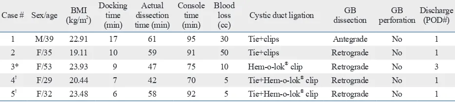
[image:4.595.58.524.593.698.2]RSSC was performed consecutively in 5 patients by a sin-gle surgeon between October 2013 and November 2013. Four patients were females and one was male; the median age of the patients was 35 years old (range: 29‒53) (Table 1). Despite strict selection criteria for patient enrollments, we observed moderate inflammatory changes around the Calot area and dilated cystic duct requiring intracorporeal tie ligation before endoscopic clipping in most cases. Addi-tionally, the surgical system induced an upward medial re-traction of the GB by the assisting surgeon, which caused a narrowing of Calot’s triangle, leading to prolonged dissec-tion time, in contrast to SFLC (Table 2). Neither bile duct injury nor bile spillage occurred during any of the opera-tions. Robotic setting (docking) time, actual dissection time, and intraoperative blood loss seemed to decrease gradually as case numbers increased (Fig. 3). There were no open conversion cases, and most of the patients were discharged
Table 1. Case Series of RSSC
Case # Sex/age (kg/mBMI 2)
Docking time (min)
Actual dissection time (min)
Console time (min)
Blood loss
(cc) Cystic duct ligation
GB
dissection perforationGB Discharge (POD#)
1 M/39 22.91 17 61 95 30 Tie+clips Antegrade No 1
2 F/35 19.11 10 59 91 50 Tie+clips Retrograde No 1
3* F/53 23.93 9 47 75 10 Hem-o-lok® clip Retrograde No 3
4†
F/29 20.44 7 42 70 5 Tie+Hem-o-lok® clip Retrograde No 1
5† F/32 23.48 6 58 92 5 Tie+Hem-o-lok® clip Retrograde No 1
RSSC, robotic single-site cholecystectomy; BMI, Body Mass Index; GB, gallbladder; POD, postoperative day.
*This case was demonstrated live at ROBOTIC SURGERY LIVE 2013, Severance Hospital, Yonsei University Health System, Seoul, Korea, October 23, 2013.
addition, intraoperative stable 3-D images, no tremor, and real-time fluorescent cholangiography provided optimal conditions for safe laparoscopic single-site cholecystectomy. Even in cases of dilated cystic duct, intracorporeal tying of the cystic duct was feasible (in fact, 4 patients out of 5 in the present series required intracorporeal tying due to dilat-ic system, this right-left orientation problem in our SFLC
[image:5.595.72.541.80.427.2]was completely solved: the surgeon’s right-hand motion in the console controlled the left-sided robotic arms, yet the ef-fector movement was noted in right side of the patient due to the curved configuration, maximizing surgeons’ ergo-nomics when performing single-site laparoscopic surgery. In
Table 2. Comparison between Initial Experiences of RSSC and SFLC
Variable RSSC (n=5) SFLC (n=20) p value
Age (yrs) 37.6±9.4 44.4±12.8 0.283
Sex (n, %) 0.824
Male 1 (20) 5 (25)
Female 4 (80) 15 (75)
BMI (kg/m2) 21.9±2.1 23.1±2.4 0.328
Diagnosis (%) 0.644
GB stone 2 (40) 11 (55)
GB polyp 2 (40) 4 (20)
Adenomyomatosis 1 (20) 5 (25)
Size of GB polyp (mm) 12.6±4.1 13.4±3.4 0.809
ASA score 1.4±0.5 1.1±0.3 0.295
Operative time (min) 132.6±25.2 39.75±15.6 0.001
Actual dissection time (min)* 53.4±8.4 32.2±13.4 0.003
Blood loss (cc) 20.0±19.7 7.3±10.1 0.225
Bile spillage during operation (n, %) 0.482
No 5 (100) 18 (90)
Yes 0 (0) 2 (10)
Drain insertion (n, %) 0 (0) 0 (0) NA
Open conversion (n, %) 0 (0) 0 (0) NA
Pain scale (VAS score)
Immediate post-operation 5.2±1.5 4.3±1.5 0.248
At discharge 1.8±0.4 1.9±0.8 0.789
Complication (n, %) NA
No 5 (100) 20 (100)
Yes 0 (0) 0 (0)
Hospital stay (days) 1.4±0.9 1.35±0.6 0.89
ASA, American Society of Anesthesiologist; VAS, Visual Analogue Scale; RSSC, robotic single-site cholecystectomy; SFLC, single-fulcrum laparoscopic cho-lecystectomy; BMI, Body Mass Index; GB, gallbladder; NA, not available.
*The actual dissection time included the period from dissection of Calot’s triangle to retrieval of the specimen.
Fig. 3. Change in actual dissection time and estimated blood loss with RSSC. (A) Serial change of actual dissection time (min) for RSSC. (B) Serial change of blood loss (mL) for RSSC. RSSC, robotic single-site cholecystectomy.
0 10 20 30 40 50 60 70
Case #1 Case #2 Case #3 Case #4 Case #5
A
Actual dissection time
Min
0 10 20 30 40 50 60
Case #1 Case #2 Case #3 Case #4 Case #5
B
Amount of blood loss
[image:5.595.76.540.474.611.2]prolonged operation time in our series.
In addition, difficult operations were expected in cases of gallbladder rupture or bleeding during procedure. In such cases, the optimal operative view could not be maintained, as the accessory port should be used for suction and manip-ulation for preventing bile spillage or for suctioning bleed-ing to ensure the operation field. GB traction could not re-main steady in those cases. Therefore, strict patient selection and careful dissection are thought to be important in taking advantage of RSSC, which is why our present series in-cludes more asymptomatic GB polyps than the SFLC series in our retrospective data set (p<0.004) (Table 2). Of course, the high cost of the robotic surgical system would be also another major obstacle to expanding RSSC to routine clini-cal practice.12 In the near future, it is highly expected that new surgical instruments, such as a right-angle dissector, as well as angulated motion of effector instruments, and the available energy source system in the Maryland dissector needs to be improved in order to be more effective and safe when used to perform single-site cholecystectomy.
In conclusion, the robotic single-site surgical system gen-erally provides a more comfortable environment and im-proved ergonomics for surgeons performing laparoscopic cholecystectomy. Special features of the robotic system, such as intraoperative fluorescence imaging and ergonomic normal hand orientation, can contribute to safe and comfort-able operation in single-site minimally invasive cosmetic surgery. However, several potential disadvantages should be considered when performing RSSC. Nevertheless, with ad-vancement of technical innovation and strict case selection, RSSC could be an alternative that may become a potential means of safe and effective minimally invasive cosmetic surgery. More experiences must be carefully performed in order to exactly address the role of RSSC in the advanced laparoscopic era.
SUPPLEMENTARY DATA
Video 1. Video clip of Robotic single-site cholecystectomy.
REFERENCES
1. Zucker KA, Bailey RW, Gadacz TR, Imbembo AL. Laparoscopic guided cholecystectomy. Am J Surg 1991;161:36-42.
2. Ouchi K, Mikuni J, Kakugawa Y; Organizing Committee, The ed cystic duct) (Table 1), indicating the possible of
expan-sion of this minimally invasive cosmetic surgery.12
Due to technical difficulties in laparoscopic single-site surgery, surgeons’ experience levels are very important in performing laparoscopic single-site cholecystectomy. It is estimated that at least about 5‒20 cases are necessary for overcoming the learning curve,13-15 and most learning curves in laparoscopic single-site cholecystectomy are thought to be overcome during surgery. On the contrary, the learning curve of robotic single-site cholecystectomy is mostly relat-ed to the robotic setting period. Once the operative setting is optimally defined, the unique characteristics of the robot-ic surgrobot-ical system make it easy to perform single-site chole-cystectomy.16,17 In addition, we just applied Firefly Fluores-cence Image in last 2 cases. It was felt that this system can enhance the safety of laparoscopic cholecystectomy to avoid unnecessary bile duct injury. We need to accumulate further experiences about it.
angiography in single-site robotic cholecystectomy (SSRC): a sin-gle-institutional prospective study. Surg Endosc 2013;27:2156-62. 10. Choi SH, Hwang HK, Kang CM, Lee WJ. Single-fulcrum laparo-scopic cholecystectomy: a single-incision and multi-port tech-nique. ANZ J Surg 2012;82:529-34.
11. Hwang HK, Choi SH, Kang CM, Lee WJ. Single-fulcrum laparo-scopic cholecystectomy in uncomplicated gallbladder diseases: a retrospective comparative analysis with conventional laparoscopic cholecystectomy. Yonsei Med J 2013;54:1471-7.
12. Vidovszky TJ, Smith W, Ghosh J, Ali MR. Robotic cholecystecto-my: learning curve, advantages, and limitations. J Surg Res 2006;136:172-8.
13. Kravetz AJ, Iddings D, Basson MD, Kia MA. The learning curve with single-port cholecystectomy. JSLS 2009;13:332-6.
14. Solomon D, Bell RL, Duffy AJ, Roberts KE. Single-port chole-cystectomy: small scar, short learning curve. Surg Endosc 2010;24:2954-7.
15. Gumbs AA, Milone L, Sinha P, Bessler M. Totally transumbilical laparoscopic cholecystectomy. J Gastrointest Surg 2009;13:533-4. 16. Spinoglio G, Lenti LM, Maglione V, Lucido FS, Priora F, Bianchi
PP, et al. Single-site robotic cholecystectomy (SSRC) versus sin-gle-incision laparoscopic cholecystectomy (SILC): comparison of learning curves. First European experience. Surg Endosc 2012; 26:1648-55.
17. Chang L, Satava RM, Pellegrini CA, Sinanan MN. Robotic sur-gery: identifying the learning curve through objective measure-ment of skill. Surg Endosc 2003;17:1744-8.
30th Annual Congress of the Japanese Society of Biliary Surgery. Laparoscopic cholecystectomy for gallbladder carcinoma: results of a Japanese survey of 498 patients. J Hepatobiliary Pancreat Surg 2002;9:256-60.
3. Kang CM, Choi GH, Park SH, Kim KS, Choi JS, Lee WJ, et al. Laparoscopic cholecystectomy only could be an appropriate treat-ment for selected clinical R0 gallbladder carcinoma. Surg Endosc 2007;21:1582-7.
4. Bucher P, Pugin F, Buchs N, Ostermann S, Charara F, Morel P. Single port access laparoscopic cholecystectomy (with video). World J Surg 2009;33:1015-9.
5. Rao PP, Bhagwat SM, Rane A, Rao PP. The feasibility of single port laparoscopic cholecystectomy: a pilot study of 20 cases. HPB (Oxford) 2008;10:336-40.
6. Chamberlain RS, Sakpal SV. A comprehensive review of single-incision laparoscopic surgery (SILS) and natural orifice translumi-nal endoscopic surgery (NOTES) techniques for cholecystectomy. J Gastrointest Surg 2009;13:1733-40.
7. Kroh M, El-Hayek K, Rosenblatt S, Chand B, Escobar P, Kaouk J, et al. First human surgery with a novel single-port robotic system: cholecystectomy using the da Vinci Single-Site platform. Surg Endosc 2011;25:3566-73.
8. Wren SM, Curet MJ. Single-port robotic cholecystectomy: results from a first human use clinical study of the new da Vinci single-site surgical platform. Arch Surg 2011;146:1122-7.