Contrast-enhanced multi-detector row spiral computed tomography (MDCT) was introduced as a promising nonin-vasive method for vascular imaging. This study examined the accuracy of this technique for detecting significant coronary artery stenoses. Both MDCT(Sensation 16, Siemens, Germany, 12 × 0.75 mm collimation and 0.42 sec rotation speed, 120 kV, 500 effective mA, and 2.7 mm/rotation table-feed) and invasive coronary angiography (CAG) were performed on 61 patients (mean age 59.2 ± 10, 44 men) who were suspected of having coronary artery disease. All patients were treated with atenolol (25 - 50 mg) prior to imaging and the heart rate was maintained below 65 beats per minutes during image acquisition. The images were reconstructed in the diastole around TI - 400 ms with a 0.5 mm increment and a 1.0 mm thickness. All coronary arteries with a diameter of 2.0 mm or more were assessed for the presence of a stenosis (> 50% luminal narrowing). Two independent radiologists who were unaware of the results of the invasive CAG evaluated the MDCT data, and the results were compared with those from the invasive CAG (interval 1- 27, mean 11 days). An evalua-tion of the CT coronary angiogram (CTCA) was possible in 58 of the 61 patients (95%). Image acquisition of the major coronary arteries including the left main trunk was available in 229 out of 244 arteries. Invasive CAG showed that 35 out of 58 patients had significant coronary artery stenoses by. patient analysis of those who could be evaluated showed that CT coronary angiography correctly classified 30 out of 35 patients as having at least 1 coronary stenosis (sensitivity 85.7%, specificity 91.3%, positive predictive value 93.8%, negative predictive value 80.8%). By analyzing each coronary artery, CAG found 62 stenotic coronary arteries in the 229 coronary arteries that could be evaluated. MDCT correctly
detected 50 out of 62 stenotic coronary arteries and an absence of stenosis was correctly identified in 156 out of 167 normal coronary arteries (sensitivity 80.6%, specificity 93.4%, positive predictive value 81.9%, negative predictive value 92.8%). The non-invasive technique of MDCT for examining the coronary artery appears to be a useful method for detecting coronary artery stenoses with a high accuracy particularly with the proximal portion and large arteries.
Key Words:Coronary artery stenoses, computed tomography, imaging, stent, MDCT
INTRODUCTION
For almost 50 years, selective coronary angio-graphy has remained the clinical “gold standard” for evaluating the coronary anatomy and defining epicardial coronary artery disease. While conven-tional invasive coronary angiography provides exceptional spatial resolution and a general map of the coronary system, it is expensive and has a small but definite risk of complications. In addi-tion, it requires either a brief hospitalization period or a period of observation for several hours after the procedure in a specialized monitoring unit. The replacement of even a fraction of these procedures with noninvasive imaging modalities would constitute an important advance in the care of patients suspected of having coronary artery disease. Currently, a number of imaging modal-ities are used for diagnosing epicardial coronary artery disease. Most identify the luminal diameter or stenosis, wall thickness, and plaque volume.1 Since 1999, multi-detector row spiral computed tomography (MDCT) scanners have been avail-able for coronary artery scanning.2 The increased
The Utility of Multi-detector Row Spiral CT for Detection
of Coronary Artery Stenoses
Jae-Youn Moon1, Namsik Chung1, Byoung Wook Choi2, Kyu Ok Choe2, Hye Sun Seo1, Young-Guk Ko1, Seok-Min Kang1, Jong-Won Ha1, Se-Joong Rim1, Yangsoo Jang1, Won-Heum Shim1, and Seung-Yun Cho1
1Division of Cardiology, Cardiovascular Hospital, Yonsei University College of Medicine, 2Division of Radiology, Yonsei
University, Seoul, Korea.
Received June 25, 2004 Accepted November 18, 2004
scan speed results in thinner collimated slice widths and an improved spatial and temporal resolution.
Recently, with the introduction of MDCT com-bined with a subsecond rotation and retrospective electrocardographic (ECG) gating, the invasive modalities have been challenged by an additional new noninvasive assessment of coronary artery stenoses.3 However, the image quality has been insufficient for the reliable detection of coronary stenoses in a substantial number of cases. Calcifi-cations often hinder an evaluation of severely dis-eased coronary segments, and the coronary ar-teries are frequently affected by motion arti-facts.4-8 In addition, it has been observed that the patient's heart rate during the scan critically influences the image quality.9,10 Therefore, heart rate control is one of the most important factors in better image acquisition. Accordingly, the cur-rent technique for image acquisition with MDCT requires pre-medication with beta-blockers.11
This study evaluated the diagnostic accuracy of MDCT angiography in determining significant coronary artery stenoses ( 50% lumen diameter narrowing in angiography) and occlusions com-pared with conventional invasive angiography in Korean patients who had their heart rate con-trolled with beta-blockers. In addition, the detec-tion rate of computed tomographic coronary an-giography (CTCA) in each coronary arterial seg-ment and the detection rate by the location of the stenotic lesion were assessed.
MATERIALS AND METHODS
Patients
The study was performed prospectively from February to July 2003. CTCA was performed on 61 patients who were suspected of having coro-nary artery disease (20 - 76 years, mean 59.3 ±10.0 years). They underwent CTCA, as well as invasive coronary angiography, over a six-month period. The average time between the two examinations was 11 days (range: 1 - 27 days). All the patients were treated with atenolol (25 - 50 mg) and four patients with heart rates higher than 70 bpm received a short-lasting beta-blocker (propranolol
40 mg) prior to imaging. Only those patients in a sinus rhythm, without implanted pacemakers or valve prostheses, and without contraindications to the administration of an iodinated contrast agent were enrolled in this study. Hemodynamicaly un-stable patients were excluded and all patients were allowed to continue concurrent medications with no additional medications except for the beta-blockers. The patients who previously had coronary artery stents inserted were included in this study. Twenty-one patients had previously undergone percutaneous transluminal coronary angioplasty with a stent implantation.
MDCT scan
CT was performed by using a 16-slice CT (Sen-sation 16, Siemens, Germany) with 12 × 0.75 mm collimation and 0.42s rotation speed. 120 kV, 500 effective mA, and 2.7 mm/rotation table-feed. Images were reconstructed in the diastole around TI -400 ms with a 0.5 mm increment and a 1.0 mm thickness.
Quantitative coronary angiography
All the data was compared with the results of the invasive coronary angiography. The invasive coronary angiograms were evaluated by a blinded independent observer using quantitative coronary angiography (QCA) and was used as the gold standard for detecting a stenosis. Lesions with a diameter reduction of 50% or more were con-sidered to be significant stenoses. In addition, the reference diameter of the lesion (vessel diameter in the non-diseased artery immediately proximal to the lesion) was documented because only those lesions with a lumen diameter 2.0 mm were included in the analysis.
Image evaluation and comparison of two exami-nations
estimation, the coronary arteries were classified according to those that could be evaluated and those that could not. In those arteries that could be evaluated, the presence of significant stenoses (exceeding 50% diameter reduction) was visually assessed and measured. The result was analyzed first by a patient evaluation i.e. how accurately the method detected a patient with coronary artery occlusive disease (CAOD). The overall sensitivity and overall specificity were checked by patient analysis. Second, the results were analyzed ac-cording to each coronary artery (LAD, LCx, RCA and Left main trunk) and the sensitivity, speci-ficity, positive predictive value and negative pre-dictive value of each coronary artery was then calculated. Finally the detection rate for stenotic lesion was evaluated depending on the location of the lesion in order to determine if it could influ-ence the detection of stenosis.
RESULTS
Baseline clinical characteristics
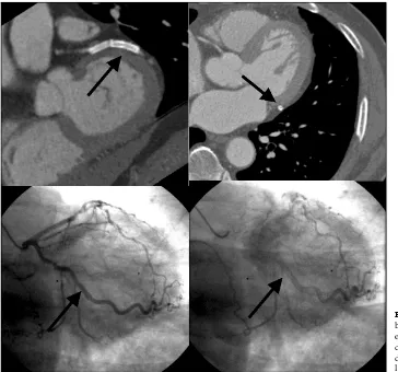
[image:3.595.52.412.393.583.2]CTCA was performed without complications in all patients (mean age: 59.3 ± 10.0 years, M : F= 44 : 17) (Fig. 1). An evaluation of CTCA was possible in 58 out of 61 patients (95%). Invasive coronary angiography indicated 38 patients with significant coronary artery stenoses (1 vessel dis-ease: 17 cases, 2 vessel disdis-ease: 11 cases, 3 vessel disease: 10 cases) and 23 patients with a normal coronary artery or minimal coronary artery stenoses (< 50% luminal narrowing). The images of the 3 patients were could not be evaluated due to blurring artifacts, which were due to an in-creased heart rate during the scanning procedures and motion artifacts. The image acquisition of the major coronary arteries including the left main trunk was available in 229 out of 244 arteries (Table 1)
Table 1. Baseline Clinical Characteristics
Age 59.3 ± 10.0
Number (M : F) 61 (44:17)
Normal coronary artery by CAG 23 (37.7%)
Coronary artery disease by CAG 1-vessel disease
2-vessel disease 3-vessel disease
38 (62.3%) 17 (27.9%) 11 (18.0%) 10 (16.4%)
[image:3.595.67.529.620.742.2]Images that could be evaluated by MDCT 58 (95.0%)
Comparison of CTCA and invasive coronary angiography
Table 2 shows a comparison between the MDCT and invasive coronary angiography. CTCA cor-rectly classified 30 out of 35 patients who had significant coronary artery stenoses as having coronary artery stenoses. Five patients who were incorrectly classified by CTCA had only one vessel stenosis. Among the remaining 5 cases, one case had a lesion of a total occlusion in the pro-ximal RCA, which was interpreted as a diminu-tive RCA. A second case with a stenotic lesion in the PL (posterolateral) branch was not detected because it was located far distal. A misdiagnosis of the third case with a proximal LAD lesion was attributed to motion artifacts. A stenotic lesion at the proximal edge of a previous RCA stent and the lesion of the diagonal branch ostium adjust to the previous proximal LAD stent were not cor-rectly assessed in the fourth and fifth case,
respec-tively. In the last 2 cases, the previously inserted stent interfered with the detection of the coronary artery lesions, known as metallic stent artifact in CTCA. Therefore, by patient analysis, CTCA cor-rectly classified 30 out of 35 patients, who could be evaluated, as having at least one coronary artery stenosis (overall sensitivity 85.7%, overall specificity 91.3%, overall positive predictive value 93.8%, overall negative predictive value 80.8% and accuracy 87.9%) (Table 2).
Accuracy of CTCA
[image:4.595.59.411.390.585.2]The sensitivity, specificity, positive predictive value and negative predictive value were com-pared by coronary vessel analysis and the results are shown in Table 3. Image acquisition of the major coronary arteries (LAD, LCx, RCA, Left main trunk) was available in 229 out of 244 (61×4) arteries. CTCA correctly detected 50 out of 62 stenotic coronary artery and an absence of
Table 2. Comparison of the CTCA and Invasive CAG by Patients Analysis
Invasive CAG
Total
Normal CAOD
CTCA Normal 21 (91.3%) 5 (14.3%) 26
CAOD 2 (8.7%) 30 (85.7%) 32
Total 23 (100.0%) 35 (100.0%) 58*
* This value was calculated in evaluable patients who could be evaluated by MDCT (n=58). CAOD, coronary artery occlusive disease (> 50% stenoses). overall sensitivity 85.7%, overall specificity 91.3%, overall positive predictive value 93.8%, overall negative predictive value 80.8% and accuracy 87.9%
[image:4.595.67.528.621.704.2]stenosis was correctly identified in 156 out of 167 normal coronary arteries (sensitivity 80.6%, speci-ficity 93.4%, positive predictive value 81.9%, negative predictive value 92.8%). In the LAD, there was a relatively high level of coronary
[image:5.595.70.527.119.244.2]ves-sels that could not be evaluated (8.2%). On the other hand, it was lowest in the LCx (4.2%). This might be due to the coronary artery calcifications, which were more frequently identified in the LAD than in the other arteries in this study group.
Table 3. Accuracy of MDCT for the Stenotic Vessels by Vessel Analysis in the Evaluable Vessels
LAD (%) LCx (%) RCA (%) Lt. main (%)
Sensitivity 86.3 81.2 73.9 100
Specificity 85.2 92.8 94.1 98.2
PPV* 79.1 81.2 89.5 50
NPV* 90.6 92.8 84.2 100
Accuracy 85.7 89.6 86.0 98.0
Unevaluable 8.2 4.9 6.6 4.9
*PPV, positive predictive value; NPV, negative predictive value.
[image:5.595.58.412.274.651.2]Previous reports4 showed the reasons why some segments could not be assessed. The presence of extensive calcifications can complicate a correct assessment of the lumen of the coronary arteries particularly the LAD and motion artifacts of the CTCA developed frequently in the RCA. Occasio-nally, small sized LCx could not be evaluated using the CTCA modality. The other values showed similar levels. Only one case had a signi-ficant stenosis in the Lt. main trunk in invasive coronary angiography.
Accuracy of CTCA for stenoses in regards to lesion location
Table 4 shows a comparison of the detection rates in each stenotic segment with regard to the lesion locations. While the relatively proximal portion were frequently detected, distal lesions and side branches were detected less frequently.
DISCUSSION
The high spatial resolution associated with
con-trast-enhanced multi-detector row coronary CT angiography may provide a great deal of infor-mation on coronary artery stenotic lesions and noninvasive images of the coronary artery wall.13 Recently, Achenbach et al.6 reported a sensi-tivity of 85% in detecting a significant stenotic coronary artery with contrast-enhanced MDCT in the interpretable native coronary arteries (luminal diameter 2 mm vessels were assessed) and only 68% of all the coronary arteries could be inter-preted. Ropers et al.11 reported a sensitivity of 92% in detecting a significant stenotic coronary artery using contrast-enhanced MDCT in coronary arteries that could be interpreted (luminal dia-meter 1.5 mm vessels were assessed). And they reported that 88% of coronary arteries could be evaluated using beta-blockade pre-medication.
[image:6.595.66.526.462.743.2]This study demonstrated that CTCA allows the detection of coronary artery stenoses and occlu-sions with a high sensitivity (80.6%) and speci-ficity (93.4%) if sufficient image quality for an evaluation can be obtained. In this study, 93.8% of all coronary arteries could be evaluated with beta- blocker pre-medication. The sensitivity and specificity were similar to those of CTCA in other
Table 4. Accuracy of CTCA in with Regards to the Lesion Locations
Location True Positve False Negative Detection rate(%)
LAD lesion
p-LAD 8 0 100.0
m-LAD 9 1 90.0
d-LAD 2 1 66.7
Diagonal br. 5 4 55.5
Intermedius br 1 0 100.0
LCx lesion
P-LCx 6 2 75.0
d-LCx 7 0 100.0
OM br. 3 2 60.0
RCA lesion
p-RCA 6 3 66.7
m-RCA 9 0 100.0
d-RCA 6 1 85.7
PD br. 2 1 66.7
PL br. 0 2 0
Lt. main 1 0 100.0
research centers6-8,11,14-16 but the portion of artery that could be interpreted was relatively higher than in the other results.
In contrast to previous studies, the CTCA re-sults were verified by QCA and all vessel seg-ments, proximal and distal segseg-ments, including side branches, were included in the analysis if the vessel diameter measured more than 2.0 mm. The cut off value of 2.0 mm means that stenoses in vessels smaller than 2.0 mm rarely constitute targets for revascularization.17,18
This study demonstrates that in the proximal and mid segments of all 3 major arteries and the left main trunk, CTCA has a high accuracy in excluding coronary artery disease and identifying significant stenoses, but CTCA is relatively limited in the diagnosis of the distal arteries, the side branches and near the previous stent legion. This suggests that stenoses of the relative large vessels were detected more easily by CTCA, suggesting the utility of MDCT as a feasible diagnostic
mo-dality and a noninvasive technique for the selec-tion of a revascularizaselec-tion target.
The limitation of CTCA was a calcification of the coronary arteries. In this study, some patients had severe coronary calcification and an accurate evaluation of the CTCA image was difficult, re-sulting in these lesions often being misdiagnosed as severe stenoses. However, in this group, some patients had only minimal luminal narrowing or did not have a severe coronary stenosis in the invasive coronary angiography. Other studies19 have reported that although this finding was a limitation for an accurate diagnosis using MDCT, the presence of any calcium was strongly sugges-tive of the presence of atherosclerosis in the coro-nary artery. Hence, patients with no significant stenoses in the invasive coronary angiography but with detectable calcification by MDCT could be diagnosed with coronary artery disease. This fact might be one of the advantages of CTCA.
[image:7.595.56.420.101.441.2]nary artery stenting is currently the most common form of non-surgical myocardial revascularization. However, the stent restenosis remains a clinical problem. Therefore, a noninvasive assessment of the stented segment in these patients is highly desirable. It was reported that EBCT (Electron Beam Computed Tomography) was successfully used for a stent patency evaluation but the stent lumen itself could not be accurately visualized.20-22 In this study, 24 of the patients previously under-went coronary stenting in 38 lesions. The radio-logists often misdiagnosed the previous stented segments and the luminal narrowing could not be accurately measured. In the presence of intra-coronary stents, high-density artifacts, combined with partial volume effects, prevent an adequate assessment of the vessel lumen within the struts of the stent. However, the patency can be assessed if it is enhanced by administering a contrast medium to the vessel segment distal to the stent.4 Although the stent lumen cannot be visualized in most stents, a reliable evaluation of the stent patency is possible by visualizing the distal flow and measuring the attenuation changes in the visible stent lumen and outside the stent. How-ever, the enhancement of the stent distal flow by the antegrade collateral flow in total occluded stents was not correctly assessed. In our study, 33 out of 38 coronary stents (21 patients) could be evaluated using these methods (86.8%) and MDCT correctly classified 31 out of these 33 stents as being either patent (28 stents) or occluded (3 stents) and only two stents were misdiagnosis. Some cases of an in-stent restenosis but with patent distal flow were also classified as being patent by MDCT. Because the stent lumen could be partially visualized in most stents, a reliable evaluation of an in-stent restenosis remains to be assessed in the future.
It is clear that the CTCA is a feasible diagnostic method for detecting coronary artery stenoses with a high sensitivity and specificity under the appropriate heart rate controls. In particular, CTCA showed a high accuracy in the detection of proximal lesion stenoses. However, because most of the segments that could not be evaluated were affected by the coronary motion and coronary artery stents, a further shortening the image acquisition window and technical advances is
needed. The non-invasive technique of MDCT for assessing the coronary artery appears to be a useful method for detecting coronary artery stenoses with a high accuracy particularly to the proximal lesions and large arteries. In addition, MDCT angiography is a rapidly developing imaging modality and further technical advances aimed at improving the diagnostic accuracy and clinical utility are expected in the future.23 MDCT angiography is expected to be a good screening modality of coronary artery disease. Besides being a non-invasive alternative, MDCT can offer addi-tional information about the spatial orientation of the vessels and can also identify and quantify the level of calcium deposition within the coronary vessel wall.
REFERENCES
1. Fayad ZA, Fuster V. Clinical imaging of the high-risk or vulnerable atherosclerotic plaque. Circ Res 2001;89: 305-16.
2. Klingenbeck-Regn K, Schaller S, Flohr T, Ohnesorge B, Kopp AF, Baum U. Subsecond multi-slice computed tomography: basics and applications. Eur J Radiol 1999; 31:110-24.
3. Knez A, Becker CR, Ohnesorge B, Haberl R, Reiser M, Steinbeck G. Noninvasive detection of coronary artery stenosis by multislice helical computed tomography. Circulation 2000;101:221-2.
4. Nieman K, Oudkerk M, Rensing BJ, Ooijen P, Munne A, Geuns RJ, et al. Coronary angiography with multi-slice computed tomography. Lancet 2001;357:599-603. 5. Kopp AF, Schroeder S, Kuettner A, Baumbach A,
Georg C, Kuzo R, et al. Non-invasive coronary angiography with high resolution multidetector-row computed tomography. Eur Heart J 2002;23:1714-25. 6. Achenbach S, Giesler T, Ropers D, Ulzheimer S, Derlien
H, Schulte C, et al. Detection of coronary artery stenoses by contrast-enhanced, retrospectively ECG-gated, multi-slice spiral CT. Circulation 2001;103:2535-8. 7. Knez A, Becker CR, Leber A, Ohnesorge B, Becker A, White C, et al. Usefulness of multislice spiral computed tomography angiography for determination of coronary artery stenoses. Am J Cardiol 2001;88:1191-4.
8. Vogl TJ, Abolmaali ND, Diebold T, Engelmann K, Ay M, Dogan S, et al. Techniques for the detection of coronary atherosclerosis: multi-detector row CT coro-nary angiography. Radiology 2002;223:212-20.
experience in 94 patients. Clin Imaging. 2002;26:106-11. 10. Giesler T, Baum U, Ropers D, Ulzheimer S, Wenkel E, Mennicke M, et al. Noninvasive visualization of coronary arteries using contrast-enhanced multidetector CT: Influence of heart rate on image quality and stenosis detection. Am J Roentgenol 2002;179:911-6. 11. Ropers D, Baum U, Pohle K, Anders K, Ulzheimer S,
Ohnesorge B, et al. Detection of coronary artery ste-noses with thin-slice multi-detector row spiral com-puted tomography and multiplanar reconstruction. Circulation 2003;107:664-6.
12. Detre KM, Wright E, Murphy ML, Takaro T. Observer agreement in evaluating coronary angiograms. Circu-lation 1975;52:979-86
13. Becker CR, Ohnesorge BM, Joseph Schoepf U, Reiser MF. Current development of cardiac imaging with multi-detector row CT. Eur J Radiol 2000;36:97-103. 14. Achenbach S, Moshage W, Ropers D, Nossen J, Daniel
WG. Value of electron-beam computed tomography for the detection of high-grade coronary artery stenoses and occlusions. N Engl J Med 1998;339:1964-71. 15. Ha JW, Cho SY, Shim WH, Chung N, Jang Y, Lee HM,
et al. Noninvasive evaluation of coronary artery bypass graft patency using three-dimensional angiography obtained with contrast-enhanced electron beam CT. Am J Roentgenol 1999;172: 1055-9
16. Nieman K, Cademartiri F, Lemos PA, Raaijmakers R, Pattynama PM, de Feyter PJ. Reliable noninvasive coronary angiography with fast submillimeter multis-lice spiral computed tomography. Circulation 2002; 106:2051-4.
17. Reddy G, Chernoff DM, Adams JR, Higgins CB, et al. Coronary artery stenoses: assessment with contrast-enhanced electron-beam CT and axial reconstructions. Radiology 1998;208:167-72.
18. Schmermund A, Rensing BJ, Sheedy PF, Bell MR, Rumberger JA. Intravenous electron-beam computed tomographic coronary angiography for segmental analysis of coronary artery stenoses. J Am Coll Cardiol 1998;31:1547-54.
19. Rumberger JA. Noninvasive coronary angiography using computed tomography Ready to kick it up another notch? Circulation 2002;106:2036-8.
20. Pump H, Mohlenkamp S, Sehnert CA, Schimpf SS, Schmidt A, Erbel R, et al. Coronary arterial stent patency: assessment with electron-beam CT. Radiology 2000;214:447-52.
21. Mohlenkamps S, Pump H, Baumgart D, Haude M, Gronemeyer DH, Seibel RM, et al. Minimally invasive evaluation of coronary stents with electron beam computed tomography:in vivoand in vitroexperience. Catheter Cardiovasc Interv 1999;217:564-571.
22. Maintz D, Juergens KU, Wichter T, Grude M, Heindel W, Fischbach R. Imaging of coronary artery stents using multislice computed tomography: in vitro eval-uation. Eur Radiol 2003;13:830-5.