INTRODUCTION
To successfully rejuvenate an aging face, contributing factors to changes associated with aging must be thoroughly assessed. The Asian face is markedly different from Caucasian face in many aspects, including skin tone, texture, elasticity, skin thick-ness, subcutaneous fat contents, and skeletal framework.1-3
Therefore, the aging process in Asian faces could be remark-ably different from that in Caucasian faces.4-6
Three-dimensional (3D) CT studies revealed that the midfa-cial skeleton shows angular changes with aging in Caucasians.7-9 These changes may play a role in the loss of soft-tissue cheek mass support, cheek mass drooping, inferior and posterior dis-placement of the inferior orbital rim, distortion of orbital rim curve, and the scleral show in the midface. Difference in facial skeletal contour is most important for distinguishing between different ethnic groups. However, ethnic differences in age-re-lated facial skeletal changes have not yet been fully establish-ed. Though several investigators have studied skeletal remod-eling in aging Caucasian faces, Asian midfacial skeletal remo-deling has not yet been reported. Understanding these inherent facial skeletal differences and applying them to rejuvenation surgery are essential for more desirable surgical outcomes.
This study aimed to analyze midfacial skeletal changes in aging Asian faces using a new method and to explore ethnic
dif-Analysis of Age-Related Changes in Asian Facial
Skeletons Using 3D Vector Mathematics on Picture
Archiving and Communication System
Computed Tomography
Soo Jin Kim, So Jung Kim, Jee Soo Park, Sung Wan Byun, and Jung Ho Bae
Department of Otorhinolaryngology-Head and Neck Surgery, Ewha Womans University, School of Medicine, Seoul, Korea.
Purpose: There are marked differences in facial skeletal characteristics between Asian and Caucasian. However, ethnic differences in age-related facial skeletal changes have not yet been fully established. The aims of this study were to evaluate age-related changes in Asian midfacial skeletons and to explore ethnic differences in facial skeletal structures with aging between Caucasian and Asian. Materials and Methods: The study included 108 men (aged 20–79 years) and 115 women (aged 20–81 years). Axial CT images with a gantry tilt angle of 0 were analyzed. We measured three-dimensional (3D) coordinates at each point with a pixel lens cursor in a picture archiving and communication system (PACS), and angles and widths between the points were calculated using 3D vector mathematics. We analyzed angular changes in 4 bony regions, including the glabellar, orbital, maxillary, and pyriform aperture re-gions, and changes in the orbital aperture width (distance from the posterior lacrimal crest to the frontozygomatic suture) and the pyriform width (between both upper margins of the pyriform aperture).
Results: All 4 midfacial angles in females and glabellar and maxillary angles in males showed statistically significant decreases with aging. On the other hand, the orbital and pyriform widths did not show statistically significant changes with aging.
Conclusion: The results of this study suggest that Asian midfacial skeletons may change continuously throughout life, and that there may be significant differences in the midfacial skeleton between both sexes and between ethnic groups.
Key Words: Facial bones, aging, computed tomography, vector, Asian Yonsei Med J 2015 Sep;56(5):1395-1400
http://dx.doi.org/10.3349/ymj.2015.56.5.1395 pISSN: 0513-5796 · eISSN: 1976-2437
Received: September 30, 2014 Revised: December 16, 2014 Accepted: December 16, 2014
Corresponding author: Dr. Jung Ho Bae, Department of Otorhinolaryngology-Head and Neck Surgery, Ewha Womans University, School of Medicine, 1071 Anyangcheon-ro, Yangcheon-gu, Seoul 158-710, Korea.
Tel: 82-2-2650-2847, Fax: 82-2-2648-5604, E-mail: [email protected] •The authors have no financial conflicts of interest.
© Copyright: Yonsei University College of Medicine 2015
ferences between Caucasian and Asian skeletal facial structures with aging.
MATERIALS AND METHODS
Data was collected in a retrospective manner from previously acquired facial CT scans at Ewha Womans University Hospital between December 2010 and April 2013. The 1.0-mm width axial images using a 64-channel multidetector computed to-mography (MDCT) (SOMATOM Sensation 64, Siemens, For-chheim, Germany) were acquired at the 120 kV and 180 mA setting, and coronal and sagittal images were reconstructed along with axial images. Scans showing either soft tissue lesions or normal findings were included in this study. Scans showing any evidence of previous facial bone trauma, surgery, or the absence of upper dentition with the exception of the premo-lars were excluded from the study.
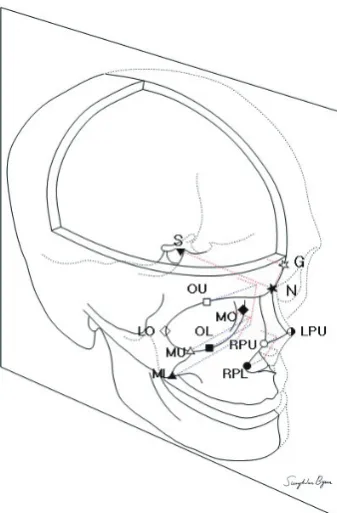
This study included a total of 223 Asians, all of whom were born and living in South Korea. For the evaluation of angular changes with aging, angular measurements of 4 bony regions (glabellar, orbital, maxillary, and pyriform aperture angles) were made using a method based on 3D vector mathematics. A line from the sella to the nasion was taken as the reference line. Each landmark as described below was identified, and a line was drawn to the reference line. Angular measurements were made as follows: the glabellar angle between the refer-ence line and a line drawn from the maximal prominrefer-ence the glabella to the nasofrontal suture; the orbital angle between the reference line and a line drawn from the most superior to the most inferior midportion of the orbit; the pyriform angle between the reference line and a line drawn from the nasal bone to the lateral inferior pyriform aperture; and the maxil-lary angle between the reference line and a line drawn from the most superior to the most inferior maxilla at the articula-tion of the inferior maxillary wing and alveolar arch. By em-ploying the methods described in Fig. 1, 3D co-ordinates of each point on axial images were measured using a pixel lens cursor in a picture archiving and communication system (PACS) re-port viewer, software version 5.0 (INFINITT Co., Ltd., Seoul,
[image:2.595.335.504.186.443.2] [image:2.595.47.540.580.703.2]Korea). Each point on an axial image was verified using recon-structed coronal and sagittal images in the PACS report viewer (Fig. 2). From given co-ordinates, the 2 vectors AB and AC were calculated. The dot product of the 2 vectors was defined as AB · AC= |AB| |AC| cos θ from geometric definition.10 This equation allows the angle θ, an angle between AB and AC to be precise-ly computed. The orbital aperture width (distance from the posterior lacrimal crest to the frontozygomatic suture) and the
Fig. 1. Points used for angular measurement. With given coordinates, dot products between the reference line (sella-nasion line) and individual lines were used to measure angles between them. The orbital aperture width (distance between the posterior lacrimal crest to the frontozygomatic su-ture) and the pyriform width (between both upper points of the pyriform apertures) were also measured. S, sella; N, nasion; G, the maximal promi-nence of the glabella; OU, the upper midpoint of the orbit; OL, the lower midpoint of orbit; MO, the medial point of the orbit; LO, the lateral point of the orbit; MU and ML, the upper and lower points of the maxillary wall at the articulation of the inferior maxillary wing and alveolar arch; RPU, the right upper point of the pyriform aperture; LPU, the left upper point of the pyriform apertrue; RPL, the right lower point of the pyriform aperture.
Fig. 2. Three-dimensional (3D) co-ordinates of the sella and nasion in a PACS as the reference line for angular measurement. The 3D co-ordinates of the sella (A) and nasion (B) on axial images were measured using a pixel lens cursor in a PACS report viewer. Each point was verified on reconstructed coro-nal (C and D) and sagittal images in the PACS report viewer. PACS, picture archiving and communication system.
C
pyriform width (between both lateral margins of the pyriform aperture) were also measured using the PACS.
Statistical analysis was performed by the IBM SPSS software version 19.0 (International Business Machines Corp., Armonk, NY, USA), and a p value of <0.05 was considered statistically significant. The Pearson correlation test was used to find any changes associated with aging, and the independent sample t test was used to identify any trends in facial aging between the age groups. This study design and experimental protocol were approved by the Institutional Review Board of our hospital (IRB No. ECT13-41A-19).
RESULTS
A total of 223 facial CT scans were analyzed (108 men, 115 wo-men). The age of the subjects ranged from 20 to 81 years. Male
and female subjects each were divided into 3 groups: the young, middle, and old age groups (Table 1).
Correlations between age and midfacial anglular measurements
All 4 midfacial angles in females and the glabellar and maxil-lary angles in males showed statistically significant decreases with aging (p<0.05). LOESS regression curves were also creat-ed to better illustrate trends (Figs. 3 and 4). Each angular mea-surement was marked on the sample 3D reconstructed image for better understanding (Fig. 5).
[image:3.595.59.557.303.713.2]Angular changes between individual age groups For both men and women, the glabellar and maxillary angles showed statistically significant decreases with aging in the young, middle, and old age groups. For males, mean glabellar angles were 69.4±6.14, 67.4±6.26, and 65.0±4.77 degrees in the Table 1. Demographic Data of the Subjects
Young (20–39) Middle (40–59) Old (60–)
Total
Male Female Male Female Male Female
Number 44 39 40 39 24 37 223
Mean age 28.6 29.6 49.8 49.1 67.5 68.4 46.9
SD 6.3 5.9 5.6 5.6 6.4 6.7
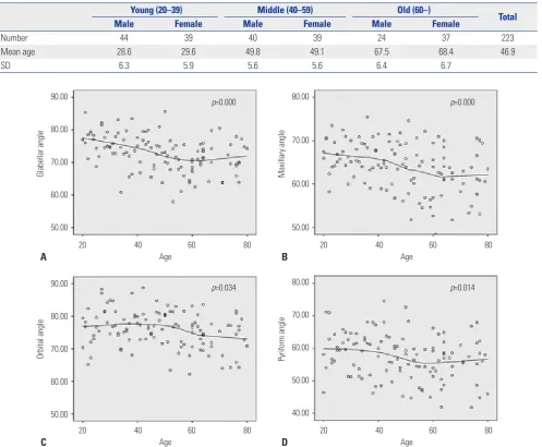
Fig. 3. LOESS regression curves illustrating the trend of changes in glabellar, maxillary, orbital, and pyriform angles based on 115 female data points. Gla-bellar (A), maxillary (B), orbital (C), and pyriform (D) angles all show statistically significant decreases with aging.
90.00
80.00
70.00
60.00
50.00
90.00
80.00
70.00
60.00
50.00
80.00
70.00
60.00
50.00
80.00
70.00
60.00
50.00
40.00
20 40 60 80
20 40 60 80
20 40 60 80
20 40 60 80 Age
Age
Age
Age
Glabellar angle
Orbital angle
Maxillary angle
Pyriform angle
A
C
B
D
p=0.000
p=0.034
p=0.000
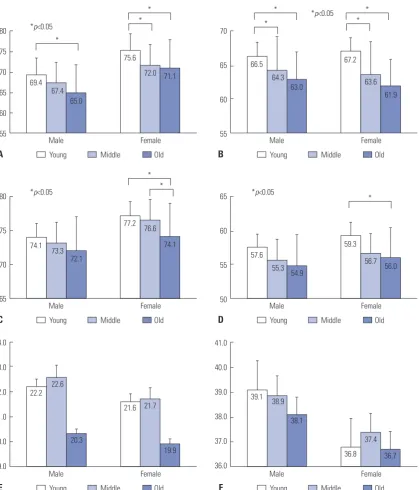
young, middle, and old age groups, respectively. For females, similar changes in the glabellar angle were observed with mean angles measuring 75.6±4.98, 72.0±5.88, and 71.1±4.51 degrees in the young, middle, and old age groups, respectively. For males, mean maxillary angles were 66.5±4.70, 64.3±4.27, and 63.0±4.10 degrees in the young, middle, and old age groups,
respectively. For females, more prominent changes in the mean maxillary angle were observed with aging in the young and middle age groups; mean maxillary angles were 67.2±5.35, 63.6±5.90, and 61.9±5.73 degrees in the young, middle, and old age groups, respectively. For females, the orbital angle showed a statistically significant decrease between the middle (76.6±5.61 degrees) and old age groups (74.1±5.18 degrees), and the pyriform angle also showed a statistically significant decrease between the young (59.3±6.52 degrees) and middle age groups (56.7±7.21 degrees) (Fig. 6A-D).
Periorbital and perinasal changes
[image:4.595.80.504.76.400.2]The orbital and pyriform widths did not show significant changes with aging. For males, mean pyriform widths were 22.2±2.43, 22.6±2.11, and 20.3±2.15 mm in the young, middle, and old age groups, respectively. For males, mean orbital wid-ths were 39.1±3.51, 38.9±3.90, and 38.1±2.35 mm in the young, middle, and old age groups, respectively. For females, mean pyriform widths were 21.6±2.11, 21.7±1.93, and 19.9±1.83 mm in the young, middle, and old age groups, respectively; while mean orbital widths were 36.8±3.67, 37.4±2.59, and 36.7±2.61 mm in the respective groups (Fig. 6E and F).
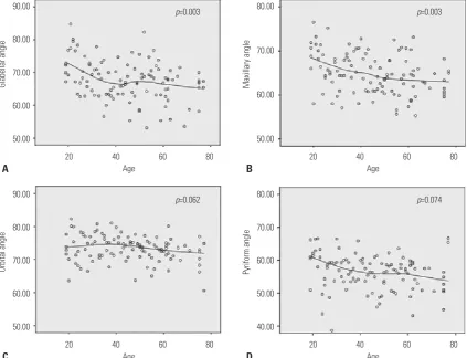
Fig. 4. LOESS regression curves illustrating the trend of changes in glabellar, maxillary, orbital, and pyriform angles based on 108 male data points. Glabel-lar (A) and maxilGlabel-lary (B) angles show statistically significant decreases with aging, while orbital (C) and pyriform (D) angles do not.
90.00
80.00
70.00
60.00
50.00
90.00
80.00
70.00
60.00
50.00
80.00
70.00
60.00
50.00
80.00
70.00
60.00
50.00
40.00
20 40 60 80
20 40 60 80
20 40 60 80
20 40 60 80 Age
Age
Age
Age
Glabellar angle
Orbital angle
Maxillary angle
Pyriform angle
A
C
B
D
p=0.003
p=0.062
p=0.003
p=0.074
Glabellar angle
Orbital angle Pyriform
angle
Maxillary angle
[image:4.595.44.280.440.610.2]DISCUSSION
In the present study, we demonstrated that Asian midfacial sk-eletons change continuously throughout life, mainly in a clock-wise angular rotation around the orbit in a right-facing cranio-facial skeleton. It can be described as Lambros’s theory, sum-marized as a rotation of the maxilla relative to the cranial base.8 Shaw and Kahn9 suggested the trend of aging midfacial skele-tal changes in 60 Caucasian subjects, and they expanded the study by increasing the sample size to 120 and by including
[image:5.595.99.519.68.559.2]measurements of upper and lower facial skeletons.11 The find-ing showed that glabellar and maxillary angles significantly de-creased, whereas the pyriform aperture significantly increased with aging in both male and female subjects. In study of Rich-ard, et al.,7 angular measurements of four bony regions (glabel-lar, orbital, maxillary, and pyriform aperture angles) were taken from 50 male and 50 female subjects, and all four the measure-ments were found to significantly decrease with aging. In our data, the orbital and maxillary angles showed less changes, the pyriform angle showed more prominent changes compared to Fig. 6. Column graphs illustrating changes in glabellar angle, maxillary angle, orbital, and pyriform aperture angles, as well as pyriform and orbital widths. Glabellar (A) and maxillary (B) angles show a statistically sinificant decrease with aging in both sexes. Orbital (C) and pyriform (D) angles show a statistically significant decrease with aging in females only. Pyriform (E) and orbital (F) widths do not show statistically significant differences between the age groups.
80
75
70
65
60
55
80
75
70
65
24.0
23.0
22.0
21.0
20.0
19.0
Male *p<0.05
*p<0.05 *p<0.05
*p<0.05
Male
Male
Young Middle Old
Young Middle Old
Young Middle Old
Young Middle Old
Young Middle Old
Young Middle Old *
*
* *
* 69.4
74.1
22.2 67.4
73.3
22.6 65.0
72.1
20.3
75.6
77.2
21.6 72.0
76.6
21.7 71.1
74.1
19.9 Female
Female
Female
70
65
60
55
65
60
55
50
41.0
40.0
39.0
38.0
37.0
36.0
Male
Male
Male
* *
* *
66.5
57.6
39.1 55.3
63.0
54.9
38.1
67.2
59.3
36.8 63.6
37.4 61.9
56.0 Female
Female
Female *
A
C
E
B
D
F
36.7 64.3
38.9
those studies conducted on Caucasians.7,9,11 These ethnic differ-ences may be explained by characteristic features of Asian fa-cial skeletons. Numerous studies revealed significant skeletal differences between Asians and Caucasians, demonstrated by cephalometric analysis.2,4,12,13 The characteristics of Asian facial skeletons can be summarized as wide and flat midface con-tours, prominent zygomas, small nasal bones, and wide man-dible angles. In particular, the relatively short midfacial skele-ton and strong zygoma in Asians may be contributing factors to less prominent angular changes in orbital and maxillary re-gions in Caucasians.
There have been markedly different results among previous studies on Caucasians. One of the reasons is the different study design between studies. Shaw and Kahn9 and Richard, et al.7 used different reconstruction protocols and measurement pro-grams. Furthermore, sample sizes were not large enough to com-pare each other.
In the present study, we increased the study population size for normal distribution and 3D vector mathematics using the PACS. Because of using these methods, we did not require 3D rendering and the angular measurement process in the recon-structed images. Compared to methods using the reconstruc-tion program, much time were saved, and relatively small dif-ferences were observed between repetition of tests. Finally, therefore, we concluded these methods are quite effective and accurate, especially in a large study population.
The female subjects tended to exhibit more midfacial angu-lar changes with aging than the male subjects. This gender dif-ference in angular changes was observed in all four regions in varying degrees. In addition, female facial skeletal remodeling seemed to occur more rapidly between the young and middle age groups than between the middle and old age groups, where-as male subjects it did not. This difference may be due to bone
remodeling by sex hormonal change during menopause.14
The orbital and pyriform widths showed no significant ch-anges with aging. These results are quite different from those of previous studies on Caucasian subjects.9,15,16 A plausible ex-planation of these results is differences in nutritional status and physique between generations. Since nutritional status has changed considerably in the Korean society over the past several decades, there have been large differences in facial skel-etal size between generations.17,18 This is a limitation of the cross-sectional design of our study. On the other hand, size dif-ferences between generations had little effect on angular ch-anges of facial skeletons.
The growing desire to rejuvenate the aging faces in Asian countries is due to the increases of aging population and so-cioeconomic status.1 Especially, people living in the Far East, including Korea, Japan, and China, have greater interest in cos-metic surgeries, including facial rejuvenation. Understanding inherent ethnic differences and applying them to rejuvena-tion surgery would certainly lead to better surgical outcomes.
Until now, there have been few studies on the facial skeletal aging process in Asians. This is the first study to show midfacial skeletal changes in Asians, which is based on a larger sample size than previous similar studies performed in Caucasians.
In conclusion, the results of this study suggest that changes in Asian midfacial skeletons may occur continuously through-out life, and show different rates and degrees depending on the regions of the face and sex, and that the most striking changes could occur in young female subjects.
REFERENCES
1. McCurdy JA Jr. Considerations in Asian cosmetic surgery. Facial Plast Surg Clin North Am 2007;15:387-97.
2. Morris DE, Moaveni Z, Lo LJ. Aesthetic facial skeletal contouring in the Asian patient. Clin Plast Surg 2007;34:547-56.
3. Wong JK, Larrabee WF Jr. Asian facial plastic surgery. Arch Facial Plast Surg 2010;12:217.
4. Shirakabe Y, Suzuki Y, Lam SM. A new paradigm for the aging Asian face. Aesthetic Plast Surg 2003;27:397-402.
5. Lam SM. Aesthetic strategies for the aging Asian face. Facial Plast Surg Clin North Am 2007;15:283-91.
6. Sykes JM. Management of the aging face in the Asian patient. Fa-cial Plast Surg Clin North Am 2007;15:353-60.
7. Richard MJ, Morris C, Deen BF, Gray L, Woodward JA. Analysis of the anatomic changes of the aging facial skeleton using computer-assisted tomography. Ophthal Plast Reconstr Surg 2009;25:382-6. 8. Pessa JE. An algorithm of facial aging: verification of Lambros’s
theory by three-dimensional stereolithography, with reference to the pathogenesis of midfacial aging, scleral show, and the lateral suborbital trough deformity. Plast Reconstr Surg 2000;106:479-88. 9. Shaw RB Jr, Kahn DM. Aging of the midface bony elements: a
three-dimensional computed tomographic study. Plast Reconstr Surg 2007;119:675-81.
10. Spiegel M, Lipschutz S, Spellman D. Vector Analysis (Schaum’S Outline). 2nd ed. New York, NY: McGraw-Hill; 2009.
11. Shaw RB Jr, Katzel EB, Koltz PF, Yaremchuk MJ, Girotto JA, Kahn DM, et al. Aging of the facial skeleton: aesthetic implications and rejuvenation strategies. Plast Reconstr Surg 2011;127:374-83. 12. Gu Y, McNamara JA Jr, Sigler LM, Baccetti T. Comparison of
cra-niofacial characteristics of typical Chinese and Caucasian young adults. Eur J Orthod 2011;33:205-11.
13. Yang DB, Chung JY. Infracture technique for reduction malar-plasty with a short preauricular incision. Plast Reconstr Surg 2004; 113:1253-61.
14. Shaw RB Jr, Katzel EB, Koltz PF, Kahn DM, Girotto JA, Langstein HN. Aging of the mandible and its aesthetic implications. Plast Re-constr Surg 2010;125:332-42.
15. Zadoo VP, Pessa JE. Biological arches and changes to the curvilin-ear form of the aging maxilla. Plast Reconstr Surg 2000;106:460-6. 16. Pessa JE, Chen Y. Curve analysis of the aging orbital aperture. Plast
Reconstr Surg 2002;109:751-5.
17. Lee MJ, Popkin BM, Kim S. The unique aspects of the nutrition tran-sition in South Korea: the retention of healthful elements in their traditional diet. Public Health Nutr 2002;5:197-203.