Oocyte Maturity in Relation to Woman’s Age in
In Vitro
Fertilization Cycles Stimulated by Single Regimen
Hee Jun Lee,
1Byung Chul Jee,
2,3Chang Suk Suh,
2,3Seok Hyun Kim,
1,2and Shin Yong Moon
1,2 1Department of Obstetrics and Gynecology, Seoul National University Hospital, Seoul;2Department of Obstetrics and Gynecology, Seoul National University College of Medicine, Seoul; 3Department of Obstetrics and Gynecology, Seoul National University Bundang Hospital, Seongnam, Korea.
Received: December 27, 2010 Revised: January 26, 2011 Accepted: February 22, 2011
Corresponding author: Dr. Byung Chul Jee, Department of Obstetrics and Gynecology, Seoul National University Bundang Hospital, 166 Gumi-ro, Bundang-gu, Seongnam 463-707, Korea.
Tel: 82-31-787-7254, Fax: 82-31-787-4054 E-mail: [email protected]
∙ The authors have no financial conflicts of interest.
© Copyright:
Yonsei University College of Medicine 2012
This is an Open Access article distributed under the terms of the Creative Commons Attribution Non-Commercial License (http://creativecommons.org/ licenses/by-nc/3.0) which permits unrestricted non-commercial use, distribution, and reproduction in any medium, provided the original work is properly cited.
Purpose: During stimulated in vitro fertilization (IVF) cycle, up to 30% of the re-covered oocytes are immature ones which have poor fertilization capacity; howev-er, the precise influencing factors are largely unknown. Here, we analyzed the as-sociation of oocyte immaturity with woman’s age in IVF cycles stimulated by single regimen. Materials and Methods: A total of one-hundred ninety five IVF cycles stimulated by recombinant FSH and GnRH antagonist protocol between 2003 and 2009 were analyzed retrospectively. The mean age of women was 34.2±4.0 (26-45 years). After triggering by exogenous hCG, an ultrasound-guided retrieval of oocytes was performed 35-36 hours later. All clinical data were strati-fied by woman’s age; group I: ≤30 (n=36), II: 31-35 (n=83), III: 36-40 (n=57), and IV: ≥41 (n=19). Results: The total retrieved oocytes, as well as immature oocytes, were significantly lower in group IV, however, the mean % of immature oocytes was significantly higher in group IV than other age groups. Oocyte immaturity tended to decrease as increasing age in women aged 40 years or less. Conclusion: In stimulated IVF cycle, much higher oocyte immaturity was noted in women aged 41 years or more.
Key Words: Oocyte maturity, age, in vitro fertilization
INTRODUCTION
During stimulation of in vitro fertilization (IVF) cycle, up to 30% of the recovered oocytes are immature ones.1-7 Reducing yield of immature oocytes is an another
important aspect in stimulated IVF cycles because they usually have lower matu-ration capacity and seldom yield transferable embryos.6 In clinical practice,
im-proving developmental competence of these rescued oocytes in vitro would be useful for women who yield only immature oocytes or show total fertilization fail-ure of in vivo matured oocytes in stimulated IVF cycles.8-12
ap-35-36 hours later. The collected cumulus-oocyte complexes were assessed by the presence or absence of a germinal vesi-cle (GV) or the first polar body (PB) by stereomicroscope (×200). In situation of unclear maturity, the oocytes were sometimes denuded by using 85 IU/mL hyaluronidase (Cook) and mechanical pipetting.
Immature oocytes were defined as oocytes that appeared arrested at either prophase I (oocytes with GV) or meta-phase I (evidence of GV breakdown but no PB visible). Im-mature oocytes were then cultured in commercial IVM me-dium (Cook-BL; Cook, Brisbane, Australia) supplemented with rFSH 75 mIU/mL (Serono), rhCG 0.5 IU/mL (Serono) and rEGF 10 ng/mL (Invitrogen, Carlsbad, CA, USA). All immature oocytes were cultured in 1 mL each of IVM me-dium up to 48 hours in an atmosphere of 5% CO2 and 95%
air with high humidity. After IVM, they were stripped with 85 IU/mL hyaluronidase and mechanical pipetting until completely denuded of their cumulus cells. If matured, they were then fertilized by conventional insemination as the same method in in vivo matured oocytes. Normal fertiliza-tion was confirmed when two distinct pronuclei were pres-ent 16-18 hours later. Embryo culture was performed as de-scribed previously.6
All data in the present study were analyzed using SPSS (Windows version 12.0). The Chi square test was used to compare two proportions. Since all data from four age groups showed a normal distribution, we used the parametric ANO-VA test to compare the means within four groups; when a significant difference identified, the Scheffe Post Hoc test was applied. A p-value of <0.05 (two-tailed) was always considered statistically significant.
RESULTS
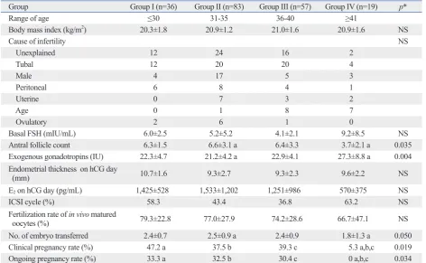
As shown in Table 1, causes of infertility, mean body mass index, and basal FSH level were not different between the four age groups. The mean antral follicle count was lowest in group IV, which was significantly different from that of group II. The dose of exogenous gonadotropins (ampoules) tended to be higher in group IV, but serum estradiol level at triggering day was similar in four groups. The fertilization rate of in vivo matured oocytes was similar among the four groups. The number of transferred embryos was lowest in group IV, and accordingly, the clinical pregnancy rate was significantly lower in group IV when compared with other age groups.
propriately. During ovarian stimulation, the oocyte popula-tion at the time of hCG may be heterogeneous, leading to retrieval of oocytes at different stages of maturation.
Currently, the mechanisms and the affecting factors to yield immature oocyte in stimulated IVF cycles are poorly understood. A high rate of oocyte immaturity was reported to be associated with young female age,5 the use of GnRH
antagonist rather than GnRH agonist,4 and urinary hCG as a
triggering agent rather than recombinant form.7 When all
IVF cycles stratified were by two patient age groups (<35 or ≥35 years), the incidence of abnormally higher oocyte immaturity (i.e. the cycles retrieving >50% immature oo-cytes) was found to be significantly higher in <35 group than ≥35 group (8.5% vs. 1.4%).5 It has also been reported
that urinary gonadotropin increases the yield of mature oo-cytes in patients with a previous cycle having a high inci-dence of oocyte immaturity by using recombinant FSH.7
We hypothesized that oocyte immaturity would be more common in older women undergoing stimulated IVF cycle because of their closer proximity to ovarian senescence and menopause. In the present study, we investigated the asso-ciation between woman’s age and oocyte immaturity in IVF cycles stimulated by single regimen: i.e. recombinant FSH (rFSH) and GnRH antagonist.
MATERIALS AND METHODS
A total of 195 IVF cycles stimulated by rFSH and GnRH antagonist during a period between 2003 and 2009 were se-lected. We obtained Institutional Review Board approval, although this was a retrospective comparative study. The mean age of women was 34.2±4.0 years with a range of 26-45 years. The infertility factors were identified as the follow-ing: tubal (n=56), unexplained (n=54), male (n=29), perito-neal (n=19), age factor (n=16), uterine (n=12), and ovulatory (n=9).
were immature ones in women aged 40 years or more.
DISCUSSION
In the present study, we attempted to investigated the rela-tion between patient’s age and the rate of immature oocytes in IVF cycles. The oocyte immaturity was the highest in women aged 40 years or more; however, oocyte immaturity The mean number of total retrieved oocyte, as well as the
[image:3.595.70.543.80.371.2]number of immature oocyte, tended to decrease as increas-ing age (Fig. 1). The resultant mean % of immature oocyte tended to be decreased as increasing age, however, this phe-nomenon was observed only in women aged 40 years or less (37.9% in group I, 32.4% in group II, and 23.3% in group III, p>0.05). The mean % of immature oocyte was the high-est in group IV with statistical significance, compared with group II and III. More than half of the retrieved oocytes
Table 1. Clinical and Laboratory Characteristics of the Study Subjects according to Female Age Group
Group Group I (n=36) Group II (n=83) Group III (n=57) Group IV (n=19) p*
Range of age ≤30 31-35 36-40 ≥41
Body mass index (kg/m2) 20.3±1.8 20.9±1.2 21.0±1.6 20.9±1.6 NS
Cause of infertility NS
Unexplained 12 24 16 2
Tubal 12 20 20 4
Male 4 17 5 3
Peritoneal 6 8 4 1
Uterine 0 7 3 2
Age 0 1 8 7
Ovulatory 2 6 1 0
Basal FSH (mIU/mL) 6.0±2.5 5.2±5.2 4.1±2.1 9.2±8.5 NS
Antral follicle count 6.3±1.5 6.6±3.1 a 6.4±3.3 3.7±2.1 a 0.035
Exogenous gonadotropins (IU) 22.3±4.7 21.2±4.2 a 22.9±4.1 27.3±8.8 a 0.004
Endometrial thickness on hCG day
(mm) 10.7±1.6 9.3±2.7 9.3±2.3 9.6±2.2 NS
E2 on hCG day (pg/mL) 1,425±528 1,533±1,202 1,251±986 570±375 NS
ICSI cycle (%) 58.3 43.4 36.8 63.2 NS
Fertilization rate of in vivo matured
oocytes (%) 79.3±22.8 77.0±27.9 74.2±28.6 66.7±47.1 NS
No. of embryo transferred 2.4±0.7 2.5±0.9 a 2.4±0.9 1.8±1.3 a 0.050
Clinical pregnancy rate (%) 47.2 a 37.5 b 39.3 c 5.3 a,b,c 0.019
Ongoing pregnancy rate (%) 33.3 a 32.5 b 30.4 c 0 a,b,c 0.034
*ANOVA test with Scheffe’s post-hoc test; same superscript means p<0.05 for each other within same row. NS, not significant.
Fig. 1. (A) The mean number of total retrieved oocytes (group I: ≤30 years old, II: 31-35 years, III: 36-40 years, IV: ≥41 years) (I-IV: p=0.001, II-IV: p=0.000, III-IV:
p=0.048, ANOVA test with Scheffe’s post-hoc test). (B) The mean number of immature oocytes (I-III: p=0.044, I-IV: p=0.008, II-IV: p=0.045). (C) The mean per-centage of immature oocytes (II-IV: p=0.049, III-IV: p=0.002).
0 0 0
2 0.5
1
10 4
1.5
20
6 2 30
8 2.5 40
10 3 50
12 3.5 60
M
ea
n
nu
m
be
r
M
ea
n
nu
m
be
r
M
ea
n
pe
rc
en
t (
%
)
Group I Group II Group III Group IV Group I Group II Group III Group IV Group I Group II Group III Group IV
9.25
3.31
37.9 9.57
3.16
32.4
7.02 2.14
23.3
2.84
1.53
52.5
[image:3.595.70.543.407.562.2]ty was similar between hCG-containing highly purified HMG and rFSH; 69% vs. 67.4%,15 and 71.2% vs. 70.8%.16
Urinary HMG also yielded a similar rate of mature oocyte compared with rFSH; 89.3% vs. 86.2%.17 Even significantly
less mature oocyte was reported by using urinary HMG compared with highly purified FSH; 80.6% vs. 88.8%.18
Therefore, currently available evidence does not support the superiority of gonadotropins including LH content, in terms of oocyte maturity.
GnRH antagonist protocol appears to have a better chance to obtain immature oocytes compared with GnRH agonist protocol.4 This may be due to greater heterogeneity in
folli-cle development in patients receiving antagonist protocol.19
In the present study, we selected single stimulation regimen (rFSH only with GnRH antagonist protocol) to eliminate possible confounding effect from variable regimens.
In conclusion, the rate of immature oocyte was not pro-portional to woman’s age; it was the highest in women ≥41 years, followed by young women ≤30 years. Further re-searches are needed regarding the identification of influenc-ing factors and the mechanisms for oocyte immaturity de-spite adequate ovarian stimulation. Furthermore, efforts should be continued to reduce its incidence in stimulated IVF cycle.
REFERENCES
1. Mandelbaum J, Junca AM, Belaisch-Allart J, Salat-Baroux J, Plachot M, Antoine JM, et al. [Oocyte maturation and intracyto-plasmic sperm injection]. Contracept Fertil Sex 1996;24:534-8. 2. Cha KY, Chian RC. Maturation in vitro of immature human
oo-cytes for clinical use. Hum Reprod Update 1998;4:103-20. 3. De Vos A, Van de Velde H, Joris H, Van Steirteghem A. In-vitro
matured metaphase-I oocytes have a lower fertilization rate but similar embryo quality as mature metaphase-II oocytes after intra-cytoplasmic sperm injection. Hum Reprod 1999;14:1859-63. 4. Nogueira D, Friedler S, Schachter M, Raziel A, Ron-El R, Smitz J.
Oocyte maturity and preimplantation development in relation to follicle diameter in gonadotropin-releasing hormone agonist or antagonist treatments. Fertil Steril 2006;85:578-83.
5. Moore AK, Arny MJ, Lynch K, Grow DR. Oocyte maturation ar-rest more common in younger patients undergoing IVF/ICSI. Fer-til Steril 2007;88(Suppl 1):S271.
6. Jee BC, Han SH, Moon JH, Suh CS, Kim SH; Seoul National University College of Medicine Assisted Reproductive Technolo-gy (SMART) Study Group. Influence of well defined protein source on in vitro maturation of human oocyte: human follicular fluid versus human serum albumin. Fertil Steril 2008;89:348-52. 7. Huddleston HG, Jackson KV, Doyle JO, Racowsky C. hMG
in-creases the yield of mature oocytes and excellent-quality embryos in patients with a previous cycle having a high incidence of oocyte tended to rather decrease as increasing age in women aged
40 years or less. In contrary to our hypothesis, the rate of immature oocytes was not strictly proportional to women’s age. This observation in part is consistent with the previous study which showed that oocyte immaturity was more fre-quent in <35-year group than ≥35-year group.5
It is highly likely that women aged 40 years or more have higher oocyte immaturity during stimulated IVF cycle be-cause ovarian senescence progresses in those group. How-ever, relatively higher oocyte immaturity was also noted in women aged 30 years or less, and the reason is still unclear. Young patients in general are high responders, therefore, they might have higher heterogeneity of follicular develop-ment. In the same context, they might have higher chance not to respond to usual dosage of hCG. Higher oocyte im-maturity in young women would be a reflection of their high follicular response, not attributing to intrinsic oocyte fac-tors. This explanation seems to be highly possible because the young women in our study yielded similar number of transferred embryos and had a higher clinical and ongoing pregnancy rate despite higher oocyte immaturity.
It was anecdotally reported that 27-year-old Turner mo-saicism showed an excellent ovarian response to stimula-tion during several IVF cycles, but a high proporstimula-tion (80%) of immature oocytes was noted.13 Since young woman with
Turner mosaicism is associated with impending premature ovarian failure, it can be assumed that infertile women who are impending ovarian failure may produce a large propor-tion of immature oocytes.
When high oocyte immaturity is noted in stimulated IVF cycle, extending duration of triggering has been recom-mended.14 In order to extend the hCG to retrieval interval,
transvaginal oocyte retrieval could be scheduled 41 hours post-hCG instead of conventional 35 hours.
Alternately, change of ovarian stimulation regimen has been suggested to be beneficial in patients with such a high proportion of immature oocyte;7 in a cohort of patients with
maturi-Hum Reprod 2002;17:1604-9.
15. Bosch E, Vidal C, Labarta E, Simon C, Remohi J, Pellicer A. Highly purified hMG versus recombinant FSH in ovarian hyper-stimulation with GnRH antagonists--a randomized study. Hum Reprod 2008;23:2346-51.
16. Baker VL, Fujimoto VY, Kettel LM, Adamson GD, Hoehler F, Jones CE, et al. Clinical efficacy of highly purified urinary FSH versus recombinant FSH in volunteers undergoing controlled ovarian stimulation for in vitro fertilization: a randomized, multi-center, investigator-blind trial. Fertil Steril 2009;91:1005-11. 17. Ng EH, Lau EY, Yeung WS, Ho PC. HMG is as good as
recombi-nant human FSH in terms of oocyte and embryo quality: a pro-spective randomized trial. Hum Reprod 2001;16:319-25. 18. Imthurn B, Macas E, Rosselli M, Keller PJ. Nuclear maturity and
oocyte morphology after stimulation with highly purified follicle stimulating hormone compared to human menopausal gonadotro-phin. Hum Reprod 1996;11:2387-91.
19. Huirne JA, Homburg R, Lambalk CB. Are GnRH antagonists comparable to agonists for use in IVF? Hum Reprod 2007;22: 2805-13.
immaturity. Fertil Steril 2009;92:946-9.
8. Kim BK, Lee SC, Kim KJ, Han CH, Kim JH. In vitro maturation, fertilization, and development of human germinal vesicle oocytes collected from stimulated cycles. Fertil Steril 2000;74:1153-8. 9. Picton HM. Oocyte maturation in vitro. Curr Opin Obstet Gynecol
2002;14:295-302.
10. Chian RC, Buckett WM, Abdul Jalil AK, Son WY, Sylvestre C, Rao D, et al. Natural-cycle in vitro fertilization combined with in vitro maturation of immature oocytes is a potential approach in in-fertility treatment. Fertil Steril 2004;82:1675-8.
11. Jurema MW, Nogueira D. In vitro maturation of human oocytes for assisted reproduction. Fertil Steril 2006;86:1277-91.
12. Oktay K, Demirtas E, Son WY, Lostritto K, Chian RC, Tan SL. In vitro maturation of germinal vesicle oocytes recovered after pre-mature luteinizing hormone surge: description of a novel approach to fertility preservation. Fertil Steril 2008;89:228.e19-22. 13. Moore AK, Lynch K, Arny MJ, Grow DR. Turner mosaicism