Possible Causes of Tooth Wear in Medieval Icelanders
Svend Richter
*, Sigfus Thor Eliasson
Faculty of Odontology, University of Iceland, Iceland
Copyright©2016 by authors, all rights reserved. Authors agree that this article remains permanently open access under the terms of the Creative Commons Attribution License 4.0 International License
Abstract
Objectives: The importance of the Icelandic Sagas as a source of information on the way of life and diet habits in Iceland and possibly other Nordic countries 1000 years ago is obvious. Extensive tooth wear in archaeological human skull material worldwide has been blamed on coarse diet. Near volcano Hekla, 66 skeletons dated from before 1104 were excavated from a graveyard. The purpose of this study was to determine the main causes of tooth wear in Icelanders 1000 years ago. Materials and methods: Available were 49 skulls for research. Two methods were used to evaluate tooth wear and seven for age estimation. An attempt was made to determine main causes of tooth wear in the light of likely diet and beverage consumption according to a computer search on food and drink customs described in the Icelandic Sagas. Results: Extensive tooth wear was seen in all groups, increasing with age. The first molars had the highest score with no difference between sexes. It had all the similarities seen in wear from coarse diet. In some instances it had similar characteristics as seen in erosion in modern Icelanders consuming excessive amounts of soft drinks. According to the Sagas, acidic whey was a daily drink and used for preservation of food in Iceland until recently. Conclusions: It is postulated that consumption of acidic drinks and food in addition to a coarse and rough diet, played a significant role in the dental erosion seen in ancient Icelanders.Keywords
Dental Erosion, Paleodontology, Icelandic Settlers1. Introduction
It is the norm to see extensive tooth wear in all ancient societies and considered to be a major cause of tooth problems[1-3]. Tooth wear studies based on skull material from archaeological excavations have mainly been attributed to factors related to the diet[4, 5]. Hardly ever is acid erosion mentioned as a possible cause of this observed tooth wear[6]. Wear is a generic term used in dentistry to describe the phenomena of attrition (proximal and occlusal inter-dental friction), abrasion (friction with the intervention of particles)
and erosion (chemical dissolution). This terminology suggests that these three phenomena act independently, whereas in fact it is more often the case that they interact simultaneously, which makes the diagnosis more difficult [3].
Skeletal remains from ancient populations can provide evidence for dental health. Investigating the ancient dentition can assist in reconstructing the lifestyle patterns and shed light on the dental and general health. Among the conditions to be looked at are ante mortem tooth loss, root abscesses, caries frequency, alveolar bone loss and occlusal tooth wear[7]. Enamel hypoplasia, hypomineralization of enamel appearing as lines or pits, particularly on incisors, can be an indication of malnutrition or infections during tooth formation[8].
Dental wear patterns are one of the most commonly utilized methods of age estimation in adult skeletal remains in archaeology. The use of this method arises from the high survival rate of teeth within archaeological contexts and the relative ease with which dental wear patterns can be observed and scored[9].
Studies by qualified dental personnel on dental health in ancient Icelanders have been almost none existent. Anthropologists and archaeologists have, however, noticed, registered and commented on excessive tooth wear without classifying or categorizing the type of wear[4, 5].
An anthropological and archaeological study was undertaken in 1931- 1939 at the Skeljastadir farm in Thjorsardalur in Iceland. Sixty six skeletons were excavated from an ancient graveyard, 5 infants, 2 children and 59 adults. Volcanic ash from the mountain Hekla, dated from 1104, covered the skeletons. The skeletons are preserved in the National Museum of Iceland. The purpose of this investigation was to undertake detailed and thorough odontological investigation on the skulls to cast a light on dental health in Viking Age Icelanders, especially prevalence and type of tooth wear[4, 7].
2. Materials and Methods
Of the 66 skeletons, 51 skulls could be used for the general dental health investigation and 915 teeth in 49 skulls were evaluated for the tooth- wear examination.
The two authors examined blinded the skulls and teeth. Agreement was more than 90% on all measurements. When there was a disagreement, an agreement was reached based on reexamination and discussion.
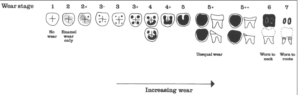
The adult skeletons were sexed using morphological characteristics from skull, mandible and in few instances, pelvis[10-14]. Seven methods were used for age estimation, five were based on tooth development[15-19], one on tooth wear[20] and one on ectodermal suture closure[21]. Two methods were used to register tooth wear. The first method was simply to register the wear into four categories: 1. no wear; 2. wear in enamel; 3. dentin exposed; 4. exposure of the pulp cavity. The second method was that developed by Brothwell, where wear was registered into 13 groups[22] as shown in Figure 1. The Brothwell method was limited to skeletons of subjects 18 years and older: 22 males, 21 females and one that could not be sexed. For statistical calculations, the scale for the wear stages was modified and numbered from 0-12 as shown in Table 1.
Tooth wear was examined with dental prope under good lighting and loupes with 2.8x magnification (Exam Vision ApS, Samsø, Denmark) and photographs with high resolution (Lester A. Dine Inc., Olympus Digital Camera
Model No C-5060, Florida, USA). For analyzing abscesses, conventional dental and radiographic examination was performed. For X-ray analysis a portable X-ray unit was used (Sirona, Helio Dent, Bensheim, Germany) and digital X-ray technology (Trophy RVG Digital X-ray Systems, Eastman Kodak Company, N.Y. USA). Statistical analyzes were made in the StatView statistical software package (StatView Software, SAS, Cary, USA) and the t-test was used for significance testing.
3. Results
A total of 1001 teeth were present in the 51 skulls examined. The number of missing teeth ante mortem in both jaws were 95 or less than 10% of teeth present, 36 in maxilla and 59 in mandible. A total of 28 individuals had lost teeth ante mortem, from one to thirteen teeth. Missing teeth post mortem were 281 and missing teeth with no information 225, which could raise the ratio of lost teeth ante mortem.
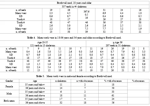
[image:2.595.64.550.400.554.2]Available for tooth wear examination were 49 skulls. Gender distribution was equal, 24 females, 24 males and 1 of unknown sex. Tooth wear for each tooth number is shown in Table 2, where 0 means no detectable wear, 1 wear in enamel, 2 wear exposing dentin and 3 wear exposing the pulp cavity.
Figure 1. Brothwell classification of molar wear. White denotes enamel, black exposed dentine. (From Mays S. The archaeology of human bones.
Routledge 2003: 59. Adapted from Brothwell 1981: figure 3.9).
Table 1. Modification of Brothwell tooth wear.
Brothwell wear stages (1981) 1 2 2+ 3- 3 3+ 4 4+ 4 5+ 5++ 6 7
Brothwell wear stages, modification 0 1 2 3 4 5 6 7 8 9 10 11 12
Table 2. Mean tooth wear for each tooth number according to the classification: 0 no wear, 1 wear in enamel, 2 dentin exposed and 3 exposure of pulp
cavity.
Mean tooth wear 915 teeth in 49 skeletons n. of teeth
Mean wear SD Tooth #
18 1,2 0,9 18
27 1,8 0,7 17
37 2,2 0,6 16
33 2,1 0,7 15
28 1,8 0,7 14
28 1,9 0,5 13
19 1,8 0,6 12
23 2,0 0,2 11
21 2,1 0,2 21
18 1,7 0,7 22
33 1,9 0,7 23
31 1,9 0,7 24
33 1,9 0,7 25
32 2,2 0,1 26
26 1,7 0,1 27
20 1,2 0,2 28 Tooth #
SD 0,9 48 0,6 47 0,4 46 0,7 45 0,5 44 0,6 43 0,5 42 2,2 41 0,3 31 0,4 32 0,6 33 0,5 34 0,7 35 0,4 36 0,4 37 0,9 38 Mean wear
Table 3. Mean tooth wear according Brothwell modification. Brothwell mod. 18 years and older
337 teeth in 44 skeletons n. of teeth
Mean wear SD Tooth # 19 2,3 0,6 18 26 4,5 0,7 17 367,6 0,5 16 31 6,9 0,4 26 24 4,4 0,6 27 16 1,8 0,5 28 Tooth # SD Mean wear
n. of teeth
[image:3.595.66.548.89.430.2]48 2,6 2,2 23 47 3,0 5,6 38 46 2,6 7,5 39 36 2,6 7,7 32 37 2,5 5,2 32 38 2,1 1,9 21
Table 4. Mean tooth wear in 18-36 years and 36 years and older according to Brothwell mod.
Age 18-35
121 teeth in 13 skeletons 207 teeth in 31 skeletons ≥ Age 36
n. of teeth Mean wear SD Tooth # 7 0,4 0,7 18 9 1,6 0,9 17 9 5,0 1,8 16 11 5,5 2,0 26 10 2,6 2,1 27 7 0,3 0,5 28 11 3,6 0,8 18 16 6,4 0,7 17 26 8,7 0,4 16 19 7,9 0,5 26 13 6,1 0,8 27 8 3,3 0,8 28 Tooth # SD Mean wear
n. of teeth
48 1,0 0,8 12 47 1,5 2,4 13 46 1,6 5,4 12 36 1,9 5,9 10 37 1,8 2,9 11 38 0,7 0,5 10 48 0,9 4,1 10 47 0,5 7,0 25 46 0,4 8,7 26 36 0,5 8,8 21 37 0,4 6,4 21 38 0,6 3,2 11
Table 5. Mean tooth wear in male and female according to Brothwell mod.
Gender Age n skeletons n with abscesses % with abscesses % abscesses
Female 35 years and below 11 2 18
36 years and above 14 7 50
Male 35 years and below 6 2 33
36 years and above 18 11 61
Both sexes 35 years and below 17 4 24
36 years and above 32 18 56
[image:3.595.62.547.544.747.2]Assessment of tooth wear according to age and sex, using the Brothwell method, is shown in Tables 3-5. The tooth wear was only measured in skulls of subjects classified as being aged 18 years and older: 21 males, 21 females and one skull that could not be sexed. Tooth wear was significantly higher in the age group 36 and older than for the 18-35 years group (p˂0,001) (Table 4), but no statistical difference between genders was noted (Table 5). Tooth wear with cuppings was registered in most molars in the younger age group and in the older age group where enamel was left (Fig. 2 and 3). Root abscesses were found in 22 skulls out of 49, or in 45% of cases. The abscesses were significantly more common in the 36 years and older group and significantly more common in males than females (p˂0,001) (Table 6) and the first molar showed the highest rate of abscesses (Table 7). Caries was almost nonexistent, being registered in just two teeth.
Table 6. Prevalence of root abscesses according to two age groups and sex.
Gender Age n skeletons n with abscesses % with abscesses % abscesses
Female 35 years and below 11 2 18
36
36 years and above 14 7 50
Male 35 years and below 6 2 33
54
36 years and above 18 11 61
Both sexes 35 years and below 17 4 24
45
36 years and above 32 18 56
Table 7. Prevalence of root abscesses presented for each tooth. Prevalence of abscesses for each tooth
1 9 3 2 1 1 1 1 5 1
18 17 16 15 14 13 12 11 21 22 23 24 25 26 27 28
48 47 46 45 44 43 42 41 31 32 33 34 35 36 37 38
4. Discussion
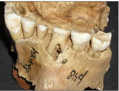
Tooth wear can be seen in archaeological material from all over the world. This wear is generally far more extensive than can be seen in current living populations. Ancient people consumed more course and rough unprocessed diet, resulting in extensive occlusal tooth wear. This wear has been shown to gradually increase with age [4, 10]. Usually the wear is most dominating on molars, starting in occlusal enamel and gradually reaching into dentin. Even though the wear reaches well into the dentin, the teeth appeared to remain functional since the odontoblastic activity prevents the wear to reach into the pulpal cavity because of secondary dentin formation[22] (Fig. 2).
Figure 2. Secondary dentin formation is seen in tooth 46. Tooth 36 has
excessive wear with pulp exposure and root abscess formation.
Figure 3. Increasing tooth wear according to time of eruption. Pulp
[image:4.595.313.552.78.261.2]exposure in tooth 16.
Figure 4. Root abscess in tooth 16 due to tooth wear.
The amount of wear can be graded by several methods. The simplest scoring of tooth wear is using only four categories; no wear, wear in enamel, wear into dentin and wear exposing the pulp cavity. When more detailed grading is preferred, several systems are available. The Brothwell method was elected to be used in the present study[14, 22] (Fig. 1). Mean wear of each tooth type can be seen in Table 2. The third molars have the least wear and the first molars the most, with the second molars in between.
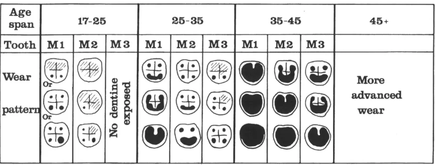
The amount of wear of the first molar indicates six years of wear when the second molar erupts. When third molar erupts, first molar has approximately twelve years wear and second molar six years wear. This can clearly be seen on Figures 3 and 4, which shows worn dentition from two angles. Different eruption times for the molars are, therefore, the basis for the age estimation. If, for example, second molar shows that it has been in the mouth for 12 years and the third for six years, it is likely that the individual has been about 24 years old. If second molar shows 18 years of wear, it is likely that the individual is around 30 years. Most of the adulthood can be determined the same way. The study by Miles indicated small differences in the wear rate between the three molars. He found the first molar to wear fastest with decreasing rate in the posterior direction. Thus, he showed that it took second molars 6,5 years and third molars 7 years to wear the same as first molars in 6 years[23]. These differences can be seen in Figure 3, explaining the age estimation method by Miles. Some studies have reported similar wear differentials[24], but others have reported equal rates of wear[25]. The Miles method was one of the methods used for age estimation in present study. Many investigators have confirmed the usefulness of tooth wear for age estimation [24, 26, 27].
[image:4.595.58.297.459.644.2]Figure 5. Estimated correspondence between adult age at death and molar wear phases for British material from Neolithic to medieval periods. (From Mays S. The archaeology of human bones. Routledge 2003: 63. Adapted from Brothwell 1981: fig. 3.9[24]).
When the tooth wear is so excessive that it opens into the pulp cavity, the effects on tooth physiology and pathology needs to be considered. Tooth wear is the cumulative loss of enamel and dentin and it has been considered natural that teeth wear throughout life. Usually the odontoblasts compensate for this wear with new, reactive, secondary dentin being laid down inside the pulp cavity, making the teeth functional and healthy[14]. Therefore, many investigators have used secondary or tertiary dentin as a foundation for developing methods for age estimation. These methods are based on radiographic measurements on formation of new dentin and shrinkage of the pulp cavity[19, 28].
Several teeth in this investigation had wear exposure into the pulp cavity. Jon Steffensen, who excavated and originally investigated this bone material, found only one definite case where bone changes indicated scurvy or more likely vitamin D deficiency[4], although scurvy was known to be a common disease in Iceland until the 19th century[5, 29]. Pindborg, supported by other authors, stated that scurvy could harm the odontoblasts, leading to irregular dentin formation that possibly wears at a faster rate[30]. Furthermore, Brothwell stated that in some instances tooth wear could be so much and so fast that the normal odontoblastic activity is not enough to compensate for the wear, resulting in an exposure into the pulp cavity[22]. Excessively worn dentition is shown in Figure 2, where tooth
number 36 has an exposure into pulp cavity, while visible secondary dentin formation has still compensated for the wear of tooth number 46. It is doubtful that vitamin C or D deficiency has resulted in exposure in only one first molar, although uneven wear is a possibility.
Root abscesses were found in almost half of the skulls. Since the caries rate was very low for the population it cannot explain the high root abscess incidence. A likely explanation is that excessive wear in some instances reached into the pulp cavity. This explanation is supported by the fact that first molars, which showed the highest score of tooth wear, (table 2) also had the greatest frequency of root abscesses (Table 7).
Figures 6
Figures 7 Figures 8
Figures 6-8. Similar appearance between the wear of medieval teeth and the erosion in young people today. Figure 8 with permission from Ulla Pallesen.
Figure 6 shows clearly that the buccal and lingual enamel surfaces show little or no tooth wear. This suggests that the observed tooth wear was not caused by gastric reflux, even though lifting and other heavy work might have caused problems such as hiatus hernia which would lead to gastric acid reaching the mouth and causing this typical pattern of tooth erosion. Although tooth erosion associated with gastric reflux disease is now quite common in Iceland, and has a typical clinical appearance, the prevalence in the medieval period is not known[33].
Iceland‘s history of food and nutrition can shed light on tooth wear in ancient Icelanders. Because of lack of salt, drying was one of two most popular ways for preserving food mostly used for fish that was air-dried and cured as stockfish[34]. Meat was also dried or smoked, but most often it was soured in lactic acid. Due to the lack of grain, there are many tales in the literature about the dietary peculiarities of the Icelanders, including that they ate dry fish instead of bread[34]. It is more than likely that in addition to coarse diet, dried foodstuffs contaminated by dust, and in Thjorsardalur volcanic ash, were responsible for the extensive tooth wear in the medieval Icelanders. Furthermore, the lack of grain
and fruits could be the explanation for the low caries rate in the skull sample.
The most characteristic Icelandic foodstuffs are two dairy products. The first whey (Icel. mysa), a watery part of milk that separates from the curds in the process of making the second major dairy product, skyr. Most of the whey was processed by pouring it into a wooden barrel with open holes in the lid for fermentation. When this process was finished mysa had turned into lactic acid, sýra, which was then mixed with water in the proportion of one part acid with 11 parts water. This drink was the everyday thirst quencher in Iceland until mid-20th century[35]. Lactose is a disaccharide sugar found in milk and makes up around 2–8% (by weight) of milk. As the whey fermented, about half of the lactose was converted to lactic acid along with other by-products[36]. The lactic acid was the most important food conservation medium in Iceland. Leathery meat was cooked and afterwards put into lactic acid to make it softer[35].
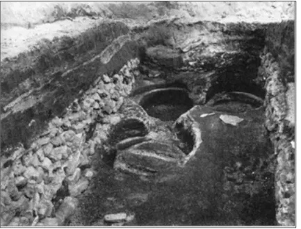
Figure 9. Barrel imprints in the storage room of the farm Stöng (from Ágústsson 1989)
Extensive studies have been made on erosion in young people in Iceland in recent years. Dr. Thorbjorg Jensdottir has been at the forefront of those who have studied the erosive effects of acidic beverages in the country. The capability of acidic beverages to erode dental enamel depends not only on the pH of the drink, but also on its buffering capacity. Her study showed that the erosive potential of whey, mysa, was very high[37]. Mysa and sýra in the mediaeval period is believed to have contained much higher concentrations of lactic acid than can be found in the same modern products manufactured under much different and more controlled conditions[36].
During the Viking period, storage of food or production of dairy products took place in small outhouses next to the longhouse. Up to the 20th century, Icelandic farms always included a room for the production of whey and skyr and large wooden barrels or storage trunks were typical features of storage rooms, as can be seen in Figure 9 from the farm Stong in Thorsardalur, not far from the archaeological site Skeljastadir.
5. Conclusions
In addition to coarse diet, dried meat and fish, probably contaminated by abrasive material, acidic beverages and acidic food, were responsible for the extensive tooth wear in
medieval Icelanders.
Acknowledgements
The study is a part of the research project Odontological investigation on archaeological human remains from Skeljastadir in Thjorsardalur which was supported by the Research Fund of the University of Iceland and the Research Fund of the Icelandic Dental Association. Thanks are extended to Professor Ulla Pallesen, Univ. of Copenhagen, Department of Odontology.
REFERENCES
[1] Molnar S. Tooth wear and culture: a survey of tooth function among some prehistoric populations. Curr Anthropol 1972;13:511-26.
[2] Whittaker DK, Davies G, Brown M. Tooth loss, attrition and temporomandibular joint changes in a Romano-British population. J Oral Rehabil 1985;12:407-19.
2012;57:214-29.
[4] Steffensen J. Knoglene fra Skeljastaðir i Þjórsárdalur. Forntida gårder i Island: meddelanden från den nordiska arkeologiska undersökningen i Island sommaren 1939 København: Munksgaard 1943:227-60.
[5] Gestsdóttir H. The palaeopathological diagnosis of nutritional disease: A study of the skeletal material from Skeljastaðir, Iceland. MSc dissertation Department of Archaeological Science, Univ of Bradford 1998.
[6] Johansson A. A cross-cultural study of occlusal tooth wear. Swed Dent J Suppl 1992;86:1-59.
[7] Þórðarson M. Skeljastaðir, Þjórsárdalur. Forntida gårder i Island: meddelanden från den nordiska arkeologiska undersökningen i Island sommaren 1939 København: Munksgaard 1943:121-336.
[8] Caufield PW, Li Y, Bromage TG. Hypoplasia-associated severe early childhood caries--a proposed definition. J Dent Res 2012;91:544-50.
[9] Millard AR, Gowland RL. A Bayesian approach to the estimation of the age of humans from tooth development and wear. Archeologia e Calcolatori 2002;13:197-210.
[10]Richter S. Odontological investigation on archaeological human remains from Skeljastadir in Thorsardalur. Thesis submitted for Master of Science degree University of Iceland, Faculty of Odontology 2005:1-66.
[11]Bass W. Human Osteology: A loboratory and Field Manual of the Human Skeleton. Specials Publication No. 2. Missouri Arcgaeological Society Colombia, Missouri 4th ed 1995. [12]Ubelaker DH. Human Skeletal Remains: Excavation, Analysis,
Interpretation, 2nd edn Washington DC: Taraxacum; 1989. [13]Duric M, Rakocevic Z, Donic D. The reliability of sex
determination of skeletons from forensic context in the Balkans. Forensic Sci Int 2005;147:159-64.
[14]Mays S. The archaeology of human bones. Routledge. Redrawn from Miles (1963: figure 10). 2003:61.
[15]Demirjian A, Goldstein H. New systems for dental maturity based on seven and four teeth. Ann Hum Biol 1976;3:411-21. [16]Haavikko K. Tooth formation age estimated on a few selected teeth. A simple method for clinical use. Proc Finn Dent Soc 1974;70:15-9.
[17]Kullman L, Johanson G, Akesson L. Root development of the lower third molar and its relation to chronological age. Swed Dent J 1992;16:161-7.
[18]Mincer HH, Harris EF, Berryman HE. The A.B.F.O. study of third molar development and its use as an estimator of chronological age. J Forensic Sci 1993;38:379-90.
[19]Kvaal SI, Kolltveit KM, Thomsen IO, Solheim T. Age estimation of adults from dental radiographs. Forensic Sci Int1995;74:175-85.
[20]Miles AEW. Dentition in the Estimation of Age. J Dent Res 1963;42:255-63
[21]Meindl RS, Lovejoy CO. Ectocranial suture closure: a revised method for the determination of skeletal age at death based on the lateral-anterior sutures. Am J Phys Anthropol 1985;68:57-66.
[22]Brothwell D. Digging up bones: The excavation, treatment and study of human skeletal remains. Cornell University Press. 1981:71-2.
[23]Miles AEW. The Miles Method of Assessing Age from Tooth Wear Revisited. Journal of Archaeological Science 2001;28:973-82.
[24]Kieser J, Preston C, Evans W. Skeletal age at death: an evaluation of the Miles method of ageing Journal of Archaeological Science 1983;10:9-12.
[25]Nowell GW. An evaluation of the miles method of ageing using the Tepe Hissar dental sample. Am J Phys Anthropol 1978;49:271-6.
[26]Tomenchuk J, Mayhall JT. A correlation of tooth wear and age among modern Igloolik eskimos. Am J Phys Anthropol 1979;51:67-77.
[27]Richards LC, Miller SL. Relationships between age and dental attrition in Australian aboriginals. Am J Phys Anthropol 1991;84:159-64.
[28]Paewinsky E, Pfeiffer H, Brinkmann B. Quantification of secondary dentine formation from orthopantomograms--a contribution to forensic age estimation methods in adults. Int J Legal Med 2005;119:27-30.
[29]Johnsen B. Food in Iceland 874-1550. Medicinhistorik Årsbok 1968 1968:1-11.
[30]Pindborg JJ. De hårde tandvævs sygdomme. Munksgaard, København 1965:142-6.
[31]Arnadottir IB, Saemundsson SR, Holbrook WP. Dental erosion in Icelandic teenagers in relation to dietary and lifestyle factors. Acta Odontol Scand 2003;61:25-8.
[32]Khan F, Young WG, Law V, Priest J, Daley TJ. Cupped lesions of early onset dental erosion in young southeast Queensland adults. Aust Dent J 2001;46:100-7.
[33]Holbrook WP, Furuholm J, Gudmundsson K, Theodors A, Meurman JH. Gastric reflux is a significant causative factor of tooth erosion. J Dent Res 2009;88:422-6.
[34]Gísladóttir H. Eldhús og matur á Íslandi. Cand Mag disertation, Univ of Iceland 1991.
[35]Mehler N. From self-sufficiency to external supply and famine: Foodstuffs, their preparation and storage in Iceland. Processing, Storage, Distribution of Food: Food in the Medieval Rural Environment RURALIA (Book 8) Publisher: Brepols Publishers 2011;8:173-86.
[36]Lanigan LT, Bartlett DW. Tooth wear with an erosive component in a Mediaeval Iceland population. Arch Oral Biol 2013;58:1450-6.