Copyright © 1998, American Society for Microbiology. All Rights Reserved.
Outbreak of Staphylococcus schleiferi Wound Infections: Strain
Characterization by Randomly Amplified Polymorphic DNA
Analysis, PCR Ribotyping, Conventional Ribotyping,
and Pulsed-Field Gel Electrophoresis
JAN KLUYTMANS,
1* HANS BERG,
1PAUL STEEGH,
1FRANC¸OIS VANDENESCH,
2JEROME ETIENNE,
2ANDALEX
VANBELKUM
3Department of Clinical Microbiology, Ignatius Hospital Breda, 4800 RK Breda,
1and Department of Medical Microbiology &
Infectious Diseases, University Hospital Rotterdam, 3015 GD Rotterdam,
3The Netherlands, and Faculte´ de Me´decine,
Laboratoire de Bacte´riologie, UPRES EA 1655, 69372 Lyon cedex 08, France
2Received 23 January 1998/Returned for modification 9 March 1998/Accepted 12 May 1998
Within a 1-year period, six surgical-site infections (SSI) caused by Staphylococcus schleiferi were observed in
the department of cardiac surgery of Ignatius Hospital, Breda, The Netherlands. Since outbreaks caused by
this species of coagulase-negative staphylococci have not been described before, an extensive environmental
survey and a case control study were performed in combination with molecular typing of the causative
microorganism in order to identify potential sources of infection. Variability, as detected by four different
genotyping methods (random amplification of polymorphic DNA [RAPD], conventional and PCR-mediated
ribotyping, and pulsed-field gel electrophoresis [PFGE] of DNA macro restriction fragments), appeared to be
limited both among the clinical isolates and among several control strains obtained from various unrelated
sources. Among unrelated strains, RAPD and PCR-mediated ribotyping identified two types only, whereas
seven different types were identified in a relatively concordant manner by conventional ribotyping and PFGE.
The latter two procedures proved to be the most useful tools for tracking the epidemiology of S. schleiferi. Four
of the outbreak-related strains were identical by both methods, and two isolates showed limited differences. In
the search for a potential source of S. schleiferi infection, two slightly different PFGE types were encountered
on several occasions in the nose of a single surgeon. These strains were, however, clearly different from the
outbreak type. In contrast, S. schleiferi cultures remained negative for two persons identified on the basis of
case control analysis. It was demonstrated that SSI caused by S. schleiferi had a clinical impact for patients
comparable to that of a wound infection caused by Staphylococcus aureus. This report describes the first
well-documented outbreak of S. schleiferi infection. A source of the outbreak was not detected.
Staphylococcus schleiferi was recognized in the late 1980s as
a new species of coagulase-negative staphylococci (CoNS) (5).
Since then, this pathogen has been recovered from several
kinds of infections in humans, e.g., brain empyema,
surgical-site infections (SSI), intravascular device-related bacteremia,
infections of implanted prosthetic material (including
pace-makers [3]), and endocarditis (4, 13). Its involvement in
uri-nary tract infections was considered to be proven in 0.7% of
404 infections caused by CoNS (18). The pathogenicity of S.
schleiferi was confirmed in a model study of abscess formation
in mice (14). S. schleiferi was shown to be more virulent than,
for instance, Staphylococcus warneri or Staphylococcus hominis.
Moreover, all S. schleiferi strains produce beta-hemolysin,
lipase, and esterase as putative virulence factors.
Little is known of the epidemiology of S. schleiferi; for this
reason, S. schleiferi strains from diverse sources have been
studied by various genotyping methods in order to define the
genetic diversity within the species. Plasmid typing appeared to
be unsuccessful because extrachromosomal elements were
present in only a small fraction of strains (8). DNA restriction
analysis with five different restriction enzymes showed no
di-vergence in a diverse group of 31 strains. Ribotyping appeared
to be more adequate in detecting genetic polymorphisms
among these isolates (8). In a preliminary pulsed-field gel
electrophoresis (PFGE) trial, a single strain of S. schleiferi was
included (20). The PFGE fingerprint obtained for this strain
clearly separated it from isolates of other species of CoNS. A
subsequent study, including five S. schleiferi strains, once again
revealed the genetic homogeneity of the species: only minor
variation was observed upon SmaI digestion of genomic DNA
and separation of the macro restriction fragments, either by
PFGE or by field inversion gel electrophoresis (15, 21, 30).
However, despite the availability of technically adequate typing
technology, the precise clinical epidemiology of S. schleiferi
remained unknown, since major outbreaks of infection due to
this species had not been described.
In our department of cardiac surgery (Ignatius Hospital,
Breda, The Netherlands), six patients nursed within the
de-partment developed SSI with S. schleiferi in 1 year. All
infec-tions involved the sternotomy site. Since outbreaks of S.
schle-iferi infection have not been reported before, an investigation
into the source of these infections was performed. This
inves-tigation involved environmental sampling, a case control study,
and molecular typing of the outbreak-related and
environmen-tal strains.
MATERIALS AND METHODS
Setting.In the cardiac surgery department of Ignatius Hospital in Breda, approximately 1,500 cardiac surgical procedures are performed each year. The department consists of an operating theater, a postoperative intensive care unit,
* Corresponding author. Mailing address: Department of Clinical
Microbiology, Ignatius Hospital Breda, P.O. Box 90158, 4800 RK
Breda, The Netherlands. Phone: 31 76 5258015. Fax: 31 76 5138636.
E-mail: jkluytmans@ignatius.nl.
2214
on May 15, 2020 by guest
http://jcm.asm.org/
and a general postoperative ward. There is an active infection control policy which includes continuous surveillance of postoperative sternal wound infec-tions. Overall, the deep SSI rate was approximately 1% during the years 1991 to 1996, and approximately half of these infections were caused by Staphylococcus
aureus.
Bacteriology.Surgical sites are routinely monitored for signs of infection, and wound sampling with sterile cotton swabs is performed whenever an infection is suspected. Prior to sampling, the surface of the wound is cleaned with a disin-fecting agent. Swabs are transported to the microbiology laboratory; for all different morphotypes of staphylococci growing in the resulting cultures, a slide agglutination test (Staphaurex Plus; Murex Diagnostics, Breukelen, The Neth-erlands) and a test for the presence of heat-stable thermonuclease are routinely performed. If these two tests are both positive the isolate is considered to be S.
aureus, and if the tests are both negative the isolates are considered to be CoNS.
If the tests are discordant, a tube coagulase test and a biochemical identification test with Api ID32 Staph (bioMerieux, Lyon, France) are performed. After the outbreak of S. schleiferi was recognized, this procedure was modified by adding the tube coagulase test to the routinely performed tests. Based on these proce-dures, S. schleiferi was identified and isolated from clinical samples of six patients from September 1995 to September 1996.
Environmental sampling.From all surgeons, anesthetists, nurses of the oper-ating theater and the postoperative wards, and technicians for extracorporeal circulation, nasal swabs were obtained for culture. From the surgeons, hands were sampled as well. After actively moving one hand in a sterile surgical glove containing sterile broth for one minute, the broth was used for culture. Both the swabs and the broths were inoculated on blood agar plates and incubated at 37°C for 48 h. The outbreak-related isolates of S. schleiferi showed characteristic beta-hemolysis after this period (10). Furthermore, during 10 surgical sessions, environmental samples were collected while a surgical procedure was being performed. Sampling methods included air settling plates and active air sam-pling.
Case control study.Prospective surveillance for SSI has routinely been per-formed in the department of cardiothoracic surgery since 1991. Criteria for the presence of SSI are those of the Centers for Disease Control and Prevention (11). The six patients with SSI caused by S. schleiferi served as experimental subjects. Two control groups were selected. First, 24 patients were randomly selected from among patients who had had operations during the time period in which the six S. schleiferi SSI cases were identified. Second, all patients who had developed SSI with S. aureus in 1995 and 1996 were selected as a control group. From S. schleiferi SSI patients and controls, the following variables were re-corded: patient identification, age, sex, height, weight, underlying diseases, im-munosuppressive drugs, diabetes mellitus, smoking habits, New York Heart Association (NYHA) score (a score for the severity of cardiovascular disease, with a range in increasing severity of 1 to 4), date of admission, date of surgery, date of discharge, surgeons (first and assistant), anesthetist, operating room nurses (first assistant and second assistant), technician for extracorporeal circu-lation, perioperative antibiotic prophylaxis, duration of surgery, duration of ex-tracorporeal circulation, operating room, volume of perioperative loss of blood, size of blood transfusion, emergency procedure, rethoracotomy, postoperative infections at other sites, and outcome (survival). The results were analyzed with Statistical Package for the Social Sciences software. S. schleiferi SSI patients were compared with controls, and crude odds ratios were determined. Statistical significance was determined with Fisher’s exact test for categorical variables or the t test for continuous variables. To find out if risk factors were independent, multiple logistic regression was performed. Statistical significance was accepted
at P of,0.05.
Molecular typing. The six outbreak-related isolates and a collection of S.
schleiferi isolates from the environmental samples were included in the analysis
(see Table 3 for a description of the strains). For comparison and validation, a reference collection consisting of 10 epidemiologically unrelated strains of di-verse geographical origin was included in all studies. All typing procedures were performed and analyzed without knowledge of the origin of the isolates (blind).
RAPD.Random amplification of polymorphic DNA (RAPD) was performed essentially as described previously (26–28). Bacteria were treated with lyso-staphin (35 mg/ml; Sigma, Zwijndrecht, The Netherlands) and subsequently lysed by the addition of guanidinium containing lysis buffer. DNA was purified by
affinity chromatography onto Celite (0.2 g/ml; Jansen Pharmaceuticals, Beerse, Belgium) according to established protocols (1). The DNA concentration in the resulting eluates was determined by agarose gel electrophoresis (Hispanagar; Sphaero Q, Leiden, The Netherlands). Samples of the DNA preparations were coelectrophoresed with known amounts of lambda DNA. After staining, the amounts were estimated by comparison of fluorescence intensities upon UV transillumination of the gel. For RAPD, SuperTaq DNA polymerase (Sphaero Q) was used, and cycling was performed in BioMed (Theres, Germany) PCR machines (model 60). Primers used were ERIC1 and ERIC2 (32) or RAPD1 and RAPD7 (25) in single amplification reactions. Amplicons were analyzed on agarose gels, and the resulting fingerprints were photographed with a charge-coupled device charge-coupled to a thermoprinter (Progress Control; Mitsubishi, Waal-wijk, The Netherlands).
PCR ribotyping.PCR ribotyping was performed according to an optimized protocol (16) based on prior publications (2, 9). The 16S-to-23S intergenic region was amplified with primers sp1 and sp2, and the amplicons were separated on agarose gels. The ribopatterns were visualized by UV transillumination after ethidium bromide staining.
Conventional ribotyping.Ribotyping was performed as described previously (8). In short, DNA was isolated from staphylococcal cultures by standard pro-cedures (19), and HindIII digests were prepared according to recommendations of the manufacturer of the restriction enzyme (Boehringer, Mannheim, Germa-ny). DNA fragments were size separated by electrophoresis and blotted onto nylon membranes. Plasmid pKK3535, containing the rrnB ribosomal operon of
Escherichia coli, was used as a probe (8). The probe was labeled with
digoxigenin-11-dUTP by random priming, and chemiluminescence was generated with Lu-migen PPD as the substrate (Boehringer). Ribotypes were finally visualized by exposure of X-ray films.
PFGE. PFGE was carried out based on protocols previously described for DNA from S. aureus or other species of CoNS (17, 24, 29). A suspension of bacteria was mixed in a 1:1 ratio with 1% InCert agarose (FMC Bioproducts, Rockland, Maine). Agarose plugs were prepared with Bio-Rad (Veenendaal, The Netherlands) casting forms and incubated with lysostaphin (Sigma). Sphero-plasts were lysed by incubating the plugs in buffer containing 1% sodium dodecyl sulfate and 1 mg of proteinase K (Boehringer) per ml. Plugs were washed six times for 30 min each in 10 mM Tris-HCl (pH 8.0)–1 mM EDTA and stored at 4°C. DNA within half a plug was digested by SmaI (Boehringer), and PFGE was carried out in 1% SeaKem GTG agarose gels (FMC Bioproducts). The buffer
consisted of 0.53Tris-borate-EDTA. Electrophoresis was performed in a
Bio-Rad CHEF Mapper. Running time was 22 h with linear ramping from 2.16 to
44.69 s at an angle of 120° (60°/260°). The voltage was 6 V/cm, gel dimensions
were 120 by 140 by 5 mm, and the temperature was set at 14°C. Gels were stained with ethidium bromide and photographed with instant Polaroid equipment. Differences in banding patterns were documented by at least two independent observers. Strains belonging to a single PFGE type should display electrophero-grams that differ in a maximum of three bands. If more differences in DNA restriction fragment migration are observed, this is considered to be a new type. This interpretation is according to general guidelines issued by an American working party (22, 23).
RESULTS
Patients and case control studies.
Between September 1995
and September 1996, six patients developed SSI with S.
schle-iferi (Table 1). This accounted for 40% of the number of SSI
observed in this particular period, which strongly underscores
the potential outbreak relatedness. The SSI rate in the year of
the outbreak was 1.0%. This was due to 15 SSI, 12 of which
were deep SSI. The deep SSI rate in this year was 0.8%. Eight
SSI were caused by S. aureus, one by Staphylococcus
epidermi-dis. All patients with an S. schleiferi SSI were males; their
average age was 63 years. S. schleiferi SSI patients were
com-parable with both control groups with regard to all
preopera-TABLE 1. S. schleiferi SSI patient data included in the case control study
aPatient
no. Date of surgery(mo/day/yr) Age(yr) Length of stay(days) S A N OR Kind of SSI Days beforeonset of SSI PFGEtype Ribotype
1
9/27/95
57
29
Yes
Yes
No
4
Deep
11
C
C
2
10/03/95
62
22
Yes
No
Yes
3
Deep
45
B
E
3
10/27/95
64
22
No
No
No
2
Deep
11
B
G
4
7/15/96
55
29
Yes
Yes
Yes
1
Superficial
10
B
E
5
7/31/96
73
28
No
Yes
Yes
2
Deep
12
B
E
6
8/29/96
69
25
No
Yes
No
3
Superficial
8
B
E
aAll patients were male. Abbreviations: S, surgeon A; A, anesthetist A; N, nurse A; OR, operating room.
on May 15, 2020 by guest
http://jcm.asm.org/
[image:2.612.50.556.81.164.2]tive variables included in the case control study (Table 2). For
the perioperative variables, a significant association was
ob-served with the presence during surgery of an anesthetist (A)
and an operating room nurse (A). Both anesthetist A and
nurse A were repeatedly sampled for S. schleiferi carriage, but
this species was never cultured. From one of the surgeons (A),
S. schleiferi was isolated repeatedly. In Table 2 it is shown that,
contrary to expectations, he was not significantly associated
with the S. schleiferi SSI patients. To control for possible
con-founding, logistic regression analysis was performed. The
pres-ence of anesthetist A, operating room nurse A, or surgeon A
and age, gender, NYHA score, and smoking habits were
en-tered into the model. Anesthetist A (P
5
0.041) and operating
room nurse A (P
5
0.018) were both identified as independent
risk factors for development of SSI with S. schleiferi when
noninfected patients were the control group. When patients
with an S. aureus SSI were considered controls, only anesthetist
A (P
5
0.002) was identified as an independent risk factor. The
pathogenicity of S. schleiferi can be deduced from Table 2. The
median postoperative length of stay was significantly longer for
patients with S. schleiferi (27 days) than for patients without
SSI (11 days) and was comparable to that of patients suffering
from S. aureus infection (26 days). The mean highest value of
C-reactive protein was higher in the group of S.
schleiferi-infected individuals than in the nonschleiferi-infected group; this
differ-ence approached statistical significance.
Molecular typing.
Molecular typing data are categorized
into two groups, based on the observed and overlapping
reso-lution of RAPD and PCR ribotyping versus Southern
hybrid-ization-based ribotyping and PFGE.
PCR ribotyping and RAPD.
PCR ribotyping and RAPD
experimental data are depicted in Fig. 1 and 2. PCR ribotyping
reveals homogeneity among the strains. Only strain 26,
en-countered as a noninvasive colonizer in the nose of a surgical
patient, differed from the other strains, in the sense that the
two smaller amplicons were lacking. RAPD analysis employing
two combinations of primers (ERIC1-ERIC2 and
RAPD1-RAPD7) in two separate assays was performed. The strain
grouping deduced from the PCR ribotyping data was
con-firmed by the RAPD tests. The only deviating fingerprint,
again, was acquired for strain 26. Furthermore, all PCR
fin-gerprints are identical, except for some minor differences in
band staining intensity.
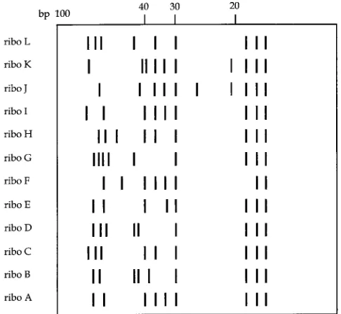
PFGE and ribotyping.
The experimental results of PFGE
analysis are depicted in Fig. 3. The numbers of DNA macro
restriction fragments vary from 13 to 15; of these, 12 are
universally present. If these results were interpreted on the
basis of guidelines issued previously (22, 23), all isolates would
have been considered clonally related. However, if single band
differences are taken into consideration as well, different types
can be distinguished. Table 3 surveys all of the typing data,
including those based on single band differences in the PFGE
electropherograms. It is comforting to note that this
classifica-tion is largely corroborated by the convenclassifica-tional ribotyping data
(Table 3). A schematic representation of the seven different
ribotyping patterns is shown in Fig. 4.
DISCUSSION
[image:3.612.58.548.80.318.2]This report describes the first documented outbreak of S.
schleiferi infection. In the laboratory, S. schleiferi is difficult to
[image:3.612.310.547.639.682.2]FIG. 1. PCR-mediated ribotyping of S. schleiferi strains collected during the present study. Strain numbers are indicated at the top of the lanes and corre-spond with those given in Table 3. Note that only for strain 26 is an aberrant DNA banding pattern observed. On the left (lane M), a 100-bp length marker is displayed; fragments with sizes of 600 and 100 bp are highlighted.
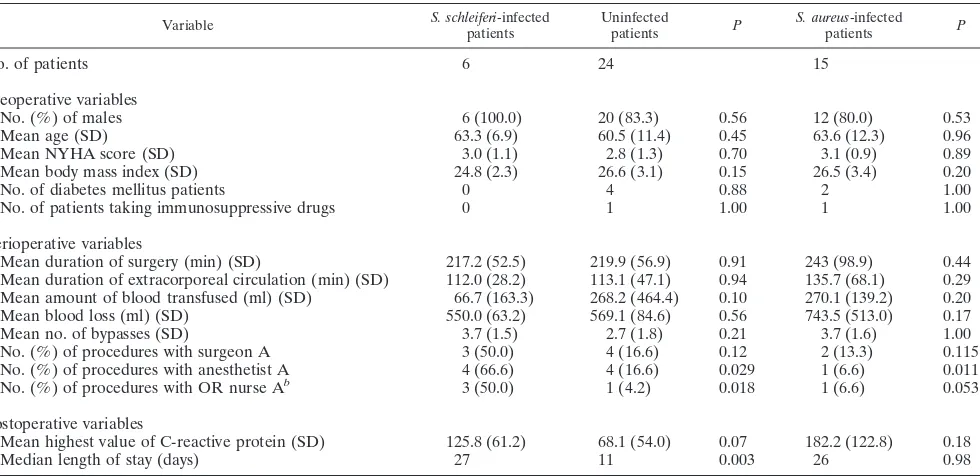
TABLE 2. Case control data
aVariable S. schleiferi-infectedpatients Uninfectedpatients P S. aureus-infectedpatients P
No. of patients
6
24
15
Preoperative variables
No. (%) of males
6 (100.0)
20 (83.3)
0.56
12 (80.0)
0.53
Mean age (SD)
63.3 (6.9)
60.5 (11.4)
0.45
63.6 (12.3)
0.96
Mean NYHA score (SD)
3.0 (1.1)
2.8 (1.3)
0.70
3.1 (0.9)
0.89
Mean body mass index (SD)
24.8 (2.3)
26.6 (3.1)
0.15
26.5 (3.4)
0.20
No. of diabetes mellitus patients
0
4
0.88
2
1.00
No. of patients taking immunosuppressive drugs
0
1
1.00
1
1.00
Perioperative variables
Mean duration of surgery (min) (SD)
217.2 (52.5)
219.9 (56.9)
0.91
243 (98.9)
0.44
Mean duration of extracorporeal circulation (min) (SD)
112.0 (28.2)
113.1 (47.1)
0.94
135.7 (68.1)
0.29
Mean amount of blood transfused (ml) (SD)
66.7 (163.3)
268.2 (464.4)
0.10
270.1 (139.2)
0.20
Mean blood loss (ml) (SD)
550.0 (63.2)
569.1 (84.6)
0.56
743.5 (513.0)
0.17
Mean no. of bypasses (SD)
3.7 (1.5)
2.7 (1.8)
0.21
3.7 (1.6)
1.00
No. (%) of procedures with surgeon A
3 (50.0)
4 (16.6)
0.12
2 (13.3)
0.115
No. (%) of procedures with anesthetist A
4 (66.6)
4 (16.6)
0.029
1 (6.6)
0.011
No. (%) of procedures with OR nurse A
b3 (50.0)
1 (4.2)
0.018
1 (6.6)
0.053
Postoperative variables
Mean highest value of C-reactive protein (SD)
125.8 (61.2)
68.1 (54.0)
0.07
182.2 (122.8)
0.18
Median length of stay (days)
27
11
0.003
26
0.98
aP values represent comparisons between S. schleiferi-infected patients and uninfected patients and between S. schleiferi-infected patients and S. aureus-infected
patients.
bOR, operating room.
on May 15, 2020 by guest
http://jcm.asm.org/
differentiate from S. aureus: the species are morphologically
similar, and S. schleiferi subsp. schleiferi produces both
clump-ing factor and heat-stable thermonuclease. For these and
maybe other reasons, this pathogen may be underreported. If
only slide coagulase testing and assessment of thermostable
nuclease were performed diagnostically, the present outbreak
would have been considered to be due to S. aureus. However,
a negative result in the tube coagulase test performed by one of
the technicians revealed that the isolate was S. schleiferi, which
was confirmed by biochemical analysis with API ID32 Staph.
Subsequent reexamination of all of the clinical S. aureus strains
from the preceding year (1995) revealed two more patients
infected with S. schleiferi (patients 1 and 2). This illustrates how
a potentially misidentified bacterial species can turn into an
“emerging” pathogen. According to standard microbiology
laboratory procedures, performance of a slide agglutination
test for clumping factor combined with a test for
thermonucle-ase is sufficient to discriminate S. aureus from CoNS. However,
S. schleiferi subsp. schleiferi would also be considered S. aureus
by this approach. Therefore, the magnitude of S. schleiferi
infection in clinical practice may be underestimated. This
ob-servation underscores the usefulness of including a tube
coag-ulase test in routine procedures to differentiate S. aureus from
other staphylococcal species.
A variety of accurate procedures for correct identification of
clinical strains of CoNS have been described lately, although
potential experimental pitfalls have been demonstrated for
“difficult” isolates (31). Gas-liquid chromatography of fatty
acids, though not yet within reach of the routine diagnostic
laboratory, appears to be reliable and robust (21). Automated
Microscan identification (Pos ID and Rapid Pos ID panels) of
species was shown to be less reliable for the infrequently
oc-curring species of CoNS (7). The procedure we used during the
present study for definitive identification of S. schleiferi (API
ID32 Staph) was demonstrated to be an accurate means of
identification. In a multicenter study, it led to 100% efficient
characterization of this species (12). This reliability was
under-scored in the present study: all S. schleiferi strains included
shared several genotypic characteristics as well, most clearly
demonstrated by the homogeneity of the RAPD and PCR
ribotyping data. This implies that the current collection of
strains (n
5
26) could serve the purpose of validating novel
species-specific identification assays, such as heat shock
pro-tein-based species identification tests (6).
It was shown previously that PFGE performed on DNA
isolated from strains of S. schleiferi detected limited genetic
heterogeneity among the strains (30). In the present study,
however, PFGE together with conventional ribotyping turned
out to be the typing combination of choice for analysis of
S. schleiferi strains. PCR ribotyping was inadequate, as was
RAPD employing the current combinations of random or
ERIC primers. The latter two procedures have been applied
successfully for other species, which indicates that S. schleiferi
may be genetically a rather homogeneous species. This
puta-tive clonality is further corroborated by strikingly similar
PFGE fingerprints (Fig. 3). Other studies have tried to
opti-mize typing methods (among them PFGE) for S. schleiferi
without satisfactory results (8, 15, 20, 30). The conclusion is
that the results to date all point toward S. schleiferi being
a rather homogeneous species genetically. Further studies
should reveal whether alternative approaches yield better
re-sults. The question of whether minor differences are relevant
can be answered only after more profound investigation into
this relatively rare species. In view of the higher heterogeneity
of unrelated strains by PFGE (six different patterns among 10
strains) than of outbreak-related strains (two patterns among 6
strains), we consider the outbreak-related strains with identical
types to be clonally related.
Four of the six S. schleiferi isolates encountered in the
car-diac surgery patients were identical with respect to the
[image:4.612.138.460.70.255.2]param-FIG. 2. RAPD analysis of S. schleiferi strains collected during the present study. The top panel shows results obtained by the combined application of primers ERIC1 and ERIC2; the fingerprints in the bottom panel were generated with the RAPD1-RAPD7 combination. On the left and between lanes 10 and 11 (lanes M), a 100-bp length marker is displayed; fragments with sizes of 900 and 400 bp are highlighted.
FIG. 3. PFGE data obtained for S. schleiferi strains collected during the present study. On the right an array of bacteriophage lambda DNA concatemers is shown; the sizes of two of the fragments are indicated; fragments differ in length by units of 50 kbp. Note that the result obtained for strain 26 is not depicted; this strain was analyzed on a separate gel, but the fingerprint strongly resembled the basic patterns shown here.
on May 15, 2020 by guest
http://jcm.asm.org/
[image:4.612.66.273.610.675.2]eters examined. Five of the six isolates had the same PFGE
pattern. This provided proof of an ongoing outbreak, and,
theoretically, a source should have been present during the
outbreak period. However, neither microbiological screening
nor detailed case control studies revealed a potential reservoir
for the outbreak-related genotype. In conclusion, it can be
stated that S. schleiferi can cause significant infections in a
clinically persistent fashion: in our case a single type was
en-countered regularly within a period of nearly a year.
Unfortu-nately, the source of infection remained enigmatic. An
appar-ent source, one of the surgeons who persistappar-ently carried S.
schleiferi in the nose, was ruled out by typing of the strain and
by the outcome of the case control study: the nasal inhabitant
differed from the outbreak strain. On the other hand, the
individuals implicated as possible sources of infection by the
case control analysis (anesthetist A and operating room nurse
A) did not carry S. schleiferi despite repeated culture assays.
Consequently, causal involvement could not be proven. This
may have been a consequence of the insensitivity of
bacterio-logical detection of S. schleiferi, in which experience is limited.
Experience is limited not only with bacteriological techniques
but also with the epidemiology of S. schleiferi. The ecological
niches of this microorganism are at present unknown. Also, it
is not known if carriage is persistent or transient. The
obser-vation of this outbreak strongly suggests that there has been a
persistent source in the department, either human or in the
inanimate environment. Unfortunately, the source of the
out-break was not identified.
[image:5.612.308.551.457.681.2]FIG. 4. Schematic representation of the different ribotyping patterns (ribo) obtained from strains included in the present study. The designations listed on the left correspond to the ribotype patterns given in Tables 1 and 3. Patterns H through L are patterns which have been observed in other S. schleiferi strains. At the top, a 100-bp length marker is indicated.
TABLE 3. Survey of origins and genotypes of S. schleiferi strains
Strain Origina Isolation
dateb ribotypePCR RAPD codeCompoundc Conventionalribotype PFGEtype
Epidemiologically unrelated international isolates
1
U.K.
1993
A
A/A
A
A
2
U.K.
1993
A
A/A
B
B
3
U.S.
1988
A
A/A
C
C
4
France
1989
A
A/A
D
D
5
France
1989
A
A/A
E
E
6
New Zealand
1989
A
A/A
C
C
7
France
1989
A
A/A
F
F
8
France
1988
A
A/A
B
B
9
France
1986
A
A/A
A
A
10
France
1984
A
A/A
E
B
Strains from patients involved in outbreak
12
Pat. 5
8-96
A
A/A
E
B
13
Pat. 3
11-95
A
A/A
G
B
14
Pat. 2
10-95
A
A/A
E
B
15
Pat. 1
10-95
A
A/A
C
C
16
Pat. 4
8-96
A
A/A
E
B
17
Pat. 6
9-96
A
A/A
E
B
Strains from surgeon A
11
Nose
10-96
A
A/A
C
G
19
Shedding
10-96
A
A/A
C
G
20
Nose
10-96
A
A/A
C
H
21
Shedding
10-96
A
A/A
C
H
22
Shedding
10-96
A
A/A
C
G
23
Shedding
10-96
A
A/A
C
H
24
Nose
10-96
A
A/A
C
H
25
Shedding
10-96
A
A/A
C
H
Strains not involved in outbreak
18
95-1-2c
d1995
A
A/A
A
C
26
Pat. nose
1997
B
B/B
E
I
aU.K., United Kingdom; U.S., United States; Pat., patient.
bDates for strains isolated from patients involved in the outbreak and from surgeon A are given as month-year.
cThe first letter in the compound RAPD code identifies the ERIC1-ERIC2 result; the second letter identifies the RAPD1-RAPD7 score. dStrain from a quality control study conducted by the Dutch Foundation for Quality Assessment of Medical Microbiological Laboratories (SKMM).
on May 15, 2020 by guest
http://jcm.asm.org/
This study shows the outbreak potential of S. schleiferi.
En-vironmental sources may be of crucial importance in the
ac-quisition of S. schleiferi infections; future studies should reveal
whether this is an exception or the rule.
REFERENCES
1. Boom, R., C. J. A. Sol, M. M. M. Salimans, C. L. Jansen, P. M. E.
Wertheim-van Dillen, and J. J. Wertheim-van der Noordaa.1990. Rapid and simple method for purification of nucleic acids. J. Clin. Microbiol. 28:495–503.
2. Cartwright, C. P., F. Stock, S. E. Beekmann, E. C. Williams, and V. J. Gill. 1995. PCR amplification of rRNA intergenic spacer regions as a method for epidemiologic typing of Clostridium difficile. J. Clin. Microbiol. 33:184–187. 3. Celard, M., F. Vandenesch, H. Darbas, J. Grando, J. Jean-Pierre, G.
Kir-korian, and J. Etienne.1997. Pacemaker infection caused by Staphylococcus
schleiferi, a member of the human preaxillary flora: four case reports. Clin.
Infect. Dis. 24:1014–1015.
4. Fleurette, J., M. Bes, Y. Brun, J. Freney, F. Forey, M. Coulet, M. E. Reverdy,
and J. Etienne. 1989. Clinical isolates of Staphylococcus lugdunensis and
Staphylococcus schleiferi: bacteriological characteristics and susceptibility to
antimicrobial agents. Res. Microbiol. 140:107–118.
5. Freney, J., Y. Brun, M. Bes, H. Meugnier, F. Grimont, P. A. D. Grimont, C.
Nervi, and J. Fleurette.1988. Staphylococcus lugdunensis sp. nov. and
Staph-ylococcus schleiferi sp. nov., two species from human clinical specimens. Int.
J. Syst. Bacteriol. 38:168–172.
6. Goh, S. H., S. Potter, J. O. Wood, S. M. Hemmingsen, R. P. Reynolds, and
A. W. Chow.1996. HSP60 gene sequences as universal targets for microbial species identification: studies with coagulase-negative staphylococci. J. Clin. Microbiol. 34:818–823.
7. Grant, C. E., D. L. Sewell, M. A. Pfaller, R. V. Bumgardner, and J. A.
Williams.1994. Evaluation of two commercial systems for identification of coagulase negative staphylococci to species level. Diagn. Microbiol. Infect. Dis. 18:1–5.
8. Grattard, F., J. Etienne, B. Pozzetto, F. Tardy, O. G. Gaudin, J. Fleurette. 1993. Characterization of unrelated strains of Staphylococcus schleiferi by using ribosomal DNA fingerprinting, DNA restriction patterns, and plasmid profiles. J. Clin. Microbiol. 31:812–818.
9. Gurtler, V. 1993. Typing of Clostridium difficile strains by PCR amplification of variable length 16S-23S rDNA spacer regions. J. Gen. Microbiol. 139: 3089–3097.
10. Hebert, G. A. 1990. Hemolysins and other characteristics that help differen-tiate and biotype Staphylococcus lugdunensis and Staphylococcus schleiferi. J. Clin. Microbiol. 28:2425–2431.
11. Horan, T. C., R. P. Gaynes, W. J. Martone, W. R. Jarvis, and T. G. Emori. 1992. CDC definitions of nosocomial surgical site infections, 1992: a modi-fication of CDC definitions of surgical wound infections. Am. J. Infect. Control 20:271–274.
12. Ieven, M., J. Verhoeven, S. R. Pattyn, and H. Goossens. 1995. Rapid and economical method for species identification of clinically significant coagu-lase-negative staphylococci. J. Clin. Microbiol. 33:1060–1063.
13. Jean-Pierre, H., H. Darbas, A. Jean-Roussenq, and G. Boyer. 1989. Patho-genicity in two cases of Staphylococcus schleiferi, a recently described species. J. Clin. Microbiol. 27:2110–2111.
14. Lambe, D. W., K. P. Ferguson, J. L. Keplinger, C. G. Gemmell, and J. H.
Kalbfleisch.1990. Pathogenicity of Staphylococcus lugdunensis, S. schleiferi and three other coagulase negative staphylococci in a mouse model and possible virulence factors. Can. J. Microbiol. 36:455–463.
15. Lina, B., F. Vandenesch, J. Etienne, B. Kreiswirth, and J. Fleurette. 1992. Comparison of coagulase negative staphylococci by pulsed field gel electro-phoresis. FEMS Microbiol. Lett. 71:133–138.
16. Martirosian, G., S. Kuipers, H. Verbrugh, A. van Belkum, and F.
Meisel-Mikolajczyk.1995. PCR ribotyping and arbitrarily primed PCR for typing strains of Clostridium difficile from a Polish maternity hospital. J. Clin. Mi-crobiol. 33:2016–2021.
17. Nur, Y. A., M. F. Q. VandenBregh, M. A. Yusuf, A. van Belkum, and H. A.
Verbrugh. 1997. Nasal carriage of multiresistant Staphylococcus aureus among health care workers and pediatric patients in two hospitals in Mo-gadishu, Somalia. Int. J. Infect. Dis. 1:186–191.
18. Ozturkeri, H., O. Kocabeyoglu, Y. Z. Yergok, E. Kosan, O. S. Yenen, and K.
Keskin.1994. Distribution of coagulase negative staphylococci, including the newly described species Staphylococcus schleiferi, in nosocomial and commu-nity acquired urinary tract infections. Eur. J. Clin. Microbiol. Infect. Dis.
13:1076–1079.
19. Renaud, F., J. Etienne, A. Bertrand, Y. Brun, T. B. Greenland, J. Freney, and
J. Fleurette.1991. Molecular epidemiology of Staphylococcus haemolyticus strains isolated in an Albanian hospital. J. Clin. Microbiol. 29:1493–1497. 20. Snopkova, S., F. Gotz, J. Doskar, and S. Rosypal. 1994. Pulsed field gel
electrophoresis of the genomic restriction fragments of coagulase negative staphylococci. FEMS Microbiol. Lett. 124:131–139.
21. Stoakes, L., M. A. John, R. Lannigan, B. C. Schieven, M. Ramos, D. Harley,
and Z. Hussain.1994. Gas-liquid chromatography of cellular fatty acids for identification of staphylococci. J. Clin. Microbiol. 32:1908–1910.
22. Tenover, F. C., R. D. Arbeit, R. V. Goering, and The Molecular Typing
Working Group of the Society for Healthcare Epidemiology of America.
1997. How to select and interpret molecular strain typing methods for epi-demiological studies of bacterial infections: a review for healthcare epide-miologists. Infect. Control Hosp. Epidemiol. 18:426–439.
23. Tenover, F. C., R. D. Arbeit, R. V. Goering, P. A. Mickelsen, B. E. Murray,
D. H. Persing, and B. Swaminathan.1995. Interpreting chromosomal DNA restriction patterns produced by pulsed-field gel electrophoresis: criteria for bacterial strain typing. J. Clin. Microbiol. 33:2233–2239.
24. Trzcinski, K., W. van Leeuwen, A. van Belkum, P. Grzewiowski, J.
Kluijt-mans, M. Sijmons, H. Verbrugh, W. Witte, and W. Hryniewicz.1997. Two clones of methicillin resistant Staphylococcus aureus in Poland. Clin. Micro-biol. Infect. 3:198–207.
25. van Belkum, A., J. Kluytmans, W. van Leeuwen, R. Bax, W. Quint, E. Peters,
A. Fluit, C. Vandenbroucke-Grauls, A. van den Brule, H. Koeleman, W. Melchers, J. Meis, A. Elaichouni, M. Vaneechoutte, F. Moonens, N. Maes, M. Struelens, F. Tenover, and H. Verbrugh.1995. Multicenter evaluation of arbitrarily primed PCR for typing of Staphylococcus aureus strains. J. Clin. Microbiol. 33:1537–1547.
26. van Belkum, A., R. Bax, and G. Prevost. 1994. Comparison of four genotyp-ing assays for epidemiological studies of methicillin resistant Staphylococcus
aureus. Eur. J. Clin. Microbiol. Infect. Dis. 13:420–424.
27. van Belkum, A., R. Bax, P. Peerbooms, W. Goessens, N. van Leeuwen, and
W. G. V. Quint.1993. Comparison of phage typing and DNA fingerprinting for discrimination of methicillin-resistant Staphylococcus aureus. J. Clin. Mi-crobiol. 31:798–803.
28. van Belkum, A., R. Bax, P. J. C. van der Straaten, W. G. V. Quint, and E.
Veringa. PCR fingerprinting for epidemiological study of Staphylococcus
aureus. J. Microbiol. Methods 20:235–247.
29. van Belkum, A., W. van Leeuwen, R. Verkooyen, S. Can Sacilik, C. Cokmus,
and H. Verbrugh.1997. Dissemination of a single clone of methicillin-resistant Staphylococcus aureus among Turkish hospitals. J. Clin. Microbiol.
35:978–981.
30. Vandenesch, F., B. Lina, C. Lebeau, T. B. Greenland, and J. Etienne. 1993. Epidemiological markers of coagulase negative staphylococci. Intensive Care Med. 19:311–315.
31. Vandenesch, F., C. Lebeau, M. Bes, G. Lina, B. Lina, T. Greenland, Y.
Benito, Y. Brun, J. Fleurette, and J. Etienne. 1994. Clotting activity in
Staphylococcus schleiferi subspecies from human patients. J. Clin. Microbiol.
32:388–392.
32. Versalovic, J., T. Koeuth, and J. R. Lupski. 1991. Distribution of repetitive DNA sequences in eubacteria and application to fingerprinting of bacterial genomes. Nucleic Acids Res. 19:6823–6831.