human frontostriatal structure, function, and
cognition
Andreas Meyer-Lindenberg, … , Joel E. Kleinman, Daniel R.
Weinberger
J Clin Invest.
2007;117(3):672-682. https://doi.org/10.1172/JCI30413.
Dopamine- and cAMP-regulated phosphoprotein of molecular weight 32 kDa (DARPP-32),
encoded by
PPP1R1B
, is a pivotal integrator of information in dopaminoceptive neurons,
regulating the response to neuroleptics, psychotomimetics, and drugs of abuse, and
affecting striatal function and plasticity. Despite extensive preclinical work, there are almost
no data on DARPP-32 function in humans. Here, we identify, through resequencing in 298
chromosomes, a frequent
PPP1R1B
haplotype predicting mRNA expression of
PPP1R1B
isoforms in postmortem human brain. This haplotype was associated with enhanced
performance on several cognitive tests that depend on frontostriatal function. Multimodal
imaging of healthy subjects revealed an impact of the haplotype on neostriatal volume,
activation, and the functional connectivity of the prefrontal cortex. The haplotype was
associated with the risk for schizophrenia in 1 family-based association analysis. Our
convergent results identify a prefrontal-neostriatal system affected by variation in
PPP1R1B
and suggest that DARPP-32 plays a pivotal role in cognitive function and possibly in the
pathogenesis of schizophrenia.
Research Article
Neuroscience
Find the latest version:
Genetic evidence implicating
DARPP-32 in human frontostriatal
structure, function, and cognition
Andreas Meyer-Lindenberg,1,2,3 Richard E. Straub,3 Barbara K. Lipska,3 Beth A. Verchinski,2,3
Terry Goldberg,3 Joseph H. Callicott,3 Michael F. Egan,3 Stephen S. Huffaker,3
Venkata S. Mattay,2,3 Bhaskar Kolachana,3 Joel E. Kleinman,3 and Daniel R. Weinberger3
1Unit for Systems Neuroscience in Psychiatry, 2Neuroimaging Core Facility, and 3Clinical Brain Disorders Branch,
Genes, Cognition, and Psychosis Program, National Institute for Mental Health (NIMH), NIH, US Department of Health and Human Services, Bethesda, Maryland, USA.
Dopamine- and cAMP-regulated phosphoprotein of molecular weight 32 kDa (DARPP-32), encoded by
PPP1R1B
, is a pivotal integrator of information in dopaminoceptive neurons, regulating the response to
neu-roleptics, psychotomimetics, and drugs of abuse, and affecting striatal function and plasticity. Despite
exten-sive preclinical work, there are almost no data on DARPP-32 function in humans. Here, we identify, through
resequencing in 298 chromosomes, a frequent
PPP1R1B
haplotype predicting mRNA expression of
PPP1R1B
isoforms in postmortem human brain. This haplotype was associated with enhanced performance on several
cognitive tests that depend on frontostriatal function. Multimodal imaging of healthy subjects revealed an
impact of the haplotype on neostriatal volume, activation, and the functional connectivity of the prefrontal
cortex. The haplotype was associated with the risk for schizophrenia in 1 family-based association analysis.
Our convergent results identify a prefrontal-neostriatal system affected by variation in
PPP1R1B
and suggest
that DARPP-32 plays a pivotal role in cognitive function and possibly in the pathogenesis of schizophrenia.
Introduction
Dopamine- and cAMP-regulated phosphoprotein of molecular weight 32 kDa (DARPP-32) was initially identified as a major target for dopamine-activated adenylyl cyclase in striatum (1). DARPP-32 acts as an amplifier of PKA- and PKG-mediated signaling when it is phosphorylated at Thr34, which converts it into an inhibitor of a multifunctional serine/threonine protein phosphatase, PP-1 (2). Conversely, phosphorylation at Thr75 by Cdk5 converts DARPP-32 into an inhibitor of PKA. Over more than 2 decades, an intense research effort has demonstrated that this dual function places DARPP-32 at a unique position as a central molecular switch, inte-grating multiple information streams and converging through a variety of neurotransmitters, neuromodulators, neuropeptides, and steroid hormones onto dopaminoceptive neurons (3). Since dopaminergic neurotransmission is critical for motivated behavior, working memory (4), and reward-related learning (5) and is impli-cated in, among other conditions, schizophrenia (6), alcoholism (7), Parkinson disease, and pathological gambling (8), DARPP-32 has received considerable attention not only in basic neuroscience but also in studies of the pathogenesis of these disorders and as a potential drug target. It has been shown that DARPP-32 mediates effects of D2 receptor stimulation (3), functions as a key node in a final common pathway of psychotomimetics in both frontal cor-tex and striatum (9), and is implicated in the mechanism of action of drugs of abuse (10).
DARPP-32 is expressed in regions receiving dopaminergic innervation. By far the highest levels are found in the neostria-tum (caudate and putamen) (11), where DARPP-32 is expressed in GABAergic medium-sized spiny neurons (12). In a topographically well-organized manner, the neostriatum receives excitatory glu-tamatergic projections from the cortex and thalamus, integrates them with monoaminergic inputs, and sends them via the globus pallidus and substantia nigra pars reticulata to the thalamus, which projects back to the cortex (13). These parallel processing loops are critical for the ongoing processing of sensorimotor, cog-nitive, and emotional information (13). In addition, a general role in learning through a reciprocal influence on cortical processing via striatopallidal-thalamocortical pathways has been suggested (14). Of particular interest for neuropsychiatry is a circuit link-ing the dorsolateral prefrontal cortex (DLPFC) with the rostral striatum (including both the caudate nucleus and the putamen rostral to the anterior commissure) (15). One function attributed to these prefrontal-striatal interactions is that of acting as a “fil-ter” of information competing for prefrontal cortical processing (16). Consequently, lesions to the neostriatal-prefrontal system at all levels in animals and in patients with various striatal disorders impair prefrontally dependent cognitive functions such as work-ing memory, set shiftwork-ing, and executive control (17); these impair-ments are characteristic of the cognitive deficits found in condi-tions such as Parkinson disease and schizophrenia (18).
Despite the considerable importance of DARPP-32 for dopami-nergic signaling and striatal function in animal models and the potentially substantial clinical implications, data demonstrating the relevance of DARPP-32 in humans are almost wholly lacking. One report found that DARPP-32 protein abundance was selec-tively reduced in the prefrontal cortex of brain tissue from patients with schizophrenia (19), and 1 recent report found no significant
Nonstandard abbreviations used: COMT, catechol-O-methyltransferase; DARPP-32,
dopamine- and cAMP-regulated phosphoprotein of molecular weight 32 kDa; DLPFC, dorsolateral prefrontal cortex; FBAT, Family-Based Association Testing; fMRI, functional MRI; FMT, face-matching task; LD, linkage disequilibrium; ROI, region(s) of interest; t-DARPP, truncated DARPP; VBM, voxel-based morphology. Conflict of interest: The authors have declared that no conflict of interest exists.
difference in mRNA levels in brains from 16 elderly schizophrenic patients compared with matched controls (20). However, given the central role of the protein in molecular mechanisms related to dopaminergic neurotransmission in striatum and its diverse pharmacological actions, we hypothesized that it would be associ-ated in humans with higher-level cognitive and emotional behav-iors implicated in cortical-striatal circuitry and function and con-tribute genetically to variation in these functions. Since genetic variation does not directly cause behavioral phenotypes but rather impacts on neuronal features that influence neural systems–level processing (21), we used a hierarchical stepwise translational genetic approach to investigate this issue. First, we identified genetic variation in PPP1R1B through resequencing. The com-mon variants, only present in noncoding DNA, were then geno-typed in a large family-based dataset of white subjects, the Clinical Brain Disorders Branch/NIMH (CBDB/NIMH) sample, identify-ing genetic variants (SNPs and haplotypes) that were associated with performance on a range of cognitive tests dependent on fron-tostriatal function. Since this suggested an impact on neuronal function, we studied, in an independent postmortem dataset, the influence of genetic variation in PPP1R1B on PPP1R1B mRNA lev-els, as transcriptional regulation is a frequently discussed mecha-nism for the impact of nonexonic genetic variation on neuronal function in humans (22). We found that the same variants that had an impact on cognitive function predicted mRNA expres-sion of PPP1R1B isoforms in postmortem human brain. Together with the cognitive data, this indicated a molecular mechanism for the functional influence of PPP1R1B genetic variation on striatal neurons and their likely interactions with prefrontal cortex. In an independent and large sample of healthy white subjects, we tested this possibility on the neural systems level using multimodal neu-roimaging. We found that these same genetic variants in PPP1R1B
had an impact on neostriatal structure and activation as well as on
structural and functional connectivity of striatum with prefrontal cortex. As an active genetic control, we studied the well-character-ized Val108/158 Met polymorphism in catechol-O-methyltransferase
(COMT). This variant is useful as a stringent control, as it is associ-ated with prefrontal cognitive function and functional brain acti-vation (23, 24) as well as schizophrenia and has been shown to have a strong impact on dopamine turnover, but preferentially in prefrontal cortex (25), not in the striatum (26). Indeed, we found that this polymorphism had no effect on striatal volume or activa-tion or PPP1R1B mRNA expression. Finally, we provide prelimi-nary evidence though analysis of the CBDB/NIMH family-based association dataset that the same variants may also be associated with risk for schizophrenia, a disease in which disturbances of dopaminergic neurotransmission and frontostriatal function play a prominent role (27, 28).
Results
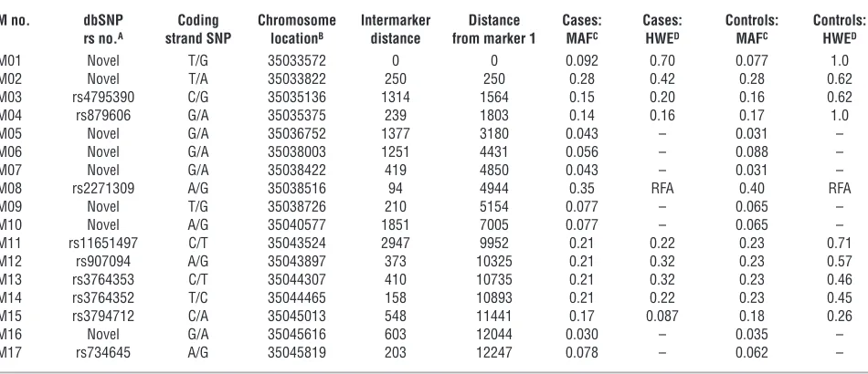
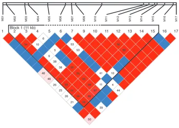
Genetic variation inPPP1R1B. Resequencing of DNA from 105 white and 44 African American probands identified 17 SNPs with minor allele frequencies in the white sample greater than 3% (Table 1). These were then genotyped in a large family-based sample of white subjects, the CBDB/NIMH family dataset. Overall, linkage disequi-librium (LD) between markers was moderately strong (Figure 1). No coding or splice site SNPs or other obvious common func-tional variants were identified. Using the method of Gabriel et al. (29), a large haplotype block spanning M02–M15 was identified (Figure 1). From the genotyped variants, to avoid multiple testing, we selected SNPs M03, M04, and M11–M15 within that block as a 7-SNP haplotype characterizing common variations in DARPP-32 for purposes of clinical association, gene expression analyses, and multimodal neuroimaging, all performed in independent datasets of white subjects. These 7 SNPs were selected because they showed the strongest individual SNP associations in the cognitive
phe-Table 1
Marker and map information in the CBDB/NIMH sample
M no. dbSNP Coding Chromosome Intermarker Distance Cases: Cases: Controls: Controls: rs no.A strand SNP locationB distance from marker 1 MAFC HWED MAFC HWED
M01 Novel T/G 35033572 0 0 0.092 0.70 0.077 1.0
M02 Novel T/A 35033822 250 250 0.28 0.42 0.28 0.62
M03 rs4795390 C/G 35035136 1314 1564 0.15 0.20 0.16 0.62
M04 rs879606 G/A 35035375 239 1803 0.14 0.16 0.17 1.0
M05 Novel G/A 35036752 1377 3180 0.043 – 0.031 –
M06 Novel G/A 35038003 1251 4431 0.056 – 0.088 –
M07 Novel G/A 35038422 419 4850 0.043 – 0.031 –
M08 rs2271309 A/G 35038516 94 4944 0.35 RFA 0.40 RFA
M09 Novel T/G 35038726 210 5154 0.077 – 0.065 –
M10 Novel A/G 35040577 1851 7005 0.077 – 0.065 –
M11 rs11651497 C/T 35043524 2947 9952 0.21 0.22 0.23 0.71
M12 rs907094 A/G 35043897 373 10325 0.21 0.32 0.23 0.57
M13 rs3764353 C/T 35044307 410 10735 0.21 0.32 0.23 0.46
M14 rs3764352 T/C 35044465 158 10893 0.21 0.22 0.23 0.45
M15 rs3794712 C/A 35045013 548 11441 0.17 0.087 0.18 0.26
M16 Novel G/A 35045616 603 12044 0.030 – 0.035 –
M17 rs734645 A/G 35045819 203 12247 0.078 – 0.062 –
M08 deviated from Hardy-Weinberg equilibrium (HWE) and was not analyzed further. SNPs with minor allele frequency (MAF) of less than 0.1 were not tested for deviation from HWE. ASingle Nucleotide Polymorphism database RefSNP accession ID number (http://www.ncbi.nlm.nih.gov/projects/SNP/); BUCSC
March 2006 assembly; CP values; Dexact P values. See Figure 1 for map of gene. RFA, removed from further analyses. Family-based association sample
[image:3.585.56.531.111.316.2]notype analyses (see below) and were in strong LD. The majority (approximately 75%) of subjects carried a very frequent haplotype (CGCACTC); 1 other haplotype (GATGTCA) had sample frequen-cies around 15%, and 3 other haplotypes were rare (Table 2).
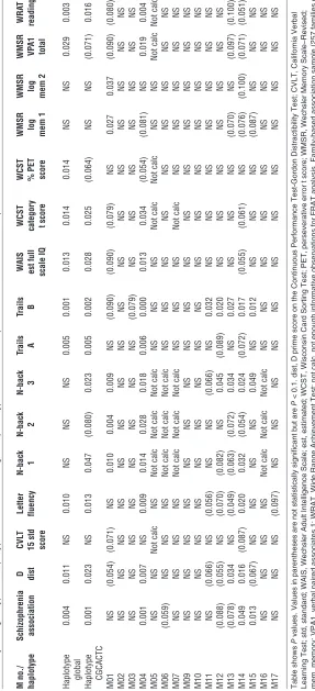
Cognitive phenotypes. In the CBDB/NIMH family dataset, to search for evidence of functional relevance of the identified genetic varia-tion in the gene, we used Family-Based Associavaria-tion Testing (FBAT) software version 1.7.2 to test for genetic association with cognitive performance in a sample of probands and their primarily unaffected siblings from our ongoing family-based study. The quantitative cog-nitive phenotypes were derived from a standard cogcog-nitive test bat-tery for which heritability and association with schizophrenia (30) have been previously established. This analysis showed significant genotype effects on a range of phenotypes (Table 3), including gen-eral intelligence, measured by IQ and Wide Range Achievement Test (WRAT) reading; working memory, as assessed by the N-back test (1- to 3-back); Wisconsin Card Sorting Test (categories completed and perseverative errors t score) and letter fluency tests; and also sequencing, response alternation, and attention, as measured by the Gordon Continuous Performance Test D', trails B and trails A. All of these tests have been related to function of cortical striatal loops (18). In contrast, no association was found with tests of episodic memory, the California Verbal Learning Test, and Wechsler Memory Scale–Revised logical memory 1 and 2, tests that are traditionally related to function of temporal-diencephalic circuitry. No other tests of association with cognitive phenotypes were performed in this study. Most significant associations were found with M04, with better performance associated with the G allele. The same domains were associated with the 7-SNP haplotype, as expected, since it con-sisted of SNPs selected for cognitive association (Table 3).
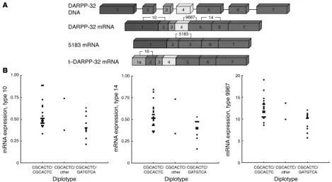
Impact of genetic variation on gene expression. To investigate the impact of the observed genetic variation on functional gene regulation affecting expression, we studied the effect of the PPP1R1B haplotype on DARPP-32 mRNA abundance in an independent sample of post-mortem human prefrontal cortices from 38 white subjects (22 con-trols and 16 subjects with schizophrenia). Several probes were used to identify known mRNA isoforms using quantitative PCR. Probe 14 identified all transcripts, probe 10 identified all transcripts except a truncated form previously described as t-DARPP (AY070271) (31), and probe 9667 identified all transcripts except for a splice variant (AK123112) missing exons 3 and 4. Probe 16 specifically identified t-DARPP, and probe 5183 identified the splice variant AK123112 (Figure 2A). In an analysis that controlled for the potentially con-founding factors of sex, smoking, diagnostic status, postmortem interval, age, and RNA quality, the PPP1R1B haplotype identified from the cognitive associations had a significant impact, or trended
Figure 1
Genetic variation in thePPP1R1B region, showing the genotyped SNPs and their LD. Top row shows relative positions of the SNPs (see Table 1
[image:4.585.109.476.80.343.2]for numbering). Below is a color-coded display of LD: strong LD (D'), red; weak LD, pink; weaker LD, white; not calculated, blue boxes. The haplotype block defined by the method of Gabrieli et al. that was used for association with biological and clinical phenotypes is marked by a black outline (output of the program Haploview). See Tables 1 and 2 and DNA collection and genotyping in Methods for details.
Table 2
Frequencies of 7-SNP haplotype in CBDB/NIMH samples
M03 M04 M11 M12 M13 M14 M15 Frequency
C G C A C T C 76.0%
G A T G T C A 14.1%
C G T G T C C 3.5%
C G T G T C A 2.1%
C A C A C T C 1.6%
toward significance, on all probes assaying the expression of the full-length mRNA (type 14, P < 0.006; type 10, P = 0.09; type 9967, P < 0.003). Expression was highest for homozygotes of the frequent (CGCACTC) haplotype, lowest for carriers of the less frequent (GATGTCA) haplotype, and intermediate for 2 subjects heterozygous for CGCACTC in combination with a rare haplotype (Figure 2B). There was no significant effect of diagnostic status, in agreement with recent observations (20). Probes specific for t-DARPP (P = 0.24) and the splice variant 5183 (P = 0.94) showed no significant association with haplotype. As a control, we ran the same analysis with the Val108/158 Met genotype in COMT. This had no significant effect on expres-sion levels of PPP1R1B. These data impli-cate a genetic mechanism for cognitive associations at the level of transcriptional regulation, splicing, or mRNA stability.
Neuroimaging sample. The observed associations of genetic variation with cognitive tests related to frontostriatal function suggest a functional impact on this neural circuit. To investigate such systems-level correlates, we studied struc-ture and function of the neostriatum and its interactions with prefrontal cortex (Figure 3) in an independent and large sample of healthy white volunteer sub-jects (96–142 subsub-jects) not included in the cognitive test sample. Many of these sub-jects participated in both structural and functional neuroimaging; details of the demographics and subject overlap appear in Supplemental Table 1 (supplemental material available online with this article; doi:10.1172/JCI30413DS1).
Brain structure. First, we used voxel-based morphometry (VBM) in 96 individuals to canvas the brain for regional volume changes related to the haplotype derived from the cognitive associations and show-ing association with mRNA expression. This revealed a bilateral relative decrease in neostriatal volume (maximum in dor-sal putamen) (Supplemental Table 2 and Figure 3A) for the frequent (CGCACTC) haplotype compared with all other haplo-types. In these subjects we then investigat-ed “structural connectivity” of the bilat-eral striatum, a measure of covariation of structural volume data that we previously found related to cortical-subcortical con-nectivity influenced by genetic variation (32). We observed a pronounced increase in this measure of putative structural con-nectivity between striatum and lateral
[image:5.585.62.349.102.731.2]frontal cortex, especially DLPFC, in carriers of the frequent haplo-type (Supplemental Table 2 and Figure 3B).
Brain function. To probe functional consequences of this effect of genetic variation on brain structure and mRNA expression, we analyzed 2 large archival functional MRI (fMRI) datasets. While neither of these tasks was specifically designed to probe striatal function, both of them show differential activation in neostriatum in the context of prefrontal cortex involvement, as predicted by the frontostriatal loop concept (13). Performance during these tasks did not correlate with PPP1R1B CGCACTC haplotype (Supplemental Table 1). First, during a well-established working memory paradigm that robustly activates DLPFC, the N-back task (33), carriers of the frequent (CGCACTC) haplotype showed significantly less reactiv-ity in the bilateral putamen (Supplemental Table 3 and Figure 3C), in regional agreement with the structural findings. Functional con-nectivity (a measure of correlation of brain activity over time) of the bilateral striatum showed a result similar to that of structural con-nectivity of the same region: concon-nectivity with prefrontal cortex was strongly increased in carriers of the frequent haplotype (Supplemen-tal Table 3 and Figure 3D). Next, we analyzed an emotional face-matching task (FMT) using threatening visual stimuli (32), which probes neural circuitry of the human emotional alerting response and engages ventrolateral prefrontal and lateral orbitofrontal cor-tices (34). Again, we observed a significant effect of haplotype on striatal activation (caudate and putamen); as in the other fMRI task, carriers of the frequent haplotype showed decreased reactivity of the putamen (Supplemental Table 4 and Figure 3E). The
analy-sis of the effects of haplotype on functional connectivity revealed a region in the left DLPFC that had increased connectivity for carriers of this haplotype (Supplemental Table 4 and Figure 3F). While it is expected that anatomical wiring and functional reactivity of the same brain circuits should bear some relationship to each other, since the structural and fMRI protocols and analyses performed here are independent acquisitions both technically and temporally and spatial normalization procedures ensure that the fMRI analyses are not confounded by the structural measures, these results provide convergent evidence for genetic impact on structure and function of frontostriatal circuitry. To exclude potential confounds by geno-type effects on performance, we analyzed the effect of PPP1R1B hap-lotype on correct responses (in percentages) and reaction time (in seconds) for the 2 tasks. No significant effects were observed (FMT, percentage correct, P = 0.24, reaction time, P = 0.27; N-back, percent-age correct, P = 0.47, reaction time, P = 0.77).
COMT control analysis in neuroimaging sample. As a genetic control, we also analyzed regional volume and functional activation in the sample of healthy white volunteers as a function of the COMT Val108/158 Met genotype, which was known for a large majority of participants (see Supplemental Table 5 for details of demograph-ics). In contrast to the pronounced effects of PPP1R1B haplotype on our striatal imaging variables, neither striatal volume nor acti-vation was significantly affected by genetic variation in COMT.
[image:6.585.57.526.81.338.2]Family-based association with schizophrenia. We performed a family-based association study for schizophrenia in 257 white families of self-declared European ancestry from the CBDB/NIMH sample.
Figure 2
Effect ofPPP1R1B haplotype on mRNA expression in postmortem human brain. (A) Exon position of primers used for quantitative PCR for
isoform amplification, showing which isoform was amplified by which probe. (B) Effect of PPP1R1B haplotypes on mRNA expression for the common isoform. The PPP1R1B haplotype has an impact on expression of all probes assaying the expression of the full-length mRNA (type 14,
P < 0.006; type 10, P = 0.09; type 9967, P < 0.003). Expression was highest for homozygotes of the frequent (CGCACTC) haplotype (21 sub-jects), lowest for carriers of the less frequent (GATGTCA) haplotype (11 subsub-jects), and intermediate for 2 subjects heterozygous for CGCACTC in combination with a rare haplotype. B is the ratio of DARPP expression to the geometric mean of expression of 3 housekeeping genes. See
We found evidence for an association between allelic variation in
PPP1R1B and schizophrenia for the common haplotype that showed association in our various analyses of normal subjects (global,
P = 0.004). The frequent haplotype CGCACTC, containing the M04 G allele, was positively associated with schizophrenia (P = 0.001), and the rare haplotype CACACTC, containing the M04 A allele, was negatively (P = 0.0003) associated with schizophrenia. For single SNPs, the strongest positive individual association (G allele,
P = 0.001) was found for SNP M04 (Table 3). Nominally significant association was also found for M14 (T allele, P = 0.049) and M15 (C allele, P = 0.031), which are in tight LD with M04 (Figure 3) and may therefore not represent an independent signal. There was also weak evidence for an association with SNP M06 (G allele, P = 0.059).
We were unable to replicate association results using the white families from the NIMH Human Genetics Initiative dataset (hap-lotype CGCACTC frequency: 0.76, P = 0.73, NS) (only 15 families had informative data).
Discussion
We used a translational genetics approach to investigate the rel-evance of DARPP-32 for aspects of human brain morphology and function. Resequencing of the gene revealed common sin-gle-nucleotide variants mostly within a single LD block, which we summarized for subsequent multilevel association analyses
in a 7-SNP haplotype. These 7 SNPs were selected because they showed strong association to cognitive phenotypes implicated in cortical-striatal function and were then used as a haplotype to test other biologic associations in independent datasets. This strategy reduces the number of association tests in a complex phenotype dataset while at the same time increasing the likeli-hood that chromosomes with causative mutations/haplotypes are contrasted with other chromosomes. All uncovered variants were noncoding, in agreement with recent findings from a lim-ited resequencing study in 50 Chinese probands (35). While it cannot be excluded that the effects observed in this study are due to several rare mutations that are coding, arose on the back-ground of a common (ancestral) haplotype, and are in LD with the analyzed SNPs, we propose that the associations with cogni-tion and brain structure and funccogni-tion observed in our dataset may reflect complex patterns of allelic heterogeneity that have an impact on expression. This is supported by the analysis of mRNA expression, which showed an impact of PPP1R1B genetic variation on the abundance of all identified full-length PPP1R1B
isoforms, compatible with the assumption of polymorphic func-tional regulatory elements. Expression was highest for subjects homozygous for the frequent (CGCACTC) haplotype and lowest for carriers of the GATGTCA haplotype. This provided a plausi-ble mechanism for an impact of the genetic variation
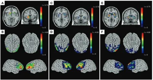
character-Figure 3
Neuroimaging analyses (structure and function) of common haplotype in PPP1R1B in control sample of white subjects. Top row shows
hap-lotype effects on volume (A) or activation (C and E) in striatum; bottom row shows haplotype effects on structural (B) and functional (D and F)
connectivity of striatum with prefrontal cortex. Structural MRI analyses (voxel-based morphometry): (A) significantly reduced volume in striatum
(P < 0.05) for carriers of the frequent (CGCACTC) haplotype; (B) greater structural connectivity between prefrontal cortex and striatum for
homozygotes for the frequent (CGCACTC) haplotype. fMRI, N-back task: (C) significantly reduced reactivity in putamen (P < 0.05) for carri-ers of the frequent (CGCACTC) haplotype; (D) greater functional connectivity between prefrontal cortex and striatum for homozygotes for the
frequent (CGCACTC) haplotype. fMRI, FMT: (E) significantly reduced reactivity in striatum (P < 0.05) for carriers of the frequent (CGCACTC) haplotype; (F) greater functional connectivity between prefrontal cortex and striatum for homozygotes for the frequent (CGCACTC) haplotype.
t color scales depict statistical significance level (t statistical value). See Supplemental Tables 2–4 for detailed statistical information and
[image:7.585.42.545.82.347.2]ized here on neuronal function, similar to observations in other psychiatric risk genes of noncoding marker haplotypes having effects on mRNA expression (36).
In an effort to bridge the gap from gene transcription to cogni-tive behavior, we studied the effects of the associated haplotype at the level of neural systems in another independent sample of healthy controls using neurobiological phenotypes plausibly connected to the cognitive phenotypes and to variation in gene expression. As reviewed, DARPP-32 is unambiguously linked to neostriatal function (11), which is, via interaction with prefrontal cortex, prominently implicated in a variety of cognitive domains (13). In agreement with the neurochemical anatomy of DARPP expression, we observed a substantial impact of genetic variation on neostriatal volume in structural neuroimaging. Volume was relatively reduced in dorsal putamen in carriers of the frequent haplotype. The dorsal putamen participates in interactions with the prefrontal cortex (13); in primates, this has been labeled the
associative loop and includes the DLPFC, much of the caudate nucleus, and the precommisural putamen as well as the ventral anterior thalamus (15). Perturbing this circuit at any point may affect its function, and indeed, there are studies in primates and rats showing that dysfunction of the striatum produces behav-ioral deficits similar to those found in dysfunction of the prefron-tal cortex (37). A genetic effect on striaprefron-tal-prefronprefron-tal interaction was confirmed by an analysis of structural connectivity of the striatum, which showed a strong increase in its correlation with prefrontal cortex in the frequent haplotype. While it is important to bear in mind that this structural connectivity reflects covaria-tion of regional volumes across subjects and does not directly quantify anatomical connections in white matter, we (32) and others (38) have previously observed patterns of structural con-nectivity that agree with known anatomical concon-nectivity and are sensitive to genetic variation (32), and studies of conditions with known impact on anatomical connectivity have shown that struc-tural covariance characterizes these processes in aging (39–41) and in schizophrenia (42). Furthermore, in the human optic system, it has been demonstrated directly that anatomically connected structures (i.e., optic tract, lateral geniculate nucleus, and primary visual cortex) covary in volume across individuals (43).
The structural findings were confirmed and extended by the results in functional imaging, where functional connectivity between striatum and prefrontal cortex was again shown to be increased in carriers of the frequent haplotype in both fMRI para-digms. Regarding striatal activation, the striatum showed less responsiveness to the environmental stimuli in carriers of the fre-quent haplotype in both tasks, suggesting that this finding indi-cates more efficient, intrastriatal processing. Indeed, the relatively smaller putaminal volume associated with this haplotype may also reflect more efficient circuitry. Since regional volume changes are taken into account when functional imaging data are spatially normalized and the main effect of task is a deactivation during working memory but an activation during face processing, these functional imaging results are not likely to be partial volume arti-facts. Again, the localization of statistical maxima was in lateral putamen (also reaching significance in caudate for the face-pro-cessing task), in agreement with the structural findings and high-lighting those regions receiving input from the DLPFC and premo-tor prefrontal cortex. It is important to note that while our tasks are cognitive and engage associative prefrontal-striatal circuitry broadly, our results highlight putamen as the component of the
structural and functional circuit DARPP most has an impact on but not necessarily as the main striatal processing station related to task performance. Taken together, the neuroimaging data there-fore show that the frequent haplotype is associated in healthy indi-viduals with more efficient intrastriatal processing combined with an increase of prefrontal cortical input onto a smaller striatum. We note that the size reduction may contribute to striatal pro-cessing efficiency as it has been proposed that increasing overlap of functional projection fields by compression of pathways into successively smaller striatal structures is a key mechanism for the integration of information streams in the basal ganglia (15). Since our analyses of function concerned 2 tasks that were designed to assess prefrontal function during emotional and working memory processing, it would be useful to extend the present study by tasks that are specifically intended to activate striatal areas engaged in other cognitive functions, such as implicit memory or reward.
These systems-level findings provide a neural mechanism for the observed pronounced impact of genetic variation in PPP1R1B
on a wide range of cognitive domains during neuropsychological testing. The imaging findings confirm the behavioral importance of DARPP-32 effects on frontostriatal processing since working memory and response alternation and attention critically depend on both prefrontal and striatal function and are disturbed in diseases thought to have an impact on these structures, such as schizophrenia and Parkinson disease (18). Improved working memory and executive capacity may also underlie the association with general intelligence (44). The range of cognitive tests affected by variation in this gene likely reflects the critical importance of frontostriatal function for core aspects of cognition (18). As an important negative, 3 memory tasks depending on hippocampal but not striatal function, the California Verbal Learning Test and Wechsler Memory Scale–Revised logical memory 1 and 2 tests, were not affected by PPP1R1B variation, suggesting a relatively specific impact on the frontostriatal circuit as well as a limited role of DARPP-32–dependent processing in neural systems for episodic memory in humans.
Our control analysis using the functional common Val108/158 Met genetic variation in COMT showed no effect on PPP1R1B
mRNA expression, volume, or functional activation of the striatum, in good agreement with human (45) and animal knockout (26) data showing that this variant has an impact on dopamine turn-over in prefrontal cortex but not striatum, where dopamine flux is mainly dependent on the dopamine transporter. These negative control findings, therefore, suggest that the biological associations with PPP1R1B found in the present study are specifically related to the integrative function of DARPP-32 in dopaminoceptive striatal neurons and not to a general effect on dopaminergic neurotrans-mission elsewhere in this circuit.
is also in or near a region implicated in risk for schizophrenia by a recent metaanalysis of whole genome linkage (46). Indeed, we found positive associations for the M04 G allele as well as for the frequent (CGCACTC) haplotype, the same alleles that had an impact on cognitive, expression, and imaging phenotypes.
This finding, however, must be regarded as preliminary since we did not have an adequately powered family-based replication sam-ple, and it is therefore difficult to know whether this is a true asso-ciation or a false positive. However, it is noteworthy that the alleles and haplotypes implicated in disease risk also predicted changes in controls mirroring observations in manifest schizophrenia. Stria-tal volumes in drug-naive, first-episode patients have been found to be decreased (47), an effect confounded later in the illness by neuroleptic treatment, which increases striatal volume (48). Imag-ing studies of patients with schizophrenia also show increased prefrontal-subcortical connectivity (49). This supports the tenta-tive clinical association by showing that the directionality of the observed genetic effects agreed with the disease phenotype.
To the degree that this genetic association with schizophrenia can be confirmed, our data lead to the provocative observation that a frequent haplotype in PPP1R1B predicts increased frontostriatal interactions that appeared beneficial (as evidenced by relatively better performance on a wide range of cognitive tasks) yet contributed to risk for schizophrenia. This raises the question of whether a genetic advantage in normal subjects may translate into a disadvantage in the context of other functional impairments also associated with schizophrenia, such as abnormal function of the prefrontal cortex. To the degree that the striatum acts as an active gating station (50), increased information flow through this regulatory cognitive sub-system is predicted to contribute to increased flexibility, working memory capacity, and control capabilities, as observed in our normal subjects (51); on the other hand, in manifest schizophrenia, which is characterized by an inefficient, fractionated pattern of activation in prefrontal cortex (33), the same information-processing constel-lation could facilitate the persistence of disorganized cortical infor-mation and contribute to an escape of dysfunctional, unmodulated information from frontostriatal loops, leading to deficient cognition and inappropriate behavioral responses (16). Stated another way, the common DARPP-32 haplotype appears associated with optimized frontal-striatal function regardless of the specific information being processed through the system. The molecular processes by which genetic and environmental information regulate the development and modification of prefrontal-striatal circuitry involve synaptic plasticity, and DARPP-32 is a key molecule for shaping plasticity in striatal neurons receiving frontal afferents (52). Further work is nec-essary to confirm or refute this speculation.
In summary, we present convergent evidence in 3 independent datasets implicating DARPP-32 in a frontostriatal neural system for executive cognition and response selection in humans. We hope that this genetic identification of a molecular target at a critical nexus of dopaminergic neurotransmission and synaptic plastic-ity gives renewed impetus to the pursuit of therapeutic strategies aimed at postsynaptic signal integration in dopaminoceptive neu-rons that might benefit a diverse range of psychiatric disorders, notably addiction and schizophrenia.
Methods
Subjects. Five independent subject samples were used in this study. (a) For the initial sequencing effort to discover genetic variants, we resequenced DNA from 105 white and 44 African American patients with
schizophre-nia, including the 9.7 kb of PPP1R1B composing the region of the gene spanned by its 7 exons and 2 kb upstream of the transcription start site. (b) For mRNA expression, postmortem brain samples were collected from 38 white subjects (22 controls and 16 schizophrenics). For genetic studies, probands with schizophrenia spectrum disorders, their unaffected siblings, and controls came from the Clinical Brain Disorders Branch Sibling Study, a study of neurobiological abnormalities related to genetic risk for schizo-phrenia (30). Only white people of European ancestry were studied to mini-mize heterogeneity and potential stratification artifacts. DNA was available for 257 white patients, 327 of their siblings, 397 parents of probands, and 243 controls. All subjects gave written informed consent and participated in the study according to the guidelines of the NIMH Institutional Review Board. Two independent samples from this genetic dataset were drawn for the present analyses. (c) Family data consisting of affected and unaffected offspring and parents were used for FBAT analyses (http://www.biostat. harvard.edu/~fbat/fbat.htm) of disease and cognitive phenotypes. (d) An independent sample of healthy volunteer subjects was selected for neuro-imaging after careful screening (30) to ensure they were free of any lifetime history of psychiatric or neurological illness, psychiatric treatment, or drug or alcohol abuse (Supplemental Table 1). The use in imaging of healthy subjects only is tailored to isolate gene effects independent of confound-ers related to illness, including chronicity, drug and alcohol use, smoking, medical treatment, and ongoing symptomatology. All available scans of subjects meeting these criteria (96–142 subjects; see Supplemental Table 1 for details) were used. (e) Finally, we also genotyped a selected sample of 67 white families from the NIMH Human Genetics Initiative (53) consist-ing of participants with the diagnosis of schizophrenia or schizoaffective disorder, depressed subtype, 1 or 2 siblings, and available parents. No other phenotypic measures were available for this sample.
DNA collection and genotyping. We used standard methods to extract DNA from white blood cells with the PUREGENE DNA purification kit (Gentra). We first resequenced material from 105 white and 44 African American patients with schizophrenia and identified 17 SNPs with minor allele fre-quencies greater than 3% in the CBDB/NIMH family white dataset (Table 1). Genotype for only 1 SNP (M08) deviated from Hardy-Weinberg equilib-rium in cases and controls; this SNP was discarded from further analyses. Genotype accuracy was assessed by regenotyping within a subsample, and reproducibility was routinely greater than 99%. We eliminated probable genotyping errors within the family dataset through non-Mendelizing transmissions and haplotype inconsistency errors via MERLIN version 1.0.1 (http://www.sph.umich.edu/csg/abecasis/Merlin/download/). Genotypes based on assayed genotyping and on sequencing were identical. We mea-sured LD between markers as indexed by the D' and R2 statistics from case
and control haplotypes with the program LDMAX within the Graphical Overview of Linkage Disequilibrium (GOLD) software package 2001 (54). To construct haplotypes for analyses of biological (functional imaging and gene expression) data, we used Haploview 3.2 software (http://www.broad. mit.edu/mpg/haploview/index.php) and assigned haplotypes to individuals using PHASE 2.1 (55). Haplotype frequencies are given in Figure 1.
also included. Selection of these cognitive phenotypes was based on evi-dence that they are related to genetic risk for schizophrenia (30). No other cognitive phenotypes were evaluated for this study. Single SNP and family-based haplotype analyses of these phenotypes were performed with FBAT version 1.7.2 using permutation testing for assessment of significance.
Gene expression. mRNA expression was studied using quantitative PCR. We employed 5 PPP1R1B probe/primer combinations on cDNA from postmortem brain samples collected from 38 white subjects (22 controls and 16 schizophrenics) after informed consent from the legal next of kin under NIMH protocol 90-M-0142. Gray matter tissue from the middle frontal gyrus was obtained from a coronal slab corresponding to the middle one-third immediately anterior to the genu of the corpus callo-sum. White matter was trimmed off. Tissue was pulverized and stored at –80°C. Total RNA was extracted from 300 mg of tissue using the TRIzol Reagent (Invitrogen). The yield of total RNA was determined by spectro-photometry, measuring absorbance at 260 nm. RNA quality was assessed with high-resolution capillary electrophoresis (Agilent Technologies), and only samples showing clearly defined, sharp 18S and 28S ribosomal peaks, 28S/18S ratios greater than 1.2, and RNA integrity numbers greater than or equal to 4.0 were included. Total RNA (4 μg) was used in 50 μl of a reverse transcriptase reaction to synthesize cDNA by using the Super-Script First-Strand Synthesis System for RT-PCR (Invitrogen). Applied Biosystems Assays-on-Demand and custom-made primers/probes (for type 5183) were used to amplify specific transcripts based on the unique exon structure of each DARPP-32 isoform (17). Three probes were based on the common exon structure as found in full-length cDNA: exons 4 and 5 (type 9967; Hs00259967), 1 and 2 (type 10; Hs00938410_m1) and 5 and 6 (type 14; Hs00938414_m1); splice variant 5183, exons 2 and 5; and t-DARPP (type 16; Hs00938416_g1). The following Applied Biosys-tems Assays-on-Demand were used for measuring the levels of control genes for normalization of DARPP-32 expression data: porphobilinogen deaminase (PBGD; Hs006009297), β2 microglobulin (B2M; Hs99999907), and β glucuronidase (GUSB; Hs99999908). Expression levels of mRNAs were measured by real-time quantitative RT-PCR, using an ABI Prism 7900 Sequence Detection System with a 384-well format (Applied Biosys-tems). Each 10 μl reaction contained 900 nM of primer, 250 nM of probe, and TaqMan Universal PCR Master Mix (Applied Biosystems) containing Hot GoldStar DNA Polymerase (Eurogentec), dNTPs with dUTP, uracil N-glycosylase, passive reference, and 100–200 ng of cDNA template. PCR cycle parameters were 50°C for 2 minutes, 95°C for 10 minutes, 40 cycles of 95°C for 15 seconds, and 59°C or 60°C for 1 minute. PCR data were acquired from Sequence Detector Software (SDS version 2.0; Applied Bio-systems) and quantified by a standard curve method using serial dilutions of pooled cDNA derived from RNA obtained from 10 to 12 control sub-jects. In each experiment, the R2 value of the curve was more than 0.99, the slope was between –3.2 and –3.5 (amplification efficiency 96%–101%), and controls containing no-template cDNA resulted in no detectable signal. All samples were measured in a single plate for each target gene, and their threshold cycle (Ct) values were in the linear range of the standard curve. All measurements were performed in triplicate. The data were normalized to the geometric mean of the control genes. All diplotypes containing at least 1 haplotype appearing in more than 1 subject were included in the analysis, leaving 34 subjects. Data were analyzed using the general lin-ear model, with DARPP-32 diplotypes as predictors. Sex, smoking status, diagnostic status, postmortem interval, age, and RNA quality were also included in the regression model to account for variables unrelated to genotype that can potentially influence gene expression (57).
Neuroimaging — tasks. Both structural and functional neuroimaging were used to characterize neural function on the systems level. During fMRI scanning, subjects completed 2 tasks. The FMT is a simple perceptual task
previously described to robustly engage the amygdala and hippocampal formation (58, 59). During 2 blocks of an emotion task, subjects viewed a trio of faces, selecting 1 of the 2 faces (bottom) that was identical to the tar-get face (top). Per block, 6 images were presented sequentially for 5 seconds each, 3 of each sex and target affect (angry or afraid) derived from a stan-dard set of pictures of facial affect. Emotion tasks alternated with 3 blocks of a sensorimotor control task in which faces were replaced with simple geometric shapes. The second task, the N-back (2-back version), probes working memory. It requires subjects to monitor a series of sequentially presented numerals randomly chosen from between 1 and 4 and, while continually receiving new numbers, press a corresponding button if the number presently on the screen is the same as that presented 2 items ear-lier. As a control task (0-back), subjects press a button corresponding to the number currently on the screen. This task has been used extensively (33) to study working memory in controls as well as patients with schizophrenia and their relatives and elicits robust DLPFC activation that is abnormal in patients. For both tasks, percentage of correct responses and reaction time were recorded. To investigate the effects of PPP1R1B haplotype on these performance parameters as potential confounds in neuroimaging, the gen-eral linear model was used with haplotypes assigned by PHASE as predic-tors and a contrast comparing the most frequent haplotype (CGCACTC) with the second most frequent haplotype (GATGTCA).
Neuroimaging: structural image processing. We acquired 3D structural MRI scans on a 1.5 Tesla GE scanner, using a T1-weighted spoiled grass sequence (repetition time[s]/excitation time[s]/number of excita-tions, 24/5/1; flip angle, 45°; matrix size, 256 × 256 voxels; field of view, 24 × 24 cm) with 124 sagittal slices (0.94 × 0.94 × 1.5 mm resolution) pre-processed as previously described (60) followed by an optimized VBM pro-tocol using customized templates (61, 62). Resulting gray matter images were smoothed with a 12-mm Gaussian kernel prior to statistical analysis. Analysis was performed on a Linux workstation (Red Hat Enterprise) as implemented in MATLAB 6.52SP2 (MathWorks) and the general linear model (63) in SPM2 software (http://www.fil.ion.ucl.ac.uk/spm). The specification of parameters for the design matrix was described in detail elsewhere (60). In brief, effects of DARPP-32 genotype on gray matter vol-ume were examined by using an analysis of covariance model adjusting for orthogonalized first- and second-order polynomials of age and sex. Haplotype effects were assessed as described below.
Neuroimaging: functional image processing. Blood oxygenation level–depen-dent (BOLD) fMRI was performed on a GE HealthCare Signa HDx 3.0T, using gradient echo-planar imaging (EPI) (24 axial slices, 4 mm thickness, 1 mm gap; TR/TE, 2,000/28 ms; field of view, 24 cm; matrix, 64 × 64 voxels). Images were processed as described previously (58, 59), using SPM99. In brief, images were realigned to the middle image of the scan run, spatially normalized into a standard stereotactic space (Montreal Neurological Institute template) using an affine and nonlinear (4 × 5 × 4 basis-func-tions) transformation, smoothed with an 8-mm full width Gaussian filter at half maximum and ratio normalized to the whole-brain global mean. Statistical images for the contrast of the emotion task versus the senso-rimotor control (for face matching) or 2-back minus 0-back (for N-back) were obtained for each subject. These maps were then analyzed for haplo-type effects as described below.
with all voxel time series, resulting in a map which contained, in each voxel, the correlation coefficient of the time series in that voxel with that of the ref-erence regions. These maps, 1 per subject, were then analyzed in a random-effects model in SPM for haplotype random-effects as described below.
Neuroimaging: structural covariance analysis. Structural covariance exam-ines structural coupling across individuals and employs a similar measure of coupling to functional connectivity, only this time not between func-tional data (fMRI time series), but between voxel-wise gray matter volume maps derived from VBM (see above). Voxels identified as significant in this approach have regional volume that is significantly positively or negatively correlated with the target area across subjects. We followed methods pub-lished previously (32). Summed volume in an anatomically defined ROI (bilateral putamen) in standardized space was computed by adding the local volume in all voxels comprising the ROI and used as a covariate of interest in a random-effects general linear model in SPM that also included nuisance covariates as described above for VBM.
Neuroimaging: second-level analysis of haplotype effects and statistical infer-ence. After neuroimages were prepared for each subject by processing as described in the previous paragraphs, for all imaging methods, the analysis of haplotype effects was performed in a second-level random-effects model as previously described (24). The group level model used 1 contrast image per subject and regressed the estimated BOLD change on each subject’s inferred haplotype frequencies. For statistical inference on the second level, we used the established methods of Gaussian random fields (GRF) theory (65). The significance threshold was set to P < 0.05, corrected for multiple comparisons within hypothesis-driven ROI, defined by using the Wake Forest University School of Medicine PickAtlas (http://www.fmri.wfubmc. edu/download.htm). We used false discovery rate (FDR), as implemented in
SPM2, a frequentist method to control for type I error rate across the brain (66). For activation and volume, we investigated the striatum (caudate, putamen, and pallidum). For connectivity with striatum, we used a pre-frontal cortex ROI encompassing Brodmann areas 9, 10, 32, 44, 45, and 46. Voxels significant at those thresholds are listed in Supplemental Tables 2–4. In Figures 1–3, all voxels affected by genotype within these regions are shown rendered on a representative control volume and surface. While all haplotypes were included in the statistical model, since comparisons with rarer haplotypes were underpowered in our imaging dataset, we compared only the most frequent haplotype (CGCACTC) and the second most fre-quent (GATGTCA) haplotype.
Association with schizophrenia. We tested for association with schizophre-nia in 2 datasets: the CBDB/NIMH family dataset (257 families) and the NIMH Human Genetics Initiative family dataset (67 families). FBAT was used, with permutation testing for assessment of significance.
Acknowledgments
We would like to thank Courtnea Rainey for assistance in design-ing and drawdesign-ing Figure 2A. This work was supported by the NIMH/Intramural Research Program.
Received for publication September 20, 2006, and accepted in revised form December 5, 2006.
Address correspondence to: Daniel R. Weinberger, NIH, Genes, Cognition and Psychosis Program, Room 4S-235, 10 Center Drive, Bethesda, Maryland 20892, USA. Phone: (301) 402-7564; Fax: (301) 480-7795; E-mail: [email protected].
1. Walaas, S.I., Aswad, D.W., and Greengard, P. 1983. A dopamine- and cyclic AMP-regulated phospho-protein enriched in dopamine-innervated brain regions. Nature. 301:69–71.
2. Hemmings, H.C., Jr., Greengard, P., Tung, H.Y., and Cohen, P. 1984. DARPP-32, a dopamine-regulated neuronal phosphoprotein, is a potent inhibitor of protein phosphatase-1. Nature. 310:503–505. 3. Svenningsson, P., et al. 2004. DARPP-32: an
inte-grator of neurotransmission. Annu. Rev. Pharmacol. Toxicol. 44:269–296.
4. Goldman-Rakic, P.S. 1998. The cortical dopamine system: role in memory and cognition. Adv. Pharmacol. 42:707–711.
5. Schultz, W. 1998. Predictive reward signal of dopa-mine neurons. J. Neurophysiol. 80:1–27.
6. Weinberger, D.R. 1987. Implications of normal brain development for the pathogenesis of schizophrenia.
Arch. Gen. Psychiatry. 44:660–669.
7. Grace, A.A. 2000. The tonic/phasic model of dopa-mine system regulation and its implications for understanding alcohol and psychostimulant craving.
Addiction. 95(Suppl. 2):S119–S128.
8. Fiorillo, C.D., Tobler, P.N., and Schultz, W. 2003. Discrete coding of reward probability and uncertain-ty by dopamine neurons. Science. 299:1898–1902. 9. Svenningsson, P., et al. 2003. Diverse
psychotomi-metics act through a common signaling pathway.
Science. 302:1412–1415.
10. Zachariou, V., et al. 2006. Phosphorylation of DARPP-32 at Threonine-34 is required for cocaine action. Neuropsychopharmacology. 31:555–562. 11. Ouimet, C.C., LaMantia, A.S.,
Goldman-Rakic, P., Goldman-Rakic, P., and Greengard, P. 1992. Immunocytochemical localization of DARPP-32, a dopamine and cyclic-AMP-regulated phospho-protein, in the primate brain. J. Comp. Neurol.
323:209–218.
12. Ouimet, C.C., et al. 1984. DARPP-32, a dopamine- and adenosine 3′:5′-monophosphate-regulated phosphoprotein enriched in dopamine-innervated
brain regions. III. Immunocytochemical localization.
J. Neurosci. 4:111–124.
13. Alexander, G.E., DeLong, M.R., and Strick, P.L. 1986. Parallel organization of functionally seg-regated circuits linking basal ganglia and cortex.
Annu. Rev. Neurosci. 9:357–381.
14. Graybiel, A.M. 2005. The basal ganglia: learn-ing new tricks and lovlearn-ing it. Curr. Opin. Neurobiol.
15:638–644.
15. Haber, S.N. 2003. The primate basal ganglia: par-allel and integrative networks. J. Chem. Neuroanat.
26:317–330.
16. Swerdlow, N.R., Geyer, M.A., and Braff, D.L. 2001. Neu-ral circuit regulation of prepulse inhibition of startle in the rat: current knowledge and future challenges.
Psychopharmacology (Berl.). 156:194–215.
17. Dunnett, S.B., Meldrum, A., and Muir, J.L. 2005. Frontal-striatal disconnection disrupts cognitive per-formance of the frontal-type in the rat. Neuroscience. 135:1055–1065.
18. Pantelis, C., et al. 1997. Frontal-striatal cognitive deficits in patients with chronic schizophrenia.
Brain. 120:1823–1843.
19. Albert, K.A., et al. 2002. Evidence for decreased DARPP-32 in the prefrontal cortex of patients with schizophrenia. Arch. Gen. Psychiatry. 59:705–712. 20. Baracskay, K.L., Haroutunian, V., and
Meador-Woodruff, J.H. 2006. Dopamine receptor signaling molecules are altered in elderly schizophrenic cortex.
Synapse. 60:271–279.
21. Meyer-Lindenberg, A., and Weinberger, D.R. 2006. Intermediate phenotypes and genetic mechanisms of psychiatric disorders. Nat. Rev. Neurosci. 7:818–827. 22. Harrison, P.J., and Weinberger, D.R. 2005.
Schizo-phrenia genes, gene expression, and neuropa-thology: on the matter of their convergence. Mol. Psychiatry. 10:40–68.
23. Egan, M.F., et al. 2001. Effect of COMT Val108/158 Met genotype on frontal lobe func-tion and risk for schizophrenia. Proc. Natl. Acad. Sci. U. S. A. 98:6917–6922.
24. Meyer-Lindenberg, A., et al. 2006. Impact of com-plex genetic variation in COMT on human brain function. Mol. Psychiatry. 11:867–877.
25. Tunbridge, E.M., Bannerman, D.M., Sharp, T., and Harrison, P.J. 2004. Catechol-o-methyltransferase inhibition improves set-shifting performance and elevates stimulated dopamine release in the rat pre-frontal cortex. J. Neurosci. 24:5331–5335. 26. Gogos, J.A., et al. 1998.
Catechol-O-methyltrans-ferase-deficient mice exhibit sexually dimorphic changes in catecholamine levels and behavior.
Proc. Natl. Acad. Sci. U. S. A. 95:9991–9996. 27. Meyer-Lindenberg, A., et al. 2002. Reduced
pre-frontal activity predicts exaggerated striatal dopa-minergic function in schizophrenia. Nat. Neurosci.
5:267–271.
28. Kellendonk, C., et al. 2006. Transient and selective overexpression of dopamine D2 receptors in the striatum causes persistent abnormalities in pre-frontal cortex functioning. Neuron. 49:603–615. 29. Gabriel, S.B., et al. 2002. The structure of
hap-lotype blocks in the human genome. Science.
296:2225–2229.
30. Egan, M.F., et al. 2001. Relative risk for cognitive impairments in siblings of patients with schizo-phrenia. Biol. Psychiatry. 50:98–107.
31. El-Rifai, W., et al. 2002. Gastric cancers overexpress DARPP-32 and a novel isoform, t-DARPP. Cancer Res. 62:4061–4064.
32. Pezawas, L., et al. 2005. 5-HTTLPR polymorphism impacts human cingulate-amygdala interactions: a genetic susceptibility mechanism for depression.
Nat. Neurosci. 8:828–834.
33. Callicott, J.H., et al. 2000. Physiological dysfunc-tion of the dorsolateral prefrontal cortex in schizo-phrenia revisited. Cereb. Cortex. 10:1078–1092. 34. Meyer-Lindenberg, A., et al. 2005. Neural correlates
of genetically abnormal social cognition in Wil-liams syndrome. Nat. Neurosci. 8:991–993. 35. Li, C.H., Liao, H.M., Hung, T.W., and Chen, C.H.
candi-date gene for schizophrenia. Schizophr. Res. 87:1–5. 36. Law, A.J., et al. 2006. Neuregulin 1 transcripts
are differentially expressed in schizophrenia and regulated by 5′ SNPs associated with the disease.
Proc. Natl. Acad. Sci. U. S. A. 103:6747–6752. 37. Partiot, A., et al. 1996. Delayed response tasks in
basal ganglia lesions in man: further evidence for a striato-frontal cooperation in behavioral adaptation.
Neuropsychologia. 34:709–721.
38. Mechelli, A., Friston, K.J., Frackowiak, R.S., and Price, C.J. 2005. Structural covariance in the human cortex. J. Neurosci. 25:8303–8310.
39. Raz, N., et al. 2005. Regional brain changes in aging healthy adults: general trends, individual differences and modifiers. Cereb. Cortex. 15:1676–1689. 40. Raz, N., et al. 2004. Aging, sexual dimorphism, and
hemispheric asymmetry of the cerebral cortex: rep-licability of regional differences in volume. Neuro-biol. Aging. 25:377–396.
41. Raz, N., et al. 1997. Selective aging of the human cerebral cortex observed in vivo: differential vulner-ability of the prefrontal gray matter. Cereb. Cortex.
7:268–282.
42. Woodruff, P.W., et al. 1997. Structural brain abnor-malities in male schizophrenics reflect fronto-tem-poral dissociation. Psychol. Med. 27:1257–1266. 43. Andrews, T.J., Halpern, S.D., and Purves, D. 1997.
Correlated size variations in human visual cortex, lateral geniculate nucleus, and optic tract. J. Neurosci. 17:2859–2868.
44. Gray, J.R., Chabris, C.F., and Braver, T.S. 2003. Neu-ral mechanisms of geneNeu-ral fluid intelligence. Nat. Neurosci. 6:316–322.
45. Lynch, D.R., et al. 2003. Lack of effect of polymor-phisms in dopamine metabolism related genes on imaging of TRODAT-1 in striatum of asymptomat-ic volunteers and patients with Parkinson’s disease.
Mov. Disord. 18:804–812.
46. Lewis, C.M., et al. 2003. Genome scan meta-analy-sis of schizophrenia and bipolar disorder, part II: Schizophrenia. Am. J. Hum. Genet. 73:34–48. 47. Keshavan, M.S., Rosenberg, D., Sweeney, J.A., and
Pettegrew, J.W. 1998. Decreased caudate volume in neuroleptic-naive psychotic patients. Am. J. Psychiatry. 155:774–778.
48. Chakos, M.H., et al. 1994. Increase in caudate nuclei volumes of first-episode schizophrenic patients taking antipsychotic drugs. Am. J. Psychiatry. 151:1430–1436.
49. Meyer-Lindenberg, A.S., et al. 2005. Regionally specific disturbance of dorsolateral prefrontal-hippocampal functional connectivity in schizo-phrenia. Arch. Gen. Psychiatry. 62:379–386. 50. Schneider, J.S. 1984. Basal ganglia role in behavior:
importance of sensory gating and its relevance to psychiatry. Biol. Psychiatry. 19:1693–1710. 51. Miller, E.K., and Cohen, J.D. 2001. An integrative
theory of prefrontal cortex function. Annu. Rev. Neurosci. 24:167–202.
52. Valjent, E., et al. 2005. Regulation of a protein phosphatase cascade allows convergent dopa-mine and glutamate signals to activate ERK in the striatum. Proc. Natl. Acad. Sci. U. S. A. 102:491–496. 53. Cloninger, C.R., et al. 1998. Genome-wide search
for schizophrenia susceptibility loci: the NIMH Genetics Initiative and Millennium Consortium.
Am. J. Med. Genet. 81:275–281.
54. Abecasis, G.R., and Cookson, W.O. 2000. GOLD — graphical overview of linkage disequilibrium.
Bioinformatics. 16:182–183.
55. Stephens, M., Smith, N.J., and Donnelly, P. 2001. A new statistical method for haplotype reconstruc-tion from populareconstruc-tion from populareconstruc-tion data. Am. J. Hum. Genet. 68:978–989.
56. Jastak, S., Wilkinson, G.S., and Jastak, J. 1984. Wide range achievement test — revised. Jastak Associates.
Wilmington, Delaware, USA.
57. Lipska, B.K., et al. 2006. Critical factors in gene expres-sion in postmortem human brain: focus on studies in schizophrenia. Biol. Psychiatry. 60:650–658. 58. Hariri, A.R., Tessitore, A., Mattay, V.S., Fera, F., and
Weinberger, D.R. 2002. The amygdala response to emotional stimuli: a comparison of faces and scenes. Neuroimage. 17:317–323.
59. Hariri, A.R., et al. 2005. A susceptibility gene for affective disorders and the response of the human amygdala. Arch. Gen. Psychiatry. 62:146–152. 60. Pezawas, L., et al. 2004. The brain-derived
neuro-trophic factor val66met polymorphism and varia-tion in human cortical morphology. J. Neurosci.
24:10099–10102.
61. Augood, S.J., Westmore, K., and Emson, P.C. 1997. Phenotypic characterization of neurotensin mes-senger RNA-expressing cells in the neuroleptic-treated rat striatum: a detailed cellular co-expres-sion study. Neuroscience. 76:763–774.
62. Ashburner, J., and Friston, K.J. 2000. Voxel-based morphometry — the methods. Neuroimage.
11:805–821.
63. Friston, K.J., et al. 1995. Statistical parametric maps in functional imaging: a general linear approach.
Hum. Brain Mapp. 2:189–210.
64. Meyer-Lindenberg, A., et al. 2001. Evidence for abnormal cortical functional connectivity during working memory in schizophrenia. Am. J. Psychiatry. 158:1809–1817.
65. Worsley, K.J., et al. 1996. A unified statistical approach for determining significant signals in images of cerebral activation. Hum. Brain Mapp.
4:58–73.