0095-1137/97/$04.00
1
0
Copyright
q
1997, American Society for Microbiology
Persistence of
Borrelia burgdorferi
in Experimentally Infected
Dogs after Antibiotic Treatment
REINHARD K. STRAUBINGER,
1* BRIAN A. SUMMERS,
2YUNG-FU CHANG,
3ANDMAX J. G. APPEL
1James A. Baker Institute for Animal Health,
1Department of Pathology,
2and Diagnostic Laboratory,
3College of Veterinary Medicine, Cornell University, Ithaca, New York 14853
Received 11 July 1996/Returned for modification 16 September 1996/Accepted 15 October 1996
In specific-pathogen-free dogs experimentally infected with
Borrelia burgdorferi
by tick exposure, treatment
with high doses of amoxicillin or doxycycline for 30 days diminished but failed to eliminate persistent infection.
Although joint disease was prevented or cured in five of five amoxicillin- and five of six doxycycline-treated
dogs, skin punch biopsies and multiple tissues from necropsy samples remained PCR positive and
B.
burg-dorferi
was isolated from one amoxicillin- and two doxycycline-treated dogs following antibiotic treatment. In
contrast,
B. burgdorferi
was isolated from six of six untreated infected control dogs and joint lesions were found
in four of these six dogs. Serum antibody levels to
B. burgdorferi
in all dogs declined after antibiotic treatment.
Negative antibody levels were reached in four of six doxycycline- and four of six amoxicillin-treated dogs.
However, in dogs that were kept in isolation for 6 months after antibiotic treatment was discontinued, antibody
levels began to rise again, presumably in response to proliferation of the surviving pool of spirochetes.
Antibody levels in untreated infected control dogs remained high.
Lyme disease, or Lyme borreliosis, in humans and domestic
animals is caused by the tick-transmitted spirochete
Borrelia
burgdorferi
. The clinical signs in humans were reviewed by
Steere (43), and those in dogs were reviewed by Levy et al.
(17). There appears to be a general agreement that
B.
burg-dorferi
persists in humans and animals for months or years, and
perhaps for life (3, 5, 29, 41), despite a strong humoral immune
response. One possible explanation for the persistence is the
failure of the host to produce borreliacidal OspA antibodies
(34, 35, 44), which are induced by vaccination but rarely
fol-lowing natural infection. Another possible explanation is the
invasion of poorly vascularized connective tissues by
spiro-chetes or even an intracellular localization (10, 13, 16, 18).
Elimination of borreliae by antibiotic treatment would be
highly desirable. Although
B. burgdorferi
is susceptible to
an-tibiotics in vitro (4, 14), there are contradictory reports as to
the in vivo efficacy of antibiotics. While some authors claim
that elimination of
B. burgdorferi
occurs after antibiotic
treat-ment of human patients (20, 25), others are less certain (30, 37,
45). The antibiotics most commonly used for the treatment of
acute Lyme disease in humans are orally administered
doxy-cycline and amoxicillin (26, 32, 40). Intravenous delivery of
antibiotics is reserved for chronic and persistent disease (8,
46).
A variety of animal models have been used to study the
efficacy of antibiotic treatments. As in humans, contradictory
results have been obtained in mice (19, 23), hamsters (14, 15),
and gerbils (31). We have developed a canine model of Lyme
borreliosis that in its clinical signs, pathology, persistence of
spirochetes, and immune response closely resembles many
as-pects of human Lyme borreliosis (3). We have used the canine
model to evaluate the effectiveness of doxycycline and
amoxi-cillin in eliminating both bacterial persistence and joint lesions
and to evaluate the effects of antibiotic therapy on the immune
response to
B. burgdorferi.
MATERIALS AND METHODS
Dogs.Specific-pathogen-free (SPF) beagles from the Baker Institute colony were used. Dogs were kept in P2 isolation units in conformance with the Animal Welfare Act and New York State Department of Health regulations. The ex-periments were initiated in 6-week-old male and female pups. The dogs were observed daily for clinical signs. The body temperature and body weight of each pup were tested daily and weekly, respectively.
Infection of dogs.The dogs were infected by placing 15 adult female and 7 male ticks onto the clipped side of each dog for a 7-day period as reported previously (3). Ticks (Ixodes scapularis) were collected by flagging in a forested area in North Salem, Westchester County, N.Y., in April and November. The infectivity of the ticks was confirmed by culturing 20 individual ground ticks from each collection in BSK-II medium for 6 weeks as reported previously (3). Of 20 ticks collected in the spring, 10 were culture positive, and of 20 ticks collected in the fall, 12 were culture positive. Two SPF beagles served as uninfected control dogs.
Antibiotic treatment.Approximately 2 months after tick exposure, by which time infection was documented byB. burgdorferiisolation and positive PCR results from skin biopsies taken at the sites of the tick bites, antibiotic treatment was initiated and maintained for a 30-day period. One dog (A95-2/5) remained uninfected. Six dogs received 50 mg (approximately 10 mg/kg of body weight) of doxycycline twice a day orally; four dogs received 100 mg (approximately 20 mg/kg) of amoxicillin three times a day orally; two dogs received the same amount of amoxicillin twice a day orally (A94-10/3 and 10/7); and six infected dogs remained untreated. After the antibiotic treatments were completed, the dogs were kept for an additional 2 (A94-10 series) to 6 (A95-2 series) months before euthanasia was performed. Additional skin punch biopsy samples were taken during this time (see Tables 1 and 2).
Isolation ofB. burgdorferi.For isolation ofB. burgdorferi, skin punch biopsy samples (4-mm diameter) were collected from dogs at approximately monthly intervals before and after antibiotic treatment and at 2-week intervals for some dogs (see Tables 1 and 2). In addition, 23 different tissues were collected from each dog at necropsy, with frequent changes of instruments to avoid cross-contamination. These tissues included synovial membranes from six joints (shoulder, elbow, and stifle), muscle and fascia from the front and hind limbs, cervical, axillary, and popliteal lymph nodes, pericardium, peritoneum, and me-ninges. Skin biopsy samples were ground in 0.2 ml of BSK-II1KR medium with a pellet pestle and placed into 6.5 ml of medium as reported previously (3). Postmortem tissues were suspended in medium in a tissue homogenizer (stom-acher; Teckmar, Cincinnati, Ohio). Three milliliters of the suspension was placed into 27 ml of prewarmed BSK-II1KR medium as reported previously (3). The medium was incubated at 348C for 5 weeks and examined at 1, 3, and 5 weeks by dark-field microscopy for the presence of live spirochetes.
PCR. (i) DNA extraction.To avoid cross contamination with DNA products from previous PCRs, extraction, amplification, and detection of DNA were carried out in separated rooms with different sets of equipment. Total DNA from skin punch biopsies, from postmortem tissues, and from cultured bacteria were extracted according to a standard protocol (36) with minor modifications. Briefly, tissues or pelleted bacteria were digested in a solution containing 100 ml of
* Corresponding author. Mailing address: James A. Baker Institute
for Animal Health, College of Veterinary Medicine, Cornell
Univer-sity, Ithaca, NY 14853. Phone: (607) 256-5637. Fax: (607) 256-5608.
E-mail: rks4@cornell.edu.
111
on May 15, 2020 by guest
http://jcm.asm.org/
proteinase K (10 mg/ml; Gibco BRL, Grand Island, N.Y.), 150ml of 5% sodium dodecyl sulfate (Amersham Life Science, Cleveland, Ohio), and 75ml ofb -mer-captoethanol (Sigma, St. Louis, Mo.) in a 1.5-ml microcentrifuge tube for 3 h. DNA was extracted with Tris-saturated phenol (pH 58.0; Amersham Life Science), chloroform, and isoamyl alcohol (Fisher Scientific, Pittsburgh, Pa.). Purified DNA was precipitated with 7.5 M ammonium acetate and 70% ethanol, dried, and dissolved in water. Concentration and purity of extracted DNA were verified at two wavelengths,l15260 nm andl25280 nm.
(ii) PCR.Previously published primer sets for the chromosomal 23S rRNA gene and for the plasmidial OspA gene were chosen for the detection ofB. burgdorferi(JS1, 59-AGAAGTGCTGGAGTCGA-39, and JS2, 59-TAGTGCTC TACCTCTATTAA-39[38]; and SL1, 59-AATAGGTCTAATAATAGCCTTAA TAGC-39, and SL2, 59-CTAGTGTTTTGCCATCTTC TTTGAAAA-39[9]). Re-actions were carried out in 50-ml volumes containing 13buffer II, 1.5 mM MgCl2, 200mM (each) deoxynucleoside triphosphates, 1.25 U ofTaqpolymerase
(Perkin Elmer, Branchburg, N.J.), 50 pmol of each primer, and 2ml of DNA solution. The 23S rRNA gene amplification included heating to 948C for 3 min, amplification during 45 cycles at 948C for 45 s and at 508C for 105 s, and extension at 728C for 2 min. The OspA gene amplification included heating to 948C for 2 min; amplification during 45 cycles at 948C for 1 min, 508C for 1 min, and 728C for 1 min; and extension at 728C for 2 min (GeneAmp PCR System 9600; Perkin Elmer). DNA fragments were separated on 1.5% agarose gels, stained with ethidium bromide, and visualized over a UV light source.
Serology.Serum samples were collected from dogs at 2-week intervals begin-ning at the time of infection. Sera were tested forB. burgdorferiantibody levels by a computerized kinetic enzyme-linked immunosorbent assay (KELA) as re-ported previously (3). In order to avoid fluctuations between tests, all sera from each group of dogs were tested together.
Histopathology.Dogs were euthanized approximately 2 (A94-10 series) or 6 (A95-2 series) months after the antibiotic treatments had been finalized. Tissues were removed from euthanized dogs, fixed in 10% buffered formalin, and pro-cessed by standard methods. Tissues included joint capsules from the shoulder, elbow, carpus, stifle, and tarsus and the cervical, axillary, and popliteal lymph nodes.
Testing for antibiotic levels in dogs.On day 2 of the antibiotic treatment and again in weekly intervals until the end of treatment, blood samples were drawn into EDTA-coated blood collection tubes from dogs at 2, 4, 6, and 8 h after treatment from all antibiotic-treated dogs and again at 10 and 12 h after treat-ment from doxycycline-treated dogs. Plasma was collected. Plasma antibiotic concentrations were determined by an agar gel diffusion bioassay as reported previously (6).Bacillus cereus was used to test for doxycycline, andBacillus stearothermophiluswas used to test for amoxicillin (Difco Laboratories, Detroit, Mich.).
RESULTS
Clinical signs.
Except for a few slight elevations in some
dogs, the dogs’ body temperatures remained normal
through-out the experiment and weight gains were in the physiological
range. One of the infected but untreated control dogs became
lame and was euthanized for necropsy on day 120 postinfection
(A95-2/10). One of the doxycycline-treated dogs (A95-2/0)
showed signs of lameness and was euthanized 210 days after
infection (109 days after the antibiotic treatment was
complet-ed).
Isolation of
B. burgdorferi.
Two months after tick exposure
and before the antibiotic treatment of the dogs was initiated,
B.
burgdorferi
was isolated from skin punch biopsy samples of all
dogs with the exception of one amoxicillin-treated dog
(A95-2/5) that remained uninfected throughout the experiment and
one infected and untreated control dog (A94-10/8) that had
positive skin biopsies at a later date (Table 1).
Within 2 weeks of initiation of the antibiotic treatment, skin
punch biopsy samples from all treated dogs became culture
negative while biopsy samples from untreated infected control
dogs remained culture positive (Table 1). Skin punch biopsy
samples from all treated dogs remained culture negative for
the remaining 2 (A94-10 series) or 6 (A95-2 series) months
until euthanasia with the exception of that from dog A95-2/1,
which had a positive biopsy 6 months after doxycycline
treat-ment.
[image:2.612.58.555.82.321.2]Attempts to isolate
B. burgdorferi
from tissues collected
post-mortem 2 (A94-10 series) or 6 (A95-2 series) months after the
antibiotic treatment had been completed were made. Results
are presented in Table 1.
B. burgdorferi
was isolated from
multiple tissues of six of six untreated infected control dogs but
from only axillary lymph nodes (near the site of tick exposure)
in one doxycycline (A94-10/2)- and one amoxicillin (A94-10/
7)-treated dog (Table 1).
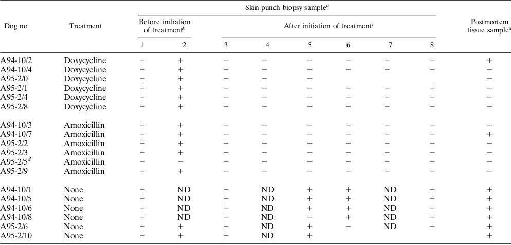
TABLE 1. Culture demonstration of persistence of
B. burgdorferi
in experimentally infected and antibiotic-treated dogs
Dog no. Treatment
Skin punch biopsy samplea
Postmortem tissue samplea
Before initiation
of treatmentb After initiation of treatmentc
1 2 3 4 5 6 7 8
A94-10/2
Doxycycline
1
1
2
2
2
2
2
2
1
A94-10/4
Doxycycline
1
1
2
2
2
2
2
2
2
A95-2/0
Doxycycline
2
1
2
2
2
2
A95-2/1
Doxycycline
1
1
2
2
2
2
2
1
2
A95-2/4
Doxycycline
1
1
2
2
2
2
2
2
2
A95-2/8
Doxycycline
1
1
2
2
2
2
2
2
2
A94-10/3
Amoxicillin
1
1
2
2
2
2
2
2
2
A94-10/7
Amoxicillin
1
1
2
2
2
2
2
2
1
A95-2/2
Amoxicillin
1
1
2
2
2
2
2
2
2
A95-2/3
Amoxicillin
1
1
2
2
2
2
2
2
2
A95-2/5
dAmoxicillin
2
2
2
2
2
2
2
2
2
A95-2/9
Amoxicillin
1
1
2
2
2
2
2
2
2
A94-10/1
None
1
ND
1
ND
1
1
ND
1
1
A94-10/5
None
1
ND
1
ND
1
1
ND
1
1
A94-10/6
None
1
ND
1
ND
1
1
ND
1
1
A94-10/8
None
2
ND
2
ND
2
1
ND
1
1
A95-2/6
None
1
1
1
ND
1
2
ND
1
1
A95-2/10
None
1
1
1
ND
1
1
a1, presence ofB. burgdorferiDNA;2, absence ofB. burgdorferiDNA; ND, not done.
bSamples 1 and 2 were taken 16 and 1 day, respectively, before initiation of treatment in group A94-10 and 33 and 5 days, respectively, before initiation of treatment
in group A95-2.
cSamples 3 to 8 were taken 12, 40, 54, 68, 82, and 97 days, respectively, after initiation of treatment in group A94-10 and 9, 23, 51, 134, 162, and 191 days, respectively,
after initiation of treatment in group A95-2.
dTick-exposed, uninfected dog.
on May 15, 2020 by guest
http://jcm.asm.org/
PCR.
Skin punch biopsy and postmortem tissues were tested
for borrelia DNA with specific primer pairs targeting the
chro-mosomal 23S rRNA gene or the OspA gene on plasmids. The
results are presented in Table 2 and Fig. 1. PCR was positive
for skin punch biopsy samples from all dogs with the exception
of dog A95-2/5 before antibiotic treatment was initiated.
Within 2 weeks of initiation of the antibiotic treatment, skin
punch biopsy samples from two of six doxycycline- and two of
five amoxicillin-treated dogs became PCR negative while
bi-opsy samples from untreated infected control dogs remained
PCR positive. However, between 51 and 134 days after
treat-ment was completed, skin punch biopsy samples from all
in-fected and antibiotic-treated dogs showed positive PCRs again
(Table 2; Fig. 1). In addition, with the exception of dog
A95-2/5, which remained uninfected throughout the experiment,
multiple tissues from three of four doxycycline-treated, two of
three amoxicillin-treated, and two of two untreated infected
dogs had positive PCRs (Table 2). Tissues from two uninfected
SPF dogs, as well as from dog A95-2/5, that served as negative
controls were PCR negative.
Joint histopathology.
No significant joint lesions were seen
in any of the amoxicillin-treated dogs by 2 (A94-10 series) or 6
months (A95-2 series) after antibiotic treatment was
com-pleted (4 and 8 months, respectively, after infection). The
joints of five of six doxycycline-treated dogs were
inconspicu-ous as well. However, one doxycycline-treated dog (A95-2/1)
had mild joint lesions by 8 months after infection. Deep in the
joint capsule of the right tarsus, light infiltrates of neutrophils,
eosinophils, and monocytes were seen. A small leukocytic
fo-cus was found in the joint capsule of the right shoulder. Joint
lesions were seen in four of six untreated infected control dogs.
Mild to severe mono- or polyarthritis with plasma cells,
lym-phocytes, and a few neutrophils was seen in dogs A94-10/1,
A94-10/5, A95-2/6, and A95-2/10. In untreated dogs, synovitis
involved more joints and infiltrates were more severe than
those for the single doxycycline-treated dog with joint lesions.
Lymph node changes (e.g., cortical hyperplasia and sinus
his-tiocytosis) were similar in all infected treated and untreated
dogs.
The joints of two uninfected SPF control dogs were free of
lesions.
Immune response.
Positive KELA levels (above 100 U)
ap-peared between 6 and 8 weeks after infection in all infected
dogs with the exception of one amoxicillin-treated dog
(A95-2/5) that remained uninfected. Thereafter, the titers in
un-treated infected control dogs increased and remained high
until the dogs were euthanized 4 to 8 months after infection
(Fig. 2C).
The antibody levels in antibiotic-treated dogs varied. KELA
units declined in all antibiotic-treated dogs by 4 weeks after
treatment was initiated and reached negative levels in four of
six doxycycline- and four of five amoxicillin-treated dogs (one
dog, A95-2/5, remained uninfected) (Fig. 2A and B). In dogs
that were kept for 6 months after 30 days of antibiotic
treat-ment, antibody levels began to rise again. Three of three
doxy-cycline-treated dogs and two of three amoxicillin-treated
dogs responded with increasing titers at that time (Fig. 2A
and B).
Antibiotic levels in plasma.
Peak concentrations in plasma
[image:3.612.57.556.81.329.2]were reached at 2 h after treatment (Fig. 3). MICs of
doxycy-cline were still present in four of four dogs by 10 h after
treatment and in two of four dogs by 12 h after treatment,
when the antibiotic was administered again (Fig. 3A). MICs of
amoxicillin were present at 4 h after treatment but not at 6 and
TABLE 2. PCR demonstration of persistence of
B. burgdorferi
in experimentally infected and antibiotic-treated dogs
Dog no. Treatment
Skin punch biopsy samplea
Postmortem tissue samplea
Before initiation of
treatmentb After initiation of treatment c
1 2 3 4 5 6 7 8 9
A94-10/2
Doxycycline
1
1
1
1
1
1
1
1
ND
A94-10/4
Doxycycline
1
1
1
1
2
1
1
2
1
ND
A95-2/0
Doxycycline
1
1
1
2
1
2
1
A95-2/1
Doxycycline
1
1
1
1
1
1
1
1
1
1
A95-2/4
Doxycycline
2
1
2
2
2
1
2
2
2
1
A95-2/8
Doxycycline
1
1
2
2
2
2
1
2
2
2
A94-10/3
Amoxicillin
1
1
1
1
2
2
1
1
ND
A94-10/7
Amoxicillin
1
1
1
2
1
1
2
1
1
ND
A95-2/2
Amoxicillin
1
2
2
2
1
1
2
1
2
2
A95-2/3
Amoxicillin
1
1
2
2
1
1
1
2
2
1
A95-2/5
dAmoxicillin
2
2
2
2
2
2
2
2
2
2
A95-2/9
Amoxicillin
1
1
1
2
1
1
2
2
1
1
A94-10/1
None
1
ND
1
ND
1
ND
1
ND
1
ND
A94-10/5
None
1
ND
1
ND
1
ND
2
ND
1
ND
A94-10/6
None
1
ND
1
ND
1
ND
1
ND
1
ND
A94-10/8
None
1
ND
1
ND
1
ND
1
ND
1
ND
A95-2/6
None
2
1
1
ND
2
ND
2
ND
1
1
A95-2/10
None
1
1
1
ND
1
1
a1, presence ofB. burgdorferiDNA;2, absence ofB. burgdorferiDNA; ND, not done.
bSamples 1 and 2 were taken 16 and 1 day, respectively, before initiation of treatment in group A94-10 and 33 and 5 days, respectively, before initiation of treatment
in group A95-2.
cSamples 3 to 9 were taken 26, 40, 54, 68, 82, 97, and 117 days, respectively, after initiation of treatment in group A94-10 and 9, 23, 51, 107, 134, 162, and 191 days,
respectively, after initiation of treatment in group A95-2.
dTick-exposed, uninfected dog.
on May 15, 2020 by guest
http://jcm.asm.org/
8 h after treatment, when treatment was repeated (Fig. 3B).
Differences in plasma antibiotic levels were minimal when
plasma antibiotic levels from different time intervals of
treat-ment were compared.
DISCUSSION
An important question regarding Lyme disease remains: is
conventional antibiotic treatment 2 to 4 weeks in duration
sufficient to eliminate disease and infection? Disseminated
Lyme disease may be associated with acute and/or long-term
[image:4.612.62.295.67.478.2]morbidity (22, 39), in which case the efficacy of the antibiotic
treatment becomes very important. Some authors claim
elim-ination of
B. burgdorferi
and disease occurs after antibiotic
treatment of human patients (20, 25) while evidence is
mount-ing that spirochetes can persist despite antibiotic therapy (12,
FIG. 2. KELA antibody units toB. burgdorferiin sera from dogs exposed to
B. burgdorferi-infected ticks on day 0. Antibiotic treatment was initiated on day 50 (A94-10 series) and day 69 (A95-2 series). Dogs were euthanized between days 133 and 150 (A94-10 series) and between days 272 and 288 (A95-2 series) after exposure. Note: antibody levels in the shaded area below 100 U are neg-ative. (A) Doxycycline-treated dogs. Note: dog A95-2/0 was euthanized on day 210 after exposure. (B) Amoxicillin-treated dogs. Note: dog A95-2/5 remained uninfected. (C) Untreated infected control dogs. Note: dog A95-2/10 was eutha-nized on day 120 after exposure.
FIG. 1. Detection ofB. burgdorferiin skin punch biopsy samples by PCR using a specific primer for the OspA gene. Lanes for all panels: M, molecular weight markers (the DNA fragment at 308 bp is specific forB. burgdorferiOspA);
1, positive control using DNA fromB. burgdorfericultures;2, negative control using water. Doxycycline (A)- and amoxicillin (B)-treated dogs. Results are shown for dogs A95-2/1 and A95-2/9 33 and 5 days before initiation of treatment (lanes 1 and 2, respectively), and 9, 23, 51, 107, 134, 162, and 191 days after initiation of treatment (lanes 3 to 9, respectively). Results are also shown for dogs A94-10/2 and A94-10/7 16 and 1 days before initiation of treatment (lanes 1 and 2, respectively), and 26, 40, 54, 68, 82, 97, and 117 days after initiation of treatment (lanes 3 to 9, respectively). (C) For dog A95-2/5 (uninfected control dog), skin biopsy samples were tested over a 224-day period (lanes 1 to 9). For dog A94-10/8 (untreated infected control dog) skin biopsy samples were tested 34, 76, 104, 132, and 167 days after infection (lanes 1 to 5).
on May 15, 2020 by guest
http://jcm.asm.org/
[image:4.612.325.549.152.652.2]30, 37, 45). Similar contradictory results have been obtained in
experimental mice (19, 23), hamsters (14, 15), and gerbils (31).
Perhaps the time allowed between treatment and testing was
not sufficient in most of these studies, since surviving borreliae
replicate slowly.
Our results for dogs are in agreement with the results for
mice reported by Malawista et al. (19), in which negative
cul-ture and PCR were results found by 30 days after termination
of treatment but two of six mice were culture positive and one
of six mice were PCR positive by 60 days after the last
treat-ment.
We previously developed an experimental model for Lyme
disease in the dog (3) in which acute arthritis with lameness as
well as persistent
B. burgdorferi
and polyarthritis without
clin-ical signs were documented for up to 1 year after tick exposure.
The study was limited to 1 year. In the present study we have
addressed the question of whether 4-week treatment with the
most commonly used antibiotics for Lyme disease in humans—
doxycycline and amoxicillin—is sufficient to eliminate infection
and disease. We conclude that antibiotics did ameliorate the
disease, as, with one exception, arthritis was not seen in treated
dogs. However,
B. burgdorferi
, as judged by culture and PCR,
appeared to persist in dogs after 30 days of treatment with
either doxycycline or amoxicillin.
B. burgdorferi
was isolated from axillary lymph nodes from
one amoxicillin- and one doxycycline-treated dog as well as
from one skin biopsy sample from a doxycycline-treated dog.
However, by PCR using primers for the
B. burgdorferi
23S
rRNA gene and for the outer surface protein A (OspA) gene,
we found messages in untreated as well as antibiotic-treated
dogs in skin punch biopsy samples and in multiple necropsy
tissue samples in three of four doxycycline- and two of three
amoxicillin-treated dogs (Table 2; Fig. 1). Tissues from two
uninfected SPF dogs and dog 95-2/5, which never became
infected, remained PCR negative.
The ultimate proof for persistence of live organisms is
iso-lation. However, Malawista et al. (19) and Persing et al. (28)
have shown that spirochetal DNA does not persist in tissues
when live borreliae are absent. They suggested that persisting
PCR positivity indicates persistent infection. Otherwise, it
would be difficult to explain why serum antibody titers initially
declined in antibiotic-treated dogs that were kept in isolation
but began to increase by 6 months after treatment (Fig. 2B and
C). An additional 2- or 3-month waiting period may have
produced more positive culture results because of the slow
replication of
B. burgdorferi.
The plasma antibiotic levels in treated dogs were adequate.
MICs in doxycycline-treated dogs were maintained throughout
the 30-day period of treatment, an important aspect because
doxycycline has a bacteriostatic, not bacteriocidal, effect.
Be-cause amoxicillin has a bacteriocidal effect, the MICs that
lasted for at least 4 h after each treatment should have been
sufficient. It is not known whether spirochetes survive
antibi-otic treatment due to inadequate antibiantibi-otic penetration of
poorly vascularized connective tissues or whether spirochetes
hide intracellularly as was shown in vitro (10, 16, 18). Further,
it is unclear whether
B. burgdorferi
develops antibiotic
resis-tance or whether additional factors play a role.
In earlier studies we concluded that spirochete isolation in
culture and PCR results were comparable for demonstrating
the presence of the infecting agent (3, 7). The PCR in those
studies was made with nested primers derived from the
B.
burgdorferi
41-kDa flagellin gene. In our present study, we have
used primers for the
B. burgdorferi
23S rRNA gene and for the
OspA gene as reported elsewhere (11, 24, 28, 33). It became
obvious that PCR using these primers is more sensitive than
isolation of
B. burgdorferi
or nested PCR using 41-kDa primers.
While
B. burgdorferi
could only be cultivated from two lymph
nodes and one skin biopsy sample from antibiotic-treated dogs,
a large number of samples were positive by PCR (Tables 1 and
2).
The persistence of
B. burgdorferi
in mammalian hosts after
antibiotic treatment is reminiscent of the persistence of other
spirochetes: treponemes and leptospires. It is well known that
antibiotic treatment of syphilis patients sometimes fails to
eradicate treponemes from the central nervous system and
other sites during late syphilis (27). Penicillin treatment is very
effective in ameliorating acute symptoms of leptospirosis in
humans and animals. However, the infecting agent persists
after treatment and kidney failure with uremia is a frequent
consequence several months later (1, 47). In contrast,
treat-ment with streptomycin (2, 42) or doxycycline (21) was
suffi-cient to eliminate persistent infection.
In conclusion, the canine model of Lyme disease seems
valuable for the detection of persistence of
B. burgdorferi
after
exposure to infected ticks and for the response to antibiotic
treatment. We have shown that treatment with high doses of
amoxicillin or doxycycline for a 30-day period was not sufficient
to eliminate the persistent infection. Although (i) the infection
rate was greatly reduced, (ii) antibody levels declined after
treatment, and (iii) joint lesions were prevented or cured,
per-sistent borreliae stimulated antibody responses again by 6
months after treatment and the possibility of clinical and
pathological relapses remained.
FIG. 3. (A) Doxycycline levels in blood plasma from doxycycline-treated dogs at times posttreatment are shown. Dogs were treated at 12-h intervals. A gel diffusion bioassay using B. cereuswas used. (B) Amoxicillin levels in blood plasma from amoxicillin-treated dogs at times posttreatment are shown. Dogs were treated at 8-h intervals. A gel diffusion bioassay usingB. stearothermophilus
was used.
on May 15, 2020 by guest
http://jcm.asm.org/
[image:5.612.65.292.70.372.2]ACKNOWLEDGMENTS
This work was supported by grant C011798 from the New York State
Department of Health.
We thank Mary Beth Matychak, Peter Harpending, and Patti Easton
for technical assistance and Dorothy Scorelle for typing the
manu-script.
REFERENCES
1.Alexander, A. D., and P. L. Rule.1986. Penicillins, cephalosporins, and tetracyclines in treatment of hamsters with fatal leptospirosis. Antimicrob. Agents Chemother.30:835–839.
2.Alt, D. P., and C. A. Bolin.1996. Preliminary evaluation of antimicrobial agents for treatment of Leptospira interrogans serovar pomona infection in hamsters and swine. Am. J. Vet. Res.57:59–62.
3.Appel, M. J. G., S. Allen, R. H. Jacobson, T.-L. Lauderdale, Y.-F. Chang, S. J. Shin, J. W. Thomford, R. J. Todhunter, and B. A. Summers.1993. Experimental Lyme disease in dogs produces arthritis and persistent infec-tion. J. Infect. Dis.167:651–664.
4.Baradaran-Dilmaghani, R., and G. Stanek.1996. In vitro susceptibility of thirty Borrelia strains from various sources against eight antimicrobial che-motherapeutics. Infection24:60–65.
5.Barthold, S. W.1995. Animal models for Lyme disease. Lab. Invest.72:127– 130.
6.Bennett, J. V., J. L. Brodie, E. J. Benner, and W. M. M. Kirby.1966. Simplified, accurate method for antibiotic assay of clinical specimens. Appl. Microbiol.14:170–177.
7.Chang, Y.-F., R. K. Straubinger, R. H. Jacobson, J. B. Kim, T. J. Kim, D. Kim, S. J. Shin, and M. J. G. Appel.1996. Dissemination ofBorrelia burg-dorferiafter experimental infection in dogs. J. Spirochetal Tick-Borne Dis.
3:80–86.
8.Dattwyler, R. J., J. J. Halperin, H. Pass, and B. J. Luft.1987. Ceftriaxone as effective therapy in refractory Lyme disease. J. Infect. Dis.155:1322–1325. 9.Demaerschalck, I., A. B. Messaoud, M. De Kesel, B. Hoyois, Y. Lobet, P.
Hoet, G. Bigaignon, A. Bollen, and E. Godfroid.1995. Simultaneous pres-ence of differentBorrelia burgdorferigenospecies in biological fluids of Lyme disease patients. J. Clin. Microbiol.33:602–608.
10. Georgilis, K., M. Peacocke, and M. S. Klempner.1992. Fibroblasts protect the Lyme disease spirochete,Borrelia burgdorferi, from ceftriaxone in vitro. J. Infect. Dis.166:440–444.
11. Goodman, J. L., J. F. Bradley, A. E. Ross, P. Goellner, A. Lagus, B. Vitale, B. W. Berger, S. Luger, and R. C. Johnson.1995. Bloodstream invasion in early Lyme disease: results from a prospective, controlled, blinded study using the polymerase chain reaction. Am. J. Med.99:6–12.
12. Hassler, D., K. Riedel, J. Zorn, and V. Preac-Mursic.1991. Pulsed high-dose cefotaxime therapy in refractory Lyme borreliosis. Lancet338:193. 13. Ha¨upl, T., G. Hahn, M. Rittig, A. Krause, C. Schoener, U. Schonherr, J. R.
Kalden, and G. R. Burmester.1993. Persistence ofBorrelia burgdorferiin ligamentous tissue from a patient with chronic Lyme borreliosis. Arthritis Rheum.36:1621–1626.
14. Johnson, R. C., C. Kodner, and M. Russell.1987. In vitro and in vivo susceptibility of the Lyme disease spirochete,Borrelia burgdorferi, to four antimicrobial agents. Antimicrob. Agents Chemother.31:164–167. 15. Johnson, R. C., C. B. Kodner, P. J. Jurkovich, and J. J. Collins.1990.
Comparative in vitro and in vivo susceptibilities of the Lyme disease spiro-cheteBorrelia burgdorferito cefuroxime and other antimicrobial agents. An-timicrob. Agents Chemother.34:2133–2136.
16. Klempner, M. S., R. Noring, and R. A. Rogers.1993. Invasion of human skin fibroblasts by the Lyme disease spirochete,Borrelia burgdorferi. J. Infect. Dis.
167:1074–1081.
17. Levy, S. A., S. W. Barthold, D. M. Dambach, and T. L. Wasmoen.1993. Canine Lyme borreliosis. Compend. Cont. Educ. Pract. Vet.15:833–848. 18. Ma, Y., A. Sturrock, and J. J. Weis.1991. Intracellular localization ofBorrelia
burgdorferiwithin human epithelial cells. Infect. Immun.59:671–678. 19. Malawista, S. E., S. W. Barthold, and D. H. Persing.1994. Fate ofBorrelia
burgdorferiDNA in tissues of infected mice after antibiotic treatment. J. In-fect. Dis.170:1312–1316.
20. Massarotti, E. M., S. W. Luger, D. W. Rahn, R. P. Messner, J. B. Wong, R. C. Johnson, and A. C. Steere.1992. Treatment of early Lyme disease. Am. J. Med.92:396–403.
21. McClain, J. B. L., W. R. Ballou, S. M. Harrison, and D. L. Steinweg.1984. Doxycycline therapy for leptospirosis. Ann. Intern. Med.100:696–698. 22. McFadden, P.1995. Lyme disease research. Science270:1419. (Letter.) 23. Moody, K. D., R. L. Adams, and S. W. Barthold.1994. Effectiveness of
antimicrobial treatment againstBorrelia burgdorferiinfection in mice. Anti-microb. Agents Chemother.38:1567–1572.
24. Moter, S. E., H. Hofmann, R. Wallich, M. M. Simon, and M. D. Kramer.
1994. Detection ofBorrelia burgdorferisensu lato in lesional skin of patients with erythema migrans and acrodermatitis chronica atrophicans by OspA-specific PCR. J. Clin. Microbiol.32:2980–2988.
25. Nadelman, R. B., J. Nowakowski, G. Forseter, S. Bittker, D. Cooper, N. Goldberg, D. McKenna, and G. P. Wormser.1993. Failure to isolateBorrelia burgdorferiafter antimicrobial therapy in culture-documented Lyme borre-liosis associated with erythema migrans: report of a prospective study. Am. J. Med.94:583–588.
26. Nowakowski, J., R. B. Nadelman, G. Forseter, D. McKenna, and G. P. Wormser.1995. Doxycycline versus tetracycline therapy for Lyme disease associated with erythema migrans. J. Am. Acad. Dermatol.32:223–227. 27. Panconesi, E., G. Zuccati, and A. Cantini.1981. Treatment of syphilis: a
short critical review. Sex. Transm. Dis.8:321–325.
28. Persing, D. H., B. J. Rutledge, P. N. Rys, D. S. Podzorski, P. D. Mitchell, K. D. Reed, B. Liu, E. Fikrig, and S. E. Malawista.1994. Target imbalance: disparity ofBorrelia burgdorferigenetic material in synovial fluid from Lyme arthritis patients. J. Infect. Dis.169:668–672.
29. Preac-Mursic, V., E. Patsouris, B. Wilske, S. Reinhardt, B. Gross, and P. Mehraein.1990. Persistence ofBorrelia burgdorferi and histopathological alterations in experimentally infected animals. A comparison with his-topathological findings in human disease. Infection18:332–341.
30. Preac-Mursic, V., K. Weber, H. W. Pfister, B. Wilske, B. Gross, A. Baumann, and J. Prokop.1989. Survival ofBorrelia burgdorferiin antibiotically treated patients with Lyme borreliosis. Infection17:355–359.
31. Preac-Mursic, V., B. Wilske, G. Schierz, E. Suess, and B. Gross.1989. Comparative antimicrobial activity of the macrolides againstBorrelia burg-dorferi. Eur. J. Clin. Microbiol. Infect. Dis.8:651–653.
32. Rahn, D. W.1992. Antibiotic treatment of Lyme disease. Postgrad. Med.
91:57–64.
33. Rijpkema, S. G. T., M. J. C. H. Molkenboer, L. M. Schouls, F. Jongejan, and J. F. P. Schellekens.1995. Simultaneous detection and genotyping of three genomic groups ofBorrelia burgdorferisensu lato in DutchIxodes ricinusticks by characterization of the amplified intergenic spacer region between 5S and 23S rRNA genes. J. Clin. Microbiol.33:3091–3095.
34. Sadziene, A., M. Jonsson, S. Bergstro¨m, R. K. Bright, R. C. Kennedy, and A. G. Barbour.1994. A bactericidal antibody toBorrelia burgdorferiis di-rected against a variable region of the OspB protein. Infect. Immun.62:
2037–2045.
35. Sambri, V., S. Armati, and R. Cevenini.1993. Animal and human antibodies reactive with the outer surface protein A and B ofBorrelia burgdorferiare borreliacidal, in vitro, in the presence of complement. FEMS Immunol. Med. Microbiol.7:67–72.
36. Sambrook, J., E. F. Fritsch, and T. Maniatis.1989. Molecular cloning: a laboratory manual, 2nd ed. Cold Spring Harbor Laboratory Press, Cold Spring Harbor, N.Y.
37. Schmidli, J., T. Hunziker, P. Moesli, and U. B. Schaad.1988. Cultivation of
Borrelia burgdorferifrom joint fluid three months after treatment of facial palsy due to Lyme borreliosis. J. Infect. Dis.158:905–906. (Letter.) 38. Schwartz, I., G. P. Wormser, J. J. Schwartz, D. Cooper, P. Weissensee, A.
Gazumyan, E. Zimmermann, N. S. Goldberg, S. Bittker, G. L. Campbell, and C. S. Pavia.1992. Diagnosis of early Lyme disease by polymerase chain reaction amplification and culture of skin biopsies from erythema migrans lesions. J. Clin. Microbiol.30:3082–3088.
39. Shadick, N. A., C. B. Phillips, E. L. Logigian, A. C. Steere, R. F. Kaplan, V. P. Berardi, P. H. Duray, M. G. Larson, E. A. Wright, K. S. Ginsburg, J. N. Katz, and M. H. Liang.1994. The long-term clinical outcomes of Lyme disease. Ann. Intern. Med.121:560–567.
40. Sigal, L. H.1992. Current recommendations for the treatment of Lyme disease. Drugs43:683–699.
41. Sigal, L. H.1994. Persisting symptoms of Lyme disease—possible explana-tions and implicaexplana-tions for treatment. J. Rheumatol.21:593–595.
42. Spradbow, P. B.1963. Chemotherapy of experimental leptospiral infection in mice. Br. J. Pharmacol.20:237–244.
43. Steere, A. C.1989. Lyme disease. N. Engl. J. Med.321:586–596.
44. Straubinger, R. K., Y.-F. Chang, R. H. Jacobson, and M. J. G. Appel.1995. Sera from OspA-vaccinated dogs, but not those from tick-infected dogs, inhibit in vitro growth ofBorrelia burgdorferi. J. Clin. Microbiol.33:2745– 2751.
45. Strle, F., V. Preac-Mursic, J. Cimperman, E. Ruzic, V. Maraspin, and M. Jereb.1993. Azithromycin versus doxycycline for treatment of erythema migrans: clinical and microbiological findings. Infection21:83–88. 46. Wahlberg, P., H. Granlund, D. Nyman, J. Panelius, and I. Seppua¨la¨.1994.
Treatment of late Lyme borreliosis. J. Infect.29:255–261.
47. Watt, G., L. P. Padre, M. L. Tuazon, C. Calubaquib, E. Santiago, C. P. Ranoa, and L. W. Laughlin.1988. Placebo-controlled trial of intravenous penicillin for severe and late leptospirosis. Lancet(i):433–435.