J Clin Invest.
2004;
114(4)
:569-581.
https://doi.org/10.1172/JCI21358
.
The transcription factor NF-
k
B is activated in a range of human cancers and is thought to
promote tumorigenesis, mainly due to its ability to protect transformed cells from apoptosis.
To investigate the role of NF-
k
B in epithelial plasticity and metastasis, we utilized a
well-characterized in vitro/in vivo model of mammary carcinogenesis that depends on the
collaboration of the Ha-Ras oncoprotein and TGF-
b
. We show here that the IKK-2/I
k
B
a
/NF-k
B pathway is required for the induction and maintenance of epithelial-mesenchymal
transition (EMT). Inhibition of NF-
k
B signaling prevented EMT in Ras-transformed epithelial
cells, while activation of this pathway promoted the transition to a mesenchymal phenotype
even in the absence of TGF-
b
. Furthermore, inhibition of NF-
k
B activity in mesenchymal
cells caused a reversal of EMT, suggesting that NF-
k
B is essential for both the induction
and maintenance of EMT. In line with the importance of EMT for invasion, blocking of NF-
k
B
activity abrogated the metastatic potential of mammary epithelial cells in a mouse model
system. Collectively, these data provide evidence of an essential role for NF-
k
B during
distinct steps of breast cancer progression and suggest that the cooperation of Ras- and
TGF-
b
–dependent signaling pathways in late-stage tumorigenesis depends critically on
NF-k
B activity.
Article
Oncology
Find the latest version:
NF-
κ
B is essential for epithelial-
mesenchymal transition and metastasis
in a model of breast cancer progression
Margit A. Huber,1,2,3 Ninel Azoitei,1 Bernd Baumann,1 Stefan Grünert,2 Andreas Sommer,2Hubert Pehamberger,3 Norbert Kraut,4 Hartmut Beug,2 and Thomas Wirth1
1Department of Physiological Chemistry, Ulm University, Ulm, Germany. 2Institute of Molecular Pathology, Vienna, Austria. 3Department of Dermatology, Vienna Medical University, Vienna, Austria. 4Department of New Chemical Entity Lead Discovery, Boehringer Ingelheim Austria GmbH, Vienna, Austria.
The transcription factor NF-
κ
B is activated in a range of human cancers and is thought to promote
tumorigenesis, mainly due to its ability to protect transformed cells from apoptosis. To investigate the role of
NF-
κ
B in epithelial plasticity and metastasis, we utilized a well-characterized in vitro/in vivo model of
mam-mary carcinogenesis that depends on the collaboration of the Ha-Ras oncoprotein and TGF-
β
. We show here
that the IKK-2/I
κ
B
α
/NF-
κ
B pathway is required for the induction and maintenance of epithelial-mesenchymal
transition (EMT). Inhibition of NF-
κ
B signaling prevented EMT in Ras-transformed epithelial cells, while
activation of this pathway promoted the transition to a mesenchymal phenotype even in the absence of TGF-
β
.
Furthermore, inhibition of NF-
κ
B activity in mesenchymal cells caused a reversal of EMT, suggesting that
NF-
κ
B is essential for both the induction and maintenance of EMT. In line with the importance of EMT for
invasion, blocking of NF-
κ
B activity abrogated the metastatic potential of mammary epithelial cells in a mouse
model system. Collectively, these data provide evidence of an essential role for NF-
κ
B during distinct steps of
breast cancer progression and suggest that the cooperation of Ras- and TGF-
β
–dependent signaling pathways
in late-stage tumorigenesis depends critically on NF-
κ
B activity.
Introduction
Cancer development and metastasis are multistep processes that involve local tumor growth and invasion followed by dissemina-tion to and re-establishment at distant sites. The ability of a tumor to metastasize is the major determinant of the mortality of cancer patients. Thus, elucidating the molecular pathways essential for tumor metastasis is of high priority in cancer biology and provides a basis for novel therapeutic targets for the development of anti-metastatic cancer treatments.
Initially discovered and studied as a major activator of immune and inflammatory functions via its ability to induce expression of genes encoding cytokines, cytokine receptors, and cell-adhesion molecules, the transcription factor NF-κB has recently been impli-cated in the control of cell proliferation and oncogenesis (reviewed in ref. 1). NF-κB transcription factors bind to DNA as hetero- or homodimers that are composed of five possible subunits in mouse and human (RelA/p65, c-Rel, RelB, p50, and p52). These proteins are characterized by their Rel homology domains, which mediate DNA binding, dimerization, and interactions with inhibitory factors known as inhibitor κB (IκB) proteins. Whereas the Rel/p65 and p50 subunits are ubiquitously expressed, the p52, c-Rel, and RelB sub-units are more functionally important in specific differentiated cell types (reviewed in ref. 2). In most unstimulated cells, NF-κB dimers are inactive because of association with IκB proteins that mask the
nuclear localization sequence of NF-κB, thereby retaining it in the cytoplasm and preventing DNA binding. Several IκB proteins are involved in the control of NF-κB activity, three of which (IκBα, IκBβ, and IκBε) act as negative regulators in a stimulus-dependent manner. Stimulation of cells, for example, by proinflammatory cytokines such as TNF-α and IL-1, results in the phosphorylation of IκB at two serine residues located within the N-terminal domain of the proteins (reviewed in refs. 3, 4). This phosphorylation of IκB results in ubiquitination of nearby lysine residues, which represents the signal for degradation by the 26S proteasome. Degradation of the IκB proteins results in the liberation of NF-κB, allowing nuclear translocation and binding to cognate DNA motifs in the regulatory regions of a host of target genes. As a consequence, transcription of these genes, which are involved in immune and inflammatory responses and regulation of apoptosis, as well as in cell growth con-trol, is induced (reviewed in refs. 3, 4). The critical step in NF-κB activation is the phosphorylation of IκB by a high-molecular-weight IκB kinase (IKK) complex. This complex consists of two kinase pro-teins, IKK-1 and IKK-2, (also called IKK-α and IKK-β, respectively), as well as a regulatory component called NF-κB essential modulator (NEMO; also called IKK-γ) (reviewed in ref. 4).
Ample evidence linking NF-κB activity to oncogenesis has accu-mulated in the past years (reviewed in refs. 1, 2). A link between aberrant NF-κB activity and cancer was initially suggested by the identification of v-Rel, a viral homolog of c-Rel, as the transform-ing oncogene of an avian retrovirus that causes aggressive tumors in chickens (5). Moreover, oncogenic viruses, such as human T cell leukemia virus I or Epstein-Barr virus, activate NF-κB as part of the transformation process (6, 7). Translocation of the NF-κB gene NF-κB2/p52 and the IκB family member Bcl-3 was observed in some lymphoid neoplasms (reviewed in ref. 8). High levels of NF-κB were shown to be essential for the transformed phenotype Nonstandard abbreviations used:
benzyloxycarbonyl-Val-Ala-Asp-fluoromethyl-ketone (Z-VAD-FMK); constitutively active (CA); electrophoretic mobility-shift assay (EMSA); epithelial-mesenchymal transition (EMT); IκB kinase (IKK); inhibitor κB (IκB); NF-κB essential modulator (NEMO); TGF-β1–activated kinase 1 (TAK1); trans-dominant (TD).
Conflict of interest: The authors have declared that no conflict of interest exists. Citation for this article: J. Clin. Invest. 114:569–581 (2004).
of Hodgkin lymphoma tumor cells (9), and mutations in the gene encoding IκBα have been detected in some cases of Hodgkin lym-phoma (10). Constitutive nuclear NF-κB activity, meanwhile, has emerged as a hallmark for many other human leukemias, lympho-mas, and solid tumors (reviewed in refs. 2, 8). Furthermore, several oncoproteins, including Ha-Ras and Bcr-Abl, are known to activate NF-κB and rely on NF-κB to mediate their transforming activity (reviewed in ref. 1). Although the exact role of NF-κB in the patho-genesis of human tumors remains to be determined, suppression of apoptosis is clearly of major importance, since inhibition of NF-κB sensitizes many tumor cells to death-inducing stimuli, including chemotherapeutic agents (reviewed in refs. 1, 2).
Whether and how NF-κB also regulates invasive responses such as epithelial-mesenchymal transition (EMT) and metasta-sis is only poorly understood. Evidence has accumulated show-ing that EMT represents an important in vitro correlate of late-stage tumor progression (11–15). As originally
defined (16), EMT involves dedifferentiation of epithelial cells to fibroblastoid, migratory, and more malignant cells, showing a profoundly altered, mesenchymal gene expression program (17, 18). EMT also occurs during embryonic development and is regulated by an elaborate interplay of signaling pathways (12, 13, 15, 16). In several epithelial cell models, includ-ing EpH4 mammary epithelial cells, oncogenic Ras protects cells from TGF-β–induced cell cycle arrest and apoptosis, thus overcoming the tumor-suppressive effects of TGF-β in early tumorigenesis. Furthermore, oncogenic Ras cooperates with TGF-β to induce EMT, a phe-notype that is stabilized by autocrine produc-tion of TGF-β (19–23) and leads to cell migra-tion and motility, which are crucial for tumor progression and metastasis (reviewed in ref. 15). The Ras downstream effector pathway required for EMT and metastasis was recently identified as the Raf/MEK/MAPK pathway, while hyper-active PI3K/Akt signaling enabled protection from TGF-β–induced apoptosis (17). In these
model systems, metastatic potential strictly correlated with the ability of epithelial tumor cells to undergo EMT (15).
[image:3.585.69.516.80.200.2]In this study, we show that NF-κB plays an essential role in the induction of EMT in Ras-transformed mammary epithelial cells. Loss of NF-κB activity abrogated protection of these cells from TGF-β–induced apoptosis, but also actively suppressed an EMT gene program, thus blocking EMT. In contrast, gain of NF-κB activity induced EMT in these Ras-transformed cells in the absence of TGF-β. Moreover, NF-κB also plays an essential role in main-taining the mesenchymal state subsequent to EMT, as its inhibi-tion caused reversal of EMT. In mice, inhibiinhibi-tion of NF-κB activ-ity strongly reduced the metastatic potential of Ras-transformed EpH4 cells. Our results indicate that NF-κB signaling is essential for EMT and metastasis in this murine mammary carcinoma model and suggest that therapeutic inhibition of NF-κB may be a useful strategy for the control of tumor invasion and metastasis.
Figure 1
NF-κB activity is induced during EMT. (A) Schematic illustrates the morphology and epithelial/mesenchymal marker redistribution or expression found in the cell types used in our study. Nontransformed EpH4 mammary epithelial cells were stably transfected with the Ha-Ras oncogene to yield transformed epithelial EpRas cells that undergo EMT upon treatment with TGF-β, resulting in mesenchymal EpRasXT cells further stabilized by an autocrine TGF-β loop. DPP-IV, dipeptidyl peptidase IV; ZO-1, zona occludens 1. (B) EMSAs of whole-cell extracts (6 μg) of exponentially growing EpRas and EpRasXT cells were performed with an NF-κB–specific probe (upper panel) and with an octamer-specific probe (Oct; lower panel) used as a control. Quantified relative DNA-binding levels are indicated below the EMSAs. Similar data were obtained using different protein extract preparations (see also Figure 2A).
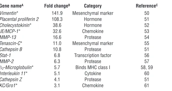
Table 1
NF-κB–regulated genes induced during EMT in the EpRas/EpRasXT cell pair
Gene nameA Fold changeB Category ReferenceC
Vimentin* 141.9 Mesenchymal marker 50
Placental proliferin 2 108.3 Hormone 51
Cholecystokinin* 38.6 Hormone 52
JE/MCP-1* 32.6 Chemokine 53
MMP-13 16.6 Protease 54
Tenascin-C* 11.0 Mesenchymal marker 55
Cathepsin B 10.8 Protease 51
Stat-1 6.8 Transcription factor 56
MMP-2 6.3 Protease 57
β2-Microglobulin* 5.7 Binds MHC class I 58, 59
Interleukin 11* 5.1 Cytokine 60
Cathepsin Z 4.1 Protease 51
KC/Gro1* 3.1 Chemokine 61
[image:3.585.243.527.526.677.2]Results
Upregulation of NF-κB signaling during EMT. EpRas cells represent onco-genic, fully polarized, Ha-Ras–transformed EpH4 mammary epithe-lial cells that undergo EMT in response to TGF-β both in tumors as well as in collagen gels, giving rise to mesenchyme-like cells (EpRasXT cells) in both cases. EpRasXT cells are characterized by a spindle-like morphology and gain of mesenchymal marker proteins, a phenotype stabilized by an autocrine TGF-β loop in vitro and in vivo (refs. 19, 20; see Figure 1A). We wanted to determine whether NF-κB might play a role in the EMT process. We therefore analyzed whole-cell extracts from exponentially growing EpRas and EpRasXT cells by electropho-retic mobility-shift assay (EMSA). We detected some NF-κB DNA-binding activity in EpRas cells even without stimulation by known inducers of NF-κB and consistently observed a 3- to 4-fold increase in NF-κB DNA-binding activity in EpRasXT cells (Figure 1B).
Based on those observations, we next asked whether NF-κB target genes were induced in mesenchymal EpRasXT cells to the same degree as their epithelial counterpart, EpRas cells. To address this question, we reanalyzed the data from a previously reported expression profile, in which we had performed microarray analy-sis of polysome-bound mRNAs to identify genes differentially expressed in EpRasXT compared with EpRas cells (Tables 2 and 3 in ref. 18). Interestingly, 13 of the 75 annotated genes upregulated during EMT had previously been described as NF-κB target genes (Table 1). In mesenchymal EpRasXT cells, several genes encoding NF-κB–regulated cytokines/chemokines (IL-11, JE/MCP-1, and
KC/Gro1), proteases (MMP-13, cathepsin B, MMP-2, and cathepsin Z), hormones (cholecystokinin and placental proliferin 2), and the tran-scription factor Stat-1, as well as β2-microglobulin were expressed at
elevated levels compared with their epithelial counterparts. Fur-thermore, NF-κB has previously been shown to directly regulate the two EMT marker genes vimentin and tenascin C (Table 1). In addition, bcl-3, a regulator of NF-κB activity (24) and previously identified as being expressed in human breast cancer (25), is like-wise induced in EpRasXT cells (18). In contrast, among the genes downregulated in EpRasXT cells, only one gene (thrombospondin 1) has been suggested to be regulated by NF-κB (ref. 26; reviewed
in ref. 27). Thus, expression profiling results clearly indicate an enriched expression of NF-κB target genes in mesenchymal EpRasXT cells, consistent with increased activity of NF-κB.
TGF-β induces NF-κB activity during EMT. Based on the observation that TGF-β signaling is essential for the induction of EMT in EpRas cells (19, 20), as well as reports that TGF-β can modulate NF-κB activ-ity in certain epithelial cells (28), we sought to test whether TGF-β can affect NF-κB activity in EpRas and EpRasXT cells. In order to characterize changes in NF-κB DNA-binding activity after stimula-tion with TGF-β1, we incubated cultures of EpRas and EpRasXT cells in the presence of TGF-β1 and monitored the levels of NF-κB DNA-binding activity by EMSA. In EpRas cells, we observed a 3- to 4-fold induction of NF-κB DNA-binding activity within 30 minutes, while we observed no response to TGF-β in EpRasXT cells (Figure 2A). The result with the EpRasXT cells was not surprising, given that the cells themselves produce TGF-β. In addition, EpRas and EpRasXT cells were transiently transfected with an NF-κB–dependent luciferase reporter (3xκB.luc). Stimulation of EpRas cells with TGF-β1 result-ed in an increase of roughly 2-fold in NF-κB transactivation activ-ity within 2–8 hours (Figure 2B), whereas no increase was noted in EpRasXT cells (data not shown). Thus, TGF-β stimulation leads to an induction of functionally active NF-κB in EpRas cells, while it does not affect the increased NF-κB activity in EpRasXT cells.
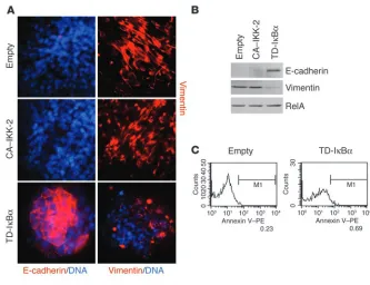
[image:4.585.78.242.84.311.2]NF-κB is essential for EMT. To study the contribution of the IKK/ IκBα/NF-κB signaling module in the regulation of EMT and metastasis, we used retroviral gene transfer to express dominant interfering mutants of this pathway in EpRas cells. Infections were performed using retroviruses expressing a trans-dominant (TD) IκBα protein (TD-IκBα, in which serine residues at posi-tions 32 and 36 are mutated to alanine residues, resulting in a nondegradable repressor), a constitutively active (CA) IKK-2 pro-tein (CA–IKK-2, in which two serine residues in the activation loop are mutated to phosphomimetic glutamic acid residues), or an empty vector control. Stably infected cells were visualized by immunofluorescence microscopy, as the retroviruses coexpress enhanced GFP. Expression of TD-IκBα and CA–IKK-2 in cells from the φNX producer line and in stably infected EpRas cells was
Figure 2
assessed by Western blot (Figure 3, A and B). This analysis revealed strong overexpression of the mutant proteins compared with that of the endogenous counterparts. As previously observed in anoth-er cellular system (29), the presence of high levels of exogenous TD-IκBα resulted in reduced expression of endogenous IκBα, most likely due to inhibition of NF-κB activity. The effects of TD-IκBα
and CA–IKK-2 expression in EpRas cells on NF-κB DNA-binding activity were analyzed by EMSA. In EpRas cells infected with TD-IκBα, no NF-κB DNA-binding activity was observed, regardless of whether cells were left unstimulated or were stimulated with TGF-β (Figure 3C), TNF-α, or PMA (data not shown). In con-trast, cells infected with CA–IKK-2 exhibited 2- to 3-fold increased DNA-binding activity in the unstimulated state (compared with unstimulated EpRas control cells), and showed a roughly 2-fold higher induction of NF-κB activity after being stimulated with TGF-β, compared with TGF-β–treated EpRas control cells. Tran-sient transfection of these cells with 3xκB.luc and subsequent luciferase assays revealed a 3- to 4-fold induction of luciferase activity in untreated EpRas cells infected with CA–IKK-2 and a more than 2-fold higher induction upon treatment with TGF-β, compared with that of unstimulated or TGF-β–treated EpRas con-trol cells, respectively. In contrast, NF-κB transcriptional activity
was virtually completely inhibited before and after treatment with TGF-β in EpRas cells expressing TD-IκBα (Figure 3D). In addi-tion, RT-PCR analysis of a subset of NF-κB–regulated target genes that are associated with EMT (Table 1) (18) was performed with the EpRas mutants before and after TGF-β–induced EMT. This analysis showed induction of MMP-13, MCP-1, and cholecystokinin
in CA–IKK-2–expressing EpRas cells in the absence of TGF-β (in the case of MCP-1, this was even comparable to the expression level in EpRasXT cells), and slightly stronger expression of these genes in the presence of TGF-β (cholecystokinin and MMP-13), whereas expression and TGF-β–induced upregulation of these genes were almost completely blocked in TD-IκBα–expressing cells (Figure 3E). Based on these results, NF-κB activates at least a subset of genes in the TGF-β–induced genetic program underlying EMT.
We then asked whether this modulation in NF-κB activity affect-ed the ability of EpRas cells to undergo EMT. On porous support (filters) allowing epithelial polarization, EpRas cells infected with empty vector (like uninfected cells) showed a fully polarized epithelial phenotype with basolateral plasma membrane expres-sion of the epithelial marker E-cadherin, but no expresexpres-sion of the mesenchymal marker vimentin (Figure 4, A–D). Treatment of these EpRas cells with TGF-β for 5 days resulted in strands of
spindle-Figure 3
[image:5.585.74.515.81.349.2]shaped, vimentin-positive cells only weakly expressing E-cadherin (Figure 4, A, B, and D). No phenotypic changes or changes in mark-er expression compared with EpRas control cells wmark-ere obsmark-erved in TD-IκBα–expressing cells in the absence of TGF-β (Figure 4, A–D). Upon treatment with TGF-β, however, a considerable proportion of these TD-IκBα–overexpressing EpRas cells with blocked NF-κB activity rapidly detached from porous supports as a consequence of cell death. The remaining cells almost completely failed to undergo EMT (Figure 4A). TGF-β–treated TD-IκBα–expressing EpRas cells lacked the strands of spindle-shaped mesenchymal cells that were abundant in empty virus–infected EpRas control cultures (Figure 4, A and B). The same cells failed to upregulate vimentin and retained high levels of E-cadherin, which was partial-ly redistributed to the cytoplasm, indicating some loss of polarity (Figure 4D). Surprisingly, CA–IKK-2–overexpressing EpRas cells
with increased NF-κB activity were able to undergo EMT at a con-siderable rate even in the absence of TGF-β (Figure 4, A and B). After cells had grown for 6 days on porous support, we observed strands of spindle-shaped, E-cadherin–negative and vimentin-positive cells that covered more than 10% of the total surface area (Figure 4, A, B, and D). When we analyzed bulk cultures of these cells by Western immunoblot, we observed a strong reduction in E-cadherin levels (Figure 4C). Consistent with these observations, we noted enhanced EMT in CA–IKK-2–expressing cells upon TGF-β
[image:6.585.82.495.79.441.2]treatment compared with that of control EpRas cells, as indicated by a higher percentage of spindle-shaped cells with cytoplasmic (depolarized) or no E-cadherin expression, and strong vimentin expression (Figure 4, A, B, and D). Importantly, very similar results were obtained when the CA-IKK-2 and TD-IκBα transgenes were introduced into V12S35Ras cells. These cells represent another
Figure 4
monogenic transformed cell line, independently generated from the original EpH4 cells and transformed by an effector mutant of a different oncogenic Ras that induces hyperactive ERK/MAPK but not PI3K signaling (17). We observed an inhibition of EMT in TD-IκBα–expressing cells and a considerable rate of spontaneous EMT in the absence of TGF-β in CA–IKK-2–expressing V12S35Ras cells that occurred even more frequently than in
CA–IKK-2–express-ing EpRas cells (see Supplemental Figure 1; supplemental material available at http://www.jci.org/cgi/content/full/114/4/569/DC1). Thus, the role of NF-κB in the regulation of EMT is not limited to a single cell line.
[image:7.585.101.480.80.502.2]A more physiological culture system to analyze epithelial cell behavior and plasticity are three-dimensional serum-free collagen I gel cultures (15). In these cultures, fully polarized EpRas control
Figure 5
TD-IκBα expressed in EpRas cells prevents EMT, whereas CA–IKK-2 induces EMT in the absence of TGF-β (analysis in collagen gels). EpRas cells expressing the empty vector control, TD-IκBα, or CA–IKK-2 were seeded into collagen gels, were allowed to form structures for 3–5 days, and were left untreated for no induction (–) or were induced to undergo EMT by the addition of TGF-β (+) for 5–6 days. (A) Left column, culture without TGF-β
cells form tubular and alveolar structures with large lumina (Fig-ure 5A). These cells show basolateral membrane (polarized) expres-sion of E-cadherin, but no vimentin expresexpres-sion (Figure 5C). Upon addition of TGF-β1, EpRas control cells underwent EMT, as shown by a spindle-shaped and migratory phenotype, loss of E-cadherin, and de novo expression of vimentin after 6 days of treatment
[image:8.585.59.527.80.501.2](Fig-ure 5, A and C). Untreated TD-IκBα cells resembled control EpRas cells in that they formed epithelial structures (tubular structures with lumina) and showed basolateral E-cadherin staining and no vimentin expression. Despite this resemblance, TD-IκBα epithelial structures appeared more compact and smaller in size (Figure 5, A and C). Upon treatment with TGF-β, epithelial structures formed
Figure 6
by TD-IκBα–expressing EpRas cells retained E-cadherin expression (Figure 5C), but rapidly disintegrated (Figure 5, A and C). Only a very small fraction of the structures (about 0.5%) formed unor-dered cell strands before disintegration (data not shown). In con-trast, untreated CA–IKK-2–expressing cells with increased NF-κB activity formed two types of structures. While a large proportion of epithelial tubular structures with lumina were apparent (Figure 5A), a significant number of the structures consisted of unordered cell strands with spindle-like cellular morphology, resembling con-trol EpRas cells treated with TGF-β (Figure 5, A and B). Interest-ingly, among the epithelial structures resembling control EpRas cells (E-cadherin positive and vimentin negative), a large percent-age had either lost or downregulated expression of CA–IKK-2, as indicated by low levels of the coregulated GFP expression (Figure 5C, bottom). In contrast, mesenchymal structures generated by untreated CA–IKK-2–expressing cells were E-cadherin negative, expressed vimentin, and showed strong GFP staining, indicative of high transgene expression (Figure 5C). Thus, high levels of CA–IKK-2 expression appear to promote EMT even in the absence of TGF-β. As expected, TGF-β treatment induced the remain-ing epithelial CA–IKK-2–expressremain-ing EpRas cells to rapidly form mesenchymal structures expressing vimentin but no E-cadherin (Figure 5, A and C). We also determined whether a strong NF-κB activator such as TNF-α would induce EMT. Indeed, we observed a small number of mesenchymal structures expressing vimentin in TNF-α–treated EpRas collagen gel cultures, even in the absence of TGF-β (Supplemental Figure 2). In conclusion, an activated NF-κB pathway plus Ras is sufficient to cause EMT, whereas inhi-bition of NF-κB activity prevents EMT, causing disintegration of structures formed in collagen.
NF-κB is required both for protection from TGF-β–induced apoptosis and for direct promotion of EMT. The previous experiments showed that NF-κB can, at least in part, substitute for TGF-β in the induction of EMT in collaboration with Ras. We next addressed the mechanistic consequence of NF-κB inhibition with respect to the prevention of EMT. Because we observed rapid disintegration of TD-IκBα– expressing structures upon TGF-β treatment, we assessed their apoptotic response under these conditions. EpRas control cells as well as TD-IκBα– and CA–IKK-2–expressing derivatives were allowed to form organotypic structures in collagen gels for 3 days. Then they were induced to undergo EMT by TGF-β or were left untreated. TGF-β–treated TD-IκBα–expressing EpRas structures showed cell disintegration due to apoptosis, as determined by in situ TUNEL staining (Figure 6A). As shown in Figure 6B, approxi-mately 55% of these cells were TUNEL positive and were thus apoptotic after 6 days of TGF-β treatment. Moreover, TD-IκBα cells showed a slight elevation in induction of apoptosis compared with control EpRas cells even in the absence of TGF-β (4.5% versus 0.6%), possibly explaining the smaller size of epithelial structures formed by TD-IκBα–expressing cells in collagen gels. Finally, EpRas cells expressing CA–IKK-2 showed low levels of apoptosis, comparable to that of EpRas control cells. We then asked whether the observed failure of TD-IκBα–expressing EpRas cells to undergo EMT (Fig-ures 4 and 5) was exclusively due to inhibition of the antiapoptotic function of NF-κB in these cells. EpRas control cells and TD-IκBα– expressing derivatives were cultured on porous support. Treatment of TD-IκBα–expressing EpRas cells with 25 μM cell-permeable caspase inhibitor benzyloxycarbonyl-Val-Ala-Asp-fluoromethyl-ketone (Z-VAD-FMK) strongly suppressed the TGF-β–induced apoptosis seen in these cells (on porous support as well as in colla-gen gels; Figure 6B), to a level that was comparable to that of EpRas control cells (data not shown). Importantly, neither mesenchymal structures (as seen in TGF-β–treated EpRas control cells) nor loss of polarized E-cadherin expression or de novo vimentin expression was observed in these Z-VAD-FMK–treated TD-IκBα–expressing EpRas cells after 4 days of stimulation with TGF-β (Figure 6, C and D). Notably, due to the significant fraction of apoptotic cells following TGF-β treatment, small areas of irregular structures were detected in the TD-IκBα–expressing EpRas cultures (data not shown). These were not observed when apoptosis was blocked by Z-VAD-FMK. In summary, NF-κB signaling is required in
[image:9.585.77.247.79.385.2]transformed cells for protection from TGF-β–induced apoptosis during EMT. Moreover, NF-κB plays an additional role as a direct regulator of the EMT program, as blockade of apoptosis does not restore the ability of TD-IκΒα–expressing EpRas cells to undergo EMT in response to TGF-β.
Inhibition of NF-κB activity in mesenchymal EpRasXT cells causes rever-sal of EMT. We next addressed whether interference with NF-κB activity would also affect the mesenchymal EpRasXT cells that have completed EMT. EpRasXT cells were again stably infected with retroviruses expressing TD-IκBα or CA–IKK-2 or with a GFP-only empty control vector. Transgene expression, as assessed by Western blot analysis, is shown in Figure 7A. EMSA showed complete inhibition of NF-κB DNA-binding activity in untreat-ed and TNF-α- or PMA-stimulated EpRasXT cells expressing TD-IκBα. The expression of CA–IKK-2 resulted only in a moder-ate (less than 2-fold) enhancement of NF-κB activity in untreated cells (Figure 7B). Transient transfection with 3xκB.luc and sub-sequent luciferase assays revealed a strong blockade of NF-κB transactivation activity in EpRasXT cells expressing TD-IκBα, while a moderate increase of luciferase activity was observed in EpRasXT cells infected with CA–IKK-2.
To test whether TD-ΙκBα was able to revert EMT, we cultured EpRasXT cells expressing TD-IκBα, CA–IKK-2, or the control vec-tor on porous support. As expected, CA–IKK-2– and control vecvec-tor– expressing EpRasXT cells showed a mesenchymal, spindle-shaped
phenotype and expressed high levels of vimentin, but no E-cadherin (Figure 8, A and B). Interestingly, however, a large per-centage of TD-IκBα–expressing cells revert-ed to an epithelial phenotype, in which the cells formed compact structures, regained marked E-cadherin expression at the plas-ma membrane, and almost completely lost expression of vimentin, as demonstrated by immunofluorescence (Figure 8A) and Western blot analysis (Figure 8B). Simi-lar results were obtained under different culture conditions (data not shown).
TD-ΙκBα–expressing EpRasXT cells showed no obvious signs of cell death when cul-tured on porous support (e.g., condensed nuclei, disintegrated cells, and detachment from porous support). Annexin V staining (Figure 8C) as well as cell cycle analysis (data not shown) of TD-IκBα –express-ing EpRasXT cells that had reverted to an epithelial phenotype demonstrated that reverted epithelial cells were still viable and healthy. These results indicate that NF-κB activity is required for maintenance of the mesenchymal phenotype of Ras-trans-formed cells that have undergone EMT.
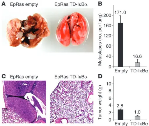
[image:10.585.40.373.79.335.2]NF-κB is required for the metastatic potential of EpRas cells in vivo. EpRas cells undergo EMT in vivo in response to endogenous TGF-β (19). TGF-β–induced EMT of EpRas cells is tightly linked to their abil-ity to form lung metastases, evident after tail vein injection of cultured EpRas cells (S. Grünert and H. Beug, unpublished data) or cells recultivated from EpRas-induced primary mammary tumors (17). Because NF-κB activity was found to be essential for both the induction and maintenance of EMT, we sought to determine whether NF-κB signaling is also required for metastatic potential induced by Ras plus TGF-β in vivo and whether inhibi-tion of NF-κB would abrogate this metastatic ability. The meta-static potential of TD-IκBα–expressing EpRas cells and EpRas control cells was assayed by injection of cultured cells into the tail vein of nude mice. Mice injected with EpRas control cells rap-idly died from numerous large metastases (on average, 3–4 weeks after tail vein injection), while mice receiving TD-IκBα –express-ing EpRas cells appeared healthy at the time of death of the mice injected with EpRas control cells. Mice injected with EpRas cells with blocked NF-κB activity showed a 2-fold decrease in lung weight (compared with that of control mice of similar age; data not shown) and had only a few small (micro-) metastases (aver-age number, 16.6 metastases per lung) by histological analysis, compared with an average number of 171.0 metastases per lung in animals injected with EpRas control cells (Figure 9, A–C). To verify that TD-IκBα–expressing EpRas cells were still able to form pri-mary tumors in a fashion similar to that of EpRas control cells, we injected the cell types described above (in each case from the same batches as used for tail vein injections) into mammary gland fat pads of nude mice. After 3 weeks, both EpRas control cells as well as EpRas cells expressing TD-IκBα formed tumors, which differed
Figure 8
mainly in size (Figure 9D). In conclusion, inhibition of NF-κB activity strongly affects the metastatic potential of EpRas cells in vivo, while primary tumor formation is affected only moderately.
Discussion
Activation of NF-κB signaling is increasingly being recognized as a key mechanism for tumorigenesis and is thought to act mainly by conferring apoptosis resistance to transformed cells. In this report, we used a well characterized combined in vitro/in vivo model of mammary carcinogenesis (EpRas) to determine the function of NF-κB in the regulation of epithelial plasticity and metastasis. First, we showed that NF-κB activity is required together with oncogenic Ras for efficient protection of mammary epithelial cells from TGF-β– induced apoptosis, as a prerequisite for these cells to undergo an EMT toward an invasive, metastatic tumor phenotype. Second, NF-κB can, in part, induce EMT in Ras-transformed cells in the absence of TGF-β, suggesting that NF-κB signaling can mediate important aspects of TGF-β signaling essential for inducing EMT. Third, NF-κB activity is necessary for cells to be maintained in a mesenchymal state, as its inhibition causes reversal of EMT; and finally, in agree-ment with its essential role in the regulation of at least these three distinct aspects of tumor progression in vitro, NF-κB signaling is required for metastasis of Ras-transformed epithelial cells in vivo.
or sensitizes cells to killing by TNF-α or anticancer drugs (30). Several studies have demonstrated that inhibition of NF-κB results in an increased sensitivity of tumor cells to cancer ther-apy–induced apoptosis. For example, delivery of a recombinant TD-IκBα to chemoresistant tumors in mouse xenograft models induced tumor regression by sensitizing them to chemothera-peutic treatment with the topoisomerase I inhibitor CPT-11 (31, 32). Our results clearly establish a critical role for NF-κB as an important regulator of the EMT gene program that goes beyond its well established function in apoptosis protection. This is based on several lines of evidence. First, NF-κB activa-tion is sufficient to induce EMT in a considerable proporactiva-tion of epithelial EpRas cells. Second, NF-κB inhibition results in a blockade of EMT (under conditions in which apoptosis was efficiently suppressed). Third, NF-κB is a critical factor for the activation of a subset of endogenous genes in the TGF-β– induced gene program. Finally, NF-κB blockade can partially revert EMT, resulting in viable and healthy epithelial cells.
Here we demonstrate for the first time to our knowledge that NF-κB plays a causal role in the induction and maintenance of EMT in Ras-transformed mammary epithelial cells, mediating an invasive/metastatic tumor phenotype. These findings add mech-anistic insight regarding the role of NF-κB in late-stage mam-mary tumorigenesis and metastasis. Our observations regard-ing a detectable or further elevated baseline activity of NF-κB in unstimulated EpRas and EpRasXT cells, respectively, are consis-tent with reports demonstrating constitutive activation of NF-κB factors in breast cancer (33, 34). Inhibition of the constitutive NF-κB activity in human breast cancer cell lines induced apoptosis (34) or led to reduced tumorigenicity (35). Furthermore, mouse mamma-ry tumor virus c-rel–transgenic mice develop late-onset mammary tumors of diverse histology (36). Interestingly, some of the tumors identified in the study by Romieu-Mourez et al. were spindle cell car-cinomas, a tumor type possibly generated by EMT (12, 22).
Recent studies have suggested that NF-κB regulates the expres-sion of multiple genes involved in tumor spread and metastasis, including those encoding MMPs, IL-8, VEGF, and CXCR4 (37, 38). Several NF-κB target genes we have reported to be induced during EMT of EpRas cells, such as those encoding various MMPs and cathepsin family members, chemokines/cytokines, tenascin-C, etc. (see Table 1 and Figure 3E), could contribute to the NF-κB– dependent metastatic capacity we observed in our study. Suppres-sion of metastasis upon blockade of NF-κB activity has also been reported in human prostate cancer cells (37), in human melano-ma cells (39), and in murine lung alveolar carcinomelano-ma cells (40). Collectively, our work and work by others clearly show the impor-tance of NF-κB signaling for tumor progression and metastasis in multiple tumor model systems. It should be noted, however, that while NF-κB contributes to oncogenesis in many cell types, its IκBα-mediated suppression in keratinocytes was recently
dem-Figure 9
Inhibition of NF-κB activity prevents metastasis of EpRas cells. (A) EpRas cells infected with empty GFP vector (Empty) or TD-IκBα were injected (5 × 10⁵ cells per injection) into the tail veins of nude mice (four animals per
cell type), and mice were analyzed for the presence of lung metastases. Note large metastatic nodules (white arrows) in the lung from the mouse injected with EpRas cells that had been infected with empty vector (EpRas empty; left). (B) Metastases in lungs similar to those shown in A were quan-tified in serial sections to determine the mean numbers of metastases per lung (four lungs per cell type). (C) Hematoxylin and eosin staining of lungs from mice injected with EpRas cells that had been infected with empty vec-tor or TD-IκBα. Note large metastases in the mouse injected with EpRas cells that had been infected with empty vector. Original magnification, ×10. (D) The same cells analyzed in A–C were injected (2 × 10⁵ cells per
[image:11.585.42.298.82.299.2]onstrated to be necessary for Ras-mediated induction of invasive epidermal tumors resembling squamous cell carcinomas (41). The role of TGF-β signaling in the oncogenic Ras/IκBα tumors, how-ever, has not been addressed.
Role of NF-κB in the cooperation of Ras- and TGF-β–dependent signal-ing pathways dursignal-ing tumor progression. Previous studies by Janda et al. (17) addressed the effect of Ras signaling pathways on epithe-lial plasticity in the EpH4/EpRas model, using Ras mutants that specifically activate only the Raf/MEK/Erk pathway (S35Ras) or the PI3K/Akt pathway (C40Ras). Furthermore, specific inhibitors that block Ras, MEK1, or PI3K were used to interfere with EMT induction or maintenance in EpRas cells (17). Both approaches showed that Ras-dependent signaling of the Raf/MEK/Erk path-way in combination with TGF-β signaling is required for EMT (17). In contrast, C40-induced PI3K/Akt signaling protected EpH4 cells from TGF-β–induced apoptosis, but failed to induce EMT. In vivo, only a Ras mutant able to activate the Raf/MEK/Erk pathway (S35Ras), but not the C40Ras mutant, was able to gen-erate metastases, strictly correlating with the potential of these mutants to induce EMT (17). A number of reports have shown that oncogenic Ras stimulates NF-κB–dependent transcription and that NF-κB is required for Ras-mediated transformation (reviewed in ref. 1). Several lines of evidence indicate that NF-κB acts in a common pathway with PI3K/Akt, leading to suppres-sion of TGF-β–induced apoptosis in Ras-transformed mammary epithelial cells. First, the rate of TGF-β–dependent apoptosis induction is very similar in TD-IκBα–expressing EpRas cells and in EpRas cells treated with a specific PI3K inhibitor (17). Second, Akt has been shown in several cellular systems to stimulate signal-ing pathways that upregulate the activity of NF-κB. Importantly, the antiapoptotic signals elicited by PDGF have been shown to require Akt-induced NF-κB transcriptional activity (42). Several reports have indicated that IKK activity is involved in the ability of Akt to stimulate NF-κB transcriptional activity (42–44), while others have found that PI3K or Akt can also stimulate NF-κB activity through signaling pathways targeting the p65 subunit of NF-κB (45). Our finding that modulating NF-κB activity has the same effects in EpRas cells and V12S35 cells would be consis-tent with the interpretation that the PI3K/Akt signaling may be upstream of NF-κB activity. Collectively, further experiments are required to elucidate how NF-κB precisely mediates Ras-depen-dent effects in the EpRas cellular system.
Our observation that TGF-β–dependent induction of EMT depends at least in part on NF-κB activity raises the question of how these signaling pathways may collaborate. Induction of NF-κB activity and transcription of target genes by TGF-β, as observed in our study, are in line with observations by Arsura et al. (28) in liver epithelial cells. TGF-β can signal in a Smad-independent manner through TGF-β1–activated kinase 1 (TAK1). Interestingly, TAK1 has been shown to directly phosphorylate the IKK complex in response to TGF-β treatment, promoting NF-κB activation (28). In addition, several Smad proteins, acting as transcription factors defined as the major responders to TGF-β signaling, can function as transcriptional coactivators through physical interaction with NF-κB subunits to stimulate transcription via κB sites (46). It will be important to further analyze at which levels these two pathways intersect to abrogate the classical functions of TGF-β in the induc-tion of apoptosis and how they cooperate in enhancing the role of TGF-β in the induction of EMT. A potential novel point of inter-section has been discovered very recently. TGF-β induces
transcrip-tion of the cell cycle inhibitor p21CIP1 by a mechanism, in which Smad proteins cooperate with FoxO transcription factors (47). Three strategies have been described to overcome this cell cycle inhibition. The first is counteraction of FoxO function by another member of the Fox transcription factor family, namely FoxG1 (47). The second is inactivation of FoxO proteins by the PI3K/Akt pathway, which results in relocalization of FoxO proteins to the cytoplasm (48). Third, in breast tumors, upregulation of IKK-2 has been observed and IKK-2 was shown to directly phosphorylate and functionally inactivate the FoxO proteins (49).
In summary, our study provides a functional dissection of the requirement for NF-κB in several aspects of breast cancer pro-gression in this combined in vitro/in vivo model system. It iden-tifies NF-κB as a pivotal regulator of the EMT process, which by itself is a critical prerequisite for metastasis. Further char-acterization of the mechanisms by which NF-κB contributes to the invasion and metastasis of mammary carcinomas and other malignant tumors will provide important targets for drug dis-covery, which should lead to new therapeutic approaches for antimetastatic cancer treatments.
Methods
Cells and cell culture. Origin and culture conditions for EpRas and EpRasXT cells were described earlier (19, 20). The generation and cul-ture conditions of φNX amphotropic retrovirus producer cells express-ing TD-IκBα, CA–IKK-2, and parental vector, coupled to the expression of a GFP–zeocin-resistance fusion gene through an internal ribosome entry site were described previously (29).
Retroviral infection of EpRas and EpRasXT cells with supernatant from φNX producer cells. EpRas and EpRasXT cells were infected with parental vector or retroviruses expressing the dominant interfering mutants as described earlier (29). Briefly, 24 hours prior to infection, EpRas and EpRasXT cells were seeded in six-well plates at a density of 2 × 10⁵ cells per well, and φNX
cells were seeded at a density of 3 × 10⁶ cells per 10-cm plate. For
infec-tion, φNX cell supernatants were obtained and filtered through a
0.45-μm filter, and 5 μg/ml polybrene (Sigma-Aldrich, St. Louis, Missouri, USA) was added to the filtrate. Thereafter, medium was removed from the EpRas and EpRasXT cells and was replaced with φNX cell superna-tants containing the retrovirus. Culture plates were centrifuged at 750 g for 2 hours, the supernatants were removed and replaced with propaga-tion medium. Then, 48 hours later, the efficiency of infecpropaga-tion was moni-tored by fluorescence microscopy (Improvision, Heidelberg, Germany) and infected cells were selected with zeocin at concentrations of 1,500
μg/ml (EpRas) and 1,700 μg/ml (EpRasXT).
NF-κB assays. Western immunoblot analysis for monitoring protein expression levels of dominant interfering NF-κB mutants was performed as described earlier (29) using antibodies specific for IκBα (sc-371; Santa Cruz Biotechnology, Santa Cruz, California, USA), IKK-2 (sc-7607; Santa Cruz Biotechnology), and RelA (sc-372; Santa Cruz Biotechnology).
NF-κB DNA-binding activity was measured by EMSA. Whole-cell extracts were prepared by the “freeze-thaw” method and EMSAs were performed as described earlier (29). In all cases, whole-cell extracts were incubated for 20 minutes at room temperature with radiolabeled double-stranded oligonucleotides containing an Igκ enhancer consensus NF-κB site, an octamer-specific site (5′-ATGCAAAT-3′), or an Sp-1–specific site (5′-ATTCGATCGGGGCGGGGCGAGC-3′), and the DNA-protein complexes formed were then separated from free oligonucleotides by electrophoresis through a native 4% polyacrylamide gel.
Semiquantitative RT-PCR. Total RNA was extracted and semiquantitative RT-PCR was carried out as described earlier (29). Mouse MMP-13 was amplified with primers 5′-CACTCCAAGGACCCAGGAGCCC-3′ (sense) and 5′-GCTGAGGGTGCAGGCGCCAGAA-3′ (antisense; 28 cycles); mouse MCP-1 was amplified with primers 5′ -CGGCTGGAGCATCCACGTGTTG-3′ (sense) and 5′-GTCTGGACCCATTCCTTCTTGGGG-3′ (antisense; 28 cycles); mouse cholecystokinin was amplified with primers 5′
-CGCAGCCGG-TAGTCCCTGCAGAA-3′ (sense) and 5′
-CCATCCAGCCCATGTAGTCCC-GG-3′ (antisense; 28 cycles); and mouse β-actin was amplified with primers 5′-GGTCAGAAGGACTCCTATGTG-3′ (sense) and 5′ -AGAGCAACATAG-CACAGCTTC-3′ (antisense; 28 cycles).
Marker analysis of cells grown on porous support. Cells were seeded on porous supports (cell culture inserts; pore size, 0.4 μm; BD, San Jose, California, USA) at densities of 0.5 × 10⁵ cells/well for EpRas cells and 1
× 105 cells/well for EpRasXT cells. In some experiments, cells were
incu-bated with the indicated concentration (see Figure 6C) of the broad-range caspase inhibitor Z-VAD-FMK (R&D Systems, Minneapolis, Min-nesota, USA). Fresh inhibitor was supplied with each medium change every other day. Cells were cultivated for 5–7 days and then either cells were subjected to immunofluorescence staining (described in detail in ref. 17) with polyclonal rabbit anti–E-cadherin (610182; 1:500 dilution; BD), monoclonal anti–mouse vimentin (Vim-13.4; V-2258; 1:500 dilu-tion; Sigma-Aldrich), and Cy3-conjugated donkey anti-mouse IgG as secondary antibody (1:1,000 dilution; Jackson ImmunoResearch Labo-ratories), or cells were lysed in buffer containing 20 mM HEPES, 25% glycerol, 0.42 M NaCl, 1.5 mM MgCl2, and 0.2 mM EDTA and subjected
to Western immunoblot analysis with antibodies specific for E-cadherin, vimentin, and RelA (as specified above). For immunofluorescence stain-ing, DAPI (0.1 ng/ml) was used for nuclear counterstaining. Digital images were collected either with a MicroMAX camera (Roper Scientific, Trenton, New Jersey, USA) on a Zeiss Axioplan 2 (Zeiss, Thornwood, New York, USA) using Metamorph software (EpRas cells; Universal Imaging Corp., Philadelphia, Pennsylvania, USA), or on a fluorescence micro-scope (Improvision) using OpenLab 3.0.2 and 3.0.3 software (EpRasXT cells; Improvision).
Collagen gel culture, marker analysis, and apoptosis assay. Serum-free, three-dimensional cultures of EpRas cells and their derivatives were performed as described earlier (17, 19). TGF-β1 (5 ng/ml; R&D Sys-tems) was added to the cultures after 2–5 days and then was supplied together with fresh medium every other day for a total of 5–7 days. In situ immunofluorescence analysis was performed to analyze three-dimen-sional structures for E-cadherin and vimentin expression, as described by Janda et al. (17). The following antibodies were used: polyclonal rabbit anti–E-cadherin (610182; 1:500 dilution; BD), monoclonal anti–mouse vimentin (Vim-13.4; V-2258; 1:500 dilution; Sigma-Aldrich), and Cy3-conjugated goat anti-mouse IgG as secondary antibody (1:3,000 dilution; Jackson ImmunoResearch Laboratories). In all cases, DAPI (0.1 ng/ml) was used for nuclear counterstaining.
For TUNEL assays, collagen gels were fixed in 4% paraformaldehyde in PBS, washed with PBS three times, and stained for apoptotic nuclei with
μm (BD). Cells were scraped off the filters, washed twice in ice-cold PBS, and resuspended in binding buffer (BD) at a density of 1 × 10⁶ cells/ml.
Then, 1 × 10⁵ cells were stained with 5 μl of phycoerythrin-conjugated
annexin V (BD) for 15 minutes, washed twice with PBS, and analyzed by flow cytometry (FACSCalibur, BD).
Tumorigenesis and metastasis assays. All animal studies have been approved by the review board of the Institute of Molecular Pathology. Athymic MF1 nude mice were used for mammary gland and tail vein injections, with 200,000 cells per 20 μl PBS for mammary gland injection (tumorigenesis assay) and 500,000 cells per 0.5 ml PBS for tail vein injection (metastasis assay) of 6- to 10-week-old female mice.
For tumorigenesis assays, mice were sacrificed when tumors of control animals had reached a size of approximately 1.0–1.5 cm in diameter (on average, 3 weeks after mammary gland injection) or if tumors ulcerated or the mice showed significant morbidity. Then, tumors were excised and col-lected in PBS and total tumor weight (from four injection sites per mouse) was determined for each individual animal.
For metastasis assays, mice were checked daily for weight loss and breath-lessness for detection of massive lung metastasis. The mice of an entire experiment were sacrificed at the time when control mice started to die from lung metastasis (on average, 3–4 weeks after tail vein injection). Lungs were excised and collected in PBS, and lung weight was determined.
For histological analysis of metastasis assays, lungs were immersed in 10% neutral buffered formalin before paraffin embedding and section-ing. Sections 5 μm in thickness were processed for hematoxylin and eosin staining and histological evaluation. Total numbers of metastases per lung were determined by collection of serial lung sections, selection of sections at approximately 0.3 mm apart, and counting of metastatic lesions, with correction for the contribution of large metastatic lesions that appeared on more than one section.
Acknowledgments
We are grateful to M. Jechlinger for providing EpRas and EpRasXT cells; B. Anic and G. Litos for excellent technical assistance; K. Stangl and P. Garin-Chesa for help with histology; the Institute of Molecular Pathology BioOptics Department for support with immunofluorescence microscopy; and K. Scharffetter-Kochanek, P. Petzlbauer, C. Hoeller, S. Maschler, and M. Herlyn for scientific discussions and useful comments. This work was in part funded by the Genome Research in Austria (GEN-AU) program (to M.A. Huber) and by the German Science Foundation (DFG SFB497/B1 to B. Baumann; SFB451/A9 to T. Wirth).
Received for publication February 17, 2004, and accepted in revised form June 22, 2004.
1. Orlowski, R.Z., and Baldwin, A.S., Jr. 2002. NF-κB as a therapeutic target in cancer. Trends Mol. Med. 8:385–389.
2. Karin, M., Cao, Y., Greten, F.R., and Li, Z.W. 2002. NF-κB in cancer: from innocent bystander to major culprit. Nat. Rev. Cancer. 2:301–310.
3. Ghosh, S., May, M.J., and Kopp, E.B. 1998. NF-κB and Rel proteins: evolutionarily conserved mediators of immune responses. Annu. Rev. Immunol. 16:225–260. 4. Karin, M., and Ben-Neriah, Y. 2000. Phosphorylation
meets ubiquitination: the control of NF-κB activ-ity. Annu. Rev. Immunol. 18:621–663.
5. Gilmore, T.D. 1999. Multiple mutations contribute to the oncogenicity of the retroviral oncoprotein v-Rel. Oncogene. 18:6925–6937.
6. Cahir-McFarland, E.D., Izumi, K.M., and Mosia-los, G. 1999. Epstein-Barr virus transformation: involvement of latent membrane protein 1-medi-ated activation of NF-κB. Oncogene. 18:6959–6964. 7. Sun, S.C. and Ballard, D.W. 1999. Persistent activa-tion of NF-κB by the tax transforming protein of HTLV-1: hijacking cellular IκB kinases. Oncogene. 18:6948–6958.
8. Rayet, B., and Gelinas, C. 1999. Aberrant rel/nfkb genes and activity in human cancer. Oncogene. 18:6938–6947.
9. Bargou, R.C., et al. 1997. Constitutive nuclear fac-tor-κB-RelA activation is required for proliferation and survival of Hodgkin's disease tumor cells. J. Clin. Invest. 100:2961–2969.
10. Cabannes, E., Khan, G., Aillet, F., Jarrett, R.F., and Hay, R.T. 1999. Mutations in the IκBα gene in Hodgkin’s disease suggest a tumour suppressor role for IκBα. Oncogene. 18:3063–3070.
11. Boyer, B., Valles, A.M., and Edme, N. 2000. Induc-tion and regulaInduc-tion of epithelial-mesenchymal transitions. Biochem. Pharmacol. 60:1091–1099. 12. Thiery, J.P. 2002. Epithelial-mesenchymal transitions
in tumour progression. Nat. Rev. Cancer. 2:442–454. 13. Thiery, J.P. 2003. Epithelial-mesenchymal
transi-tions in development and pathologies. Curr. Opin. Cell Biol. 15:740–746.
14. Petersen, O.W., et al. 2003. Epithelial to mesenchymal transition in human breast cancer can provide a nonmalignant stroma. Am. J. Pathol. 162:391–402.
15. Grünert, S., Jechlinger, M., and Beug, H. 2003. Diverse cellular and molecular mechanisms con-tribute to epithelial plasticity and metastasis. Nat. Rev. Mol. Cell Biol. 4:657–665.
16. Hay, E.D. 1995. An overview of epithelio-mesenchymal transformation. Acta Anat. (Basel). 154:8–20. 17. Janda, E., et al. 2002. Ras and TGFβ cooperatively
regulate epithelial cell plasticity and metastasis: dissection of Ras signaling pathways. J. Cell Biol. 156:299–313.
18. Jechlinger, M., et al. 2003. Expression profiling of epithelial plasticity in tumor progression. Oncogene. 22:7155–7169.
19. Oft, M., et al. 1996. TGF-β1 and Ha-Ras collabo-rate in modulating the phenotypic plasticity and invasiveness of epithelial tumor cells. Genes Dev. 10:2462–2477.
20. Oft, M., Heider, K.H., and Beug, H. 1998. TGFβ sig-naling is necessary for carcinoma cell invasiveness and metastasis. Curr. Biol. 8:1243–1252.
21. Lehmann, K., et al. 2000. Raf induces TGFβ produc-tion while blocking its apoptotic but not invasive responses: a mechanism leading to increased malig-nancy in epithelial cells. Genes Dev. 14:2610–2622. 22. Oft, M., Akhurst, R.J., and Balmain, A. 2002.
Metas-tasis is driven by sequential elevation of H-ras and Smad2 levels. Nat. Cell Biol. 4:487–494.
23. Gotzmann, J., et al. 2002. Hepatocytes convert to a fibroblastoid phenotype through the cooperation of TGF-β1 and Ha-Ras: steps towards invasiveness. J. Cell. Sci. 115:1189–1202.
24. Dechend, R., et al. 1999. The Bcl-3 oncoprotein
acts as a bridging factor between NF-κB/Rel and nuclear co-regulators. Oncogene. 18:3316–3323. 25. Cogswell, P.C., Guttridge, D.C., Funkhouser, W.K.,
and Baldwin, A.S., Jr. 2000. Selective activation of NF-κB subunits in human breast cancer: poten-tial roles for NF-κB2/p52 and for Bcl-3. Oncogene. 19:1123–1131.
26. Adolph, K.W., Liska, D.J., and Bornstein, P. 1997. Analysis of the promoter and transcription start sites of the human thrombospondin 2 gene (THBS2). Gene. 193:5–11.
27. Pahl, H.L. 1999. Activators and target genes of Rel/NF-κB transcription factors. Oncogene. 18:6853–6866.
28. Arsura, M., et al. 2003. Transient activation of NF-κB through a TAK1/IKK kinase pathway by TGF-β1 inhibits AP-1/SMAD signaling and apoptosis: implications in liver tumor formation. Oncogene. 22:412–425.
29. Huber, M.A., et al. 2002. The IKK-2/IκBα/ NF-κB pathway plays a key role in the regulation of CCR3 and eotaxin-1 in fibroblasts. A critical link to dermatitis in IκBα -deficient mice. J. Biol. Chem. 277:1268–1275.
30. Kucharczak, J., Simmons, M.J., Fan, Y., and Gelinas, C. 2003. To be, or not to be: NF-κB is the answer— role of Rel/NF-κB in the regulation of apoptosis. Oncogene. 22:8961–8982.
31. Wang, C.Y, Cusack, J.C., Jr., Liu, R., and Baldwin, A.S., Jr. 1999. Control of inducible chemo-resis-tance: enhanced anti-tumor therapy through increased apoptosis by inhibition of NF-κB. Nat. Med. 5:412–417.
32. Cusack, J.C., Jr., Liu, R., and Baldwin, A.S., Jr. 2000. Inducible chemoresistance to 7-ethyl-10-[4-(1- piperidino)-1-piperidino]-carbonyloxycamptoth-ecin (CPT-11) in colorectal cancer cells and a xeno-graft model is overcome by inhibition of nuclear factor-κB activation. Cancer Res. 60:2323–2330. 33. Nakshatri, H., Bhat-Nakshatri, P., Martin, D.A.,
Goulet, R.J., Jr., and Sledge, G.W., Jr. 1997. Consti-tutive activation of NF-κB during progression of breast cancer to hormone-independent growth. Mol. Cell. Biol. 17:3629–3639.
34. Sovak, M.A., et al. 1997. Aberrant nuclear
factor-κB/Rel expression and the pathogenesis of breast cancer. J. Clin. Invest. 100:2952–2960.
35. Romieu-Mourez, R., et al. 2001. Roles of IKK kinases and protein kinase CK2 in activation of nuclear fac-tor-κB in breast cancer. Cancer Res. 61:3810–3818. 36. Romieu-Mourez, R., et al. 2003. Mouse mammary
tumor virus c-rel transgenic mice develop mam-mary tumors. Mol. Cell. Biol. 23:5738–5754. 37. Huang, S., Pettaway, C.A., Uehara, H., Bucana, C.D.,
and Fidler, I.J. 2001. Blockade of NF-κB activity in human prostate cancer cells is associated with sup-pression of angiogenesis, invasion, and metastasis. Oncogene. 20:4188–4197.
38. Helbig, G., et al. 2003. NF-κB promotes breast cancer cell migration and metastasis by inducing the expression of the chemokine receptor CXCR4. J. Biol. Chem. 278:21631–21638
39. Huang, S., DeGuzman, A., Bucana, C.D., and Fidler, I.J. 2000. Nuclear factor-κB activity corre-lates with growth, angiogenesis, and metastasis of human melanoma cells in nude mice. Clin. Cancer Res. 6:2573–2581.
40. Andela, V.B., Schwarz, E.M., Puzas, J.E., O’Keefe, R.J., and Rosier, R.N. 2000. Tumor metastasis and the reciprocal regulation of prometastatic and antimetastatic factors by nuclear factor kappa B. Cancer Res. 60:6557–6562.
41. Dajee, M., et al. 2003. NF-κB blockade and onco-genic Ras trigger invasive human epidermal neoplasia. Nature. 421:639–643.
42. Romashkova, J.A., and Makarov, S.S. 1999. NF-κB is a target of AKT in anti-apoptotic PDGF signal-ling. Nature. 401:86–90.
43. Kane, L.P., Shapiro, V.S., Stokoe, D., and Weiss, A. 1999. Induction of NF-κB by the Akt/PKB kinase. Curr. Biol. 9:601–604.
44. Ozes, O.N., et al. 1999. NF-κB activation by tumour necrosis factor requires the Akt serine-threonine kinase. Nature. 401:82–85.
45. Madrid, L.V., Mayo, M.W., Reuther, J.Y., and Baldwin, A.S., Jr. 2001. Akt stimulates the transactivation potential of the RelA/p65 Subunit of NF-κB through utilization of the IκB kinase and activation of the mitogen-activated protein kinase p38. J. Biol. Chem. 276:18934–18940.
46. Lopez-Rovira, T., Chalaux, E., Rosa, J.L., Bartrons, R., and Ventura, F. 2000. Interaction and func-tional cooperation of NF-κB with Smads. Tran-scriptional regulation of the junB promoter. J. Biol. Chem. 275:28937–28946.
47. Seoane, J., Le, H.-V., Shen, L., Anderson, S.A., and Massague, J. 2004. Integration of Smad and forkhead pathways in the control of neuroepithelial and glio-blastoma cell proliferation. Cell. 117:211–223. 48. Brunet, A., et al. 1999. Akt promotes cell survival
by phosphorylating and inhibiting a forkhead transcription factor. Cell. 96:857–868.
49. Hu, M.C.-T., et al. 2004. IκB kinase promotes tumorigenesis through inhibition of forkhead FoxO3a. Cell. 117:225–237.
50. Lilienbaum, A., Duc Dodon, M., Alexandre, C., Gaz-zolo, L., and Paulin, D. 1990. Effect of human T-cell leukemia virus type I tax protein on activation of the human vimentin gene. J. Virol. 64:256–263. 51. Li, X., et al. 2002. IKKα, IKKβ, and NEMO/IKKγ are
each required for the NF-κB–mediated inflammatory response program. J. Biol. Chem. 277:45129–45140. 52. Katsel, P.L., and Greenstein, R.J. 2001.
Identifica-tion of overlapping AP-2/NF-κB-responsive ele-ments on the rat cholecystokinin gene promoter. J. Biol. Chem. 276:752–758.
53. Ueda, A., et al. 1994. NF-κB and Sp1 regulate tran-scription of the human monocyte chemoattractant protein-1 gene. J. Immunol. 153:2052–2063. 54. Liacini, A., et al. 2003. Induction of matrix
metalloproteinase-13 gene expression by TNF-α is mediated by MAP kinases, AP-1, and NF-κB tran-scription factors in articular chondrocytes. Exp. Cell Res. 288:208–217.
55. Mettouchi, A., et al. 1997. The c-Jun-induced trans-formation process involves complex regulation of tenascin-C expression. Mol. Cell. Biol. 17:3202–3209. 56. Tian, B., and Brasier, A.R. 2003. Identification of a
nuclear factor κB-dependent gene network. Recent Prog. Horm. Res. 58:95–130.
57. Philip, S., Bulbule, A., and Kundou, G.C. 2001. Osteopontin stimulates tumor growth and acti-vation of promatrix metalloproteinase-2 through nuclear factor-κB-mediated induction of mem-brane type 1 matrix metalloproteinase in murine melanoma cells. J. Biol. Chem. 276:44926–44935. 58. Israel, A., et al. 1989. TNF stimulates
expres-sion of mouse MHC class I genes by inducing an NF-κB-like enhancer binding activity which dis-places constitutive factors. EMBO J. 8:3793–3800. 59. Israel, A., Yano, O., Logeat, F., Kieran, M., and
Kou-rilsky, P. 1989. Two purified factors bind to the same sequence in the enhancer of mouse MHC class I genes: one of them is a positive regulator induced upon differentiation of teratocarcinoma cells. Nucleic Acids Res. 17:5245–5257.
60. Bitko, V., Velazquez, A., Yang, L., Yang, Y.C., and Barik, S. 1997. Transcriptional induction of mul-tiple cytokines by human respiratory syncytial virus requires activation of NF-κB and is inhibited by sodium salicylate and aspirin. Virology. 232:369–378. 61. Ohmori, Y., Fukumoto, S., and Hamilton, T.A.