4275
Introduction
Classical transplantation studies in chick embryos first revealed that the mes/metencephalic boundary (MHB, also referred to as the isthmus) has an organizing activity for the mesencephalon and the metencephalon (reviewed by Joyner et al., 2000; Simeone, 2000; Nakamura, 2001; Martinez, 2001; Wurst and Bally-Cuif, 2001; Rhinn and Brand, 2001). Presumptive diencephalon transplanted near the isthmus changed its fate to the tectum (Nakamura et al., 1986; Nakamura and Itasaki, 1992), and the isthmic region transplanted into the diencephalon and rhombencephalon induced tectum and cerebellum, respectively (Martinez et al., 1991; Martinez et al., 1995). Fgf8-soaked beads are able to mimic the isthmus, indicating that Fgf8 is a crucial isthmic-organizing molecule for the mesencephalon (Crossley et al., 1996; Martinez et al., 1999; Shamim et al., 1999). This notion is supported by genetic studies of Fgf8 mutant mice and zebrafish, in which the mes/metencephalic development is disrupted (Meyers et al., 1998; Reifers et al., 1998).
There are eight Fgf8 isoforms identified so far (Crossley and Martin, 1995; MacArthur et al., 1995b). Among these, Fgf8a and Fgf8b are expressed in the chick isthmus (Sato et al., 2001). Fgf8a and Fgf8b have different organizing activities in vivo. Transgenic mice in which Fgf8a was misexpressed under the control of a Wnt1 enhancer showed overgrowth of the
di-mesencephalic region (Lee et al., 1997). By contrast, the mesencephalon of Fgf8b-transgenic mice under the control of a Wnt1 enhancer, showed metencephalic properties (Liu et al., 1999), and Fgf8b-soaked beads can induce the development of cerebellar structures in the diencephalon and mesencephalon (Martinez et al., 1999). Moreover, Fgf8b misexpressed by in-ovo electroporation completely transformed the fate of the mesencephalic alar plate to become cerebellum. Although Fgf8a and Fgf8b exerted completely different effects, lower doses of Fgf8b exerted similar effects to those of Fgf8a (Sato et al., 2001; Liu et al., 2003). This result, together with the result that Fgf8b has stronger transforming activity than Fgf8a for NIH3T3 cells in vitro (MacArthur et al., 1995a) indicates that the differing effects of Fgf8 may be attributable to differences in the intensity of the signals.
It is thus of great interest to learn how the Fgf8 signal is transduced in the metencephalon and mesencephalon for their fate decision. In many systems, the signal through a receptor tyrosine kinase (RTK) is transduced through the Ras-extracellular-signal-regulated kinase (ERK) pathway to induce cellular responses, such as proliferation and differentiation (reviewed by Katz and McCormick, 1997; Rommel and Hafen, 1998). Since the Fgf receptor (Fgfr) is also a tyrosine kinase, we focused our attention on the Fgf-Ras-ERK signaling pathway. We first examined ERK activation in chick neural
The mes/metencephalic boundary (isthmus) is an organizing center for the optic tectum and cerebellum. Fgf8 is accepted as a crucial organizing signal. Previously, we reported that Fgf8b could induce cerebellum in the mesencephalon, while Fgf8a transformed the presumptive diencephalon into mesencephalon. Since lower doses of Fgf8b exerted similar effects to those of Fgf8a, the type difference could be attributed to the difference in the strength of the signal. It is of great interest to uncover mechanisms of signal transduction pathways downstream of the Fgf8 signal in tectal and cerebellar development, and in this report we have concentrated on the Ras-ERK pathway. In normal embryos, extracellular-signal-regulated kinase (ERK) is activated at the site where Fgf8 mRNA is expressed. Fgf8b activated ERK while Fgf8a or
a lower dose of Fgf8b did not activate ERK in the mes/metencephalon. Disruption of the Ras-ERK signaling pathway by a dominant negative form of Ras (RasS17N) changed the fate of the metencephalic alar plate from cerebellum to tectum. RasS17Ncanceled the effects of Fgf8b, while co-transfection of Fgf8a and RasS17Nexerted additive effects. Disruption of Fgf8b, not Fgf8a, by siRNA resulted in posterior extension of the Otx2 expression domain. Our results indicate that the presumptive metencephalon receives a strong Fgf8 signal that activates the Ras-ERK pathway and differentiates into the cerebellum.
Key words: Tectum, Cerebellum, Fgf8, Isthmus, Cell signaling, Ras, ERK, Chick
Summary
The Fgf8 signal causes cerebellar differentiation by activating the
Ras-ERK signaling pathway
Tatsuya Sato1,2,* and Harukazu Nakamura1,2,†
1Department of Molecular Neurobiology, Graduate School of Life Sciences, Tohoku University, Seiryo-machi 4-1, Aoba-ku, Sendai 980-8575, Japan
2Institute of Development, Aging and Cancer, Tohoku University, Seiryo-machi 4-1, Aoba-ku, Sendai 980-8575, Japan *Present address: Skirball Institute of Biomolecular Medicine, New York University School of Medicine, 540 First Avenue, New York, NY 10016, USA
†Author for correspondence (e-mail: nakamura@idac.tohoku.ac.jp)
Accepted 28 May 2004
Development 131, 4275-4285
Published by The Company of Biologists 2004 doi:10.1242/dev.01281
tube using an anti-di-phosphorylated ERK antibody, and found that ERK was activated strongly in Fgf8 mRNA-expressing regions. We also found that Fgf8b could activate ERK more strongly than Fgf8a in the mes/metencephalon. Misexpression of a dominant-negative form of Ras (RasS17N) was carried out
to disrupt the Ras-ERK pathway. RasS17Nchanged the fate of
the metencephalic alar plate from cerebellum to tectum. Application of siRNA against Fgf8b by electroporation resulted in posterior extension of the Otx2 expression domain. We propose that a strong Fgf8 signal activates the Ras-ERK pathway and ultimately results in cerebellar differentiation.
Materials and methods
Probes, cDNAs and expression vectors
cDNAs for En1, Fgf8, Pax2/5 and Gbx2 were cloned as described previously (Araki and Nakamura, 1999; Itasaki and Nakamura, 1996; Sato et al., 2001; Okafuji et al., 1999; Funahashi et al., 1999; Katahira et al., 2000). cDNAs for Otx2 and Wnt1 were kind gifts of Drs Kitamura and Wassef, respectively. An amino-terminal HA tagged dominant-negative form of Ras was obtained by the following procedures: the pCMV-RasN17 vector (Clontech) was digested with
EcoRI and BamHI, then the fragments were subcloned into the
pBluescript II SK(–) vector. Next, PCR was performed using this DNA construct as a template. The sequences of primers were: 5′ - CATGGATCCATGTACCCATACGACGTCCCAGACTACGCAATG-ACGGAATATAAGCTGG-3′ and 5′ -AATTAACCCTCACTAAA-GGG-3′ (T7 primer). PCR products were subcloned into the pBluescript vector and sequenced. Fgf8a, Fgf8b and HA-RasS17N
cDNAs were inserted into the pMiwIII expression vector (Araki and Nakamura, 1999), a derivative of pMiwSV, which has the chick–actin promoter and RSV enhancer (Suemori et al., 1990; Wakamatsu et al., 1997).
In-ovo electroporation
In-ovo electroporation was carried out as described previously (Funahashi et al., 1999; Nakamura et al., 2000; Nakamura and Funahashi, 2001). Briefly, fertilized chicken embryos were incubated in humid conditions at 38°C for 30-36 hours to reach 7-10-somite stages, corresponding to stage 9-10 (Hamburger and Hamilton, 1951). DNA solution was injected into the lumen of the neural tube. The electrodes (Unique Medical Imada, Natori, Japan) were placed on the vitelline membrane at a distance of 4 mm, then a rectangular pulse of 25 V, 50 ms was charged four times by the electroporator (CUY21, Tokiwa Science, Fukuoka, Japan). To monitor the ectopic expression, the GFP expression vector (pCA-GAP-GFP) (Niwa et al., 1991; Moriyoshi et al., 1996) was mixed in the DNA solution (0.35µg/µl). Since DNA is negatively charged, only the anode side of the neural tube is transfected. The other side is used as a control. In some cases, the cathode (Unique Medical Imada) was inserted into the lumen of the neural tube and a rectangular pulse of 15 V, 25 ms was given three times to transfect efficiently in the ventral side of the neural tube.
Bead implantation
AG1-X2 ion-exchange resin beads (BioRad) were washed with DMSO three times, then incubated for 20 minutes with 10 mM SU5402 (Calbiochem) in DMSO. An SU5402-soaked bead was implanted in the isthmus. At 1 or 2 hours after implantation, embryos were fixed with 4% paraformaldehyde/PBS.
siRNA to specifically silence Fgf8a and Fgf8b
Recently, it was shown that siRNA could specifically disrupt target mRNA by introducing siRNA expression vector (Katahira and Nakamura, 2003). Since the difference between Fgf8a and Fgf8b is
only the presence of 33 bases in Fgf8b, target sequence specific for
Fgf8a and Fgf8b siRNA is limited, and was determined as shown in
Fig. 7A. The 19-mer sense and antisense siRNA sequences were linked with nine nucleotide spacer (TTCAAGAGA) as a loop, and six T and A bases were added as the terminal signal to the 3′end of the forward oligonucleotides, and 5′end of the reverse oligonucleotides, respectively. EcoRI and ApaI restriction site was added to the 5′and 3′end of the reverse oligonucleotides, respectively. The forward and reverse oligonucleotides were annealed, and were inserted into the pSilencer 1.0-U6 (Ambion).
In-situ hybridization
Whole-mount in-situ hybridization was carried out according to the method of Bally-Cuif et al. (Bally-Cuif et al., 1995). RNA probes were labeled with digoxigenin (DIG) according to the manufacturer’s protocol (Promega). Alkaline phosphatase (AP)-conjugated anti-DIG antibody (Roche) was colored with nitroblue tetrazolium chloride (NBT) and 5-bromo-4-chloro-3-indolyl-phosphate (BCIP).
Immunohistochemistry
For whole-mount immunohistochemistry, the following monoclonal antibodies were used as primary antibodies: anti-En2 antibody, 4D9 (American Type Culture Collection), anti-di-phosphorylated ERK (Sigma), anti-neurofilament antibody, 3A10 (Developmental Studies Hybridoma Bank) and anti-HA antibody (Boehringer). For detection of En2 and activated ERK, horseradish peroxidase (HRP)-conjugated anti-mouse IgG (Jackson) and biotinylated anti-mouse IgG were used as secondary antibodies, respectively. For detection of neurofilament, Cy3-conjugated anti-mouse IgG (Jackson) was used as a secondary antibody. For detection of HA-tag, HRP-conjugated anti-rat IgG was used. Immunoreactivity for activated ERK was detected using the ABC-Elite system (Vector Laboratories). DAB (3,3′-diaminobenzidine) was adopted as the chromogen for HRP.
Histology
Embryos were fixed in 4% paraformaldehyde and embedded in Historesin (Leica). Serial sections at 5µm were stained with hematoxylin and eosin.
Results
ERK activation in the neural tube
ERK activation by FGf8 signaling
Implantation of an SU5402 bead in the isthmic region was carried out to examine whether ERK activation is dependent on Fgf signaling. SU5402 specifically inhibits the kinase activity of Fgf receptors, but not that of other RTKs (Mohammadi et al., 1997). Observation at 1 or 2 hours after implantation of an SU5402-soaked bead revealed that ERK activation was prevented (n=11/13) (Fig. 2A). Thus, we concluded that ERK activation in the MHB is dependent on Fgf signaling.
We then proceeded to test whether Fgf signaling could truly activate ERK. For this purpose, misexpression of Fgf8a and Fgf8b on the right side of the brain vesicles was carried out. As reported previously (Sato et al., 2001), Fgf8b instructs the mesencephalic alar plate to differentiate into the cerebellum (Fig. 2B,C), while Fgf8a changes the fate of the diencephalic alar plate to the tectum (Fig. 2D). Fgf8a and Fgf8b exerted different effects on ERK activation. When Fgf8a was transfected into diencephalon, mesencephalon and metencephalon (Fig. 2E), ectopic activation of ERK was observed only in the diencephalon (n=7/9) (Fig. 2F). However, the site of ERK activation coincided with that of Fgf8b misexpression (n=9/9) (Fig. 2G). We have previously reported that a weaker Fgf8b signal exerted similar effects to Fgf8a, both in terms of morphological effects and of specific molecular marker expression (Sato et al., 2001). Misexpression of Fgf8b at a concentration (0.01µg/µl) that exerts Fgf8a-type
effects resulted in activation of ERK only in the diencephalon, as was seen in the misexpression of Fgf8a (n=6/9) (Fig. 2H).
Misexpression of a dominant-negative form of Ras causes differentiation of the tectum instead of the cerebellum
Since ERK was activated by Fgf8b, we assumed that the Ras-ERK signaling pathway plays a pivotal role in mes/metencephalic development. In order to check this assumption, we misexpressed a dominant-negative form of Ras (RasS17N), which was shown to disrupt the Ras-ERK pathway
(Feig and Cooper, 1988). Indeed, the activation level of ERK was decreased after misexpression of RasS17N by in-ovo
electroporation in 7-10-somite stage embryos (n=8/11) (Fig. 3A). Misexpression of RasS17Nexerted drastic effects on the
development of the presumptive metencephalon (Fig. 3B-D). At E10.5 (HH36-37), a large swelling with a smooth surface was observed in the metencephalic region on the experimental side (n=7/7) (Fig. 3B,D). On the control side, the swelling displayed typical fissures of the cerebellum (Fig. 3B,C), and had an external granular layer (egl), which is also characteristic of the cerebellum at E10.5 (Fig. 3E,F). On the experimental side of the metencephalic region, the swelling lacked the external granular layer (Fig. 3E,G). Instead, the swelling had a structure similar to that of the tectum. The proper tectum at E10.5 has ten layers in addition to the neuroepithelium (Fig. 3H). In the structure on the experimental side of the metencephalic region (Fig. 3G), we could discern nine of the layers characteristic of the tectum. The outermost layer x, which is composed of optic fibers, could not be discerned. Layer x was not differentiated, possibly because all the optic fibers had already projected to the proper tectum and hence could not reach the ectopic tectum. These results indicate that Ras signaling is needed for differentiation of the cerebellum, and that disruption of Ras signaling converts the fate of the metencephalic alar plate to differentiate to the tectum. The posterior part of the swelling at the experimental side consisted of cerebellar structure.
We then examined the fate of the basal plate of the RasS17N
-misexpressing metencephalon. The oculomotor and trochlear nerves are landmarks of the ventral mesencephalon and isthmus, respectively. Whole-mount immunohistochemistry with anti-neurofilament antibody revealed that the oculomotor nerve trunk originates from the ventral mesencephalon of the control side and runs toward the external ocular muscles (Fig. 3I, III). Trochlear nerve fibers arise from the nucleus, and run dorsally along the MHB to decussate at the dorsal midline (Fig. 3,I, IV). In RasS17N-transfected embryos, swellings were
[image:3.612.63.284.71.308.2]discerned in the metencephalon (n=7/12). In the example shown in Fig. 3J,K, a rather large swelling and a smaller, more caudal swelling were discerned (Fig. 3J, arrow and arrowhead). In this embryo, very few nerve fibers were discerned at the proper trochlear nerve site. Main trochlear nerve fibers arose from the posterior portion of the large ectopic swelling (Fig. 3J, arrowheads). Nerve fibers also arose from the posterior portion of the small swelling (Fig. 3J, arrow), and these fibers merged into a main bundle while running dorsally. In some cases, small swellings differentiated in the metencephalic region (Fig. 3N), and nerve fibers arose from several points and ran dorsally (Fig. 3M). This kind of phenotype may be obtained when transfection of RasS17N was not enough to
Fig. 1. ERK activation in the neural tube. (A) Whole-mount in-situ hybridization for Fgf8 at the 8-somite stage (HH9+).
(B-E) Distribution of activated ERK revealed
change the fate of the entire metencephalon. These results suggest that an additional isthmus was formed caudal to the ectopic swelling due to the misexpression of RasS17N. In
another case, a nerve trunk originated from the ventral metencephalon and ran a similar course to the oculomotor nerve (Fig. 3L, arrowheads, n=4/7). Thus, we hypothesize that disruption of the Ras signal converts the fate of both the alar and basal plates of the metencephalon to that of the mesencephalon.
Alteration of gene expression by disruption of Ras signaling
We examined the effects of the misexpression of RasS17Non
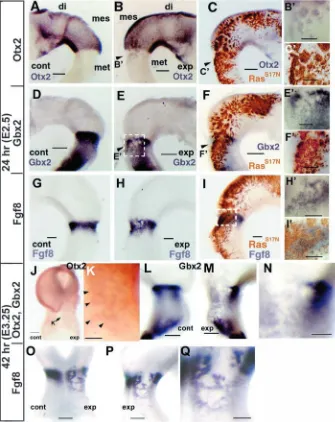
molecular markers for the mesencephalon and the metencephalon. In normal embryos, the homeobox genes, Otx2 and Gbx2, are expressed in the mesencephalon and the metencephalon, respectively (Simeone et al., 1992; Bally-Cuif et al., 1995; Millet et al., 1996; Bouillet et al., 1995; Niss and Leutz, 1998; Shamim and Mason, 1998; Hidalgo-Sanchez et al., 1999). It has been suggested that repressive interaction between Otx2 and Gbx2 determines the MHB, and that Fgf8 mRNA is induced at the interface of Otx2 and Gbx2 expression overlapping with Gbx2 expression (Millet et al., 1999; Broccoli et al., 1999; Katahira et al., 2000; Li and Joyner, 2001; Ye et al., 2001). At 24 hours after electroporation of RasS17N(E2.5,
HH17), induction of Otx2 and repression of Gbx2 in the metencephalon were observed (Otx2; n=9/9, Gbx2; n=7/8) (Fig. 4A-F). Expression of Fgf8 was also repressed by RasS17N, but was induced in the caudal part
of its expression belt so that the Fgf8 expression belt became wider (n=3/3) (Fig. 4G-I).
At 42 hours after electroporation (E3.25), boundary of patchy expression of Otx2 in the metencephalic region became blurred, and the Otx2-free area became narrower (n=11/13) (Fig. 4J,K). However, repression of Gbx2 just posterior to the proper mesencephalon became wider (Fig. 4L-N). Corresponding to the change of the manner of Gbx2 repression, the wide Fgf8-free region appeared just caudal to the proper mesencephalon (n=4/4). One or two Fgf8 expression line(s) appeared caudal to the Fgf8-free region (n=4/4) (Fig. 4O-Q). If we consider that Otx2 is induced in the RasS17N-expressing site, Fgf8 may have been induced at the border of the Otx2 and Gbx2 expression domains (Broccoli et al., 1999; Millet et al., 1999; Katahira et al., 2000; Ye at al., 2001).
Next, we examined the effect on Wnt1 expression. At E2.5, Wnt1 was expressed in the dorsal midline of the mesencephalon and in the caudal mesencephalon. Since the dorsal midline of the metencephalon does not express Wnt1, it is a good marker to discriminate between metencephalon and mesencephalon (Fig. 5A, control side). After disruption of Ras signaling, Wnt1 was induced in the dorsal metencephalon on the experimental side (n=3/4) (Fig. 5A,B). These effects of RasS17Non marker
gene expression also support the notion that disruption of Ras signaling changes the fate of the metencephalon to that of the mesencephalon.
Effects of the misexpression of RasS17Non Pax2/5, En1/2
expression were also examined. These molecules and Fgf8 are shown to be in a positive feedback loop for their expression. This feedback loop may help to maintain the organizing activity of the isthmus (reviewed by Nakamura, 2001). Expression of Pax2/5 and En1/2 was repressed in the area where RasS17Nwas misexpressed (Pax2, n=3/3; Pax5, n=4/4;
En1, n=4/4; En2, n=4/4) (Fig. 5C-V), suggesting that the Ras signaling pathway is necessary for maintenance of expression of these molecules in the mes/metencephalic region.
The Ras signaling pathway functions downstream of FgF8b, but not Fgf8a
[image:4.612.42.370.72.285.2]Morphological and gene expression analyses indicate that the Ras signaling pathway plays an important role in mes/metencephalic fate determination. To ascertain if the Ras signaling pathway functions at the downstream of the FgF8b signal, we carried out co-transfection of Fgf8b with RasS17N. If Ras functions at the downstream of the Fgf8b signal, co-transfection of RasS17Nwith Fgf8b may cancel the effects of
Fig. 2. ERK activation by Fgf8 signal. (A) Immunohistochemistry with anti-dpERK 1 hour after insertion of an SU5402-soaked bead (asterisk). ERK activation is repressed by SU5402, an inhibitor of the Fgf receptor. (B) Dorsal and (C) lateral view of an E14.5 brain after misexpression of Fgf8b. Instead of the tectum, cerebellum has differentiated in the mesencephalic region (arrow). (D) Dorsal view of an E6.5 brain after misexpression of
Fgf8a. The tectum enlarged because the fate of the diencephalic alar plate was changed to
Fgf8b misexpression. Conversely, if Ras does not transduce the Fgf8b signal, co-transfection may exert additive effects. After co-transfection, some large swellings were observed on the experimental side of the mes/metencephalon of E10.5 embryos (n=4/4) (Fig. 6A,B). Histologically, these swellings showed a tectal structure (compare Fig. 6C,D with 6E,F), in agreement with our prediction. The anterior part of the presumptive metencephalon differentiated into the tectum (Fig. 6A,E). In the posterior part of the presumptive metencephalon, target genes of ERK may have been already turned prior to expression of the introduced gene product, explaining why cerebellar differentiation may have occurred in this region.
Co-transfection of Fgf8a and RasS17N exerted additive effects (n=4/4), whereby there was differentiation of the ectopic tectum in the diencephalon (Fig. 6G,H) and in the metencephalon (Fig. 6I). These data suggest that activation of Ras signaling is necessary for cerebellar development, but is not crucial for tectal differentiation.
Differential silencing of Fgf8a and Fgf8b by siRNA method
We previously showed that Fgf8b could change the fate of the mesencephalon to the metencephalon (Sato et al., 2001), and have shown in the present study that disruption of the Ras-ERK
signaling pathway resulted in the fate change of the metencephalon to the mesencephalon. These results suggest that the Fgf8b signal activates the Ras-ERK signal pathway to organize the metencephalic differentiation. To confirm this notion, we tried differential disruption of Fgf8a and Fgf8b by the siRNA method. Since the vector-based-siRNA method has been realized recently (Katahira and Nakamura, 2003), we introduced the siRNA expression vectors to the metencephalon and mesencephalon by electroporation (Fig. 7A).
Massive degradation of Fgf8 mRNA could not be detected after Fgf8b-siRNA application, but downregulation of Fgf8 mRNA to some extent could be detected (n=5/7) (Fig. 7B-E). Degradation of Fgf8 mRNA by Fgf8a-siRNA could not be detected (n=8/8) (Fig. 7F-I). Efficient degradation of Fgf8 mRNA could be observed after application of a mixture of Fgf8a- and Fgf8b-siRNA (n=3/5) (Fig. 7J-M). Since the Fgf8 probe hybridizes to both Fgf8a and Fgf8b mRNA, disruption of each Fgf8 mRNA after application of each siRNA may be more than we could observe.
[image:5.612.50.374.70.447.2]Next we checked the effects of differential disruption of Fgf8a and Fgf8b by siRNA on ERK activation. The activation level of ERK was decreased after electroporation of FGF8b-siRNA (n=7/10) (Fig. 7N) and of both Fgf8a- and Fgf8b-siRNA (n=7/11) (Fig. 7P). Fgf8a-Fgf8b-siRNA alone did not affect
Fig. 3. Fate change of the metencephalon to the mesencephalon by RasS17Nmisexpression.
(A) Repression of ERK activity by misexpression of RasS17N. (B-D) Views from the dorsal (B), left
(C) and right (D) of the E10.5 chick brain after misexpression of RasS17N. The tectum
differentiated ectopically in place of the cerebellum (arrow on B,D). Posterior part differentiated into the cerebellum according to the original program (arrowheads on D). (E) A transverse section of the brain at the level of (E) indicated on (A). (F,G) Higher magnification of the cerebellum and ectopic tectum indicated as (F,G, respectively), on (D). The external granular layer is differentiating on the control side (arrowhead on F). (H) Higher magnification of the proper tectum. The ectopic tectum has nine layers (G), while the proper tectum has ten layers (H). (I,J,L,M) Whole-mount
immunohistochemistry with anti-neurofilament antibody on E4 embryos. (K,N) Bright field of (J,M, respectively). An arrow and arrowhead on (K) indicate a large ectopic swelling and a small one in the metencephalon, respectively. In normal embryos (I), oculomotor (III) and trochlear (IV) nerves are discernible. In RasS17N-transfected
embryos (J), trochlear nerve fibers arise from the posterior part of the large swelling and run dorsally (arrowheads). Additional nerve fibers arise from the posterior part of the small swelling (arrow). These fibers merge while running dorsally. In some cases, a nerve trunk originates from the ventral metencephalon and run a similar course to the oculomotor nerve (arrowheads on L). In another embryo that is transfected with RasS17N, several additional nerve fibers could
the activation level of ERK (n=11/14) (Fig. 7O). RasS17Nmore
effectively repressed ERK activation than Fgf8b-siRNA (compare Fig. 3A with Fig. 7N). The difference may be due to the fact that Fgf8 exerts its effects non-cell autonomously but that RasS17N exerts its effects cell autonomously. If Fgf8
mRNA is degraded by siRNA in some cells, the Fgf8 signal from the adjacent intact cells may take its place. However, RasS17Nshuts off the downstream Ras signal of the transfected
cell.
The effects of siRNA on Otx2 expression were examined, since Otx2 misexpression in the metencephalon changes its fate to the mesencephalon (Katahira et al., 2000). Transfection of Fgf8b-siRNA resulted in induction of Otx2 expression in the isthmic region (n=4/14) (Fig. 7Q-S); that is, the Otx2 expression domain extended caudally, although the effect is very subtle because of the above-mentioned reason. Transfection of Fgf8a-siRNA did not affect Otx2 expression (n=9/9) (Fig. 7T-V). The effect of Fgf8b-siRNA on Otx2 expression also suggests that disruption of Fgf8 mRNA may have occurred more than we could detect.
Discussion
Our study has demonstrated that: (1) ERK was activated at sites of Fgf8 mRNA expression; (2) misexpression of Fgf8b activated ERK and induced ectopic cerebellum in the mesencephalon; (3) misexpression of Fgf8a or a lower dose of Fgf8b activated ERK only in the diencephalon, where ectopic tectum differentiated; (4) disruption of Ras signaling by a dominant-negative form of Ras changed the fate of the metencephalic alar plate from cerebellar to tectal development; (5) co-electroporation of a dominant-negative form of Ras with Fgf8b canceled the Fgf8b effects, while co-electroporation with Fgf8a exerted additive effects; and (6) distinct disruption of Fgf8b by siRNA resulted in repression of ERK activity, and in a caudal shift of the Otx2 expression domain.
[image:6.612.45.380.75.497.2]Eight isoforms of Fgf8 have been identified to date (Crossley and Martin, 1995; MacArthur et al., 1995b), with Fgf8a and Fgf8b being expressed in the isthmus (Sato et al., 2001). Fgf8a and Fgf8b possess different organizing activities for brain development. Fgf8b-soaked beads implanted in the presumptive diencephalon induce cerebellar structures closest
Fig. 4. Effects of RasS17Nmisexpression on
Otx2, Gbx2 and Fgf8. Effects on Otx2
(A-C,J,K), Gbx2 (D-F,L-N) and Fgf8 (G-I,O-Q). Misexpression site of RasS17N(brown in
C,F,I) is assessed by immunohistochemistry against HA-tag. Otx2, Gbx2 and Fgf8 expression is represented as blue by in-situ hybridization. Twenty-four and 42 hours after electroporation (A-I and J-Q, respectively). Left (control) side of the brain vesicles (A,D,G,L) and right (transfected) side (B,C,E,F,H,I,M,P). Dorsal view (J,O). Higher magnification (B’,C’,E’,F’,H’,I’,K,N,Q). The rectangular area on (E) corresponds to (E’). By 24 hours after electroporation, Otx2 was induced in the metencephalon by RasS17N
misexpression (B,C,B’,C’). Gbx2 was repressed in the metencephalon by RasS17N
misexpression (E,F,E’,F’). Endogenous Fgf8 expression was also repressed, but was induced in the periphery of RasS17N
expression in the metencephalon (H,I,H’,I’). By 42 hours after electroporation, the boundary of patchy expression of Otx2 in the metencephalic region became blurred (J,K), and Otx2 expression area expanded (arrowheads in K). Repression of Gbx2 and Fgf8 just posterior to the proper
mesencephalon became wider, leaving a Gbx2- and Fgf8-free region (L-Q). di, diencephalon; mes, mesencephalon; met, metencephalon; cont, control side; exp, experimental side. Scale bars: 200µm (A-C,D-F,G-I,J,L,M,O,P), 100 µm
to the bead with tectum around the mini cerebellum (Martinez et al., 1999). Transgenic mice in which Fgf8b was misexpressed under the control of a Wnt1 enhancer changed the property of the presumptive diencephalon and mesencephalon to that of the metencephalon (Liu et al., 1999). Moreover, misexpression of Fgf8b by electroporation completely changed the fate of the mesencephalic alar plate so that it differentiated into cerebellum (Sato et al., 2001; Liu et al., 2003). Misexpression of Fgf8a caused expansion of the midbrain (Lee et al., 1997; Sato et al., 2001). Although Fgf8a and Fgf8b show different organizing activities, lower doses of Fgf8b exert similar effects to those of Fgf8a; the tectum was induced around the mini cerebellum in Fgf8b-bead implantation experiments, and electroporation with lower doses of Fgf8b exerted similar effects to those seen with Fgf8a (Sato et al., 2001; Liu et al., 2003). These results suggest that the difference in organizing activity between Fgf8a and Fgf8b is attributable to the difference in the intensity of the signal. This notion was further confirmed by the results of this study. Misexpression of Fgf8b at 1µg/µl resulted in activation of ERK at Fgf8b misexpressing sites throughout the diencephalon
and metencephalon, while misexpression of Fgf8b at 0.01 µg/µl or Fgf8a at 1 µg/µl resulted in activation of ERK in only a portion of the diencephalon.
Ras, a member of a group of small GTP-binding proteins, is activated downstream of various RTKs and activates Raf, Mek and ERK in turn (reviewed by Katz and McCormick, 1997; Rommel and Hafen, 1998). Since ERK was activated in the isthmus and sites of Fgf8b misexpression, we assumed that activation of the Ras-ERK pathway is necessary for metencephalic fate determination. Fate change of the alar plate is easily identifiable because of the distinct structures of the tectum and cerebellum. As expected, disruption of the Ras-ERK pathway by a dominant-negative form of Ras (RasS17N)
in the alar plate of the metencephalon caused its fate change to the tectum. To follow the development of the basal plate, we paid attention to the oculomotor and trochlear nerves. In RasS17N-transfected embryos, nerve fibers running a similar
course to that of the trochlear nerve arose from the caudal end of the ectopic swelling(s) in the metencephalic region. Additional nerve trunks similar to the oculomotor nerve originated from the ventral metencephalon in some cases. Moreover, at 24 hours after electroporation of RasS17N, induction of Otx2 and repression of Gbx2
in the metencephalon occurred. Thus, we concluded that disruption of Ras signaling by a dominant-negative form of Ras converted the fate of the presumptive metencephalon to that of the mesencephalon. As in the case of Otx2 misexpression (Katahira et al., 2000), Otx2 induction was patchy at first, but a large tectum differentiated in the most effective case. Repression of Gbx2 and Fgf8 was also patchy at first (24 hours after electroporation), but a wide region in which Gbx2 and Fgf8 were not expressed appeared just posterior to the proper mesencephalon (42 hours after electroporation). Fgf8 expression line(s) were established posterior to the Fgf8-free region. The results indicate that regulation of Otx2, Gbx2 and Fgf8 expression may have taken place. Thus new Fgf8 line(s) may have served as a new organizer, and most of the presumptive r1 region may have changed its property to that of the mesencephalon. In some cases, fate change to mesencephalon may have occurred patchily because a number of trochlear nerves differentiated in the metencephalic region (see Fig. 3M,N).
[image:7.612.49.342.326.737.2]Further evidence to support the hypothesis that
Fig. 5. Effects of RasS17Nmisexpression on Wnt1, Pax2,
Pax5, En1, En2 and Fgf8. Effects on Wnt1 (A,B), Pax2
(C-G), Pax5 (H-L), En1 (M-Q), En2 (R-V). Brown, immunohistochemical staining against HA-tag. Blue, signal for in-situ hybridization. Dorsal view (A,B), Left (control) side of the brain vesicles (C,H,M,R), right (transfected) side (D,E,I,J,N,O,S,T). Higher
magnification (F,G,K,L,P,Q,U,V). The areas enclosed by the dashed line on (E,J,O,T) corresponds to (G,L,Q,V), respectively. Wnt1 was induced in the dorsal
metencephalon (arrowheads on A). Expression of Pax2/5 and En1/2 were repressed by RasS17Nmisexpression.
the Ras-ERK pathway is activated by Fgf8b to result in metencephalic differentiation comes from co-transfection studies with RasS17Nand Fgf8b. If the Ras-ERK pathway does indeed transduce the Fgf8 signal, then co-transfection may cancel the Fgf8 signal. However, if the Ras-ERK pathway does not transduce the Fgf8 signal, co-transfection may exert additive effects. Accordingly, co-transfection of RasS17N and Fgf8b canceled the effects of Fgf8b misexpression, while co-transfection of Fgf8a and RasS17Ncaused differentiation of the ectopic tectum in the diencephalon and in the metencephalon, displaying the additive effects of Fgf8a and RasS17N misexpression. Distinct disruption of Fgf8a and Fgf8b also supports the notion that Fgf8b activates Ras-ERK signaling pathway to organize cerebellar differentiation. Disruption of Fgf8b by its specific siRNA resulted in a decrease in the activation level of Erk, and in caudal extension of the Otx2 expression domain. siRNA for Fgf8a did not affect the activity of ERK. In conclusion, the results indicate that Fgf8b functions as the organizer for the metencephalon by activating the Ras-ERK pathway.
Since Fgf8 mutant mice or zebrafish show disruption of the mes/metencephalon (Meyers et al., 1998; Reifers et al., 1998; Chi et al., 2003), it was thought that Fgf8 might also be necessary for the development of the mesencephalon. However, our results show that disruption of the Ras signaling pathway did not affect the fate of the presumptive mesencephalon, and actually changed the fate of the presumptive metencephalon to that of the mesencephalon. This suggests that Ras signaling does not play a role in fate
determination of the mesencephalon. To accord our assumption, animal cap assay indicated that the PLCγsignaling pathway through Fgf receptor IV (FgfR4) is responsible for the fate decision of the mesencephalon (Umbhauer et al., 2000). However, it was suggested that FgfR1 is the receptor for the Fgf8 signal in the isthmus region (Liu et al., 2003; Trokovic et al., 2003). So far, it is not reported that FgfR4 is expressed in the isthmic region as suggested (Walshe and Mason, 2000). Further study is needed to determine what signaling pathway is responsible for the mesencephalic determination.
In the mesencephalon, the Fgf8-Ras-ERK signaling pathway may be involved in rostrocaudal polarity formation. For the rostrocaudal polarity formation, it is suggested that En confers caudal property to the tectal anlage so that the rostrocaudal polarity of the tectum is determined according to a gradient of En (Itasaki and Nakamura, 1996; Friedman and O’Leary, 1996). ERK is activated in a gradient in the mesencephalon, as revealed by whole-mount immunohistochemistry. The gradient corresponds to that of En2 expression at the 14-somite stage, when the rostrocaudal axis is still plastic. Pax2/5, En1/2 and Fgf8 act in a positive feedback loop (reviewed by Nakamura, 2001). Disruption of Ras signaling caused repression of Pax2/5 and En1/2 expression. Thus, the Ras-ERK pathway, which is activated by Fgf8, may play a crucial role in formation of the rostrocaudal polarity of the tectum.
[image:8.612.44.368.74.402.2]In normal embryos around the 8-somite stage, ERK was activated in the region where Fgf8 mRNA was expressed. In the metencephalon, ERK became inactivated by the 14-somite stage, while it remained activated in the mesencephalon. This
Fig. 6. Co-transfection of Fgf8b/a with
RasS17N. (A-F) Dorsal view (A) and lateral
view (B), parasagittal section at the line indicated on (A) as (C) and (E) (C,E) of E10.5 brain co-electroporated with Fgf8b and
RasS17Nat the 10-somite stage. (D,F) higher
magnification of (C,E, respectively). The swelling in the mesencephalic region and anterior metencephalic region of the experimental side have sulci on the surface, and look like cerebellum (A,B).
Histologically, however, the swelling in the mesencephalon and the anterior
metencephalon of the experimental side do not have an external granular layer, which is seen in the cerebellum of the control side (C,D), and has tectal architecture (E,F). The posterior part of the metencephalic region consists of cerebellar structure (arrowheads on E). Antero-posterior direction is indicated by the double-headed arrow on (B,E). Dorsal view (G,I) and horizontal section (H) of the brain co-electroporated with Fgf8a and RasS17N.
indicates that the gene expression cascade favoring cerebellar differentiation has proceeded by the 10-somite stage, meaning that the fate of the metencephalon is determined by this time. This notion is supported by ectopic transplantation studies that show that while the rostrocaudal axis of the mesencephalon is not fixed at the 10-somite stage, the fate of the mesencephalon and metencephalon is already determined (Nakamura et al., 1986; Nakamura et al., 1988; Ichijo et al., 1990; Matsuno et al., 1990).
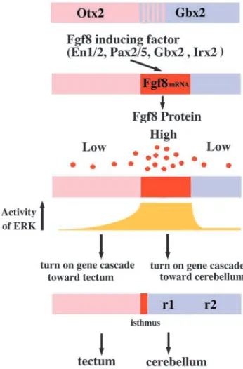
Focusing on mechanisms of mes/metencephalic development (Fig. 8), expression of Fgf8 mRNA is induced at the interface of Otx2 and Gbx2 expression, overlapping with Gbx2 expression; that is, at the presumptive metencephalon (Millet et al., 1999; Broccoli et al., 1999; Hidalgo-Sanchez et
[image:9.612.53.360.247.741.2]al., 1999; Irving and Mason, 2000; Katahira et al., 2000; Ye et al., 2001; Garda et al., 2001; Li and Joyner, 2001; Martinez-Barbera et al., 2001; Li et al., 2002). Consequently, the presumptive metencephalic region may receive a strong Fgf8 signal, in turn activating the Ras-ERK pathway, which may result in turning on the gene cascade favoring development of the cerebellum. The cascade might be turned on before the 10-somite stage, because ERK becomes strongly activated at around the 8-somite stage, its activity gradually weakening thereafter. Our results correspond well to the classical transplantation experiments that show that the fate of the mesencephalon and metencephalon is determined before the 10-somite stage. In the mesencephalon, the gene expression cascade toward cerebellar differentiation may not be turned on
Fig. 7. Distinct disruption of Fgf8a and Fgf8b by siRNA. (A) Alignment of partial sequence of
Fgf8a and Fgf8b mRNA. The target sequence of
Fgf8a- and Fgf8b-siRNA is underlined with red and green, respectively. The number indicates the number from the start codon. (B-E) Effects of Fgf8b-siRNA on Fgf8 expression. We could detect disruption of Fgf8 mRNA to some extent. Since the probe used for in-situ hybridization hybridized to both Fgf8a and Fgf8b, disruption of
Fgf8b may be more than we can detect.
(F-I) Effects of Fgf8a-siRNA on Fgf8 expression. We could not detect the effect of siRNA. This may be due to the fact that Fgf8b is
predominantly expressed in the isthmus. (J-M) Application of both Fgf8a- and Fgf8b-siRNA. Electroporation of both Fgf8a- and Fgf8b-siRNA resulted in distinct reduction of
Fgf8 mRNA in the isthmus. (N-P) Effects of
siRNA on the activation of ERK. Fgf8b-siRNA decreased the activation level of ERK (N), although Fgf8a-siRNA hardly affected ERK activity (O). (Q-S) Effects of Fgf8b-siRNA on
Otx2 expression. The arrows represent the caudal
border of the Otx2 expression domain. Application of Fgf8b-siRNA by electroporation resulted in a caudal shift of the Otx2 expression domain; in other words, the mesencephalon extended caudally. (T-V) Effects of Fgf8a-siRNA on Otx2 expression. Fgf8a-siRNA did not affect
Otx2 expression. Dorsal view (B,F,J,N-Q,T),
because Otx2 is expressed there. Fgf17 and Fgf18 that are induced by Fgf8 together with Fgf8a may regulate proliferation of the mesencephalon and metencephalon (Xu et al., 2000; Liu et al., 2003). In the mesencephalon, ERK activity remains in a gradient distribution after the 10-somite stage, and may contribute to the determination of the rostrocaudal axis of the tectum.
We thank Drs K. Kitamura and M. Wassef for the Otx2 and Wnt1 probes, respectively, Drs K. Moriyoshi and J. Miyazaki for pCA-GAP-GFP, Drs H. Takeda and M. Shinya for helpful suggestions for examining the effect of anti-di-phosphorylated ERK, and Drs Y. Wakamatsu, I. Araki, S. Sugiyama and E. Matsunaga and members of our laboratory for discussions and critical reading of the manuscript. This work was supported by the grants from the Ministry of Education, Culture, Sports, Science and Technology and from the Mitsubishi Foundation. T.S. is a recipient of JSPS Research Fellowships for Young Scientists.
References
Araki, I. and Nakamura, H. (1999). Engrailed defines the position of dorsal
di-mesencephalic boundary by repressing diencephalic fate. Development
126, 5127-5135.
Bally-Cuif, L., Cholley, B. and Wassef, M. (1995). Involvement of Wnt-1 in
the formation of the mes/metencephalic boundary. Mech. Dev. 53, 23-34.
Bouillet, P., Chazaud, C., Oulad-Abdelghani, M., Dollé, P. and Chambon, P. (1995). Sequence and expression pattern of the Stra7 (Gbx-2)
homeobox-containing gene induced by retinoic acid in P19 embryonal carcinoma cells.
Dev. Dyn. 204, 372-382.
Broccoli, V., Boncinelli, E. and Wurst, W. (1999). The caudal limit of Otx2
expression positions the isthmic organizer. Nature 401, 164-168.
Chi, C. L., Martinez, S., Wurst, W. and Martin, G. R. (2003). The isthmic
organizer signal FGF8 is required for cell survival in the prospective midbrain and cerebellum. Development 130, 2633-2644.
Christen, B. and Slack, J. M. (1999). Spatial response to fibroblast growth
factor signalling in Xenopus embryos. Development 126, 119-125.
Corson, L. B., Yamanaka, Y., Lai, K. M. and Rossant, J. (2003). Spatial
and temporal patterns of ERK signaling during mouse embryogenesis.
Development 130, 4527-4537.
Crossley, P. H. and Martin, G. R. (1995). The mouse Fgf8 gene encodes a
family of polypeptides and is expressed in regions that direct outgrowth and patterning in the developing embryo. Development 121, 439-451.
Crossley, P. H., Martinez, S. and Martin, G. R. (1996). Midbrain
development induced by FGF8 in the chick embryo. Nature 380, 66-68.
Feig, L. A. and Cooper, G. M. (1988). Inhibition of NIH 3T3 cell proliferation
by a mutant ras protein with preferential affinity for GDP. Mol. Cell. Biol.
8, 3235-3243.
Friedman, G. C. and O’Leary, D. D. (1996). Retroviral misexpression of
engrailed genes in the chick optic tectum perturbs the topographic targeting of retinal axons. J. Neurosci. 16, 5498-5509.
Funahashi, J., Okafuji, T., Ohuchi, H., Noji, S., Tanaka, H. and Nakamura, H. (1999). Role of Pax5 in the regulation of a mid-hindbrain
organizer’s activity. Dev. Growth Differ. 41, 59-72.
Gabay, L., Seger, R. and Shilo, B. Z. (1997). In situ activation pattern of
Drosophila EGF receptor pathway during development. Science 277, 1103-1106.
Garda A. L., Echevarria, D. and Martinez, S. (2001). Neuroepithelial
co-expression of Gbx2 and Otx2 precedes Fgf8 co-expression in the isthmic organizer. Mech. Dev. 101, 111-118.
Hamburger, V. and Hamilton, H. (1951). A series of normal stages in the
development of the chick embryo. J. Morphol. 88, 49-92.
Hidalgo-Sanchez, M., Simeone, A. and Alvarado-Mallart, R. M. (1999).
Fgf8 and Gbx2 induction concomitant with Otx2 repression is correlated with midbrain-hindbrain fate of caudal prosencephalon. Development 126, 3191-3203.
Ichijo, H., Fujita, S., Matsuno, T. and Nakamura, H. (1990). Rotation of
the tectal primordium reveals plasticity of target recognition in retino-tectal projection. Development 110, 331-342.
Irving, C. and Mason, I. (2000). Signaling by FGF8 from the isthmus patterns
anterior hindbrain and establishes the anterior limit of Hox gene expression.
Development 127, 177-186.
Itasaki, N. and Nakamura, H. (1996). A role for gradient en expression in
positional specification on the optic tectum. Neuron 16, 55-62.
Joyner, A. L., Liu, A. and Millet, S. (2000). Otx2, Gbx2 and Fgf8 interact
to position and maintain a mid-hindbrain organizer. Curr. Opin. Cell Biol.
12, 736-741.
Katahira, T. and Nakamura, H. (2003). Gene silencing in chick embryos
with vector-based small interfering RNA system. Dev. Growth Differ. 45, 361-367.
Katahira, T., Sato, T., Sugiyama, S., Okafuji, T., Araki, I., Funahashi, J. and Nakamura, H. (2000). Interaction between Otx2 and Gbx2 defines the
organizing center for the optic tectum. Mech. Dev. 91, 43-52.
Katz, M. E. and McCormick, F. (1997). Signal transduction from multiple
Ras effectors. Curr. Opin. Genet. Dev. 7, 75-79.
Lee, S. M., Danielian, P. S., Fritzsch, B. and McMahon, A. P. (1997).
Evidence that FGF8 signaling from the midbrain-hindbrain junction regulates growth and polarity in the developing midbrain. Development 124, 959-969.
Li, J. Y. and Joyner, A. L. (2001). Otx2 and Gbx2 are required for refinement
and not induction of mid-hindbrain gene expression. Development 128, 4979-4991.
Li, J. Y., Lao, Z. and Joyner, A. L. (2002). Changing requirements for Gbx2
in development of the cerebellum and maintenance of the mid/hindbrain organizer. Neuron 36, 31-43.
Liu, A., Li, J. Y. H., Bromleigh, C., Lao, Z., Niswander, L. A. and Joyner, A. L. (2003). FGF17 and FGF18 have different midbrain regulatory
properties from FGF8b or activated FGF receptors. Development 130, 6175-6185.
Liu, A., Losos, K. and Joyner, A. L. (1999). FGF8 can activate Gbx2 and
transform regions of the rostral mouse brain into a hindbrain fate.
[image:10.612.75.248.74.337.2]Development 126, 4827-4838.
MacArthur, C. A., Lawshe, A., Shankar, D. B., Heikinheimo, M. and Shackleford, G. M. (1995a). FGF-8 isoforms differ in NIH3T3 cell
transforming potential. Cell Growth Differ. 6, 817-825.
MacArthur, C. A., Lawshe, A., Xu, J., Santos-Ocampo, S., Heikinheimo, M., Chellaiah, A. T. and Ornitz, D. M. (1995b). FGF-8 isoforms activate
receptor splice forms that are expressed in mesenchymal regions of mouse development. Development 121, 3603-3613.
Martinez, S. (2001). The isthmic organizer and brain regionalization. Int. J.
Dev. Biol. 45, 367-371.
Martinez, S., Crossley, P. H., Cobos, I., Rubenstein, J. L. R. and Martin, G. R. (1999). FGF8 induces formation of an ectopic isthmic organizer and
isthmocerebellar development via a repressive effect on Otx2 expression.
Development 126, 1189-1200.
Martinez, S., Marin, F., Nieto, M. A. and Puelles, L. (1995). Induction of
ectopic engrailed expression and fate change in avian rhombomeres: intersegmental boundaries as barriers. Mech. Dev. 51, 289-303.
Martinez, S., Wassef, M. and Alvarado-Mallart, R. M. (1991). Induction of
a mesencephalic phenotype in the 2-day-old chick prosencephalon is preceded by the early expression of the homeobox gene en. Neuron 6, 971-981.
Martinez-Barbera, J. P., Signore, M., Boyl, P. P., Puelles, E., Acampora, D., Gogoi, R., Schubert, F., Lumsden, A. and Simeone, A. (2001).
Regionalisation of anterior neuroectoderm and its competence in responding to forebrain and midbrain inducing activities depend on mutual antagonism between OTX2 and GBX2. Development 128, 4789-4800.
Matsuno, T., Ichijo, H. and Nakamura, H. (1990). Regulation of the
rostrocaudal axis of the optic tectum: histological study after rostrocaudal rotation in quail-chick chimeras. Dev. Brain Res. 58, 265-270.
Meyers, E. N., Lewandoski, M. and Martin, G. R. (1998). An Fgf8 mutant
allelic series generated by Cre- and Flp-mediated recombination. Nature
Genet. 18, 136-141.
Millet, S., Bloch-Gallego, E., Simeone, A. and Alvarado-Mallart, R. M.
(1996). The caudal limit of Otx2 gene expression as a marker of the midbrain/hindbrain boundary: a study using in situ hybridisation and chick/quail homotopic grafts. Development 122, 3785-3797.
Millet, S., Campbell, K., Epstein, D. J., Losos, K., Harris, E. and Joyner, A. L. (1999). A role for Gbx2 in repression of Otx2 and positioning the
mid/hindbrain organizer. Nature 401, 161-164.
Mohammadi, M., McMahon, G., Sun, L., Tang, C., Hirth, P., Yeh, B. K., Hubbard, S. R. and Schlessinger, J. (1997). Structures of the tyrosine
kinase domain of fibroblast growth factor receptor in complex with inhibitors. Science 276, 955-960.
Moriyoshi, K., Richards, L. J., Akazawa, C., O’Leary, D. D. and Nakanishi, S. (1996). Labeling neural cells using adenoviral gene transfer
of membrane-targeted GFP. Neuron 16, 255-260.
Nakamura, H. (2001). Regionalization of the optic tectum: combinations of
gene expression that define the tectum. Trends Neurosci. 24, 32-39.
Nakamura, H. and Funahashi, J. (2001). Induction of DNA into chick
embryos by in ovo electroporation. Methods 24, 43-48.
Nakamura, H. and Itasaki, N. (1992). Expression of en in the prosencephalon
heterotopically transplanted into the mesencephalon. Dev. Growth Differ. 34, 387-391.
Nakamura, H., Nakano, K. E., Igawa, H. H., Takagi, S. and Fujisawa, H.
(1986). Plasticity and rigidity of differentiation of brain vesicles studied in quail-chick chimeras. Cell Differ. 19, 187-193.
Nakamura, H., Takagi, S., Tsuji, T., Matsui, K. A. and Fujisawa, H. (1988).
The prosencephalon has the capacity to differentiate into the optic tectum: analysis by chick-specific monoclonal antibodies in quail-chick-chimeric brains. Dev. Growth Differ. 30, 717-725.
Nakamura, H., Watanabe, Y. and Funahashi, J. (2000). Misexpression of
the genes in the brain vesicles by in ovo electroporation. Dev. Growth Differ.
42, 199-201.
Niss, K. and Leutz, A. (1998). Expression of the homeobox gene GBX2
during chicken development. Mech. Dev. 76, 151-155.
Niwa, H., Yamamura, K. and Miyazaki, J. (1991). Efficient selection for
high-expression transfectants with a novel eukaryotic vector. Gene 108, 193-199.
Okafuji, T., Funahashi, J. and Nakamura, H. (1999). Roles of Pax-2 in
initiation of the chick tectal development. Brain Res. 116, 41-49.
Reifers, F., Bohli, H., Walsh, E. C., Crossley, P. H., Stainier, D. Y. R. and Brand, M. (1998). Fgf8 is mutated in zebrafish acerebellar (ace) mutants
and is required for maintenance of midbrain-hindbrain boundary development and somitogenesis. Development 125, 2381-2395.
Rhinn, M. and Brand, M. (2001). The midbrain–hindbrain boundary
organizer. Curr. Opin. Neurobiol. 11, 34-42.
Rommel, C. and Hafen, E. (1998). Ras-a versatile cellular switch. Curr. Opin.
Genet. Dev. 8, 412-418.
Sato, T., Araki, I. and Nakamura, H. (2001). Inductive signal and tissue
responsiveness defining the tectum and the cerebellum. Development 128, 2461-2469.
Shamim, H. and Mason, I. (1998). Expression of Gbx-2 during early
development of the chick embryo. Mech. Dev. 76, 157-159.
Shamim, H., Mahmood, R., Logan, C., Doherty, P., Lumsden, A. and Mason, I. (1999). Sequential roles for Fgf4, En1 and Fgf8 in specification
and regionalisation of the midbrain. Development 126, 945-959.
Shinya, M., Koshida, S., Sawada, A., Kuroiwa, A. and Takeda, H. (2001).
Fgf signalling through MAPK cascade is required for development of the subpallial telencephalon in zebrafish embryos. Development 128, 4153-4164.
Simeone, A., Acampora, D., Gulisano, M., Stornaiuolo, A. and Boncinelli, E. (1992). Nested expression domains of four homeobox genes in
developing rostral brain. Nature 358, 687-690.
Simeone, A. (2000). Positioning the isthmic organizer where Otx2 and Gbx2
meet. Trends Genet. 16, 237-240.
Suemori, H., Kadokawa, Y., Goto, K., Araki, I., Kondoh, H. and Nakatsuji, N. A. (1990). Mouse embryonic stem cell line showing
pluripotency of differentiation in early embryos and ubiquitous beta-galactosidase expression. Cell. Differ. Dev. 29, 181-186.
Trokovic, R., Trokovic, N., Hernesniemi, S., Pirvola, U., Vogt Weisenhorn, D. M., Rossant, J., McMahon, A. P., Wurst, W. and Partanen, J. (2003).
FGFR1 is independently required in both developing mid- and hindbrain for sustained response to isthmic signals. EMBO J. 22, 1811-1823.
Umbhauer, M., Penzo-Méndez, A., Clavilier, L., Boucaut, J.-L. and Riou, J.-F. (2000). Signaling specificities of fibroblast growth factor receptors in
early Xenopus embryo. J. Cell Sci. 113, 2865-2875.
Wakamatsu, Y., Watanabe, Y., Nakamura, H. and Kondoh, H. (1997).
Regulation of the neural crest fate by N-myc: promotion of ventral migration and neuronal differentiation. Development 124, 1953-1962.
Walshe, J. and Mason, I. (2000). Expression of FGFR1, FGFR2 and FGFR3
during early neural development in the chick embryo. Mech. Dev. 90, 103-110.
Wurst, W. and Bally-Cuif, L. (2001). Neural plate patterning: upstream and
downstream of the isthmic organizer. Nat. Rev. Neurosci. 2, 99-108.
Xu, J., Liu, Z. and Ornitz, D. M. (2000). Temporal and spatial gradients of
Fgf8 and Fgf17 regulate proliferation and differentiation of midline cerebellar structures. Development 127, 1833-1843.
Ye, W., Bouchard, M., Stone, D., Liu, X., Vella, F., Lee, J., Nakamura, H., Ang, S. L., Busslinger, M. and Rosenthal, A. (2001). Distinct regulators
control the expression of the mid-hindbrain organizer signal FGF8. Nature