D
E
V
E
LO
P
M
E
N
T
INTRODUCTION
The generation of differentiated cell types and positioning them correctly within a developing structure are the two key processes in pattern formation. In the social amoeba Dictyostelium, these two processes are much simplified, relative to higher organisms, but most of the same fundamental principles apply and many of the signalling components are conserved (Kessin, 2001).
Developing cells aggregate together and organise themselves into a fruiting body. Midway through development, approximately one-fifth of the cells in each aggregate differentiate as prestalk cells, while the remainder differentiate as prespore cells. There are two principal sub-classes of prestalk cells, pstA cells and pstO cells (Early et al., 1993; Jermyn et al., 1989). PstA cells occupy the front half of the prestalk region, pstO cells occupy the rear half. The pstO-specific and pstA-pstO-specific reporter constructs, normally used to identify these two cell types, derive from the promoter of the ecmA gene (Williams et al., 1987; Early et al., 1993). PstO cells use cap-site distal sequences of the promoter (the ecmO region, Fig. 1), while pstA cells use cap-site proximal sequences (the ecmA region).
The best-characterised inducer of cell-type divergence is the chlorinated hexaphenone DIF-1 (Kay and Jermyn, 1983; Morris et al., 1987). In monolayer assay, DIF-1 induces prestalk and stalk cell differentiation at the expense of prespore differentiation (Early and Williams, 1988; Kopachik et al., 1985; Town et al., 1976; Williams et al., 1987). DIF-1 is produced by the prespore cells as part of a negative-feedback loop that regulates the pstO to prespore cell ratio (Kay and Thompson, 2001).
The ecmAgene is directly induced by DIF-1 (Williams et al., 1987) and in the dmtA– strain, a mutant that is defective in DIF-1 biosynthesis, the pstA-specific marker (ecmA:lacZ) is expressed but the pstO-specific marker (ecmO:lacZ) is not (Thompson and Kay, 2000). These facts suggest that DIF-1 is the physiological
inducer of pstO cell differentiation but not of pstA cell differentiation. The situation is, however, complicated by the fact that some pstO markers are expressed in dmtA– slugs (Maeda et al., 2003).
The dimA and dimB genes both encode bZIP transcription factors that are required for DIF-1 responsiveness (Thompson et al., 2004; Huang et al., 2006; Zhukovskaya et al., 2006). DimB binds to the ecmA promoter when DIF-1 is added to cells but the two known DimB-binding sites lie outwith the characterised pstO-specific region (Zhukovskaya et al., 2006). Previous mutational analyses of the ecmApromoter identified a 132-nucleotide sub-segment (Fig. 1) that directs pstO-specific gene expression (Early et al., 1993; Kawata et al., 1996). When a G-box, a non-cell-type-specific promoter element (reviewed by Schnitzler et al., 1994), was fused to the multimerised form of a distal sub-region of the 132-mer expression was weakly DIF-inducible. Prestalk-specific gene expression and DIF-inducibility were both abolished when the TTGA repeats, within the distal domain, were subjected to point mutation.
The TTGA repeats bind in vitro to the Dictyostelium STAT protein Dd-STATa (Kawata et al., 1997) but Dd-STATa is not the in vivo activator (Mohanty et al., 1999; Araki et al., 1998). There is a DIF-regulated STAT, Dd-STATc, but it also is not required for pstO cell differentiation (Fukuzawa et al., 2001). Thus, identification of the TTGA repeats in the ecmO promoter region led, circuitously, to the identification of the DIF-1:Dd-STATc pathway. However, identifying the inductive pathway that is responsible for pstO cell differentiation remains key to understanding Dictyosteliumpattern formation. We have therefore analysed the pstO-specific region of the ecmApromoter further and present evidence that it is regulated by a novel MYB protein.
MATERIALS AND METHODS
Cell growth, transformation and development
Dictyostelium discoideumstrain Ax2 was grown axenically and transformed as described (Watts and Ashworth, 1970; Pang et al., 1999). Transformants were selected at G418 concentrations of 10 g/ml when cells were to be used for DIF-1 induction assays and 50 g/ml G418 when staining whole mounts for -galactosidase activity (Dingermann et al., 1989). When first
Regulation of
Dictyostelium
prestalk-specific gene expression
by a SHAQKY family MYB transcription factor
Masashi Fukuzawa, Natasha V. Zhukovskaya, Yoko Yamada, Tsuyoshi Araki and Jeffrey G. Williams*
PstA and pstO cells are the two major populations in the prestalk region of the Dictyosteliumslug and DIF-1 is a low molecular weight signalling molecule that selectively induces pstO cell-specific gene expression. The two cell types are defined by their differential use of spatially separated regions of the ecmApromoter. Additionally, there are anterior-like cells (ALCs) scattered throughout the rear, prespore region of the slug. They, like the pstO cells, use a cap-site distal ecmApromoter segment termed the ecmO region. When multimerised, a 22-nucleotide subsegment of the ecmO region directs expression in pstA cells, pstO cells and ALCs. It also directs DIF-inducible gene expression. The 22-nucleotide region was used to purify MybE, a protein with a single MYB DNA-binding domain of a type previously found only in a large family of plant transcription factors. Slugs of a mybE-null (mybE–) strain express an ecmAO:lacZfusion gene (i.e. a reporter construct containing the ecmA and ecmO promoter regions) in pstA cells but there is little or no expression in pstO cells and ALCs. The ecmAgene is not induced by DIF-1 in a mybE- strain. Thus, MybE is necessary for DIF-1 responsiveness and for the correct differentiation of pstO cells and ALCs.
KEY WORDS: DIF-1, Dictyostelium, Prestalk cells, SHAQKY family, MYB transcription factor
Development 133, 1715-1724 (2006) doi:10.1242/dev.02327
University of Dundee, MSI/WTB Complex, Dow Street, Dundee DD1 5EH, UK.
*Author for correspondence (e-mail: j.g.williams@dundee.ac.uk)
D
E
V
E
LO
P
M
E
N
T
isolated, the mybE-null (mybE–) strain grew poorly. Fortunately, the growth problem was overcome by culturing the cells on a plastic substratum in the presence of a heat-killed bacterial food source. After continued passage, the cells spontaneously increased their growth rate. Initially, the mybE– strain also took longer than normal to develop. Again, continued passage alleviated this problem.
DIF induction assay
Transformants, bearing lacZconstructs and selected at a G418 concentration of 10 g/ml, were harvested from growth, washed and resuspended in stalk medium [10 mM KCl, 2 mM NaCl, 1 mM CaCl2, 10 mM MES (pH 6.2)]
containing 5 mM cAMP and 50 M cerulenin, with or without 100 nM DIF. The polyketide synthesis inhibitor, cerulenin, acts as an inhibitor of endogenous DIF-1 synthesis and use of a low G418 concentration was found to be necessary to prevent a high background level of lacZexpression in the parental control. In some experiments, cells were pre-incubated for 1 hour before addition of cAMP; similar results were obtained with both procedures.
Cell suspension was added into the wells of a 96-well tissue culture plate and incubated overnight at 22°C. Cells were lysed in 100 l buffer [100 mM HEPES (pH 8.0), 1 mM MgSO4, 5 mM DTT, 2% Triton X-100] containing
1 mM CPRG (Roche). The plate was incubated at 37°C until the colour developed. The -galactosidase activity was measured at 595 nm. Averages and standard deviations were calculated from multiple biological experiments, where each assay condition was analysed in duplicate or in some cases quadruplicate.
DIF induction assays and analysis of ecmAgene expression by RT-PCR
Cells were harvested from growth, washed and plated at densities varying between 104 and 105cells/cm2in stalk medium [10 mM KCl, 2
mM NaCl, 1 mM CaCl2, 10 mM MES (pH 6.2)] and incubated at 22°C
for 1 hour. The medium was changed to stalk salts, cerulenin, at concentrations between 50 M and 150 M, and 5 mM camp, then incubated for a further 6 hours. Then the medium was removed and replaced with stalk medium, the same concentration of cerulenin (Kay, 1998) and concentrations of DIF-1 up to 100 nM. The plates were incubated at 22°C for 16 hours and RNA was extracted using an RNeasy kit (Qiagen) and analysed by RT-PCR using a ‘TITANIUM’ One-Step RT-PCR kit (BD Biosciences). The ecmA primers are: forward, CCAATTAGCTGTCCAAAACC; reverse, GCAATCACCTTTACC-TCCTG. They generate a 480 nucleotide fragment; IG7, a constitutively expressed mRNA, was used as control.
Nuclear extract preparation and gel retardation assay
Nuclear extracts were prepared from slug stage cells and gel retardation assays performed as previously (Kawata et al., 1996). The probe was made by annealing complementary oligonucleotides with BamHI cohesive ends, derived from the 30-mer sequence (Fig. 2A) (gatccTTTATTTAAA-CAGTTACACCCCACAATTTTg), followed by labelling with [␣-32P]
dATP. The CP2 wild-type and mutant oligonucleotides, that were used as G-box/CAE competitors, were: parental, gatccCGGGTGTGTTAAGTTA-GGGGTGGGTTTTATAg; mutant, gatccCGGGTGTGTTAAGTTAGGG-cTcGGTTTTATAg.
Protein purification and mass spectrometry
Nuclear extract derived from 3.5⫻1011slug cells was partially purified by
precipitation with 40% (w/v) ammonium sulfate, followed by heparin sepharose chromatography. It was then loaded onto a DNA affinity column bearing an oligonucleotide corresponding to the 22-mer. This was synthesized as a duplicate copy, annealed with the complementary strand, multimerized by ligation and coupled to sepharose beads. Bound proteins were eluted with 0.4 M KCl. The eluted proteins were further purified through a second round of binding on the affinity column, concentrated and loaded onto an SDS-polyacrylamide gel. After staining with Colloidal Blue Staining Kit (Invitrogen), protein bands were excised from the gel and digested, in-gel, with trypsin. The resulting peptides were analysed by MALDI-TOF (Matrix-Assisted Laser Desorption/Ionisation) mass spectrometry.
Antibody generation
A rabbit polyclonal antibody was generated and affinity purified, using the C-terminal 15 amino acids of MybE as immunogen. The antibody recognizes MybE but it also detects unrelated proteins. Therefore, it was useful only for western transfer analysis.
Gene disruption
A ClaI fragment of 2.3 kb containing most of the mybE-coding region was cloned into pGEM7 (Promega) and a hygromycin resistance cassette was inserted between two BalII sites, located at nt+574 and nt+2079 relative to the ATG initiation codon. Transformants were isolated clonally and screened for gene disruption by PCR and western blotting.
Expression of a MybE protein fragment in E. coli
The region of mybE-encoding the MYB domain (amino acids 487-818) was cloned in pGEX 5X-1 (Pharmacia). It was expressed in E. colistrain BL21 Codonplus RIL (Stratagene), as a GST-fusion protein, and purified using glutathione-sepharose (Amersham).
RESULTS
Identification of a minimal ecmO promoter fragment
The smallest ecmO promoter sub-fragment known to direct pstO-specific gene expression is 132 nucleotides in length (Kawata et al., 1996). We first showed that a 94-mer, centred within the 132-mer, also directs pstO-specific gene expression when fused to heterologous basal promoter elements (Fig. 1, Fig. 2A). As would be predicted from previous results (Kawata et al., 1996), deletion of distal sequences, to create a 62-mer that lacks the Dd-STATa-binding site, eliminates all detectable pstO-specific gene expression at the slug stage (data not shown). However, this construct is active in culminants, where it directs expression in the upper cup cells (Fig. 2A) – a population that derives directly from the pstO cells (Jermyn and Williams, 1991). This observation led us to focus on sequences contained within the 62-mer.
Cap-site proximal elements within the minimal ecmO region are necessary and sufficient for prestalk-specific gene expression
The cap-site distal half of the 62-mer is composed of runs of T residues, interspersed with a few A residues (Fig. 2A). Deletion of this region, to yield a 30-mer construct, eliminates biological activity (Fig. 2A). Multimerising a small promoter element sometimes allows it to function in a sequence context where a single copy of the
ATG -1694
-1212
-531 -1080
+41
pstO cells
pstA cells
132-mer
Dd-STATa MybE
-1191 -1098
94-mer minimal pstO region
pstA
AACaGTT TTGtCAA TTGAaTTGA
pstO
[image:2.612.314.567.555.683.2]ecmA
D
E
V
E
LO
P
M
E
N
T
element is ineffective (Kawata et al., 1996; Powell-Coffman et al., 1994). Therefore, we analysed constructs containing a multimer fused to heterologous basal promoter elements. The multimerised element contains the central 22 nucleotides of the 30-mer (Fig. 3A). The fourfold multimer of the 22-mer (Fig. 3A) directs strong, generic (i.e. pstA, pstO and ALCs) prestalk-specific gene expression (Fig. 3B).
As with most Dictyostelium promoter regions, the 22-mer sequence is extremely AT-rich but two features stood out: a 7-nucleotide imperfect dyad, containing a G and a C residue, and an adjacent C-rich region (Fig. 2A). These two regions were point-mutated within the context of the 94-mer. Mutations 1 and 2, respectively, change the two inner and two outer C residues within the C-rich region into A residues, while mutation 3 changes the C
and G residues of the dyad into an A and a T residue (Fig. 4A). In contrast to the unmutated 94-mer, all three mutant forms display scattered, non-cell-type-specific staining (Fig. 4A). Thus, prestalk-specific gene expression requires both the dyad and the adjacent C-rich sequence.
The 22-mer sequence is both necessary and sufficient for DIF-1 inducibility
[image:3.612.50.409.59.318.2]Cells transformed with the lacZconstructs described above were tested for DIF-1 inducibility in a monolayer assay. The 94-mer construct displays a twofold increase in expression in the presence of DIF-1 (Fig. 2B). This is a lower induction ratio than with longer fragments and presumably reflects the absence of elements needed for high expression. Nonetheless, it is reproducible and the 62-mer Fig. 2. Deletion analysis of the ecmO region. (A) Structure of the minimal ecmO region and expression patterns for two deletion mutants. The structure of the minimal 94 nucleotide ecmO promoter region (see Fig. 1) and of two 5⬘-3⬘deletion mutants is shown. The STATa-binding site is underlined, as are the MybE dyad and the adjacent C-rich region. These three promoter fragments were cloned upstream of basal promoter elements in a lacZreporter construct and their spatial expression patterns determined at the indicated stages of development. (B) Analysis of DIF inducibility of the 94-mer and the two deletion mutants. Transformants of the three constructs analysed in A were rendered DIF-competent and either induced or not induced with DIF. The
lacZexpression level is presented without normalisation, so that absolute
expression levels can be compared between the three constructs.
Fig. 3. Analysis of the multimerised 22-mer lacZconstruct. (A) Structure of the 4⫻22-mer
lacZconstruct. A fourfold repeated version of a 22-mer sequence, derived from within the 30-mer (Fig. 2A), was synthesised as a single oligonucleotide with BamHI cohesive ends. This was cloned into the vector Actin15⌬Bam:gal, which provides basal transcription sequences. (B) Expression patterns for the 4⫻22-mer lacZ
construct. Stable transformant cells of the 4⫻ 22-mer lacZconstruct in Ax2 were developed to the indicated stages of development and stained for
[image:3.612.52.398.514.738.2]D
E
V
E
LO
P
M
E
N
T
and 30-mer constructs show no significant increase in activity with DIF-1 (Fig. 2B). Point mutations in either the dyad element or the C-rich sequence eliminate DIF-1 inducibility of the 94-mer construct (Fig. 4B). Moreover, when multimerised, the sequences contained within the 22-mer are sufficient to direct strong DIF-1 inducible gene expression (Fig. 3C).
We also analysed dimA–, the DIF-1 non-responsive mutant that contains a disrupted bZIP gene (Thompson et al., 2004), using the 4x22-mer:lacZ fusion as a reporter. The dimA– strain is unresponsive to DIF-1 (Fig. 3D). This provides additional evidence that sequence elements within the 22-mer lie at the end of a DIF-1 response pathway.
Identification of MybE as the predominant 22-mer binding protein
When the 30-mer (employed in preference to the 22-mer for technical reasons related to the stability of short AT-rich duplexes) is used as a probe in gel retardation with nuclear extracts prepared from slug cells, one major retarded complex and two minor complexes are observed (respectively marked with an arrow and arrowheads in Fig. 5A). Unlabelled 30-mer, used as a competitor, inhibits formation of the major complex but the minor complexes are relatively unaffected. Variants of the 30-mer, containing the point mutations analysed in the biological experiments described above (Fig. 4A,B), are ineffective as competitors for the major complex (Fig. 5A). Hence, it is a specific complex.
In order to identify interacting protein(s), a slug nuclear extract was twice purified on a multimerised 22-mer affinity column (Fig. 5B). The final eluate contains many protein species and the 10 most abundant of these were identified by mass spectrometry (Fig. 5B; data not shown). Many are predicted to be RNA-binding proteins, presumably sticking non-specifically to the affinity column, but the two highest molecular weight species are MYB family members.
MYB transcription factors contain one to three copies (termed R1 to 3) of a highly characteristic DNA-binding domain (reviewed in Lipsick, 1996). These are typically just over 50 amino acids in length. Each forms a helix-turn-helix in solution and R2 and R3 intercalate into the major groove of DNA (Ogata et al., 1994). There are three previously characterised DictyosteliumMYBs, all containing three MYB domains (Stober-Grasser et al., 1992; Otsuka and van Haastert, 1998; Guo et al., 1999). We have therefore named the two new MYB proteins MybD and MybE, and the genes that encode them mybDand
mybE (annotated as DDB0220512 and DDB0216342 in
http://dictybase.org/).
In all of the extracts analysed, MybE was the strongest staining species on the final preparative gel (e.g. Fig. 5B). MybD was present at a lower apparent abundance and in one preparation it was not detectable. It seemed probable, therefore, that the binding of MybD to the 22-mer was in some way artefactual. This led us to concentrate further effort on MybE.
Although most MYB proteins contain two or three MYB domains a large number of plant MYB transcription factors contain only one MYB domain (reviewed by Jin and Martin, 1999). Apart from the presence of one MYB domain, the principal difference between these and orthodox MYB domains is that the amino acids surrounding and including the third of the regularly spaced tryptophan residues, which characterize canonical MYB domains, is replaced by the consensus sequence SH[AL]QKY[RF]. They are therefore sometimes termed the ‘SHAQKY’ family (InterPro Accession Number IPR006447).
[image:4.612.53.355.461.743.2]An alignment of MybE with several members of the plant family is presented in Fig. 6A. The presence of the sequence SHGQY, at a precisely analogous position to the SHAQKY consensus in the plant MYBs, confirms both MybE and MybD as members of the SHAQKY family. Analysis of the genome sequence reveals five
Fig. 4. Analysis of point mutations within the 22-mer region determined within the context of a 94-mer construct.(A) Expression patterns for the mutant 94-mer constructs. The control construct contains the minimal, 94 nucleotide ecmO promoter region, upstream of basal promoter elements in a lacZ
reporter construct. Three analogous constructs were made, bearing the point mutations indicated by lower case letters and described in the text. The spatial expression patterns of the four constructs were determined at the slug stage. (B) Analysis of DIF-1 inducibility of the mutant 94-mer constructs. The four constructs analysed in A were rendered DIF-competent and either induced or not induced with DIF-1. The lacZ
D
E
V
E
LO
P
M
E
N
T
additional members of the family in Dictyosteliumbut one of them, MybH, contains both a SHAQKY class Myb domain and an orthodox Myb domain (Fig. 6B).
Characterisation of the MybE DNA-binding site Having identified MybE as the protein likely to bind the 22-mer in vivo, we analysed the protein-DNA interactions in detail. The C-rich region is essential for biological activity and for competition activity against the major retardation complex in band-shift experiments, using the 30-mer probe (Fig. 4A,B; Fig. 5A). One possible explanation for this fact is that a second protein binds to the C-rich region and acts synergistically with MybE. G box/CAE elements act by binding the transcription factor GBF (Schnitzler et al., 1994) and are known to synergise
with other promoter elements (e.g. Kawata et al., 1996; Powell-Coffman et al., 1994). It was therefore important to determine whether the C-rich sequence in the 30-mer functions via a G box.
This was first analysed using a prototypic G box, from the promoter of the cprBgene (Hjorth et al., 1988; Pears et al., 1985; Pears and Williams, 1987), as competitor in a band shift assay (Fig. 5A). The cprBG box is not an effective competitor against the 30-mer probe.
[image:5.612.56.379.63.316.2]We also generated a recombinant protein in E. coli, comprising the approximate C terminal half of MybE and encompassing the entire MYB domain, and studied its binding to the 30-mer. The E. coli-derived protein binds to the 30-mer and comparative competition experiments, using the dyad and the C-rich region
Fig. 5. Detection and purification of specific DNA-binding activities directed against the 30-mer.(A) Gel retardation assay using the 30-mer as probe. A partially purified nuclear extract from slug stage cells was bound to radioactively labelled, 30-mer
oligonucleotide (Fig. 2A) with: (1) no competitor (0); (2) the 30-mer wild-type (wt); (3) three mutant forms of the 30-mer (mut 1-3, Fig. 4A and as indicated); or (4) a wild type and a mutant form of a G box oligonucleotide. The major complex (indicated by an arrow) is efficiently competed by the wild-type oligonucleotide but not by the three mutant forms or the G boxes. There are also two minor complexes (indicated by arrowheads) that show the same behaviour as the major complex. (B) Purification of proteins that bind to a 22-mer DNA affinity column. Slug nuclear extracts were purified as shown schematically and the twice affinity-purified proteins were separated by SDS gel electrophoresis. The bands indicated by letters were excised and the proteins identified by mass spectrometry.
Arabidopsis myb CCA1 Arabidopsis myb T25K16.6 Oryza myb P0011G08.21 Arabidopsis myb F22L4.6 DdMYBe
0 5 0
4 0
3 0
2 0
1
W T E E E HNR F I E A L R L Y G RAWQK I E E H VA T K T A VQ I R S H A Q KF FS K VE K W T E D E H E R F L E A LR L Y G RAWQ R I E E H I G T K T A VQ I R S H A Q KF FT KF GK WS E E E H E R F LDA L I MY G RDW K K I E E H V G T K T T I Q I R S H A Q K Y F L K VQK W T E Q E HD KF L E A L H LF DRDW K K I K A F V GS K T V I Q I R S H A Q K Y F L K VQK W T T ED D EKFA E AY N KY GK SW KT I H S HL P DK T R E QV Q S HGQ - YL I R I GK
SHAQKY
*
*
mybE DDB0218344 mybD DDB0187565 mybF DDB0205370 mybG DDB0188105 mybH DDB0204301 mybI DDB0188286 mybJ DDB0167668
W T T ED D E K FA E AY N K Y G K - SW KT I H SHL P DK T R E Q VQ S HGQ - YL I R I GK W T Q E ED E K M A Q L Y N K Y G K - SW KA I H SHF D D K T R E Q VQ S HGQ - YL I R I GK W T L E EE E L Y K EV F N H Y G K - NW KK I K THF P DKS K S Q VT S HGQ - YL I K I NK W T D E EH Q K FL E AL T L F D R - DW KK I E S FVG S K T V I Q I R S H A Q K Y F I K V QK W T K E EE R L FV E AY K L YDK - D NKK I Q EH VK T K T I LQ VR S H A Q KF A L K L EK W T P E EH S R F I E AL S K Y GH K D V KS I S Q YVS T R N P TQ VR T H A Q K Y F L R I D R W T K E EH I R FL N G I Q I HG KG AW KE I A Q FVG T R TP TQ I Q S H A Q K Y Y L RQ K Q Arabidopsis CCA1
Arabidopsis T25K16.6 Oryza Poo11G08.21 Arabidopsis F22L4.6 mybE
+
+
Contains two myb domainsA
[image:5.612.58.542.517.679.2]B
D
E
V
E
LO
P
M
E
N
T
separately, were performed (Fig. 7). Because they are very short elements, each was synthesised in two copies as a direct repeat. A similarly duplicated version of the 30-mer was used as a control. The duplicated 30-mer competes very efficiently but the duplicated C-rich region and the duplicated dyad both fail to compete (Fig. 7). Additionally, all three point mutations within the 30-mer (Fig. 5A), two of which alter the C-rich region, render it ineffective as a competitor (Fig. 7). In combination, these data indicate that the C-rich region is as essential for binding to MybE as the dyad element.
The mybEgene is developmentally regulated and cell-type proportioning is highly aberrant in a
mybEnull strain
RT-PCR, with RNA extracted during growth and at various times during development, was used to assay mybEexpression (Fig. 8A). There is a low but finite level of expression during growth. During development there are two peaks of expression, one early during aggregation the second during early culmination.
A mybE-null strain was generated, by replacing most of the coding region sequence with a hygromycin resistance cassette. A large number of randomly selected clones were analysed by PCR and apparent disruptants were subjected to western transfer analysis using a polyclonal antibody directed against a C terminal peptide of MybE. It detects a protein of approximately 100 kDa, the size expected for MybE (Fig. 8B).
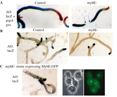
One of the strains analysed, the mybE– strain, did not detectably express mybE. This strain grew and developed poorly when first isolated but after a period in culture it grew and developed well. MybE– and control strains were co-transformed with ecmAO:lacZ, a generic prestalk marker, and also with pspA:gus. The latter construct contains the promoter of pspA, a gene that encodes a
prespore protein, coupled to -glucoronidase. Sequential staining with a -glucoronidase chromogen, X-gluc, and with the  -galactosidase substrate salmon-gal yields a red prestalk region and a blue prespore region (Early et al., 1993).
Migrating mybE– slugs are initially much longer and thinner than normal slugs and they subsequently break up into many small fragments. Fig. 9A shows the separated fragments of a mybE– slug, with just one complete break point, but many slugs contain multiple incipient break points. With slugs of the control strain, prespore cells occupy the rear 80% of slug length and prestalk cells occupy the front 20%. In the mybE– slugs proportioning is extremely variable from slug to slug and presumably depends upon the number of splitting events that had occurred, prior to fixation. The large number of extremely long slugs that predominantly comprise prespore cells, and of fragments that are entirely comprised of prespore cells (Fig. 9A), shows that the prestalk-prespore ratio is greatly distorted in favour of prespore differentiation; however, this was not quantitatively assessed.
[image:6.612.313.544.347.598.2]Analysis of slugs stained for -galactosidase alone allows better visualisation of the prestalk sub-populations. MybE– slugs show strong ecmAO:lacZ expression in the very front of the slug, with only a few scattered staining cells in what would normally be the pstO region (Fig. 9B). The heavily stained, anterior region corresponds to the pstA region of a normal slug. In such slugs,
Fig. 7. Analysis of the DNA binding properties of the MYB domain of MybE. A C terminus-proximal segment of MybE containing the MYB domain was expressed in E. coliand purified using a C terminal GST tag. Recombinant protein (500 ng) and various competitors (5 pmole) were used for gel retardation assay with a 32
P-labeled 30-mer probe. Extracts were pre-incubated in the presence of: (1) no competitor; (2) the 30-mer wild-type (wt); (3) three mutant forms of the 30-mer (mut 1-3, Fig. 4A and as indicated); (4) duplicated forms of the 30-mer, the dyad region and the C-rich region. The arrow indicates the major complex and we assume that the higher mobility forms, which mirror the behaviour of the major complex, contain degraded forms of the C terminal region of MybE.
Fig. 8. Developmental time course of mybEexpression and identification of a mybE–strain. (A) RT-PCR analysis of a developmental time course. Cells were subjected to development on water agar and total cellular RNA was isolated at the indicated stages. The morphological stages reached at the selected times indicated were: 6 hours streaming, 8 hours loose aggregates, 10 hours tight
aggregates, 12 hours standing slugs, 14 hours slugs, 16 hours early-mid culminants and 20 hours mature culminants. The samples were analysed by RT-PCR for mybEmRNA. As a positive control the same samples were analysed for expression of the constitutively active Ig7
[image:6.612.52.245.432.612.2]D
E
V
E
LO
P
M
E
N
T
stained for -galactosidase alone, the ALCs are much more easily visualised and, remarkably, there are almost no stained ALCs in the null strain (Fig. 9B).
Because only one null strain was isolated, it was important to confirm that the phenotypic characteristics are due to MybE inactivation. Therefore, the null strain was transformed with a fusion gene containing the MybE-coding region; linked at its C terminus to GFP and under the control of a semi-constitutive actin promoter. This was a co-transformation with the ecmAO:lacZ marker. The resultant transformants grew and developed well, the slugs were normally proportioned and the ecmAO:lacZ gene displays the normal expression pattern – the hallmark feature being the large number of ALCs that express ecmAO:lacZ (Fig. 9C). Thus, the phenotype observed for the null strain is due to inactivation of mybE.
MybE is essential for normal expression in the major prestalk cell sub-types
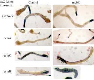
Differentiation of the prestalk cell sub-types in the null strain was further investigated using additional reporter constructs.
(1) The 4⫻22-mer:lacZconstruct serves as a generic prestalk marker that, like ecmAO:lacZ, is expressed in all anterior prestalk cells and in the ALCs. As would have been predicted from the behaviour of ecmAO:lacZitself, the 4⫻22-mer:lacZ construct is expressed in pstA cells but not in pstO cells and ALCs (Fig. 10).
(2) EcmA:lacZ is a marker for pstA differentiation. In the control, after very short times of staining, its expression is restricted to the pstA cells but a finite level of staining in pstO and ALCs becomes apparent after longer periods of incubation (Fig. 10; data not shown). In the null strain, after a short time of staining, expression is almost entirely restricted to the pstA cells (Fig. 10).
(3) EcmO:lacZ is expressed in the band of pstO cells that make up the rear half of the prestalk zone and in the ALCs. In the mybE– strain there is no band of pstO cells. This confirms the result obtained using the entire ecmAO:lacZ construct (Fig. 9B). However, there are stained cells in the rear of the slug, in the approximate position normally occupied by the rearguard cells – a subset of the ALCs that is sporadically lost from the back of the migrating slug.
(4) ecmBencodes a protein that is closely related to EcmA but in the slug stage it is strongly expressed in a small cone of prestalk cells near the tip: the pstAB cells. In slugs of the mybE– strain, ecmB:lacZ is expressed in scattered cells throughout the slug but there is no cone of pstAB cells (Fig. 10). At culmination, ecmBis activated in prestalk cells as they enter the stalk tube. It is also activated in the upper cup, the lower cup and the basal disc (Jermyn et al., 1996). In the mybE– strain at culmination, ecmB:lacZ is strongly expressed in the stalk and there are stained cells in the positions of the upper cup, lower cup and basal disc (data not shown).
MybE is essential for DIF-1 inducible expression of
ecmA
[image:7.612.50.427.58.370.2]Parental and mybE– mutant cells expressing either the ecmAO:lacZ or the 4⫻22:lacZconstruct were induced with DIF-1 in monolayer assay and lacZactivity was assayed. There is strong induction for both constructs in the parental cells but the two fusion constructs are not induced in the mybE– strain (Fig. 11A). For ecmAO:lacZ, this result was confirmed at the RNA level; by inducing cells with several concentrations of DIF-1 and monitoringlacZ expression using RT-PCR (Fig. 11B). Using the same RNA samples but with ecmA primers, RT-PCR was used to show that the endogenous ecmAgene is, as expected, also non DIF-1 inducible in the mybE– strain (Fig. 11B).
Fig. 9. Analysis of expression directed by the complete ecmA promoter in control cells, mybE– cells and mybE– cells expressing a MybE:GFP fusion gene.(A) The ecmAO-lacZconstruct and the pspA gus constructs were co-transformed into Ax2 cells and mybE– cells, and their spatial expression patterns were determined, by double staining at the slug stage. (B) Cells transformed as in A were analysed at the slug stage for lacZ expression alone, i.e. for the activity of the ecmAO promoter fragment. (C) MybE– cells were co-transformed with a MybE:GFP fusion construct under the transcriptional control of a semi-consitutive promoter and with ecmAO-lacZ. The panels on the right show a phase-contrast image of growing cells juxtaposed with the GFP fluorescence image of the same field. The panel on the left shows slugs, derived from the same population, and stained for lacZ
D
E
V
E
LO
P
M
E
N
T
DISCUSSION
The minimal promoter element that directs prestalk-specific gene expression binds MybE The minimal ecmAO promoter region that mediates prestalk-specific expression is the fourfold multimerised form of the 22-mer. This is, to our knowledge, the smallest Dictyostelium promoter subregion shown to direct cell-type specific gene expression. Interestingly, although the 22-mer derives from a region that directs expression only within pstO cells, its multimerised form directs expression in pstO cells, pstA cells and ALCs. This implies a close relationship, at the transcriptional level, between the signalling pathways directing differentiation of the two major prestalk cell subtypes.
A dyad within the 22-mer fits the MYB-binding site consensus and is almost identical to a sequence element identified within a subregion of the G6P isomerase promoter that directs prestalk-specific expression. Point mutational analysis showed that this sequence is necessary for prestalk-specific expression but the element was not further characterised (Tabata et al., 2001). As would have been predicted from the sequence of the dyad, the multimerised 22-mer binds in vitro to a MYB protein: MybE.
There is also an essential, C-rich region located downstream of the dyad. There are examples of DNA-binding sites for MYB proteins that extend downstream of the conserved region (e.g. Howe et al., 1991) and the C-rich region in the 22-mer is located very close to the dyad. Hence, it may form part of an extended binding site for MybE.
Similarities between the mybE-strain and other
Dictyosteliummutants
The mybE– strain initially grew and developed poorly but, after a period in culture, growth and development improved. It is unclear whether the improvement in development was simply the result of higher cell viability or whether secondary mutations acquired during the adaption process suppressed developmental defects. Therefore, the developmental phenotype we describe must be regarded as a
minimal phenotype. However, this phenotype fits a pattern; because there are strong similarities between the mybE– strain and known mutants in DIF-1 signalling.
The morphological phenotype of the mybE– strain is very similar to that of the dimA– and dimB–, DIF response, and dmtA–, DIF bio-synthesis, mutants (Thompson et al., 2004; Thompson and Kay, 2000; Huang et al., 2006; Zhukovskaya et al., 2006); all form long thin slugs that frequently split into fragments. The other, striking similarity between the dmtA–, dimA– and mybE– strains is that all three are selectively defective in the expression of ecmAO:lacZand ecmO:lacZin pstO cells [the situation with DimB is unclear because it showed such a difference in an Ax3 background (Huang et al., 2006) but not in an AX2 background (Zhukovskaya et al., 2006)]. It will be of interest to determine the signalling relationships between MybE and the DimA and DimB proteins.
Other proportioning mutants also display a change in the extent of the pstO region while retaining a normally sized pstA zone (Han and Firtel, 1998; Chung et al., 1998; Nelson et al., 2000; Ennis et al., 2000). However, the mybE– strain has a more severe defect than any of these strains. It raises the question of whether the mybE- mutant has: (1) a pstO region with no ecmA gene expression but otherwise normal patterning; (2) a pstO region with no expression of ecmAand altered expression patterns of other prestalk-specific genes, or (3) no pstO region. In order to distinguish between these possibilities it will be necessary to analyse additional pstO markers (Maeda, 2003).
MybE is required for the normal differentiation of ALCs
[image:8.612.52.375.60.336.2]A notable feature of the mybEnull, not to our knowledge previously described for any other mutant, is the virtual absence of ALCs that express ecmA promoter-derived markers. The fact that ecmA expression in both pstO and ALCs is greatly reduced is of interest because prior studies suggest a close link between the ALCs and the pstO cells; pstO-specific constructs, derived from the ecmApromoter,
Fig. 10. Comparison of the expression of prestalk markers in parental and mybE– cells. The 4⫻22-mer-lacZ,ecmA-lacZ, ecmO-lacZ
D
E
V
E
LO
P
M
E
N
T
are also expressed in ALCs and the two populations continually interchange position within the slug (Early et al., 1993; Abe et al., 1994). There are ALCs in the mybE– slugs, as detected by neutral red staining (J.G.W., unpublished). However, the ALCs population is composed of multiple prestalk subtypes (Jermyn and Williams, 1991; Jermyn et al., 1996) and the neutral red-positive cells in the mutant are presumably one or more of the non-ecmA-expressing subtypes.
MybE and the induction of prestalk cell differentiation by DIF-1
In addition to the virtual absence of ecmAgene expression in pstO and ALCs and the similarities to other DIF signalling mutants, several pieces of evidence combine to imply a role for MybE in mediating DIF-1 induction of ecmA gene expression: (1) the multimerised 22-mer directs DIF-inducible gene expression; (2) MybE binds in vitro to the multimerised 22-mer; (3) mutations that disrupt this binding eliminate DIF inducibility; (4) MybE is required for DIF-mediated induction of ecmA expression in vivo.
All these facts are best explained if MybE directly mediates the induction of ecmAgene expression by DIF-1. If so, MybE could be activated in some way by DIF-1 or, alternatively, it could be a constitutive component of the transcriptional complex. Precedent gives no clear guidance on these possibilities because there is no canonical pathway of MYB protein activation. There are several examples of phosphorylation of MYB proteins, but modification is effected by diverse kinases and no general mechanism of regulation has emerged (Winn et al., 2003; Kanei-Ishii et al., 2004; Oelgeschlager et al., 1995; Ramsay et al., 1995; Leverson et al., 1998; Andersson et al., 2003; Sugano et al., 1998).
Interestingly, however, there are precedents for SHAQKY proteins mediating induction by a diffusible molecules; the PHR1 protein of Arabidopsisactivates gene expression in response to phosphate deprivation (Rubio et al., 2001) and the three OsMYBS proteins of rice mediate gibberelin and sugar regulation of ␣ -amylase gene expression (Lu et al., 2002). Unusually for Myb proteins, PHR1 functions as a dimer (Rubio et al., 2001) and yeast two-hybrid analysis shows that MybE also forms homodimers (M.F. and J.G.W., unpublished data). Given these similarities, it will be of interest to determine whether DIF-1 and MybE form part of a pathway that resembles a plant signalling pathway.
We thank Drs I. Sarafimidis and R. R. Kay for kindly providing details of the DIF-1 assay, and the local proteomics facility for their invaluable assistance. This work was supported by Wellcome Trust Program Grant 053640/Z to J.G.W.
References
Abe, T., Early, A., Siegert, F., Weijer, C. and Williams, J. G.(1994). Patterns of cell movement within the Dictyosteliumslug revealed by cell type-specific, surface labeling of living cells. Cell77, 687-699.
Andersson, K. B., Kowenz-Leutz, E., Brendeford, E. M., Tygsett, A. H., Leutz, A. and Gabrielsen, O. S.(2003). Phosphorylation-dependent down-regulation of c-Myb DNA binding is abrogated by a point mutation in the v-myb oncogene. J. Biol. Chem. 278, 3816-3824.
Araki, T., Gamper, M., Early, A., Fukuzawa, M., Abe, T., Kawata, T., Kim, E., Firtel, R. A. and Williams, J. G.(1998). Developmentally and spatially regulated activation of a DictyosteliumSTAT protein by a serpentine receptor. EMBO J. 17, 4018-4028.
Chung, C. Y., Reddy, T. B. K., Zhou, K. M. and Firtel, R. A.(1998). A novel, putative MEK kinase controls developmental timing and spatial patterning in Dictyosteliumand is regulated by ubiquitin-mediated protein degradation. Genes Dev. 12, 3564-3578.
Dingermann, T., Reindl, N., Werner, H., Hildebrandt, M., Nellen, W., Harwood, A., Williams, J. and Nerke, K.(1989). Optimization and in situ detection of Escherichia colibeta-galactosidase gene expression in Dictyostelium discoideum. Gene 85, 353-362.
Early, A. E. and Williams, J. G.(1988). A Dictyosteliumprespore-specific gene is transcriptionally repressed by DIF in vitro. Development 103, 519-524.
Early, A. E., Gaskell, M. J., Traynor, D. and Williams, J. G.(1993). Two distinct populations of prestalk cells within the tip of the migratory Dictyosteliumslug with differing fates at culmination. Development 118, 353-362.
Ennis, H. L., Dao, D. N., Pukatzki, S. U. and Kessin, R. H.(2000). Dictyostelium amoebae lacking an F-box protein form spores rather than stalk in chimeras with wild type. Proc. Natl. Acad. Sci. USA97, 3292-3297.
Fukuzawa, M., Araki, T., Adrian, I. and Williams, J. G.(2001). Tyrosine phosphorylation-independent nuclear translocation of a DictyosteliumSTAT in response to DIF signaling. Mol. Cell 7, 779-788.
Guo, K., Anjard, C., Harwood, A., Kim, H. J., Newell, P. C. and Gross, J. D.
(1999). A myb-related protein required for culmination in Dictyostelium. Development 126, 2813-2822.
Han, Z. and Firtel, R. A.(1998). The homeobox-containing gene Wariai regulates anterior-posterior patterning and cell-type homeostasis in Dictyostelium. Development125, 313-325.
Hjorth, A., Datta, S., Khanna, N. C. and Firtel, R. A.(1988). Analysis of cis and trans elements involved in cAMP-inducible gene expression in Dictyostelium discoideum. Dev. Genet. 9, 435-454.
Howe, K. M., Watson, R. J., Reakes, C. F., McMahon, J., Garcia, A., LaMontagne, K., Reavis, D., Stober-Grasser, U. and Lipsick, J. S.(1991). Nucleotide preferences in sequence-specific recognition of DNA by c-myb protein. Nucleic Acids Res. 19, 3913-3919.
Huang, E., Blagg, S. L., Keller, T., Katoh, M., Shaulsky, G. and Thompson, C.
(2006). bZIP transcription factor interactions regulate DIF responses in Dictyostelium. Development133, 449-458.
Jermyn, K. A. and Williams, J. G.(1991). An analysis of culmination in Dictyosteliumusing prestalk and stalk-specific cell autonomous markers. Development 111, 779-787.
Jermyn, K. A., Duffy, K. T. and Williams, J. G.(1989). A new anatomy of the prestalk zone in Dictyostelium. Nature 340, 144-146.
Jermyn, K. A., Traynor, D. and Williams, J. G.(1996). The initiation of basal disc formation in Dictyostelium discoideum is an early event in culmination. Development 122, 753-760.
Jin, H. and Martin, C.(1999). Multifunctionality and diversity within the plant MYB-gene family. Plant Mol. Biol. 41, 577-585.
Kanei-Ishii, C., Ninomiya-Tsuji, J., Tanikawa, J., Nomura, T., Ishitani, T., Kishida, S., Kokura, K., Kurahashi, T., Ichikawa-Iwata, E., Kim, Y. et al.
[image:9.612.51.302.58.255.2](2004). Wnt-1 signal induces phosphorylation and degradation of c-Myb protein via TAK1, HIPK2, and NLK. Genes Dev. 18, 816-829.
Fig. 11. Analysis of DIF induction of ecmAin a control and a mybE–mutant. (A) Expression of promoter constructs. The mybE– strain and a control random integrant strain, both transformed with the indicated ecmApromoter fusion constructs, were assayed for DIF inducibility in the monolayer assay. Quadruplicate wells were analysed for each assay condition and this result is typical of four separate biological experiments. (B) RT-PCR analysis of the ecmAO:lacZgene and the endogenous ecmAgene. Control and mybE– cells, transformed with ecmAO:lacZ, were induced with DIF-1 in a monolayer assay using a cell density of 105/cm2and a cerulenin concentration of 50 M. Total
cellular RNA was extracted and the abundance of the lacZ, ecmAand
D
E
V
E
LO
P
M
E
N
T
Kawata, T., Early, A. and Williams, J. G.(1996). Evidence that a combined activator-repressor protein regulates Dictyosteliumstalk cell differentiation. EMBO J. 15, 3085-3092.
Kawata, T., Shevchenko, A., Fukuzawa, M., Jermyn, K., Totty, N. F., Zhukovskaya, N. V., Sterling, A. E., Mann, M. and Williams, J. G.(1997). SH2 signaling in a lower eukaryote: a STAT protein that regulates stalk cell differentiation in Dictyostelium. Cell 89, 909-916.
Kay, R. R.(1998). The biosynthesis of differentiation-inducing factor, a chlorinated signal molecule regulating Dictyostelium development. J. Biol. Chem. 273, 2669-2675.
Kay, R. R. and Jermyn, K. A.(1983). A possible morphogen controlling differentiation in Dictyostelium. Nature 303, 242-244.
Kay, R. R. and Thompson, C. R. L.(2001). Cross-induction of cell types in Dictyostelium: evidence that DIF-1 is made by prespore cells. Development 128, 4959-4966.
Kessin, R. H.(2001). Dictyostelium. Evolution, Cell Biology, and the Development of Multicellularity. Cambridge: Cambridge University Press.
Kopachik, W. J., Dhokia, B. and Kay, R. R.(1985). Selective induction of stalk-cell-specific proteins in Dictyostelium. Differentiation 28, 209-216.
Leverson, J. D., Koskinen, P. J., Orrico, F. C., Rainio, E. M., Jalkanen, K. J., Dash, A. B., Eisenman, R. N. and Ness, S. A.(1998). Pim-1 kinase and p100 cooperate to enhance c-Myb activity. Mol. Cell 4, 417-425.
Lipsick, J. S.(1996). One billion years of Myb. Oncogene 13, 223-235.
Lu, C. A., Ho, T. H., Ho, S. L. and Yu, S. M.(2002). Three novel MYB proteins with one DNA binding repeat mediate sugar and hormone regulation of alpha-amylase gene expression. Plant Cell 14, 1963-1980.
Maeda, M., Sakamoto, H., Iranfar, N., Fuller, D., Maruo, T., Ogihara, S., Morio, T., Urushihara, H., Tanaka, Y. and Loomis, W. F.(2003). Changing patterns of gene expression in Dictyosteliumprestalk cell subtypes recognized by in situ hybridization with genes from microarray analyses. Eukaryot. Cell2, 627-637.
Mohanty, S., Jermyn, K. A., Early, A., Kawata, T., Aubry, L., Ceccarelli, A., Schaap, P., Williams, J. G. and Firtel, R. A.(1999). Evidence that the DictyosteliumDd-STATa protein is a repressor that regulates commitment to stalk cell differentiation and is also required for efficient chemotaxis. Development
126, 3391-3405.
Morris, H. R., Taylor, G. W., Masento, M. S., Jermyn, K. A. and Kay, R. R.
(1987). Chemical structure of the morphogen differentiation inducing factor from Dictyostelium discoideum. Nature 328, 811-814.
Nelson, M. K., Clark, A., Abe, T., Nomura, A., Yadava, N., Funair, C. J., Jermyn, K. A., Mohanty, S., Firtel, R. A. and Williams, J. G.(2000). An F-Box/WD40 repeat-containing protein important for Dictyosteliumcell-type proportioning, slug behaviour, and culmination. Dev. Biol. 224, 42-59.
Oelgeschlager, M., Krieg, J., Luscher-Firzlaff, J. M. and Luscher, B.(1995). Casein kinase II phosphorylation site mutations in c-Myb affect DNA binding and transcriptional cooperativity with NF-M. Mol. Cell. Biol. 15, 5966-5974.
Ogata, K., Morikawa, S., Nakamura, H., Sekikawa, A., Inoue, T., Kanai, H., Sarai, A., Ishii, S. and Nishimura, Y.(1994). Solution structure of a specific DNA complex of the Myb DNA-binding domain with cooperative recognition helices. Cell 79, 639-648.
Otsuka, H. and van Haastert, P. J. M.(1998). A novel Myb homolog initiates Dictyosteliumdevelopment by induction of adenylyl cyclase expression. Genes Dev. 12, 1738-1748.
Pang, K. M., Lynes, M. A. and Knecht, D. A.(1999). Variables controlling the expression level of exogenous genes in Dictyostelium. Plasmid 41, 187-197.
Pears, C. J. and Williams, J. G.(1987). Identification of a DNA sequence element required for efficient expression of a developmentally regulated and cAMP-inducible gene of Dictyostelium discoideum. EMBO J. 6, 195-200.
Pears, C. J., Mahbubani, H. M. and Williams, J. G.(1985). Characterization of two highly diverged but developmentally coregulated cysteine proteinases in Dictyostelium discoideum. Nucleic Acids Res. 13, 8853-8866.
Powell-Coffman, J. A., Schnitzler, G. R. and Firtel, R. A.(1994). A GBF-binding site and a novel AT element define the minimal sequences sufficient to direct prespore-specific expression in Dictyostelium discoideum. Mol. Cell. Biol. 14, 5840-5849.
Ramsay, R. G., Morrice, N., Van Eeden, P., Kanagasundaram, V., Nomura, T., De Blaquiere, J., Ishii, S. and Wettenhall, R.(1995). Regulation of c-Myb through protein phosphorylation and leucine zipper interactions. Oncogene 11, 2113-2120.
Rubio, V., Linhares, F., Solano, R., Martín, A. C., Iglesias, J., Leyva, A. and Paz-Ares, J.(2001). A conserved MYB transcription factor involved in phosphate starvation signaling both in vascular plants and in unicellular algae. Genes Dev. 15, 2122-2133.
Schnitzler, G. R., Fischer, W. H. and Firtel, R. A.(1994). Cloning and characterization of the G-box binding factor, an essential component of the developmental switch between early and late development in Dictyostelium. Genes Dev. 8, 502-514.
Stober-Grasser, U., Brydolf, B., Bin, X., Grasser, F., Firtel, R. A. and Lipsick, J. S.(1992). The myb DNA-binding domain is highly conserved in Dictyostelium discoideum. Oncogene 7, 589-596.
Sugano, S., Andronis, C., Green, R. M., Wang, Z. Y. and Tobin, E. M.(1998). Protein kinase CK2 interacts with and phosphorylates the Arabidopsiscircadian clock-associated 1 protein. Proc. Natl. Acad. Sci. USA 95, 11020-11025.
Tabata, K., Matsuda, Y., Viller, E., Masamune, Y., Katayama, T. and Yasukawa, H.(2001). Myb-binding site regulates the expression of
glucosamine-6- phosphate isomerase in Dictyostelium discoideum. Dev. Growth Differ. 43, 583-589.
Thompson, C. R. L. and Kay, R. R.(2000). The role of DIF-1 signaling in Dictyosteliumdevelopment. Mol. Cell 6, 1509-1514.
Thompson, C. R. L., Fu, Q., Buhay, C., Kay, R. R. and Shaulsky, G.(2004). A bZIP/bRLZ transcription factor required for DIF signaling in Dictyostelium. Development 131, 513-523.
Town, C. D., Gross, J. D. and Kay, R. R.(1976). Cell differentiation without morphogenesis in Dictyostelium discoideum. Nature 262, 717-719.
Watts, D. J. and Ashworth, J. M.(1970). Growth of myxamoebae of the cellular slime mould Dictyostelium discoideumin axenic culture. Biochem. J. 119, 171-174.
Williams, J. G., Ceccarelli, A., McRobbie, S., Mahbubani, H., Kay, R. R., Early, A., Berks, M. and Jermyn, K. A.(1987). Direct induction of Dictyostelium prestalk gene expression by DIF provides evidence that DIF is a morphogen. Cell
49, 185-192.
Winn, L. M., Lei, W. and Ness, S. A.(2003). Pim-1 phosphorylates the DNA binding domain of c-Myb. Cell Cycle 2, 258-262.
Zhukovskaya, N. V., Fukuzawa, M., Yamada, Y., Araki, T. and Williams, J. G.