763
Introduction
A significant degree of conservation of the molecular pathways involved in endoderm formation is becoming apparent, although differences between species are also observed (Reiter et al., 2001; Xanthos et al., 2001) (for reviews, see Loose and Patient, 2004; Maduro and Rothman, 2002; Shivdasani, 2002; Stainier, 2002). In the amphibian Xenopus laevis, important players are the maternal T-box transcription factor, VegT, and a panoply of other regulatory proteins including TGFβs, Bix/Mix, GATA, Pitx2 and Sox (Clements and Woodland, 2003; Ecochard et al., 1998; Faucourt et al., 2001; Henry and Melton, 1998; Weber et al., 2000; Yasuo and Lemaire, 1999; Zhang and King, 1996). A two-step model for endoderm formation has been proposed in which, in the first step, at early blastula, VegT cell-autonomously induces Sox17 and Mix.1, as well as Nodal-related members of the TGFβ family; in the second step, which requires cell-cell contact and occurs during late blastula stages, the Nodal-related molecules act to reinforce and maintain endodermal gene expression (Chang and Hemmati-Brivanlou, 2000; Clements and Woodland, 2003; Engleka et al., 2001; Yasuo and Lemaire, 1999). During the first phase, VegT is also thought to induce a hypothetical
repressor, which may function to inhibit endodermal gene expression in cells that escape the endodermal layer during gastrulation (Clements and Woodland, 2003).
The roles played by individual GATA factors in vertebrate endoderm formation are currently unclear (Rossant et al., 2003; Soudais et al., 1995; Weber et al., 2000; Yasuo and Lemaire, 1999). Over-expression of either GATA4 or 5 in Xenopus presumptive ectoderm can induce endoderm markers, and mutations in GATA5 in zebrafish, known as faust, lead to their reduced expression in embryos (Reiter et al., 1999; Reiter et al., 2001; Shoichet et al., 2000; Weber et al., 2000). However, while GATA4–/– mice die by day 9.5 dpc and exhibit severe defects in the closure of the gut, and embryoid bodies differentiated from GATA4–/–ES cells display a defect in the differentiation of visceral yolk sac endoderm, the GATA5 null mutation is not an early embryonic lethal mutation in the mouse, giving rise only to abnormalities in the female genitourinary tract (Kuo et al., 1997; Molkentin et al., 1997; Molkentin et al., 2000; Narita et al., 1997). The other GATA factor associated with endoderm formation is GATA6: null mice die at day 5.5-6.5 dpc from defects in extra-embyonic endoderm, and recent evidence suggests that the expression of
The individual contributions of the three vertebrate GATA factors to endoderm formation have been unclear. Here we detail the early expression of GATA4, 5 and 6 in presumptive endoderm in Xenopus embryos and their induction of endodermal markers in presumptive ectoderm. Induction of HNF3β by all three GATA factors was abolished when protein synthesis was inhibited, showing that these inductions are indirect. In contrast, whereas induction of Sox17αand HNF1βby GATA4 and 5 was substantially reduced when protein synthesis was inhibited, induction by GATA6 was minimally affected, suggesting that GATA6 is a direct activator of these early endodermal genes. GATA4 induced GATA6 expression in the same assay and antisense morpholino oligonucleotides (MOs), designed to knock down translation of GATA6, blocked induction of Sox17α and HNF1β by GATA4, suggesting that GATA4 induces these genes via GATA6 in
this assay. All three GATA factors were induced by activin, although GATA4 and 6 required lower concentrations. GATA MOs inhibited Sox17α and HNF1β induction by activin at low and high concentrations in the order: GATA6>GATA4>GATA5. Together with the timing of their expression and the effects of GATA MOs in vivo, these observations identify GATA6 as the predominant GATA factor in the maintenance of endodermal gene expression by TGFβ signaling in gastrulating embryos. In addition, examination of gene expression and morphology in later embryos, revealed GATA5 and 6 as the most critical for the development of the gut and the liver.
Key words: Activin, Animal caps, Cycloheximide, Emetine, Endoderm, GATA factors, Gut, HNF1β, HNF3β, Liver, Nodal-related, Sox17α, TGFβ, Transcription, VegT, Xenopus
Summary
GATA4, 5 and 6 mediate TGF
β
maintenance of endodermal gene
expression in Xenopus embryos
Boni Anatole Afouda*, Aldo Ciau-Uitz†and Roger Patient†,‡
Institute of Genetics, University of Nottingham, Queen’s Medical Centre, Nottingham NG7 2UH, UK
*Present address: School of Medical Sciences, Cell and Developmental Biology Research Programme, Institute of Medical Sciences, University of Aberdeen, Aberdeen AB25 2ZD, UK
†Present address: MRC Molecular Haematology Unit, Weatherall Institute of Molecular Medicine, University of Oxford, John Radcliffe Hospital, Headington, Oxford OX3 9DS, UK
‡Author for correspondence (e-mail: roger.patient@imm.ox.ac.uk)
Accepted 10 December 2004
Development 132, 763-774
Published by The Company of Biologists 2005 doi:10.1242/dev.01647
Research article
De
GATA6 seen in the inner cell mass (ICM) of the blastocyst as early as 3.5 dpc may represent future primitive endoderm, identifying GATA6 as a very early determinant of this germ layer (Koutsourakis et al., 1999; Morrisey et al., 1998; Rossant et al., 2003). Consistent with important roles for GATA4 and 6 in specifying primitive endoderm, ectopic expression in mouse ES cells of GATA4 or 6, but not downstream genes like HNF4, is sufficient to drive primitive endoderm differentiation (Fujikura et al., 2002).
The observation that activins are expressed in the ICM of mouse blastocysts at the same time as GATA6, and the reported induction of GATA4 and GATA5 by activin in Xenopus presumptive ectoderm, suggest that GATA factors may mediate TGFβ/Nodal signalling during endoderm formation (Albano et al., 1993; Ariizumi et al., 2003; Hudson et al., 1997; Rossant et al., 2003; Weber et al., 2000). This has been suggested previously in Xenopus, but their precise roles remain unclear (Clements et al., 1999; Clements and Woodland, 2003; Yasuo and Lemaire, 1999). For example it is unclear whether each GATA factor has a distinct function or whether they represent a family of redundant genes. A knowledge of their direct targets would be informative in this regard.
In this study, we explore the roles of XenopusGATA4, 5 and 6 in endoderm formation using over-expression and antisense oligonucleotide approaches. We demonstrate that, like XenopusGATA5 (Weber et al., 2000), both GATA4 and the long isoform of GATA6 are potent inducers of the early endoderm markers, Sox17αand HNF1β, and the later marker, HNF3β, also known as FoxA2, in Xenopus presumptive ectoderm. In addition, using protein synthesis inhibitors, we show that GATA6 is a direct activator of both Sox17α and HNF1β, whereas induction of these genes by GATA4 and 5 is either only partly direct or, in the case of GATA4 on Sox17α, dependent on GATA6. By blocking production of these GATA factors in vivo, or in presumptive ectoderm induced to form endoderm by TGFβ signalling, we confirm that they are required for full expression of endodermal markers and for proper formation of the gut and its outgrowths. These results suggest that TGFβ signaling works through these GATA factors to maintain endodermal gene expression, and they identify GATA6 as a major player in gut development and a direct activator of target genes.
Materials and methods
ConstructsGATA4-5 and short and long GATA6 (Accession no. AY395755) expression constructs have previously been described (Peterkin et al., 2003; Weber et al., 2000). Haemagglutinin (HA)-tagged GATA4-5 plasmids were made by inserting PCR amplified fragments of GATA4-5 in the XbaI and XhoI sites of a host vector, pβUT2-HA. pβUT2-HA was created by inserting the 74 bp HA tag into the BamHI-KpnI sites of pβUT2 (Sykes et al., 1998) (M. Gering and R.P., unpublished). The following primer sequences were used for the amplification of the coding regions of GATA4: 5′ GAT CCC TCG AGC AGC TAA GAC CAG GTT GTT CC 3′for the reverse primer and 5′GGA TCT CTA GAA CCA GGG GAT CAG GAT GTA TC 3′for the forward primer and for GATA5: 5′GAT CCC TCG AGC GGC AAG TGC CAG CGC GCA CC 3′for the reverse primer and 5′GGA TCT CTA GAA CCA GGT GGT AGG AAG GCT CAG CC 3′for the forward primer. The HA-tagged long GATA6 was made by using pβUT3-HA instead of pβUT2-HA as previously described
(Peterkin et al., 2003). For in vitro transcription, the plasmids were EcoRI-linearised and T3-transcribed for GATA4-5 and SfiI-linearised and T3-transcribed for GATA6.
GATA4-, 5- and 6-GR were made by fusing their coding regions to the region encoding the hormone-binding domain of human glucocorticoid receptor in the BglII site of the host vector, pSP64T-GR previously described (Tada et al., 1997). The sequences of primers for generating the coding regions were the following: GATA4: 5′GAT CCA GAT CTA GCT AAG ACC AGG TTG TTC C 3′for the reverse primer and 5′GGA TCA GAT CTA CCA GGA TGT ATC AGA GTA TAG C 3′for the forward primer, and for GATA5: 5′GAT CCA GAT CTG GCA AGT GCC AGC GCG CAC C 3′for the reverse primer and 5′GGA TCA GAT CTA CCA GGA TGC CCA GCC GGC CTA CTC C 3′ for the forward primer and for long GATA6: 5′AGC CAA GGC CAA AGC ACA 3′for the reverse primer and 5′GGA TCA GAT CTA CCA GGA TGG ACC TGA GTG 3′for the forward primer. All the fusion plasmids were SalI-linearised and SP6-transcribed. All the constructs were checked by restriction digestion and by sequencing. Injected RNAs were synthesised using T3 or SP6 polymerase mMESSAGE mMACHINE kits (Ambion) according to the manufacturer’s instructions.
Embryos and explants
Xenopusembryos were obtained and cultured as previously described (Weber et al., 2000). They were injected at the one-cell stage into the animal pole for animal cap assays or into the vegetal pole for whole embryo assays. RNAs and morpholinos (MOs) were injected in water (4 nl). In cap explants, the amount of each MO injected was 5 ng per embryo (determined from titration experiments), or 10 ng in the presence of activin. In whole embryos, 10-40 ng of each MO was injected per embryo. Animal cap explants were dissected and cultured as previously described (Weber et al., 2000). Cycloheximide treatment was as previously described (Tada et al., 1997). Emetine was dissolved in water and treatment was similar to cycloheximide but used at 100 µg/ml final concentration. Incorporation of radiolabelled methionine was measured by spotting protein extracts onto Whatman 3MM paper and precipitating with 10% trichloroacetic acid (TCA). Unincorporated radiolabelled methionine was removed by boiling in 5% TCA and chemiluminescence was counted.
Protein analysis
Protein extraction and western blot analysis were as previously described (Peterkin et al., 2003), except that the cap explants were collected when the sibling whole embryos reached stage 12.
RNA analysis
Whole-mount in situ hybridisation and in situ hybridisation on embryo sections were conducted as previously described (Ciau-Uitz et al., 2000). The abundance of RNAs was determined using quantitative real-time RT-PCR as described (Peterkin et al., 2003). Amounts relative to the housekeeping RNA, ornithine decarboxylase (ODC), were expressed as a ratio either to stage 6.5 (Fig. 1) or to uninjected animal caps or embryos (Figs 2-5). For the sequences of primers and probes used see Table 1.
Results
GATA4, 5 and 6 are expressed in the presumptive endoderm in Xenopus embryos
Temporal expression profiles of GATA4, 5 and 6 in Xenopus embryos have been reported using either semi-quantitative RT-PCR, RNAse protection or whole-mount in situ hybridisation (Gove et al., 1997; Jiang and Evans, 1996; Weber et al., 2000; Yasuo and Lemaire, 1999). GATA5 was detected as a maternal transcript and all studies agree that zygotic transcription of all
De
three GATA factors has started by stage 10.5, at the onset of gastrulation, although GATA4 and 5 increases have also been reported at earlier blastula stages, with some disagreement over the precise stage when GATA4 expression commences. Spatially, expression appears to be in the vegetal cells fated to form endoderm, however, only GATA5 has been analysed by in situ hybridisation to sectioned embryos. We therefore re-investigated the temporal expression profiles of GATA4, 5 and 6 using the quantitative and more sensitive real-time RT-PCR method, and the spatial expression of GATA4 and 6 using in situ hybridisation to sectioned embryos (Fig. 1). Sox17αwas used as a comparator in the real-time RT-PCR (Fig. 1A). Although its expression begins at stage 8 (4-fold compared to stage 6.5 embryos), the level increases dramatically at stage 9 (80-fold over stage 6.5) when GATA4 and 6 expression begins (3- and 6-fold over stage 6.5, respectively), while zygotic GATA5 expression begins later (stage 10) (Fig. 1B). Thus, GATA4 and 6 expression coincides with the major upregulation of Sox17α.
Using in situ hybridisation on sections, we previously showed that GATA5 is expressed in the presumptive sub-blastoporal endoderm in Xenopus embryos (Weber et al., 2000). Using the same technique to investigate the spatial expression profiles of GATA4 and 6, we show that at stage 10, before the onset of gastrulation, both GATA4 and 6 are expressed in the yolky vegetal cells (Fig. 1C), which are fated to form endoderm. The expression pattern is very similar to that seen for GATA5 (Weber et al., 2000). However, although we could not detect expression of GATA5 in the supra-blastoporal endoderm, both GATA4 and 6 are expressed there (Fig. 1C, immediately above the arrowheads). We therefore conclude that, while GATA4, 5 and 6 are all expressed in the non-involuting endoderm, only GATA4 and 6 are expressed in the involuting endoderm. Thus, the pattern of expression of GATA4 and 6 at stage 10 is similar to the pan-endodermal marker, Sox17 (Engleka et al., 2001; Hudson et al., 1997). After gastrulation has begun (stages 10.5-11), the
supra-blastoporal endoderm is negative for both GATA4 and 6 (Fig. 1C, immediately above the arrowheads). In contrast, Sox17 expression in these cells continues (Engleka et al., 2001; Hudson et al., 1997). It therefore appears that GATA4 and 6 are expressed in the pre-involuting endoderm early in gastrulation but not later.
Differences in intensity of expression do exist between the GATA factors in the presumptive sub-blastoporal endoderm [compare Fig. 1C with the work of Weber et al. (Weber et al., 2000)]. Thus, while GATA5 is strongest in the centre of the vegetal hemisphere all the way from the pole to the floor of the blastocoel, GATA4 and 6 are stronger towards the edges of this group of cells. Similarly, whereas GATA5 expression was not seen in any cells in the animal cap, GATA6 and to a lesser extent GATA4 expression was detected there at stage 10, albeit weakly. The significance of these observations is however unclear at the present time.
Overall, we conclude that GATA4, 5 and 6 are expressed in Xenopus embryos early enough to play roles in endoderm formation. Although the pan-endodermal gene, Sox17α, is expressed before GATA4 and 6, their expression coincides with the major increase in Sox17α expression seen later and the maintenance of its expression. Their lack of expression in the supra-blastoporal endoderm after the onset of gastrulation suggests that they are only transiently involved in such maintenance in the posterior involuting endoderm.
Differential induction of early endoderm markers by GATA4, 5 and 6 in Xenopus presumptive ectoderm
[image:3.612.45.554.83.321.2]We have shown previously that Xenopus GATA4 and 5 can induce endoderm markers in the presumptive ectoderm of Xenopusanimal caps using a semi-quantitative RT-PCR assay (Weber et al., 2000). In these experiments, only the short isoform of XenopusGATA6 was available and this was unable to induce endodermal markers in this assay. We therefore asked if the long isoform of XenopusGATA6 (Brewer et al., 1999; Peterkin et al., 2003) has this activity. Equal concentrations of
Table 1. Primer pairs (R,F) and probes (P) used for real-time RT-PCR
Gene Sequence Reference
Sox 17α R: 5′CCA CGA CTT GCC AAG CAT CT 3′ Accession number AJ001730 F: 5′GAT CCG CCG GCC TAT GA 3′
P: 5′ACG AGC GCA AGA GAC TGG CAC AGC 3′
HNF1β R: 5′GAT CCA TGC GAT AGT AGA GAA TTC AA 3′ New F: 5′CAA AAA TTG GCT ATG GAT GCC TAT A 3′
P: 5′CTG GCC CAT CAC ACC CCC ACA A 3′
HNF3β R: 5′TTG CTC CGA GGA CAT GAG ATT 3′ New F: 5′ GTC CCA CCT CAA ACC AGA ACA 3′
P: 5′CAT TAT TCT TTC AAC CAC CCA TTT TCC ATC AA 3′
GATA4 R: 5′GCC GCA ACA TCA GGA CTT TT 3′ New F: 5′GAA GCT TCG TCC TTC ACA CAA CT 3′
P: 5′TGG AAA CCC AAC TTG TCA GTG GAT AAA CCC 3′
GATA5a R: 5′TTC AGC AAC GGA TCC ATT TCT 3′ New F: 5′CTC AAT GCC TTC TGA AGG ATA CAA 3′
P: 5′AAA ATA ACC GGT GCC GCG TCC AG 3′
GATA6b R: 5′GAA CTG AGA TTG TCG CTC TAT GTA TAT GTA T 3′ New F: 5′TGG AAG GAA ATG TGA CCC TCA T 3′
P: 5′TTT CCA GAT GAC ATG CTG TCT CTC AAC TGC 3′
ODC R: 5′GCA GCC ACT GCC AAC ATG 3′ Accession number X56316 F: 5′CTG CCG CCT CAG TGT GAA 3′
P: 5′ACC CTT AAA ACA AGC AGG CTG CTT CTG GA 3′
De
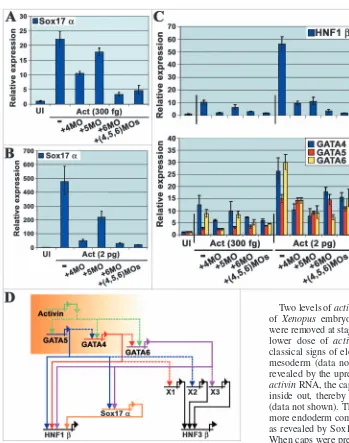
mRNAs encoding GATA4, 5 and 6 were injected into the animal poles of one-cell embryos. Animal cap explants were removed from injected embryos when they reached stage 8.5-9 and cultured until the sibling embryos reached stage 12. Harvested animal caps were divided into two for determination of protein expression by Western blot and endodermal gene induction by quantitative, real-time RT-PCR. Both short and long isoforms of GATA6 were synthesised, with the short isoform representing roughly one-third of the total GATA6 protein present (Fig. 1D). However, when gel loading was taken into account, the long isoform was judged to be present at equivalent levels to GATA4 and 5, enabling a comparison to
be made between the GATA factors for their capacities to induce endodermal gene expression. The extracted RNA was probed for the expression of three genes, Sox17α, HNF1β and HNF3β, expressed in endoderm at stage 12. Expression of Sox17α is always restricted to endoderm, while HNF1β is also expressed in mesodermal derivatives from gastrula stages (Demartis et al., 1994; Hudson et al., 1997) (see Fig. 5). HNF3β expression is restricted to the anterior and posterior endoderm from early gastrula stages (Suri et al., 2004). Differences in the strength of induction of these endodermal genes were observed between the GATA factors (Fig. 1E,F). GATA4 and 6 induced both Sox17α and HNF3βwith similar efficiencies and slightly more effectively than GATA5, already shown to be a potent inducer of endoderm markers and used here as a positive control (Weber et al., 2000). For HNF1β, while all three GATA factors induced significant expression, GATA4 was the most effective (Fig. 1E). We therefore conclude that the long isoform of GATA6 is a potent inducer of endoderm, like GATA4 and 5, and that the three GATA factors differ in their contributions to the expression of different endodermal genes.
GATA6 is a direct activator of Sox17αand HNF1β
[image:4.612.44.348.71.583.2]To determine if GATA4, 5 or 6 were activating Sox17α, HNF1βor HNF3βdirectly in these assays, we built fusions to the glucocorticoid receptor (GR) ligand-binding domain (Tada et al., 1997; Watanabe and Whitman, 1999). In the presence of the inducing hormone and protein synthesis inhibitors, which prevent secondary gene inductions, only direct targets for GATA4, 5 or 6 should be activated in explants previously injected with GATA4-GR, GATA5-GR or GATA6-GR RNA. GATA4-GRRNA was injected into the animal pole of one-cell embryos, cap explants were removed around the mid-blastula transition (MBT) and subsets were treated with the hormone, dexamethasone (Dex), and/or the protein synthesis inhibitor, cycloheximide (CHX), using the conditions previously published (Tada et al., 1997) (Fig. 2A-C). These conditions
Fig. 1.Expression profiles of GATA4, 5 and 6 and their differential induction of endodermal markers in cap explants. Xenopusembryos were collected at a range of early developmental stages and total RNA was extracted for real-time RT-PCR, monitoring Sox17α(A) and GATA4-6 (B). (C) In situ
hybridisation on serial sections of stages 10, 10.5 and 11 embryos, using GATA4 and 6 as probes.
Arrowheads mark the dorsal blastopore lip. (D) HA-tagged XenopusGATA4-6 synthesised in animal caps detected with rat monoclonal anti-HA antibody and anti-rat polyclonal antibody conjugated to peroxidase. LG6: long GATA6, SG6: short GATA6. G4 and G5: GATA4 and 5. Erk2 is a MAPK detected using rabbit polyclonal IgG and anti-rabbit antibody conjugated to horseradish peroxidase, as a loading control.
(E,F) Induction of Sox17α, HNF1β(E) and HNF3β (F) revealed by real-time RT-PCR. Error bars represent standard deviations from the mean of three
measurements of the same cDNA. Each experiment was repeated three times.
De
inhibit protein synthesis by around 95% as measured by incorporation of radiolabelled methionine (Fig. 2A). We also used Western blot analysis to check that protein synthesis was being inhibited in our assays, detecting the fusion proteins via their haemagglutinin (HA) tags (Fig. 2B). Significant inhibition was seen in samples treated with CHX, with residual protein presumably deriving predominantly from synthesis prior to the addition of CHX.
Expression of Sox17α, HNF1βand HNF3βin GATA4-GR injected caps was monitored by real-time RT-PCR (Fig. 2C). Super-induction of specific genes by CHX has been reported previously (Tadano et al., 1993; Yasuo and Lemaire, 1999) and we observed it here for Sox17αto a much greater extent than for HNF1βor HNF3β. Taking account of this, the data suggest that Sox17α may not be a direct target for GATA4 because the level of induction with CHX+Dex was not significantly
different from that with CHX alone, even though Dex alone gave a robust induction (Fig. 2C). Similarly, for HNF1β and HNF3β, comparison of CHX+Dex with Dex alone shows that induction was clearly suppressed by CHX, suggesting that the bulk of their expression requires the activities of factors induced by GATA4, whose synthesis has been blocked by CHX (Fig. 2C). The residual Dex-induced expression over CHX alone seen for HNF1β however, suggests a small direct contribution to the activity of this promoter from GATA4. The results for Sox17α and HNF1β with CHX were confirmed using another protein synthesis inhibitor, emetine (Edwards and Mahadevan, 1992), which, although a slightly less efficient inhibitor of protein synthesis (85-90% versus 95%, data not shown), does not cause super-induction (Fig. 2D). Here both the suppression of Dex-induced Sox17α expression and the residual HNF1β expression were clearly apparent in the presence of emetine. We therefore conclude that GATA4 induction of Sox17α and HNF3βis indirect, while that of HNF1βis partly direct (Fig. 2E).
We took the same approach to determine if Sox17α, HNF1β or HNF3βare direct targets for GATA5 in this assay (Fig. 2A-D). Inhibition of protein synthesis by CHX was similarly efficient and CHX again super-induced Sox17α (Fig. 2A-C). However, while the outcomes for HNF1β and HNF3β were very similar to that seen for GATA4, induction of Sox17α by GATA5-GR in the presence of Dex was increased compared to CHX super-induction alone, unlike for GATA4-GR (Fig. 2C). The conclusions for Sox17αand HNF1βwere again confirmed using emetine as the inhibitor (Fig. 2D). We therefore conclude that activation of Sox17αand HNF1βby GATA5 in this assay is partly direct while that of HNF3βis indirect (Fig. 2E).
[image:5.612.44.373.71.550.2]The approach was repeated for GATA6 (Fig. 2A-D). Inhibition of protein synthesis by CHX was similarly efficient and CHX again super-induced Sox17α(Fig. 2A-C). However, in contrast to the data for GATA4, and more strikingly than for GATA5, induction of Sox17α by GATA6-GR in the presence of Dex was clearly visible over and above CHX super-induction (Fig. 2C). Indeed the magnitude of the combined
Fig. 2.Testing GATA4, 5 and 6 for direct induction of endoderm markers in animal caps. Animal caps expressing HA-tagged Xenopus GATA4, 5 or 6 fused to the glucocorticoid receptor ligand-binding domain cultured in the presence or absence of dexamethasone (Dex) and/or cycloheximide (CHX) (A-C) or Emetine (EM) (D). Radiolabelled methionine
incorporation measured by TCA precipitation (A) and gel electrophoresis (B). Real-time RT-PCR of Sox17α, HNF1βor HNF3β(C,D). For error bars and Erk2 see legend to Fig. 1. (E) The data are represented as a simplified network of interactions, where Xs represent hypothetical intermediate factors, dotted lines are possibly indirect inductions and solid lines are direct inductions.
De
inductions appeared additive, suggesting that GATA6 induction of Sox17α was largely unaffected by protein synthesis inhibition. In a similar fashion to Sox17α, HNF1βinduction by GATA6 was clearly undiminished by the blocking of protein synthesis by CHX (Fig. 2C). In contrast, as seen for both GATA4 and 5, HNF3βinduction was completely blocked by CHX (Fig. 2C). Again, with emetine as the inhibitor, the conclusions for Sox17αand HNF1βwere confirmed (Fig. 2D). We therefore conclude that GATA6 is a direct activator of Sox17αand HNF1βbut acts indirectly on HNF3βin this assay (Fig. 2E).
GATA4 induction of Sox17αand HNF1βdepends on
GATA6: GATA factor interdependence
It has been shown that GATA factors act in a cascade in the process of endoderm formation in C. elegans (Maduro and Rothman, 2002). In addition, GATA4 and 6 induce each other as well as endoderm in mouse ES cells (Fujikura et al., 2002). Furthermore, Sox17α can induce GATA4-6 in Xenopus
embryos (Clements et al., 2003). It therefore seems likely that mutual transactivation may be part of endoderm induction by GATA4, 5 and 6. We therefore monitored GATA4, 5 and 6 expression in animal caps injected with equal amounts of GATA4, 5 or 6 RNAs (Fig. 3A). Primers and probes were designed against the 3′ UTRs of Xenopus GATA4, 5 and 6 in order to allow distinction between the endogenous transcripts and the injected RNAs. The data show that GATA4-6 can induce GATA4, albeit weakly (3-4 fold), with GATA4 and 6 being slightly better at it than GATA5 (Fig. 3A). Induction of GATA5 by itself was more efficient (6-7-fold), by GATA4 less so (2-3 fold) and by GATA6 very inefficient (Fig. 3A). Induction of GATA6 by GATA4 was the most efficient (9 fold), with GATA6 slightly worse (7-fold) and GATA5 worse still (3-fold) (Fig. 3A). Thus, of the inductions tested, the induction of GATA6 by GATA4 was the strongest, raising the possibility that the induction of Sox17αby GATA4 may be via GATA6.
To test this, we adopted an antisense approach, using morpholino oligonucleotides (MOs) to efficiently inhibit translation of specific mRNAs (for a review, see Heasman, 2002). We designed MOs against GATA4, 5 and 6 and injected them into the animal poles of one-cell embryos along with 50 pg of GATA4, 5or 6RNAs. Explants were removed at stage 8.5, cultured until sibling whole embryos reached stage 12 and subjected to Western blot and real-time RT-PCR. The minimal level of each MO, required to block translation of its cognate RNA and thereby induction of Sox17α was determined by titration (data not shown). To determine the specificity of the MOs, we injected equal amounts of GATA4, 5 or 6mRNAs into the animal poles of one-cell embryos along with GATA4, 5 or 6 MOs, and monitored inhibition of translation. The MOs were demonstrated to be very specific: GATA4 translation was completely inhibited by its cognate MO but not at all by its non-cognate MOs, and likewise for GATA5 and 6 translation (Fig. 3B).
With respect to endodermal gene induction, the GATA4 MO efficiently blocked induction of Sox17α and HNF1β by its cognate mRNA as expected (Fig. 3C). However, whereas the GATA5 MO had no effect, the GATA6 MO also severely inhibited induction of these genes by GATA4mRNA injection
Fig. 3. GATA4 induces early endoderm markers via GATA6. Stage 12 animal caps injected with HA-tagged Xenopus GATA4, 5 or 6 (50 pg) with or without GATA4, 5 or 6 MOs (5 ng). Protein production (B) and real-time RT-PCR for GATA4, 5 and 6 (A) and Sox17α, HNF1β(C,D). LG6: long GATA6, SG6: short GATA6, G5: GATA5, G4: GATA4. For Erk2 and error bars, see Fig. 1 legend. GATA factors represent some of the hypothetical intermediates (‘X’) in Fig. 2E (E). Xs, dotted and solid lines, as in Fig. 2 legend.
De
[image:6.612.46.390.70.517.2](Fig. 3C), even though translation of the GATA4mRNA was not affected (Fig. 3B). These data suggest that GATA4 induces Sox17αand HNF1βby inducing GATA6 in animal cap assays. In contrast the inductions of Sox17αand HNF1βby GATA5 and 6 were significantly less affected by the non-cognate MOs, although some reduction of HNF1βinduction by GATA6 was observed for the GATA4 MO, and Sox17α and HNF1β induction by GATA5 was reduced to ~50% by either GATA4 MO or GATA6 MO (Fig. 3C,D). Overall, these data are consistent with the cycloheximide experiments and identify GATA6 as the intermediate in the induction of Sox17α and HNF1βby GATA4, and GATA4 and 6 as intermediates in one of the induction pathways for these genes by GATA5 (Fig. 3E).
Full induction of Sox17αand HNF1βby activin requires GATA4, 5 and 6 in the order:
GATA6>GATA4>GATA5
It has recently been shown that Sox17α expression is initially induced by the T-box transcription factor, VegT, but that maintenance of expression depends on TGFβ signalling
(Clements and Woodland, 2003). The most likely TGFβ molecules responsible in the Xenopus embryo are the Nodal family, Xnr1, 2, 4, 5 and 6, and derrière, which are expressed around the same time as GATA4, 5 and 6. In view of the evidence presented here that GATA6 is a strong candidate for a direct activator of Sox17α, we asked whether GATA6, like GATA4 and 5 (Ariizumi et al., 2003; Weber et al., 2000), is induced by activin (a mimic for Nodal signalling), and if the induction of Sox17α or HNF1β by activin is via GATA4, 5 or 6. Two levels of activinRNA were injected into the animal pole of Xenopus embryos at the one-cell stage, and animal caps were removed at stage 8.5 and cultured until stage 12. With the lower dose of activin RNA (300 fg), animal caps showed classical signs of elongation caused by the induction of dorsal mesoderm (data not shown), and endoderm was induced as revealed by the upregulation of Sox17α(Fig. 4A). At 2 pg of activinRNA, the caps became white due to turning themselves inside out, thereby presenting their endoderm on the outside (data not shown). These caps contained an order of magnitude more endoderm compared to caps injected with 300 fg activin, as revealed by Sox17αexpression (Fig. 4B compared to 4A). When caps were pre-injected with MOs to GATA4, 5 and/or 6, their activin-induced morphology and level of Sox17α expression were affected to varying degrees. At the low dose of activin, the GATA6 MO restored animal cap morphology to the uninjected phenotype and reduced Sox17α expression nearly to background, suggesting that GATA6 is required for many of the effects of activin at this dose (Fig. 4A and data not shown). In contrast, the GATA4 and 5 MOs had substantially less effect on cap morphology and Sox17αexpression (Fig. 4A and data not shown). In the case of GATA5, this presumably reflects the fact that its expression was substantially less induced at this level of activin, compared to GATA4 and 6 (Fig. 4C, lower panel). Thus, GATA6 is the main GATA factor mediating activin signalling at low doses.
[image:7.612.42.391.67.510.2]At the high dose of activin, both GATA4 and 6 MOs returned the cap morphology to a more uninjected phenotype, and substantially reduced Sox17αexpression (Fig. 4B and data not shown). The GATA5 MO had a greater effect on cap morphology and Sox17α expression than at the low dose of activin, but still significantly less of an effect than for the GATA4 and 6 MOs (Fig. 4B). Expression of all three GATA factors was induced at this level of activin, although GATA4
Fig. 4.Activin induces Sox17αvia GATA4, 5 and 6 in animal caps. Stage 12 animal caps injected with activin RNA and GATA4, 5 or 6 MOs. Real-time RT-PCR for Sox17α(A,B) or HNF1βand endogenous GATA4, 5 and 6 (C). Data summarised in (D). Shaded box represents the temporal and/or spatial activin/nodal gradient of activity thought to develop in early embryos. Xs, dotted and solid lines as in Fig. 2 legend.
De
and 6 were still more abundant than GATA5 (Fig. 4C, lower panel). This could partly explain why the GATA4 and 6 MOs had more dramatic effects on Sox17α expression than the GATA5 MO. At these doses of activin, expression of both GATA4 and 6 revealed cross- and auto-dependence, while GATA5 expression was relatively unaffected by perturbation of any of the three GATA factors (Fig. 4C, lower panel).
The data obtained for HNF1β were similar to those described for Sox17α, except for greater contributions from both GATA4 and 5, consistent with their direct contributions to the expression of this gene (Fig. 4C, upper panel). Overall, these data identify GATA6 as the main player in induction of the endodermal genes studied at low doses of activin, with some support from GATA4 and very little from GATA5 (Fig. 4D). At higher doses of activin, although GATA6 is still the most active, GATA4 and 5 make greater contributions, with GATA4 still more important than GATA5 (Fig. 4D).
GATA4, 5 and 6 are required for full endodermal gene expression and gut formation in vivo
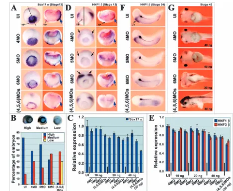
[image:8.612.61.530.72.456.2]In order to determine the roles played by GATA4, 5 and 6 in endoderm induction in vivo, we injected MOs into the vegetal hemispheres of one- or two-cell embryos. The MOs were injected separately or in combination, and their effects on endoderm formation were monitored by in situ hybridisation and real time RT-PCR using endodermal gene probes, and also by observing the morphology of the gut (Fig. 5). Injected embryos, which developed more slowly than their uninjected siblings, were collected at stages 12 (judged by blastopore size) and 34 for in situ hybridisation analysis, and at stage 43 for assessment of gut morphology. Visual assessment of Sox17α expression by whole mount in situ hybridisation revealed a clear reduction in a majority of embryos in the yolk plug (YP) region by GATA6 MO and when all three GATA MOs were injected together, with less obvious reductions by GATA4 or 5 Fig. 5.GATA4, 5 and 6 are required for development of the endoderm in vivo. (A,B,D) In situ hybridisation of Sox17α(A,B) and HNF1β(D) in stage 12 embryos injected vegetally with GATA4, 5 or 6 MOs (10-40 ng). (A,D) First column shows typical whole mount images and second column shows internal staining in halved embryos, black line indicating the plane of section. Yolk plug (YP) and involuted endoderm (blue arrows) are indicated. Black arrows in (D) indicate mesodermal expression. The percentages of embryos expressing high (normal), medium or low levels of Sox17αare represented in a histogram (B). (C,E) Real time RT-PCR analysis of Sox17α(C) and HNF1βand HNF3β(E). For error bars see Fig. 1 legend. (F) In situ hybridisation of HNF1βin stage 34 embryos. Position of the liver anlagen is indicated. (G) General gut morphology in stage 43 embryos. Embryos injected with 40 ng GATA6 MO did not survive to stage 43 and therefore the 20 ng phenotype is shown. The single embryo surviving up to stage 43 of the 30 ng GATA4, 5 and 6 MO injection is shown.
De
MOs (Fig. 5A,B). Embryos were scored according to the numbers seen in each of three categories: normal (‘high’), reduced (‘medium’) and substantially reduced (‘low’) expression of Sox17α (Fig. 5B). For uninjected embryos (n=26), 81% had high expression, 18% had medium expression and less than 1% had low expression. In contrast, for embryos injected with all three MOs (n=20), the high-, medium- and low-expressing embryo proportions were 3, 57 and 40% respectively. For embryos injected with only one of the GATA4, 5 or 6 MOs (n=25, 22 or 22), the percentages of high-, medium- and low-expressing embryos fell between these two extremes, with the relative magnitude of the effects being in the order GATA6 MO>GATA4 MO>GATA5 MO as seen for inhibition of marker induction by activin. When stained embryos were cut in half to reveal expression in the involuted endoderm, a reduction of Sox17α expression was apparent for all three MOs individually as well as all three together (Fig. 5A, blue arrows).
As an alternative measure, we carried out real-time RT-PCR on RNA extracted from whole embryos at the same stages (Fig. 5C, n=5). At the highest level of MO injected (40 ng), Sox17α expression was reduced to 75% by GATA4 MO alone, to 85% by GATA5 MO alone and to 65% by GATA6 MO alone, reflecting the hierarchical order of activity of the three GATA factors in our earlier assays. When all three MOs were injected together, the level of Sox17αexpression was reduced to 55%. We therefore conclude that all three GATA factors contribute to Sox17αexpression in vivo, with GATA6 making the greatest contribution followed by GATA4 as seen in the in vitro animal cap assays.
Whole mount in situ hybridisation was less informative for endodermal HNF1β expression, because of the low level of expression around the yolk plug (YP) and the masking of expression in the involuted endoderm, even in cleared embryos, by mesodermal expression (Fig. 5D, black arrows). Therefore, to assess the effects of GATA MO injection on the expression of HNF1β in involuted endoderm, embryos were cut in half (Fig. 5D, blue arrows). All three MOs individually and together significantly reduced this expression (6/6 embryos in each case), although increases in staining in the blastocoel floor for all three MOs and in the disrupted Brachet’s cleft region for the GATA6 MO were also apparent for reasons at present unknown. The conclusions of the in situ hybridisation analyses for HNF1βwere broadly supported by real-time RT-PCR and similar results were obtained for HNF3β (Fig. 5E). We therefore conclude that, as seen for Sox17α expression, HNF1βand HNF3β expression in stage 12 embryos depends on GATA factor activity. However, the stronger effect of the GATA6 MO seen for Sox17αwas less evident for these other two endodermal genes, presumably reflecting their reduced dependence on GATA6 in the in vitro assays.
The effects of GATA factor depletion on later development of the gut and its outgrowths were studied at stage 34 by whole mount in situ hybridisation using HNF1β as a probe and at stage 43 by morphological analysis (Fig. 5F,G). At stage 34, HNF1βis expressed strongly in the liver and more weakly in the underlying foregut and hindgut (Fig. 5F) (Demartis et al., 1994). Expression is also strong in the forming pronephros and pronephric duct, which derive from mesoderm. GATA MO injection led to severe reduction of expression in the liver for GATA6 MO and for all three MOs together (7/7 embryos in
each case), and an only slightly less severe reduction for GATA5 MO (5/7 embryos), whereas GATA4 MO had little effect (2/8 embryos showed a small reduction in expression of HNF1β) (Fig. 5F). Specificity for the effect on the liver was demonstrated by the continued strong expression of HNF1βin the pronephros and pronephric duct in all the injected embryos. The effects on the more weakly expressing gut were harder to assess at stage 34, however by stage 43, the effects became apparent as loss of gut coiling and in extreme cases a reduction in the amount of tissue. Thus, for the GATA6 MO, 24/29 embryos injected with 20 ng had less gut tissue and no coiling, while all those injected with 40 ng died (Fig. 5G). Most of the embryos injected with all three MOs died, but the single surviving embryo had very little remaining gut tissue (Fig. 5G). Injection of the GATA5 MO resulted in a complete loss of gut coiling in 5/21 embryos, coiling defects in 8/21 (Fig. 5G) and apparently normal guts in 8/21 embryos. GATA4 MO had very little effect with only 1/25 embryos displaying abnormal gut coiling. We therefore conclude that with respect to effects on gut formation and coiling, and on the liver gene expression tested, GATA factor requirements are in the order: GATA6>GATA5>GATA4.
Discussion
In this study we explore the roles of the transcription factors, GATA4, 5 and 6, in the formation of endoderm and its derivatives. Although all three factors are involved, GATA6 appears to be a key direct activator in the process, with GATA4 and 5 playing more minor roles, in part acting through GATA6. Our results suggest that, during the signal-dependent maintenance phase of endoderm formation, TGFβ signalling acts through GATA4, 5 and 6 to maintain expression of genes essential for the development of the gut and its derivatives in vivo.
GATA4, 5 and 6 are expressed appropriately for a role in maintenance of endoderm
The data on the timing of GATA4, 5 and 6 expression during early Xenopus development have been somewhat conflicting (Gove et al., 1997; Jiang and Evans, 1996; Weber et al., 2000; Yasuo and Lemaire, 1999). In addition, their roles in the complex genetic regulatory network leading to endoderm formation are unclear (Clements et al., 2003; Loose and Patient, 2004; Xanthos et al., 2001; Yasuo and Lemaire, 1999). In order to gain more insight, we re-investigated the temporal and spatial distribution of these factors relative to Sox17αand in comparison to the data already in the literature. Sox17α expression was first detected at stage 8 in comparison to GATA4 and 6 at stage 9 and zygotic GATA5 at stage 10. This timing of Sox17α expression correlates with previously published accounts and its proposed initial activation by the maternal T-box protein, VegT (Clements et al., 2001; Engleka et al., 2001; Hudson et al., 1997; Xanthos et al., 2001; Yasuo and Lemaire, 1999). The later expression of GATA4, 5 and 6 implicates these factors in the maintenance of Sox17α expression rather than its initial induction. The presence of a low level of maternal GATA5 mRNA agrees with previous reports, although the timing of zygotic expression does not (Jiang and Evans, 1996). This could possibly reflect the greater accuracy of the real-time RT-PCR method.
The spatial distribution at stage 10 of GATA4 and 6, but not
De
GATA5, correlates well with the expression pattern of Sox17 (Engleka et al., 2001; Hudson et al., 1997). Thus, GATA4, GATA6 and Sox17 are co-expressed in the supra- and sub-blastoporal endoderm at this time, whereas GATA5 is restricted to the sub-blastoporal endoderm (Weber et al., 2000). However, by stage 11, none of the GATA factors are co-expressed with Sox17 in the supra-blastoporal endoderm. Thus, while GATA4 and 6 could be maintaining Sox17 expression in the pre-involuted endoderm early, they are not involved later. All three GATA factors, however, could be involved in maintaining Sox 17 expression throughout the sub-blastoporal endoderm. Clearly other factors are involved in regulating Sox17 in the pre-involuted endoderm later. Candidate genes include the homeobox-containing transcription factor, Mixer, and the paired-like homeobox gene, Pitx2 (Engleka et al., 2001; Faucourt et al., 2001; Henry and Melton, 1998). Consistent with such a role for Pitx2, available data suggest that it induces endoderm in a GATA-independent manner. Overall we conclude that the expression profiles of GATA4-6 implicate them in the maintenance of the endodermal programme in a subset of endodermal cells.
GATA factor redundancy?
The similar temporal and spatial expression profiles of GATA4 and 6 raised the possibility that they carry out redundant functions in elaborating the endodermal gene expression programme. Consistent with this notion, both transcription factors induce expression of the same early endodermal genes in cap explants, albeit with different efficiencies. However, we show here that much of GATA4’s activity in this regard depends on the expression of GATA6. Indeed we found no evidence for GATA4 acting directly on Sox17α, in contrast to GATA6. This is a striking observation in view of the majority of the literature pointing to GATA factors binding to the same DNA sequence. Presumably the context on the Sox17α promoter is critical. This is one of the clearest demonstrations yet that apparent redundancy amongst GATA factors may reflect distinct roles in the pathways concerned. An important parenthetical point about GATA6 here is that, despite both long and short isoforms being synthesised in vivo (Brewer et al., 1999; Brewer et al., 2002; Peterkin et al., 2003), only the long isoform was able to induce endoderm markers (this study) (Weber et al., 2000).
Models have been proposed in the literature describing gene networks involved in the formation of endoderm in Xenopus embryos and mouse embryonic stem cells (Clements and Woodland, 2003; Fujikura et al., 2002; Loose and Patient, 2004; Xanthos et al., 2001; Yasuo and Lemaire, 1999). Our data agree with these models, but place GATA6 between GATA4 and to some extent GATA5, and at least two of their endoderm targets. By using inducible versions of GATA4, 5 and 6 in the presence of protein synthesis inhibitors, we were able to distinguish the direct or indirect actions of these transcription factors on Sox17α, HNF1β and HNF3β. Cycloheximide has been used successfully in the past for this purpose (Clements et al., 2003; Tada et al., 1997; Watanabe and Whitman, 1999). However, for Sox17α, superinduction was observed. This could have been a consequence of blocking the synthesis of the hypothetical endoderm inhibitor in the cap explants. If that had been the case, the superinduction should not have been observed with drug concentrations too low to
block protein synthesis. However, Sox17αwas still induced at such drug concentrations (data not shown), suggesting that cycloheximide acts through a different pathway in inducing Sox17. Thus our data add Sox17α to the previously reported GATA4and Gscas genes induced by cycloheximide (Tadano et al., 1993; Yasuo and Lemaire, 1999). The induction of GATA4 by cycloheximide does not affect the interpretation of our data as these endogenous induced GATA4 transcripts would not be translated. An explanation as to why the superinduction was greater when GATA4-GR was present, is the activation of the p38 MAP kinase pathway by cycloheximide, which could lead to phosphorylation of GATA4 (but not GATA5 or 6) thereby overriding the interaction with HSP90 (Charron et al., 2001; Kitta et al., 2003; Liang et al., 2001). This cannot explain all of the observed superinduction though, because superinduction was seen, albeit at a lower level, with GATA5 and 6 which do not contain the p38 MAPK target sequence. Because the greater superinduction in the presence of GATA4 could have exaggerated the apparent block to induction by cycloheximide, we repeated the experiment with a different protein synthesis inhibitor, emetine, which does not superinduce, and confirmed the indirect nature by which GATA4 induces Sox17α. Thus our data indicate a direct and central role for GATA6 in the genetic network orchestrating endodermal programming.
GATA4, 5 and 6 and the maintenance of endoderm
marker expression by TGFβ
A two-step model for the formation of endoderm has been proposed in which in the initial phase, in early blastulae, the maternal T-box protein VegT initiates endoderm formation by directly inducing Sox17 and nodal-related gene expression, followed by the maintenance of Sox17 in the second phase by the previously induced nodal-related proteins (Clements et al., 1999; Clements and Woodland, 2003; Yasuo and Lemaire, 1999). The second phase correlates well with the time when GATA4 and 6 expression begins, raising the obvious question as to how these two different families of proteins are connected. TGFβs, including the nodal-related proteins, induce GATA factors along with other endoderm-associated genes (this study) (Ariizumi et al., 2003; Chang and Hemmati-Brivanlou, 2000; Hyde and Old, 2000; Kofron et al., 1999; Weber et al., 2000). A dose-response relationship is seen with all three of the GATA factors and here we reveal differences between them, with sensitivity to induction by activin in the order GATA6>GATA4>GATA5. Differences such as these are likely to contribute to the region-specific expression observed in the embryo (see Fig. 1) (Weber et al., 2000). Furthermore, differences such as these are likely to mean that different GATA factors mediate TGFβ responses at different concentrations of TGFβ. In support of this suggestion, we find that the effects of low concentrations of activin on animal caps are inhibited by lost GATA function in the order GATA6>GATA4>GATA5.
At still lower concentrations of activin, Sox17 is induced without GATA factor induction (Ariizumi et al., 2003; Hudson et al., 1997; Weber et al., 2000). The explanation must be that, at lower concentrations, TGFβs induce endoderm via GATA independent pathways. These are likely to involve Smad proteins (Germain et al., 2000; Massague, 1998; Massague and Chen, 2000), possibly in partnership with Pitx2 or Mixer
De
(Faucourt et al., 2001; Henry and Melton, 1998). Such alternative pathways presumably also explain the incomplete inhibition by GATA6 morpholinos of Sox17α expression at low activin concentrations in our experiments.
Overall the data obtained here identify GATA4, 5 and 6 as mediators of TGFβ (most likely nodal) signalling in elaboration of the endoderm programme during the late blastula and gastrula stages in Xenopus laevis. At low concentrations of nodal signalling, in early blastulae, Sox17α is induced in a GATA-independent manner. However, as nodal builds up in the sub-blastoporal and early pre-involuting endoderm, our data suggest that GATA4 and 6, initially alone and then together with GATA5, directly maintain expression of endodermal gene expression. The consequences of GATA factor depletion for formation and coiling of the gut, and for gene expression in the liver, are greatest for GATA6, while being milder for GATA5, with GATA4 having very little effect. Together with the faust(GATA5) mutation in zebrafish (Reiter et al., 1999; Reiter et al., 2001), these data strongly implicate GATA5 and 6 in the development of definitive endoderm. With mouse null mutant data implicating GATA4 and 6 in primitive, extra-embryonic endoderm formation (Molkentin, 2000; Rossant et al., 2003), this branch of the GATA family clearly plays a central role in the development of all endoderm.
We would like to thank Paul Roach, Matt Loose and Tessa Peterkin for assistance with real-time RT-PCR, network diagrams and injections, and Hugh Woodland for the activin construct. We also thank Matt Loose, Andrew Johnson, Maggie Walmsley and the reviewers for their very helpful comments on the manuscript. This work was funded by The Wellcome Trust.
References
Albano, R. M., Groome, N. and Smith, J. C. (1993). Activins are expressed in preimplantation mouse embryos and in ES and EC cells and are regulated on their differentiation. Development117, 711-723.
Ariizumi, T., Kinoshita, M., Yokota, C., Takano, K., Fukuda, K., Moriyama, N., Malacinski, G. M. and Asashima, M. (2003). Amphibian in vitro heart induction: a simple and reliable model for the study of vertebrate cardiac development. Int. J. Dev. Biol. 47, 405-410.
Brewer, A., Gove, C., Davies, A., McNulty, C., Barrow, D., Koutsourakis, M., Farzaneh, F., Pizzey, J., Bomford, A. and Patient, R. (1999). The human and mouse GATA-6 genes utilize two promoters and two initiation codons. J. Biol. Chem. 274, 38004-38016.
Brewer, A., Nemer, G., Gove, C., Rawlins, F., Nemer, M., Patient, R. and Pizzey, J. (2002). Widespread expression of an extended peptide sequence of GATA-6 during murine embryogenesis and non-equivalence of RNA and protein expression domains. Gene Expr. Patterns2, 123-131.
Chang, C. B. and Hemmati-Brivanlou, A. (2000). A post-mid-blastula transition requirement for TGFbeta signaling in early endodermal specification. Mech. Dev. 90, 227-235.
Charron, F., Tsimiklis, G., Arcand, M., Robitaille, L., Liang, Q., Molkentin, J. D., Meloche, S. and Nemer, M. (2001). Tissue-specific GATA factors are transcriptional effectors of the small GTPase RhoA. Genes Dev. 15, 2702-2719.
Ciau-Uitz, A., Walmsley, M. and Patient, R. (2000). Distinct origins of adult and embryonic blood in Xenopus. Cell102, 787-796.
Clements, D. and Woodland, H. R. (2003). VegT induces endoderm by a self-limiting mechanism and by changing the competence of cells to respond to TGF-beta signals. Dev. Biol. 258, 454-463.
Clements, D., Friday, R. V. and Woodland, H. R. (1999). Mode of action of VegT in mesoderm and endoderm formation. Development126, 4903-4911.
Clements, D., Rex, M. and Woodland, H. R. (2001). Initiation and early patterning of the endoderm. Int. Rev. Cytol. 203, 383-446.
Clements, D., Cameleyre, I. and Woodland, H. R. (2003). Redundant early and overlapping larval roles of Xsox17 subgroup genes in Xenopus
endoderm development. Mech. Dev. 120, 337-348.
Demartis, A., Maffei, M., Vignali, R., Barsacchi, G. and Disimone, V.
(1994). Cloning and developmental expression of LFB3/HNF1 beta transcription factor in Xenopus laevis. Mech. Dev. 47, 19-28.
Ecochard, V., Cayrol, C., Rey, S., Foulquier, F., Cailliol, D., Lemaire, P. and Duprat, A. M. (1998). A novel XenopusMix-like gene milk involved in the control of endomesodermal fates. Development125, 2577-2585.
Edwards, D. R. and Mahadevan, L. C. (1992). Protein synthesis inhibitors differentially superinduce c-fos and c-jun by three distinct mechanisms: lack of evidence for labile repressors. EMBO J. 11, 2415-2424.
Engleka, M. J., Craig, E. J. and Kessler, D. S. (2001). VegT activation of Sox17 at the midblastula transition alters the response to nodal signals in the vegetal endoderm domain. Dev. Biol. 237, 159-172.
Faucourt, M., Houliston, E., Besnardeau, L., Kimelman, D. and Lepage, T. (2001). The Pitx2 homeobox protein is required early for endoderm formation and nodal signalling. Dev. Biol. 229, 287-306.
Fujikura, J., Yamato, E., Yonemura, S., Hosoda, K., Masui, S., Nakao, K., Miyazaki, J.-I. and Niwa, H. (2002). Differentiation of embryonic stem cells is induced by GATA factors. Genes Dev. 16, 784-789.
Germain, S., Howell, M., Esslemont, G. M. and Hill, C. S. (2000). Homeodomain and winged-helix transcription factors recruit activated Smads to distinct promoter elements via a common Smad interaction motif.
Genes Dev. 14, 435-451.
Gove, C., Walmsley, M., Nijjar, S., Bertwistle, D., Guille, M., Partington, G., Bomford, A. and Patient, R. (1997). Over-expression of GATA-6 in
Xenopusembryos blocks differentiation of heart precursors. EMBO J. 16, 355-368.
Heasman, J. (2002). Morpholino oligos: making sense of antisense? Dev. Biol.
243, 209-214.
Henry, G. L. and Melton, D. A. (1998). Mixer, a homeobox gene required for endoderm development. Science281, 91-96.
Hudson, C., Clements, D., Friday, R. V., Stott, D. and Woodland, H. R.
(1997). Xsox17alpha and -beta mediate endoderm formation in Xenopus.
Cell91, 397-405.
Hyde, C. E. and Old, R. W. (2000). Regulation of the early expression of the
Xenopusnodal-related 1 gene, Xnr1. Development127, 1221-1229.
Jiang, Y. and Evans, T. (1996). The Xenopus GATA-4/5/6 genes are associated with cardiac specification and can regulate cardiac-specific transcription during embryogenesis. Dev. Biol. 174, 258-270.
Kitta, K., Day, R. M., Kim, Y., Torregroza, I., Evans, T. and Suzuki, Y. J.
(2003). Hepatocyte growth factor induces GATA-4 phosphorylation and cell survival in cardiac muscle cells. J. Biol. Chem. 278, 4705-4712.
Kofron, M., Demel, T., Xanthos, J., Lohr, J., Sun, B., Sive, H., Wright, C., Wylie, C., Heasman, J. and Osada, S. (1999). Mesoderm induction in
Xenopusis a zygotic event regulated by maternal VegT via TGFbeta growth factors. Development126, 5759-5770.
Koutsourakis, M., Langeveld, A., Patient, R., Beddington, R. and Grosveld, F. (1999). The transcription factor GATA-6 is essential for early extraembryonic development. Development126, 723-732.
Kuo, C. T., Morrisey, E. E., Anandappa, R., Sigrist, K., Lu, M. M., Parmacek, M. S., Soudais, C. and Leiden, J. M. (1997). GATA4 transcription factor is required for ventral morphogenesis and heart tube formation. Genes Dev. 11, 1048-1060.
Liang, Q. R., Wiese, R. J., Bueno, O. F., Dai, Y. S., Markham, B. E. and Molkentin, J. D. (2001). The transcription factor GATA4 is activated by extracellular signal-regulated kinase 1-and 2-mediated phosphorylation of serine 105 in cardiomyocytes. Mol. Cell. Biol. 21, 7460-7469.
Loose, M. and Patient, R. (2004). A genetic regulatory network for Xenopus
mesendoderm formation. Dev. Biol. 271, 467-478.
Maduro, M. F. and Rothman, J. H. (2002). Making worm guts: the gene regulatory network of the Caenorhabditis elegansendoderm. Dev. Biol. 246, 68-85.
Massague, J. (1998). TGF-beta signal transduction. Annu. Rev. Biochem. 67, 753-791.
Massague, J. and Chen, Y. G. (2000). Controlling TGF-beta signaling. Genes Dev. 14, 627-644.
Molkentin, J. D. (2000). The zinc finger-containing transcription factors GATA-4,-5, and -6 – ubiquitously expressed regulators of tissue-specific gene expression. J. Biol. Chem. 275, 38949-38952.
Molkentin, J. D., Lin, Q., Duncan, S. A. and Olson, E. N. (1997). Requirement of the transcription factor GATA4 for heart tube formation and ventral morphogenesis. Genes Dev. 11, 1061-1072.
Molkentin, J. D., Tymitz, K. M., Richardson, J. A. and Olson, E. N. (2000). Abnormalities of the genitourinary tract in female mice lacking GATA5.
Mol. Cell. Biol. 20, 5256-5260.
De
Morrisey, E. E., Tang, Z., Sigrist, K., Lu, M. M., Jiang, F., Ip, H. S. and Parmacek, M. S. (1998). GATA6 regulates HNF4 and is required for differentiation of visceral endoderm in the mouse embryo. Genes Dev. 12, 3579-3590.
Narita, N., Bielinska, M. and Wilson, D. B. (1997). Cardiomyocyte differentiation by GATA-4 deficient embryonic stem cells. Development
124, 3755-3764.
Peterkin, T., Gibson, A. and Patient, R. (2003). GATA-6 maintains BMP-4 and Nkx2 expression during cardiomyocyte precursor maturation. EMBO J.
22, 4260-4273.
Reiter, J., Alexander, J., Rodaway, A., Yelon, D., Patient, R., Holder, N. and Stainier, D. (1999). Gata5 is required for the development of the heart and endoderm in zebrafish. Genes Dev. 13, 2983-2995.
Reiter, J. F., Kikuchi, Y. and Stainier, D. Y. R. (2001). Multiple roles for Gata5 in zebrafish endoderm formation. Development128, 125-135.
Rossant, J., Chazaud, C. and Yamanaka, Y. (2003). Lineage allocation and asymmetries in the early mouse embryo. Philos. Trans. R. Soc. Lond. Ser. B 358, 1341-1349.
Shivdasani, R. A. (2002). Molecular regulation of vertebrate early endoderm development. Dev. Biol. 249, 191-203.
Shoichet, S. A., Malik, T. H., Rothman, J. H. and Shivdasani, R. A. (2000). Action of the Caenorhabditis elegansGATA factor END-1 in Xenopus
suggests that similar mechanisms initiate endoderm development in ecdysozoa and vertebrates. Proc. Natl. Acad. Sci. USA97, 4076-4081.
Soudais, C., Bielinska, M., Heikinheimo, M., MacArthur, C. A., Narita, N., Saffitz, J. E., Simon, M. C., Leiden, J. M. and Wilson, D. B. (1995). Targeted mutagenesis of the transcription factor GATA-4 gene in mouse embryonic stem cells disrupts viceral endoderm differentiation in vitro.
Development121, 3877-3888.
Stainier, D. Y. R. (2002). A glimpse into the molecular entrails of endoderm formation. Genes Dev. 16, 893-907.
Suri, C., Haremaki, T. and Weinstein, D. C. (2004). Inhibition of mesodermal fate by XenopusHNF3beta/FoxA2. Dev. Biol. 265, 90-104.
Sykes, T. G., Rodaway, A. R. F., Walmsley, M. E. and Patient, R. K. (1998). Suppression of GATA factor activity causes axis duplication in Xenopus.
Development125, 4595-4605.
Tada, M., Oreilly, M. A. J. and Smith, J. C. (1997). Analysis of competence and of Brachyury autoinduction by use of hormone-inducible Xbra.
Development124, 2225-2234.
Tadano, T., Otani, H., Taira, M. and Dawid, I. B. (1993). Differential induction of regulatory genes during mesoderm formation in Xenopus laevis
embryos. Dev. Genet. 14, 204-211.
Watanabe, M. and Whitman, M. (1999). FAST-1 is a key maternal effector of mesoderm inducers in the early Xenopus embryo. Development 126, 5621-5634.
Weber, H., Symes, C., Walmsley, M. E., Rodaway, A. R. F. and Patient, R. K. (2000). A role for GATA5 in Xenopus endoderm specification.
Development127, 4345-4360.
Xanthos, J. B., Kofron, M., Wylie, C. and Heasman, J. (2001). Maternal VegT is the initiator of a molecular network specifying endoderm in
Xenopus laevis. Development128, 167-180.
Yasuo, H. and Lemaire, P. (1999). A two-step model for the fate determination of presumptive endodermal blastomeres in Xenopusembryos.
Curr. Biol. 9, 869-879.
Zhang, J. and King, M. L. (1996). XenopusVegT RNA is localised to the vegetal cortex during oogenesis and encodes a novel T-box transcription factor involved in mesodermal patterning. Development122, 4119-4129.