INTRODUCTION
In order to perform particular functions, cells become specialised and develop specific shapes and internal organisations. Despite the great diversity of cell shapes and cellular contexts, recent work reveals common themes during cell polarization, whether in unicellular organisms such as yeast or in highly complex tissues of vertebrates (Nelson, 2003). First, cells respond to an intrinsic or extrinsic cue to define one or several axes of polarity and then divide their membrane into functional sub-domains along those axes. The polarization of the membrane is then relayed to their internal organization by the activity of the microtubule and actin cytoskeleton (Shulman and St Johnston, 1999).
This unity extends at the molecular level as surprisingly few protein complexes are responsible for the polarization of most cell types studied. These include mainly the Par genes [par-1, par-3 (bazooka,baz), par-4(lkb1), par-5(14-3-3), par-6and aPKC], the Crumbs-Stardust-dPatj complex (Crb-Sdt-dPatj) and the Scribble-Lethal giant larvae-Discs Large complex (Scrib-Lgl-Dlg) (for a review see Macara, 2004). More recently, a novel group of proteins, called the Yurt/Coracle group, was shown to be required for the maintenance of epithelial polarity but not for the initial step of polarization (Laprise et al., 2009). The function and relationships of each of these genes, however, have only been deciphered in a small
number of cell types and significant differences have already emerged (Macara, 2004; Suzuki and Ohno, 2006). Furthermore, the function of each member of these complexes has not yet been tested comprehensively in most model systems.
In Drosophila, the anterior-posterior and dorso-ventral axes of polarity are set up during oogenesis by the asymmetric localization of bicoid, oskarand gurkenmRNAs within a single cell called the oocyte (Riechmann and Ephrussi, 2001). The localization of these transcripts depends on the polarized organization of the cytoskeleton, and thus on the polarity of the oocyte itself. During mid-oogenesis, the microtubule cytoskeleton is mainly organized by the activity of the Par-1 kinase at the posterior cortex of the oocyte and by the activity of the Par-3–Par-6–aPKC complex at the anterior cortex. The current model suggests that these complementary localizations are established by mutual inhibitions, with Par-1 phosphorylating Par-3 to prevent oligomerization, and with aPKC phosphorylating Par-1 to exclude it from the cortex (Benton and St Johnston, 2003b). In support of this model, expression of non-phosphorylatable forms of Par-1 or Par-3 is sufficient to mislocalize them to the entire cortex and consequently, to disrupt the polarization of the oocyte during mid-oogenesis (Benton and St Johnston, 2003b; Doerflinger et al., 2006). In addition, it has been recently proposed that aPKC also phosphorylates and inactivates Lgl at the anterior cortex, which would indirectly promote Par-1 localization at the posterior of the oocyte (Tian and Deng, 2008). Accordingly, expression of a non-phosphorylatable form of Lgl recruits Par-1 throughout the oocyte cortex. The earliest known asymmetry in this phosphorylation cascade is the localization of the Par-1 N1S isoform at the posterior of the oocyte (Doerflinger et al., 2006). This initial bias depends, however, on a previous signalling relay between the oocyte and the follicle cells, as Par-1 N1S fails to localize in gurkenmutant oocytes (Doerflinger et al., 2006). Indeed, at stage 6 of oogenesis, gurkenmRNA is translated at the posterior Development 137, 815-824 (2010) doi:10.1242/dev.045013
© 2010. Published by The Company of Biologists Ltd
1Institut Jacques Monod, CNRS-Universite Paris Diderot, Bât. Buffon –15 rue Hélène Brion, 75205 Paris cedex 13, France. 2Institute Curie, Department of Genetics and Developmental Biology (CNRS-UMR3215, Inserm-U934), 26 rue d’Ulm, 75248 Paris, Cedex 05, France.
*Present address: Medical Research Council LMCB, Cell Biology Unit, University College of London, Gower Street, London, WC1E 6BT, UK
†These authors contributed equally to this work ‡Author for correspondence (jean-rene.huynh@curie.fr)
Accepted 18 December 2009 SUMMARY
Most cell types in an organism show some degree of polarization, which relies on a surprisingly limited number of proteins. The underlying molecular mechanisms depend, however, on the cellular context. Mutual inhibitions between members of the Par genes are proposed to be sufficient to polarize the C. elegansone-cell zygote and the Drosophilaoocyte during mid-oogenesis. By contrast, the Par genes interact with cellular junctions and associated complexes to polarize epithelial cells. The Par genes are also required at an early step of Drosophilaoogenesis for the maintenance of the oocyte fate and its early polarization. Here we show that the Par genes are not sufficient to polarize the oocyte early and that the activity of the tumor-suppressor gene lethal giant larvae(lgl) is required for the posterior translocation of oocyte-specific proteins, including germline determinants. We also found that Lgl localizes asymmetrically within the oocyte and is excluded from the posterior pole. We further demonstrate that phosphorylation of Par-1, Par-3 (Bazooka) and Lgl is crucial to regulate their activity and localization in vivo and describe, for the first time, adherens junctions located around the ring canals, which link the oocyte to the other cells of the germline cyst. However, null mutations in the DE-cadheringene, which encodes the main component of the zonula adherens, do not affect the early polarization of the oocyte. We conclude that, despite sharing many similarities with other model systems at the genetic and cellular levels, the polarization of the early oocyte relies on a specific subset of polarity proteins.
KEY WORDS: Axis determination, Cell polarity, Drosophila, Oogenesis
lethal giant larvae
is required with the
par
genes for the
early polarization of the
Drosophila
oocyte
Pierre Fichelson1,*,†, Marlène Jagut1,2,†, Sophie Lepanse1, Jean-Antoine Lepesant1and Jean-René Huynh1,2,‡
D
E
V
E
LO
P
M
E
N
of the oocyte, sending a signal to the adjacent follicle cells, inducing them to become posterior (Gonzalez-Reyes et al., 1995; Roth et al., 1995). As a consequence, these cells send an unknown signal back to the oocyte to recruit Par-1 N1S to the posterior cortex and trigger the reorganization of the microtubule cytoskeleton. The posterior localization of gurkenmRNA is itself established even earlier, at stage 1 of oogenesis (also called region 3 of the germarium), tracing the initial asymmetry to the very first steps of the egg chamber formation (Huynh and St Johnston, 2004). Loss-of-function experiments have shown that again the full set of Par genes and the microtubule cytoskeleton were required at these early stages for the localization of mRNAs, proteins and centrosomes from the anterior to the posterior cortex of the oocyte (Benton et al., 2002; Cox et al.,
2001a; Cox et al., 2001b; Huynh et al., 2001a; Huynh et al., 2001b; Martin and St Johnston, 2003). However, it remains unknown whether the phosphorylation cascade described above is involved or whether Lgl and other members of polarity complexes are required in the germline to polarize the oocyte at these early stages.
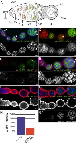
[image:2.612.52.322.227.730.2]Drosophilaoogenesis begins in region 1 of the germarium when a germline stem cell divides asymmetrically to produce a posterior cystoblast and a new stem cell at the anterior (reviewed in Huynh and St Johnston, 2004) (Fig. 1A). The cystoblast then undergoes precisely four rounds of mitosis with incomplete cytokinesis to form a cyst of 16 germline cells, which are all interconnected by stable cytoplasmic bridges called ring canals. Only one cell becomes the oocyte, while the other 15 cells develop into nurse
Fig. 1. Over-expression of a non-phosphorylatable form of Par-1 disrupts the early polarization of the oocyte.(A)The early steps of oogenesis. An egg chamber comprises 16 germline cells surrounded by follicle cells (FC). The germarium is divided into four regions along the anterior-posterior axis (1, 2a, 2b and 3). Germline stem cells (GSCs) reside at the tip of the germarium (left) in a microenvironment created by the cap cells (Cap) and terminal filament cells (TF). GSCs produce cystoblasts, which divide four times and generate germline cysts of 16 cells connected by ring canals. The GSCs and cystoblasts contain a spectrosome (red circles), which develops into a branched fusome that orients cystoblast divisions. In region 2a, cytoplasmic proteins (green), mRNAs, mitochondria and the centrosomes (blue circles) progressively accumulate at the anterior of the oocyte. In region 2b, the minus-ends of the microtubules (MT) are focused in the oocyte and the plus-ends extend through the ring canals into the nurse cells. The follicle cells (grey) start to migrate and surround the germline cells. As the cyst moves to region 3, the oocyte adheres to the posterior follicle cells and repolarises along its anterior-posterior axis, with the MT minus-ends and specific cytoplasmic components now localized at the posterior cortex (green crescent). (B)Ovariole overexpressing Par-1-GFP (green) under the control of mat-Tub-Gal4 and nos-Gal4 and stained for Orb (red) and DAPI (blue). Par-1-GFP (arrow) accumulates on the fusome during early oogenesis. The localization of Orb to the posterior of the oocyte is delayed (arrowheads). (B⬘)The DAPI channel is shown on its own. The arrow indicates the karyosome. (C)Ovariole overexpressing Par-1-AEM-GFP (green) under the control of mat-Tub-Gal4 and stained for Orb (red). The nucleus of the oocyte is shown with an asterisk. (C⬘)The Orb channel is shown on its own. Orb remains at the anterior of the oocyte (arrows). (D)Ovariole overexpressing Par-1-AEM-GFP (green) under the control of mat-Tub-Gal4 and stained for -Tubulin (red). Par-1-AEM-GFP is localized at the ring canals and the centrosomes remain at the anterior of the oocyte. The asterisks indicate the nucleus of the oocyte. (D⬘)The -Tubulin channel is shown on its own. Centrosomes remain at the anterior of the oocyte (arrows). (E)Ovariole overexpressing Par-1-AEM-GFP (green) under the control of mat-Tub-Gal4 and stained for the meiotic marker C(3)G (red) and DAPI (blue). The oocyte entered meiosis (arrowhead) but soon lost the expression of C(3)G and became polyploid (arrow). (E⬘)The DAPI channel is shown on its own. (F)Wild-type (mat-Tub -Gal4) ovariole stained for -Tubulin (red) and DAPI (blue). (F⬘)The
-Tubulin channel is shown on its own. (G)Ovariole
overexpressing Par-1-AEM-GFP (green) under the control of mat-Tub-Gal4 and stained for -Tubulin (red) and DAPI (blue). (G⬘)The
-Tubulin channel is shown on its own. (G⬙)The Par-1-AEM-GFP channel is shown on its own. (H)Quantification of the pixel intensity of the -Tubulin channel in wild-type (mat-Tub-Gal4) egg chambers and in egg chambers overexpressing Par-1-AEM-GFP
under the control of mat-Tub-Gal4.
D
E
V
E
LO
P
M
E
N
cells. During these divisions, a cytoplasmic structure called a fusome anchors one pole of each mitotic spindle and is thought to be instrumental in the selection and polarization of the oocyte (de Cuevas and Spradling, 1998; Huynh, 2006). Centrosomes, oocyte-specific proteins such as Orb and the assembly of the synaptonemal complex (SC) serve as markers of oocyte differentiation as they are progressively restricted to only one cell by the end of region 2a. In region 2b of the germarium, these components remain associated with the fusome remnants and thus accumulate at the anterior of the oocyte to form a Balbiani body close to the four ring canals (Cox and Spradling, 2003). When the oocyte reaches region 3, all of the components of the Balbiani body disassociate and move around the oocyte nucleus to form a crescent at the posterior cortex. This movement is the first sign of anterior-posterior polarity in the oocyte, and is a crucial step in the maintenance of its identity. Mutations in any of the five Drosophila Par genes (par-1, par-3, par-4, par-5, par-6) and aPKCdisrupt this polarization step, as oocyte-specific components accumulate in the oocyte but remain at the anterior and never translocate to the posterior cortex (Benton et al., 2002; Cox et al., 2001a; Cox et al., 2001b; Huynh et al., 2001a; Huynh et al., 2001b; Martin and St Johnston, 2003). These defects lead to an early arrest of oogenesis sometimes followed by the formation of egg chambers with 16 nurse cells and no oocyte, although the initial selection of the oocyte is not affected.
In this study, we have performed loss-of-function experiments for every gene of the main polarity complexes and expressed non-phosphorylatable forms of key members of those complexes during early oogenesis. Overall, our results demonstrate that, despite sharing many similarities with other model systems at the genetic and cellular levels, the polarization of the early oocyte shows significant differences.
MATERIALS AND METHODS Fly stocks
The strongest available alleles for each gene were used during this study: armXK22(Peifer and Wieschaus, 1990), armXP33(Peifer and Wieschaus,
1990),baz815-8(McKim et al., 1996),crb11A22(Bilder et al., 2003), dlgm52,
dlg18, dlg2(Woods and Bryant, 1991),lgl4(Mechler et al., 1985),l(2)gl4W3
(Bilder et al., 2000), Patjdre1 (Bhat et al., 1999),sdtXP96 (Muller and
Wieschaus, 1996),scrib1,scrib5,scrib6(Zeitler et al., 2004),shgR69(Godt
and Tepass, 1998),shgP34-1(Tepass et al., 1996). The germline clones were
generated using the FLP/FRT technique (Chou and Perrimon, 1992), using a nlsGFP recombined onto FRT[9-2], FRT40A, FRTG13, FRT79D or FRT82B (Bloomington Stock Center). Clones were induced by heat-shocking third instar larvae at 37°C for 2 hours on 3 consecutive days except for lgland scribmutant clones, which were heat-shocked only once for 1 hour at 37°C at late third instar. Overexpression experiments were performed using the Gal4/UASp system (Brand and Perrimon, 1993) with the nanos -Gal4 (nos-Gal4) (Van Doren et al., 1998) driver and the maternal-tubulin -Gal4 (mat-Tub-Gal4) driver described by Januschke (Januschke et al., 2002). We used the following transgenic lines for expression Par-3 and Par-1: Baz-S151A,S1085A-GFP (Benton and St Johnston, 2003b), UASp-Baz-S151A-GFP (Benton and St Johnston, 2003b), UASp-Baz-S1085A-GFP (Benton and St Johnston, 2003b), UASp-Par-1-N1S-UASp-Baz-S1085A-GFP (Huynh et al., 2001b) and UASp-Par-1-AEM-GFP (Doerflinger et al., 2006).
Staining procedures
Antibody staining and Hoechst staining were performed according to standard protocols. The antibodies used were mouse anti-Orb at 1:250 (6H8 and 4H8 from DSHB Iowa University) (Lantz et al., 1994), mouse anti- -Tubulin (Sigma) at 1:100, rabbit anti-Par-1 at 1:5000 (Shulman et al., 2000), rabbit anti-Par-3/Bazooka at 1:500 (gift from Andreas Wodarz, Gottingen, Germany), rat anti-DE-Cadherin (D-CAD2) at 1:20, rabbit anti-C(3)G at 1:1000 (Hong et al., 2003).
RESULTS
Expression of a non-phosphorylatable form of Par-1 is sufficient to affect both oocyte polarity and fate
To test whether the phosphorylation of Par-1 is important for the regulation of its function during early oogenesis, we expressed a form of Par-1 mutated in the apical-lateral exclusion motif (AEM) domain, in which a conserved threonine is replaced by an alanine (Doerflinger et al., 2006). This mutated form of Par-1 was shown to ectopically localize to the apical side of the follicle cells and all around the oocyte cortex at stage 7-9, disrupting the anterior-posterior axis at this stage (Doerflinger et al., 2006). We used the Gal4/UAS system to express Par-1-AEM-GFP and Par-1-GFP with two different germline-specific drivers: nanos-Gal4, which is strongly expressed in regions 1 and 2, and fading in region 3; and mat-Tub-Gal4, which comes on strongly in region 3 onward. We found that both tagged proteins were expressed at similar levels and showed similar localization on the fusome (arrow, Fig. 1B) and at the cell cortex and ring canals (Fig. 1B,D). However, using the mat-Tub-Gal4 driver, we found that egg chambers expressing Par-1-AEM did not develop past stage 4. Orb and the centrosomes localized into the oocyte but they remained at the anterior of the nucleus (100%, n95) (Fig. 1C-D⬘). The SC was correctly restricted to one cell but disappeared quickly (n50) (arrowhead, Fig. 1E). DNA staining revealed that the oocyte became polyploid, although not to the same extent as in the nurse cell nuclei (arrow, Fig. 1E⬘). By contrast, overexpression of the Par-1-AEM form with the nanos-Gal4 driver disrupted the early polarization of the oocyte in only 8% (n52) of the egg chambers, suggesting that region 3 is most sensitive to the activity of Par-1 (data not shown). Strong overexpression of a wild-type form of Par-1 (N1S) using both drivers at the same time was also able to disrupt the early anterior-posterior polarity, although much less dramatically (Fig. 1B; data not shown). Only half of the egg chambers (53%, n55) showed a delay in the posterior localization of Orb (arrowhead, Fig. 1B) and defects in the formation of the karyosome. Moreover, in contrast to the overexpression of Par-1-AEM, these defects were transient, and most egg chambers (80%, n51) proceeded through oogenesis normally after stage 6 (Fig. 1B; data not shown).
The phenotypes induced by Par-1-AEM expression are similar to those found in flies fed with colcemid, a microtubule-depolymerizing drug, or in flies mutant for hypomorphic alleles of dhc64C, which encodes the heavy chain of the minus-end-directed molecular motor Dynein (McGrail et al., 1995; Swan et al., 1999; Theurkauf, 1994). A recent study further showed that excessive and ectopic activity of Par-1 is sufficient to depolymerize oocyte microtubules during mid-oogenesis (Tian and Deng, 2009). We thus compared the levels of microtubule staining in wild-type germarium and in germarium expressing the non-phosphorylatable form of Par-1 (Fig. 1F,G). We found that microtubules were greatly reduced in egg chambers expressing Par-1-AEM compared with those of the wild type (Fig. 1F⬘,G⬘). Quantification of these results showed a decrease of more than 50% in pixel intensity in mutant egg chambers stained with an anti--Tubulin antibody (n50; Fig. 1H). We conclude that the loss of microtubules is the likely cause of the phenotypes induced by the expression of a non-phosphorylatable form of Par-1.
Overall, our results demonstrate that the regulation of Par-1 activity by phosphorylation is crucial for the early steps of egg chamber development, as previously shown during
mid-oogenesis.
D
E
V
E
LO
P
M
E
N
A non-phosphorylatable form of Par-3 is not sufficient to disrupt the early polarization of the oocyte
The requirement for Par-3 during early oogenesis has precluded the analysis of its loss at later stages, i.e. during mid-oogenesis. Nevertheless, overexpression of a non-phosphorylatable form of Par-3 during mid-oogenesis is sufficient to disrupt oocyte polarity. Indeed, it was shown that Par-1 inhibits Par-3 oligomerization and interaction with aPKC by phosphorylating two serines at position 151 and 1085 (Benton and St Johnston, 2003b). A non-phosphorylatable form of Par-3 (Baz-S151A,S1085A) spreads ectopically to the lateral cortex in follicle cells and to the posterior cortex of the oocyte at stage 7-9, disrupting the anterior-posterior polarity of the oocyte at this stage (Benton and St Johnston, 2003b). To test whether inhibition of Par-3 is required during early oogenesis, we expressed Baz-S151A,S1085A-GFP in the germarium both with nanos-Gal4 and mat-Tub-Gal4 drivers (Fig.
2C,D). We found that Baz-S151A,S1085A-GFP localized in circles around the ring canals (arrow, Fig. 2C⬙), at the junctions between the germline stem cells and cap cells (arrowhead, Fig. 2C⬙) and at the germline cell cortex (Fig. 2D,D⬙), which is identical to the localization of wild-type Baz-GFP (Fig. 2B⬘) and endogenous Par-3 (Fig. 2A). However, Orb and the centrosomes localized normally at the posterior of the oocyte (n50) when Baz-S151A,S1085A-GFP was expressed under the control of nanos-Gal4 or mat-Tub-Gal4 (asterisks, Fig. 2C-D⬘; data not shown). In contrast to later stages of oogenesis, these non-phosphorylatable forms of Par-3 are thus insufficient to disrupt the early polarization of the oocyte.
[image:4.612.54.467.265.569.2]In the follicular epithelium, Baz-S151A,S1085A-GFP is able to rescue par-3-null homozygous clones and localizes apically in the absence of endogenous Par-3 (Benton and St Johnston, 2003b). We thus tested whether Baz-S151A,S1085A-GFP could rescue the early requirement for Par-3 in the germarium (Fig. 2E). We found that in germline clones homozygous mutant for a null allele of par-3 and
Fig. 2. Overexpression of a wild-type and non-phosphorylatable form of Par-3 does not affect the polarization of the oocyte.(A) Wild-type ovariole stained for Par-3 (Baz). Par-3 is enriched at the junction between GSCs and cap cells (arrowhead) and is localized at the ring canals (arrow). (B)Ovariole overexpressing a wild-type form of Baz-GFP (green) under the control of the nos-Gal4. Orb (red) localizes normally into the oocyte (asterisk) and is relocated at the posterior cortex. (B⬘)The Baz-GFP channel is shown on its own. Baz-GFP is enriched at the junction between GSCs and cap cells (arrowhead) and is localized at the ring canals (arrow). (C)Ovariole overexpressing Baz-S151A,S1085A-GFP (green) under the control of nos-Gal4. Orb (red) localizes normally into the oocyte (asterisk) and is relocated at the posterior cortex. (C⬘)The Orb channel is shown on its own. The oocyte is indicated with an asterisk. (C⬙)The Baz-S151A,S1085A-GFP channel is shown on its own. Baz-S151A,S1085A-GFP is enriched at the junction between GSCs and cap cells (arrowhead) and is localized at the ring canals (arrow). (D)Ovariole overexpressing Baz-S151A,S1085A-GFP (green) under the control of the mat-Tub-Gal4 driver. Orb (red) localizes normally into the oocyte (asterisk) and is relocated at the posterior cortex. DNA is in blue. The oocyte is shown with an asterisk. (D⬘)The Orb channel is shown on its own. The oocyte is indicated with an asterisk. (D⬙)The Baz-S151A,S1085A-GFP channel is shown on its own. (E)Ovariole overexpressing Baz-S151A,S1085A-GFP (green) under the control of the mat-Tub-Gal4 driver in wild-type or par-3mutant egg chambers and stained for Orb (red). Wild-type egg chambers express nlsGFP (green) and mutant egg chambers are identified by their lack of nlsGFP. DNA is in blue. The oocyte is outlined by a white dotted line. Orb fails to be relocated posteriorly in egg chambers expressing only Baz-S151A,S1085A-GFP (arrow). (E⬘)The GFP channel is shown on its own. Baz-S151A,S1085A-GFP localizes to the ring canals in the presence of endogenous Par-3 (arrowhead) but not in its absence (arrow). (E⬙)The Orb channel is shown on its
own. Orb remains at the anterior of the oocyte in egg chambers expressing only Baz-S151A,S1085A-GFP (arrow).
D
E
V
E
LO
P
M
E
N
expressing Baz-S151A,S1085A-GFP, Orb failed to translocate to the posterior cortex and mutant egg chambers were arrested at stage 2-3 (arrow, Fig. 2E-E⬙) – a phenotype identical to par-3 loss-of-function. This non-phosphorylatable form of Par-3 is thus unable to rescue these early stages of oogenesis. We further noticed that Baz-S151A,S1085A-GFP did not localize at the cortex in par-3germline clones (compare arrow and arrowheads in Fig. 2E⬘), indicating that these phosphorylation sites are required for Par-3 localization at the cortex in the germline. Baz-S151A,S1085A-GFP might thus still be active in the germline, but unable to reach the cortex in the first place when endogenous Par-3 is absent, in contrast to the follicular epithelium.
Lethal giant larvae is required for the early polarization of the oocyte
Par proteins interact with the Crumbs and Scribble complexes to establish the apical-basal polarity of many epithelial cells (Goldstein and Macara, 2007; Suzuki and Ohno, 2006). To test whether these complexes were required for the early polarization of the oocyte, we induced germline clones mutant for null alleles of each member of the two complexes. As previously reported, clones of mutant follicle cells for dlg, scribor lglinduce dramatic over-proliferations of the somatic cells, which prevents the analysis of the function of these genes in the germline (Bilder et al., 2000; Goode and Perrimon, 1997; Li et al., 2008; Manfruelli et al., 1996) (Fig. 3D). To minimize this problem, we used a mild heat-shock regime to induce a low number of mutant clones. We then dissected the ovaries more than 7 days after the heat-shocks and analyzed egg chambers in which only the germline was mutant. We found that in lgl4mutant germline
cysts, Orb, the centrosomes and the SC were restricted to one cell, indicating that the selection of the oocyte was not affected (n32) (Fig. 3A,B; data not shown). However, Orb and the centrosomes rarely translocated to the posterior pole (22% at the posterior, n32). We further noticed that the penetrance of these phenotypes increased with time, suggesting that the perdurance of the Lgl protein allowed young females to produce normal-looking eggs. Identical results
were obtained with lgl4W3, a second independent allele of lgl. These
defects are highly similar to phenotypes induced by mutations in any of the Par genes.
By contrast, we found that germline clones mutant for several alleles of scribble– scrib1,scrib5andscrib6(Lighthouse et al., 2008)
– and several alleles of dlg– dlgm52, dlg18and dlg2– developed past
these early stages and went on to produce normal-looking eggs (n23 and n32, respectively; Fig. 3C,E). These results demonstrate that Lgl, but not Dlg or Scrib, are required with Par proteins for the early polarization of the oocyte.
We also induced germline clones mutant for the strongest available alleles of crumbs, stardustand dPatj. We found that in all mutant cysts analyzed, Orb and the centrosomes localized to the posterior cortex of the oocyte, as in wild-type egg chambers (Fig. 3F,G; data not shown). We thus conclude that the Crb-Sdt-dPatj complex does not play an essential role in the early polarization of the oocyte.
Taken together, these results show that the early polarization of the oocyte shares a genetic requirement for Lgl, 1 and Par-3–Par-6–aPKC in common with the follicular and embryonic epithelia.
Phosphorylation regulates the asymmetric localization of Lgl within the oocyte
[image:5.612.53.470.467.632.2]The phosphorylation of Lgl by aPKC was recently shown to play an important function during the polarization of the oocyte at mid-oogenesis (Tian and Deng, 2008). Although the overexpression of a wild-type form of Lgl triggers some polarization defects at stage 7, the expression of a non-phosphorylatable form of Lgl [named Lgl-3A, in which three aPKC phosphorylation sites were mutated to alanine (Betschinger et al., 2003)] completely disrupts the establishment of the anterior-posterior axis. By contrast, the expression of wild-type Lgl-GFP, Lgl-3A-GFP or Lgl-3A (untagged) during early oogenesis did not induce any visible polarization defects. Using both nanos-Gal4 and mat-Tub-Gal4 drivers, we found that Orb and the centrosomes (Fig. 4A-D; data not
Fig. 3. Lethal giant larvae is required for the early polarization of the oocyte, but not Scribble, Discs Large, Stardust or Crumbs.
(A-G)Mutant clones are labelled by the lack of GFP (green). (A)lgl4germline clone stained for -Tubulin (red). The centrosomes remain localized at
the anterior of the oocyte (arrow), whereas they are already at the posterior cortex in a younger wild-type cyst (arrowhead). The oocyte is outlined by a white dotted line. (B)lgl4germline clone. The SC (red) is correctly restricted to one cell and is maintained for several stages. The mutant cyst
nuclei are not pyknotic (DNA in blue). (C)scrib1germline clone. Orb (red) is relocated to the posterior of the oocyte (arrow). The oocyte is outlined
by a white dotted line. The DNA is in blue. (D)scrib1germline and somatic clone (FCC, follicle cells clone). Orb (red) is relocated to the posterior of
the oocyte. The oocyte is indicated by an asterisk. The DNA is in blue. scrib1follicle cells overproliferate and become multi-layered (arrow). (E)Orb
(red) is correctly localized at the posterior of the oocyte in Dlgm52germline clones. The oocyte is indicated by an asterisk. The DNA is in blue. (F)Orb
(red) is correctly localized at the posterior of the oocyte in sdtXP96germline clones. The oocyte is indicated by an asterisk. (G)Orb (red) is correctly
localized at the posterior of the oocyte in crb11A22germline clones. The oocyte is indicated by an asterisk.
D
E
V
E
LO
P
M
E
N
shown) localized normally at the posterior of the oocyte (n40; arrowheads, Fig. 4C⬘,D⬘). These results indicate that phosphorylation of Lgl is not crucial for the establishment of the anterior-posterior axis in the germarium.
However, we noticed that Lgl-GFP was specifically absent from the posterior cortex of the oocyte from stage 1 to stage 6 of oogenesis, whereas it was uniformly distributed in the nurse cells (arrows, Fig. 4A⬘,C⬙). By contrast, the non-phosphorylatable form of Lgl was more cytoplasmic and present at the posterior cortex of the oocyte (arrows, Fig. 4B⬘,D⬙). We further found that egg chambers expressing Lgl-GFP driven by mat-Tub-Gal4, failed to develop past stage 6-7 (n30, 100%; Fig. 4E-E⬙). These egg chambers had small oocytes lacking Orb with a partially polyploid nucleus (asterisk and arrow, Fig. 4E⬘,E⬙). Expression of Lgl-3A (but not Lgl-3A-GFP) also induced an arrest at stage 5-6, indicating that this form is active, but it did not affect the oocyte polarity and identity (data not shown). These results show that although the phosphorylation of Lgl is required for its asymmetric localization within the oocyte, only the wild-type form of Lgl is able to disrupt the oocyte identity when overexpressed.
Semi-circular adherens junctions form around the ring canals
Our results so far show that the early polarization of the oocyte shares some similarities at the genetic level with the polarization of the follicular and embryonic epithelia, although the molecular regulation might differ. A major event during epithelial polarization is the formation of intercellular junctions, and in particular adherens
junctions. DE-Cadherin and Armadillo (Drosophila-catenin) are major components of adherens junctions (AJs) and were shown to localize as rings around ring canals in the germarium, suggesting the presence of AJs between germ cells (Gonzalez-Reyes and St Johnston, 1998) (arrows, Fig. 5A). Interestingly, we further showed that Par-3 also colocalizes on these rings. To investigate whether genuine AJs form around the ring canals, we analyzed ultra-thin sections of wild-type germaria by electron microscopy (EM). We found structures with the characteristic appearance of AJs, i.e. they showed electron-dense undercoats and a parallel orientation of cell membranes (Fig. 5B,C). Their widths ranged from 100 nm to 220 nm, which is similar to mature zonula adherens (ZA) in embryonic epithelia (Tepass and Hartenstein, 1994). These AJs ran along the ring canals on several serial sections but rarely formed a full circle, as ring canals were found associated with only one AJ on single sections (Fig. 5D). They appeared in early region 2a, after the four divisions, and became more common toward region 3. By contrast, we did not find any structure similar to the septate junctions found basally to the AJs in epithelial cells.
[image:6.612.51.320.62.405.2]We also did not find a clear pattern in the distribution of AJs within germline cysts, indicating that AJ localization is probably dynamic. The localization of the AJs was confirmed by confocal microscopy of a greater number of samples (n20) using a transgene expressing a GFP-tagged DE-Cadherin and immunostainings against endogenous DE-Cadherin (n20; Fig. 5A). To test a potential function of the AJs in the polarization of the oocyte, we induced germline clones mutant for strong and intermediate alleles of DE-cadherin(shgR69, shgP34-1) and armadillo
Fig. 4. Lgl is asymmetrically localized in the oocyte in a phosphorylation-dependent manner.(A)Ovariole
overexpressing Lgl-GFP (green) under the control of nos-Gal4. Orb (red) is correctly relocated at the posterior of the oocyte (arrow). The oocyte is indicated by an asterisk. The DNA is in blue. (A⬘)The Lgl-GFP channel is shown on its own. Lgl-GFP is excluded from the posterior cortex of the oocyte (arrow). (B)Ovariole
overexpressing Lgl3A-GFP (green) under the control of nos-Gal4. Orb (red) is correctly relocated at the posterior of the oocyte (arrow). The oocyte is indicated by an asterisk. The DNA is in blue. (B⬘)The Lgl-3AGFP channel is shown on its own. Lgl-3AGFP fails to be excluded from the posterior cortex of the oocyte (arrow) and is also present in the cytoplasm. (C)Ovariole overexpressing Lgl-GFP (green) under the control of mat-Tub-Gal4. Orb (red) is correctly relocated at the posterior of the oocyte (arrowheads). The oocyte is indicated by an asterisk. The DNA is in blue. (C⬘)The Orb channel is shown on its own. Orb is correctly relocated at the posterior of the oocyte (arrowheads). (C⬙)The Lgl-GFP channel is shown on its own. Lgl-GFP is excluded from the posterior cortex of the oocyte (arrows). (D)Ovariole overexpressing Lgl3A-GFP (green) under the control of mat-Tub-Gal4. Orb (red) is correctly relocated at the posterior of the oocyte. The oocyte is indicated by an asterisk. The DNA is in blue. (D⬘)The Orb channel is shown on its own. Orb is correctly relocated at the posterior of the oocyte (arrowhead). (D⬙)The Lgl-3AGFP channel is shown on its own. Lgl-3AGFP fails to be excluded from the posterior cortex of the oocyte (arrows) and is also present in the cytoplasm. (E)Egg chamber overexpressing Lgl-GFP (green) under the control of mat-Tub-Gal4 fails to develop past stage 6-7. Orb (red) is not detected in the oocyte (indicated by an asterisk). The DNA is in blue. (E⬘)The Lgl-GFP channel is shown on its own. The oocyte is indicated by an asterisk. (E⬙)The DNA channel is shown on its own. The oocyte becomes partially polyploid (arrow), which contrasts with wild-type karyosome at younger stages (arrowhead).
D
E
V
E
LO
P
M
E
N
(armXK22, armXP33). We found that in all cases (n102), Orb and the
centrosomes translocated normally to the posterior of the oocyte, indicating that the oocyte can polarize without DE-Cadherin and -catenin (data not shown). These results are consistent with reports showing that DE-Cadherin and Armadillo are not required to initiate the polarization of either follicle cells or embryonic epithelial cells (Harris and Peifer, 2004; Harris and Peifer, 2005; Tanentzapf et al., 2000). Alternatively, additional cadherins or adhesive systems in the Drosophilagermarium could compensate for the absence of DE-Cadherin.
Lethal giant larvae is required to restrict Par-3 localization to the adherens junctions
To investigate the relationship between Par-1, the Par-3–Par-6–aPKC complex, AJs and Lgl during the polarization of the oocyte, we analyzed their localization in wild-type and mutant germaria. We have previously shown that the localization of 1, 3 and Par-6 are all independent from one another (Huynh et al., 2001a). Here, we found that Par-1 localization on the fusome is also independent of DE-Cadherin and Lgl (Fig. 6A,A⬘; data not shown). By contrast, Par-3 and DE-Cadherin stainings at the AJs were greatly reduced in lglmutant germline clones (completely absent in 64%, n33 and 54%, n34, respectively) (Fig. 6B,E). Par-3 and DE-Cadherin localization appeared more diffuse at the cortex and in the cytoplasm. These data indicate that Lgl is required to restrict Par-3 and DE-Cadherin to the AJs. We further found that DE-Cadherin localized normally in par-3mutant clones, whereas Par-3 staining was reduced or absent in shgR69mutant cysts (84%, n33) (Fig.
6C,D). These results suggest that Par-3 localization on the AJs might not be essential for its polarizing activity, as we showed that the
oocyte polarizes normally in shgR69 clones. Alternatively, the
reduced amount of Par-3 on the AJs in shgR69mutant clones might
be sufficient to polarize the oocyte.
DISCUSSION
Redundant mechanisms polarize germline and
somatic cells in Drosophilaovaries
[image:7.612.54.298.60.308.2]One general strategy to establish polarity within a cell is to create non-overlapping membrane domains along one specific axis. In most cell types, four complexes are involved in the formation of these domains: (1) Par-3–Par-6–aPKC, (2) Crb-Sdt-dPatj, (3) Scrib-Lgl-Dlg and (4) Par-1 (Henrique and Schweisguth, 2003; Macara, 2004). However, our results and recent reports show that the activities and interactions of these complexes depend on the cellular context (Suzuki and Ohno, 2006). In single-cell systems that lack intercellular junctions, such as the C. elegansembryo and vertebrate hippocampal neurons, mutual inhibitions between the Par-3–Par-6–aPKC complex and Par-1 (PAR-2) seem sufficient to establish polarity (Munro, 2006; Solecki et al., 2006). By contrast, in the follicular epithelium, the Crb-Sdt-dPatj complex acts redundantly Fig. 5. Semi-circular adherens junctions form around the ring
canals.(A)Immunostaining for DE-Cadherin of a wild-type ovariole. Composite image of several z-sections. (B-D)Ultra-thin sections of wild-type germarium analysed by electron microscopy. AJ, adherens junction; Bb, Balbiani body; ir, inner rim; N, oocyte nucleus; Oo, oocyte; or, outer rim; RC, ring canal.
Fig. 6. Lethal giant larvae and DE-Cadherin are required to restrict Par-3 (Baz) localization to the adherens junctions.
(A-E⬘)Germline clones are labelled by the lack of GFP (green). (A)lgl4
germline clone (dotted line). Par-1 (red) localizes normally on the fusome (arrow). (A⬘)Par-1 channel on its own. (B)lgl4germline clone
(dotted line). Par-3 (red) is no longer localized at the ring canals. (B⬘)Par-3 channel on its own. (C)shgR69germline clone (dotted line).
Par-3 (red) is no longer localized at the ring canals. (C⬘)Par-3 channel on its own. (D)baz815-8germline clone (dotted line). DE-Cadherin (red)
localizes normally at the ring canals. (D⬘)DE-Cadherin channel on its own. (E)lgl4germline clone (dotted line). DE-Cadherin (red) is no longer
localized at the ring canals. (E⬘)DE-Cadherin channel on its own.
D
E
V
E
LO
P
M
E
N
[image:7.612.331.537.60.363.2]with Par-3–Par-6–aPKC to define the apical side, whereas the Scrib-Lgl-Dlg complex cooperates with Par-1 on the lateral cortex. Consistent with this redundancy, expression of non-phosphorylatable forms of either Par-3 or Par-1 is not able to disrupt the apical-basal polarity of the follicle cells, although they both localize ectopically (Benton and St Johnston, 2003b; Doerflinger et al., 2006). In this study, we show that the early polarization of the oocyte is an intermediate case. Our results suggest that the Crb-Sdt-dPatj complex does not act redundantly with the Par-3–Par-6–aPKC complex as it is not required, whereas Lgl could function with Par-1. Consistent with this hypothesis, we showed that the expression of a non-phosphorylatable form of Par-1 is able to disrupt the early polarization of the oocyte, whereas a non-phosphorylatable form of Par-3 is not able to counter the activities of both Lgl and Par-1. In addition, we found that Par-1 localizes on the fusome independently of Lgl further suggesting that both could act in parallel pathways.
Phosphorylation regulates the localization and activity of Par-1, Par-3 and Lgl during early oogenesis
Our results show that phosphorylation plays a crucial role to regulate the activity and localization of Par-1, Par-3 and Lgl during early oogenesis, but to different extents in each case. Par-1 phosphorylation might not be crucial for its localization within the germarium as we found that the non-phosphorylatable and wild-type forms of Par-1 had a similar localization, although Par-1-AEM appears a bit more cytoplasmic. Its overexpression, however, induces very strong and penetrant polarity defects in the germline. These results contrast with the follicular epithelium, where Par-1-AEM-GFP localizes ectopically to the apical membrane but does not affect the polarization of those cells (Doerflinger et al., 2006). Thus, the main function of Par-1 phosphorylation in the germarium might be to downregulate its kinase activity. In addition, we showed that the microtubule cytoskeleton is a probable target of Par-1 activity, as we observed a strong reduction in microtubules in oocytes expressing Par-1-AEM.
The localization of endogenous Par-3, wild-type Par-3-GFP and non-phosphorylatable Par-3 (Baz-S151A,S1085A-GFP) also appear identical. However, Baz-S151A,S1085A was unable to localize properly in the absence of the endogenous Par-3. This failure is probably due to the inability of this mutant form of Par-3 to homodimerize (Benton and St Johnston, 2003a). Phosphorylation thus plays an important role for Par-3 localization. This non-phosphorylatable form of Par-3 is, however, still active, as it is able to rescue par-3-null homozygous clones in the follicular epithelium and is also sufficient to induce polarity defects in the oocyte when expressed at later stages of oogenesis (Benton and St Johnston, 2003b). In this latter case, Baz-S151A,S1085A was expressed in the presence of the endogenous Par-3 and was able to reach the cortex of the oocyte and localize ectopically to the posterior pole. Our results suggest that this ectopic Par-3 is probably made of heterodimers of endogenous and non-phosphorylatable forms of Par-3. Our data further show clear differences with the follicular epithelium, where expression of the non-phosphorylatable form of Par-3 in par-3mutant clones not only rescues the absence of endogenous protein, but also localizes properly at the apical side only (Benton and St Johnston, 2003b). This could be a consequence of redundant mechanisms in the follicle cells as discussed above.
We found that Lgl localization is strikingly asymmetric in the early oocyte as it is completely absent from the posterior cortex from stage 1 to stage 5-6 of oogenesis. By contrast, it becomes specifically
enriched at the posterior cortex from stage 7 onward (Tian and Deng, 2008). To our knowledge, it is the first time that such an asymmetry is described within the germline so early during oogenesis for any protein. We further demonstrated that this asymmetric localization depends on Lgl phosphorylation, as Lgl-3A localizes around the entire oocyte cortex. Phosphorylation thus plays an important role for Lgl localization. Surprisingly, the ectopic Lgl-3A localization is not sufficient to disrupt the early polarization of the oocyte. By contrast, the same Lgl-3A construct induces much stronger polarity defects than wild-type Lgl when overexpressed in embryonic neuroblasts or in the oocyte at later stages of oogenesis (Betschinger et al., 2003; Tian and Deng, 2008). One possible explanation is that at least one of the mutated Serine in Lgl-3A is also required for Lgl activity during the early stages of oogenesis. Another possibility is that an unknown redundant pathway is able to counteract Lgl activity in the germarium.
One question remaining from our work is the relationship between the localization of Par-1, Par-3 and Lgl, and their function. It is difficult to relate Par-1 localization on the fusome in region 1 of the germarium and the polarization defects induced by its absence in region 3. Furthermore, we show here that Par-3 localizes around the ring canals with DE-Cadherin and Armadillo on genuine AJs, which are structures playing key roles in the polarization of many epithelia. However, the absence of Par-3 on these junctions in DE-Cadherin mutant clones does not perturb the polarization of the oocyte. The relevant localization of Par-3 for its polarizing activity in the germarium thus remains unknown. Finally, although Lgl localization is clearly asymmetric, excessive or ectopic localization only affect oogenesis after the oocyte becomes polarized.
The microtubule cytoskeleton is a downstream effector required for the early polarization of the oocyte and the maintenance of its identity
Several arguments strongly point to the microtubule cytoskeleton and associated proteins as key targets of the polarity complexes in the germarium: (1) short treatments of microtubules depolymerizing drugs allow the restriction of cytoplasmic proteins into the oocyte in region 2 of the germarium but disrupt their localization to the posterior of the oocyte in region 3 (Vaccari and Ephrussi, 2002); (2) the Orb protein localizes into the oocyte in hypomorphic combinations of dhc64C, which encodes the heavy chain of the minus-end-directed molecular motor Dynein, but fails to translocate to the posterior pole of the oocyte (McGrail et al., 1995; Vaccari and Ephrussi, 2002); (3) In strong allelic combinations ofBicaudal D, a binding partner of the dynein-dynactin complex, Orb and centrosomes also fail to migrate to the posterior of the oocyte (Bolivar et al., 2001; Huynh and St Johnston, 2000). Furthermore, in all three cases, the oocyte becomes polyploid later on and reverts to the nurse cell fate. We show here that overexpression of Par-1-AEM strongly reduces the level of microtubules and induces identical phenotypes. This confirms that the earliest step of oocyte polarization depends on microtubules and is consistent with the well-established function of Par-1 mammalian homologues, the MARKs, in destabilizing microtubules. It is, however, more difficult to explain why the oocyte nucleus becomes polyploid and why the oocyte loses its identity. Although the Par proteins could have a separate function within the nucleus, depolymerizing the microtubules leads to an identical phenotype, which rather suggests that polyploidization of the oocyte nucleus is a direct consequence of the absence of microtubules. A failure to polarize is, however, not sufficient to induce polyploidization of the oocyte nucleus. Indeed, several mutants were found to retain Orb at the anterior of the oocyte
D
E
V
E
LO
P
M
E
N
but did not produce egg chambers with 16 nurse cells (Morris et al., 2003). The link between the cytoplasmic polarization of the oocyte and its nuclear identity thus remains unclear and is an exciting line for future investigations.
Acknowledgements
We thank S. Claret and A. Guichet for the initial observation that Par-1-AEM arrests oogenesis early, H. Doerflinger and A. Guichet for comments on the manuscript and D. St Johnston, E. Knust, U. Tepass, D. Bilder, Y. N. Jan, J. Knoblich, W. M. Deng, the Bloomington Stock Center and the DSHB for fly stocks and antibodies. P.F. and M.J. performed the analysis of loss-of-function mutants. S.L. performed the electron microscopy analysis. J.-A.L. analyzed the data. J.-R.H. conceived, designed and performed the experiments and wrote the paper. P.F. is supported by an Association pour la Recherche sur le Cancer (ARC) post-doctoral fellowship. J.-A.L., J.-R.H. and S.L. are funded by the CNRS. M.J. is supported by an MRT and ARC PhD fellowship. The J.-R.H. laboratory is supported by CNRS, Institut Curie, ARC # 3802, and ANR # 06-JCJC-0092-01.
Competing interests statement
The authors declare no competing financial interests.
References
Benton, R. and St Johnston, D.(2003a). A conserved oligomerization domain in drosophila Bazooka/PAR-3 is important for apical localization and epithelial polarity. Curr. Biol.13, 1330-1334.
Benton, R. and St Johnston, D.(2003b). Drosophila PAR-1 and 14-3-3 inhibits Bazooka/Par-3 to establish complementary cortical domains in polarized cells.
Cell115, 691-704.
Benton, R., Palacios, I. M. and St Johnston, D.(2002). Drosophila 14-3-3/PAR-5 is an essential mediator of PAR-1 function in axis formation. Dev. Cell3, 659-671.
Betschinger, J., Mechtler, K. and Knoblich, J.(2003). The Par complex directs asymmetric cell division by phosphorylating the cytoskeletal protein Lgl. Nature
422, 326-330.
Bhat, M. A., Izaddoost, S., Lu, Y., Cho, K. O., Choi, K. W. and Bellen, H. J. (1999). Discs Lost, a novel multi-PDZ domain protein, establishes and maintains epithelial polarity. Cell96, 833-845.
Bilder, D., Li, M. and Perrimon, N.(2000). Cooperative regulation of cell polarity and growth by Drosophila tumor suppressors. Science289, 113-116. Bilder, D., Schober, M. and Perrimon, N.(2003). Integrated activity of PDZ
protein complexes regulates epithelial polarity. Nat. Cell Biol.5, 53-58. Bolivar, J., Huynh, J. R., Lopez-Schier, H., González, C., St Johnston, D. and
González-Reyes, A.(2001). Centrosome migration into the Drosophilaoocyte is independent of BicD, egland of the organisation of the microtubule cytoskeleton. Development128, 1889-1897.
Brand, A. and Perrimon, N.(1993). Targeted gene expression as a means of altering cell fates and generating dominant phenotypes. Development118, 401-415.
Chou, T. and Perrimon, N.(1992). Use of a yeast site-specific recombinase to produce female germline chimeras in Drosophila. Genetics131, 643-653. Cox, D. N., Lu, B., Sun, T., Williams, L. T. and Jan, Y. N.(2001a). Drosophila
par-1 is required for oocyte differentiation and microtubule organization. Curr. Biol.
11, 75-87.
Cox, D. N., Seyfried, S. A., Jan, L. Y. and Jan, Y. N.(2001b). Bazooka and atypical protein kinase C are required to regulate oocyte differentiation in the Drosophila ovary. Proc. Natl. Acad. Sci. USA98, 14475-14480.
Cox, R. T. and Spradling, A. C.(2003). A Balbiani body and the fusome mediate mitochondrial inheritance during Drosophila oogenesis. Development130, 1579-1590.
de Cuevas, M. and Spradling, A. C.(1998). Morphogenesis of the Drosophila fusome and its implications for oocyte specification. Development125, 2781-2789.
Doerflinger, H., Benton, R., Torres, I. L., Zwart, M. F. and St Johnston, D. (2006). Drosophila anterior-posterior polarity requires actin-dependent PAR-1 recruitment to the oocyte posterior. Curr. Biol.16, 1090-1095.
Godt, D. and Tepass, U.(1998). Drosophila oocyte localization is mediated by differential cadherin-based adhesion. Nature395, 387-391.
Goldstein, B. and Macara, I.(2007). The PAR proteins: fundamental players in animal cell polarization. Dev. Cell13, 609-622.
Gonzalez-Reyes, A. and St Johnston, D.(1998). The Drosophila AP axis is polarised by the cadherin-mediated positioning of the oocyte. Development
125, 3635-3644.
Gonzalez-Reyes, A., Elliott, H. and St Johnston, D.(1995). Polarization of both major body axes in Drosophila by gurken-torpedo signalling. Nature375, 654-658.
Goode, S. and Perrimon, N.(1997). Inhibition of patterned cell shape change and cell invasion by Discs large during Drosophila oogenesis. Genes Dev.11, 2532-2544.
Harris, T. J. and Peifer, M.(2004). Adherens junctiondependent and -independent steps in the establishment of epithelial cell polarity in Drosophila. J. Cell Biol.167, 135-147.
Harris, T. J. and Peifer, M.(2005). The positioning and segregation of apical cues during epithelial polarity establishment in Drosophila. J. Cell Biol.170, 813-823. Henrique, D. and Schweisguth, F.(2003). Cell polarity: the ups and downs of
the Par6/aPKC complex. Curr. Opin. Genet. Dev.13, 341-350.
Hong, A., Lee-Kong, S., Iida, T., Sugimura, I. and Lilly, M. A.(2003). The p27cip/kip ortholog dacapo maintains the Drosophila oocyte in prophase of meiosis I. Development130, 1235-1242.
Huynh, J. R.(2006). Fusome as a cell-cell communication channel of Drosophila ovarian cyst. In Cell-Cell Channels(ed. F. Baluska, D. Valkmann and P. W. Barlow), pp. 217-235. New York: Springer-Landes Biosciences.
Huynh, J. R. and St Johnston, D.(2000). The role of BicD, Egl, Orb and the microtubules in the restriction of meiosis to the Drosophila oocyte. Development
127, 2785-2794.
Huynh, J. R. and St Johnston, D.(2004). The origin of asymmetry: early polarisation of the Drosophila germline cyst and oocyte. Curr. Biol.14, R438-R449.
Huynh, J. R., Petronczki, M., Knoblich, J. A. and St Johnston, D.(2001a). Bazooka and PAR-6 are required with PAR-1 for the maintenance of oocyte fate in Drosophila. Curr. Biol.11, 901-906.
Huynh, J. R., Shulman, J. M., Benton, R. and St Johnston, D.(2001b). PAR-1 is required for the maintenance of oocyte fate in Drosophila. Development128, 1201-1209.
Januschke, J., Gervais, L., Dass, S., Kaltschmidt, J. A., Lopez-Schier, H., St Johnston, D., Brand, A. H., Roth, S. and Guichet, A.(2002). Polar transport in the Drosophila oocyte requires Dynein and Kinesin I cooperation. Curr. Biol.
12, 1971-1981.
Lantz, V., Chang, J., Horabin, J., Bopp, D. and Schedl, P.(1994). The Drosophila orb RNA-binding protein is required for the formation of the egg chamber and establishment of polarity. Genes Dev.8, 598-613.
Laprise, P., Lau, K., Harris, K., Silva-Gagliardi, N., Paul, S., Beronja, M., Beitel, G., McGlade, C. and Tepass, U.(2009). Yurt, Coracle, Neurexin IV and the Na1,K1-ATPase form a novel group of epithelial polarity proteins. Nature459, 1141-1145.
Li, Q., Xin, T., Chen, W., Zhu, M. and Li, M.(2008). Lethal(2)giant larvae is required in the follicle cells for formation of the initial AP asymmetry and the oocyte polarity during Drosophila oogenesis. Cell Res.18, 372-384.
Lighthouse, D. V., Buszczak, M. and Spradling, A. C.(2008). New components of the Drosophila fusome suggest it plays novel roles in signaling and transport.
Dev. Biol.317, 59-71.
Macara, I. G.(2004). Parsing the polarity code. Nat. Rev. Mol. Cell. Biol.5, 220-231.
Manfruelli, P., Arquier, N., Hanratty, W. and Semeriva, M.(1996). The tumor suppressor gene, lethal(2)giant larvae (1(2)g1), is required for cell shape change of epithelial cells during Drosophila development. Development122, 2283-2294.
Martin, S. G. and St Johnston, D.(2003). A role for Drosophila LKB1 in anterior-posterior axis formation and epithelial polarity. Nature421, 379-384. McGrail, M., Ludmann, S. and Hays, T. S.(1995). Analysis of cytoplasmic dynein
function in Drosophilaoogenesis. Mol. Biol. Cell6, 886-886. McKim, K. S., Dahmus, J. B. and Hawley, R. S.(1996). Cloning of the
Drosophila melanogaster meiotic recombination gene mei-218: a genetic and molecular analysis of interval 15E. Genetics144, 215-228.
Mechler, B. M., McGinnis, W. and Gehring, W. J.(1985). Molecular cloning of lethal(2)giant larvae, a recessive oncogene of Drosophila melanogaster. EMBO J.
4, 1551-1557.
Morris, J. Z., Navarro, C. and Lehmann, R.(2003). Identification and analysis of mutations in bob, doa and eight new genes required for oocyte specification and development in Drosophila melanogaster. Genetics164, 1435-1446. Muller, H. A. and Wieschaus, E.(1996). armadillo, bazooka, and stardust are
critical for early stages in formation of the zonula adherens and maintenance of the polarized blastoderm epithelium in Drosophila. J. Cell Biol.134, 149-163. Munro, E. M.(2006). PAR proteins and the cytoskeleton: a marriage of equals.
Curr. Opin. Cell Biol.18, 86-94.
Nelson, W. J.(2003). Adaptation of core mechanisms to generate cell polarity.
Nature422, 766-774.
Peifer, M. and Wieschaus, E.(1990). The segment polarity gene armadillo
encodes a functionally modular protein that is the Drosophilahomolog of human plakoglobin. Cell63, 1167–1176.
Riechmann, V. and Ephrussi, A.(2001). Axis formation during Drosophila oogenesis. Curr. Opin. Genet. Dev.11, 374-3783.
Roth, S., Neuman-Silberberg, F. S., Barcelo, G. and Schüpbach, T.(1995). cornichon and the EGF receptor signaling process are necessary for both anterior-posterior and dorsal-ventral pattern formation in Drosophila. Cell81,
Shulman, J. M. and St Johnston, D.(1999). Pattern formation in single cells.
Trends Cell Biol.9, M60-M64.
Shulman, J. M., Benton, R. and St Johnston, D.(2000). The Drosophila homolog of C. elegans PAR-1 organizes the oocyte cytoskeleton and directs oskar mRNA localization to the posterior pole. Cell101, 377-388. Solecki, D. J., Govek, E. E., Tomoda, T. and Hatten, M. E.(2006). Neuronal
polarity in CNS development. Genes Dev.20, 2639-2647.
Suzuki, A. and Ohno, S.(2006). The PAR-aPKC system: lessons in polarity. J. Cell Sci.119, 979-987.
Swan, A., Nguyen, T. and Suter, B.(1999). Drosophila Lissencephaly-1 functions with Bic-D and dynein in oocyte determination and nuclear positioning. Nat. Cell Biol.1, 444-449.
Tanentzapf, G., Smith, C., McGlade, J. and Tepass, U.(2000). Apical, lateral, and basal polarization cues contribute to the development of the follicular epithelium during Drosophila oogenesis. J. Cell Biol.151, 891-904.
Tepass, U. and Hartenstein, V.(1994). The development of cellular junctions in the Drosophila embryo. Dev. Biol.161, 563-596.
Tepass, U., Gruszynski-DeFeo, E., Haag, T., Omatyar, L., Torok, T. and Hartenstein, V.(1996). shotgun encodes Drosophila E-cadherin and is
preferentially required during cell rearrangement in the neurectoderm and other morphogenetically active epithelia. Genes Dev.10, 672-685.
Theurkauf, W.(1994). Microtubules and cytoplasm organization during Drosophila oogenesis. Dev. Biol.165, 352-360.
Tian, A.-G. and Deng, W.-M.(2008). Lgl and its phosphorylation by aPKC regulate oocyte polarity formation in Drosophila. Development135, 463-471. Tian, A.-G. and Deng, W.-M.(2009). Par-1 and Tau regulate the anterior-posterior
gradient of microtubules in Drosophila oocytes. Dev. Biol. 327, 458-464. Vaccari, T. and Ephrussi, A.(2002). The fusome and microtubules enrich Par-1 in
the oocyte, where it effects polarization in conjunction with Par-3, BicD, Egl, and dynein. Curr. Biol. 12, 1524-1528.
Van Doren, M., Williamson, A. L. and Lehmann, R.(1998). Regulation of zygotic gene expression in Drosophila primordial germ cells. Curr. Biol. 8, 243-246. Woods, D. F. and Bryant, P. J.(1991). The discs-large tumor suppressor gene of
Drosophila encodes a guanylate kinase homolog localized at septate junctions.
Cell66, 451-464.
Zeitler, J., Hsu, C. P., Dionne, H. and Bilder, D.(2004). Domains controlling cell polarity and proliferation in the Drosophila tumor suppressor Scribble. J. Cell Biol. 167, 1137-1146