Heike Nitschke,a,bPeter Slickers,cElke Müller,cRalf Ehricht,cStefan Moneckea,c
Institute for Medical Microbiology and Hygiene, Technische Universität Dresden, Dresden, Germanya
; Department of Obstetrics and Gynecology, Elblandklinikum Meißen, Meissen, Germanyb
; Alere Technologies GmbH, Jena, Germanyc
Streptococcus agalactiae
frequently colonizes the urogenital tract, and it is a major cause of bacterial septicemia, meningitis, and
pneumonia in newborns. For typing purposes, a microarray targeting group B streptococcus (GBS) virulence-associated markers
and resistance genes was designed and validated with reference strains, as well as clinical and veterinary isolates. Selected isolates
were also subjected to multilocus sequence typing. It was observed that putative typing markers, such as alleles of the alpha-like
protein or capsule types, vary independently of each other, and they also vary independently from the affiliation to their
multilo-cus sequence typing (MLST)-defined sequence types. Thus, it is not possible to assign isolates to sequence types based on the
identification of a single distinct marker, such as a capsule type or
alp
allele. This suggests the occurrence of frequent genomic
recombination. For array-based typing, a set of 11 markers (
bac
,
alp
,
pil1
locus,
pepS8
,
fbsB
, capsule locus,
hylB
,
abiG
-I/-II plus
Q8DZ34
,
pil2
locus,
nss
plus
srr
plus
rogB2
, and
rgfC/A/D/B
) was defined that provides a framework for splitting the tested 448
S.
agalactiae
isolates into 76 strains that clustered mainly according to MLST-defined clonal complexes. There was evidence for
region- and host-specific differences in the population structure of
S. agalactiae
, as well as an overrepresentation of strains
re-lated to sequence type 17 among the invasive isolates. The arrays and typing scheme described here proved to be a convenient
tool for genotyping large numbers of clinical/veterinary isolates and thus might help obtain insight into the epidemiology of
S.
agalactiae
.
S
treptococcus agalactiae
, also commonly referred to as group B
streptococcus (GBS), belongs to the gastrointestinal and
gen-itourinary flora in humans and can cause, e.g., urinary tract
infec-tions. More importantly, it can be found in the vagina, and up to
20 to 40% of pregnant women are colonized (
1–4
).
S. agalactiae
is
a major cause of septicemia, meningitis, and pneumonia in
new-borns (
5
). Mother-to-child transmission may lead to neonatal
in-fection in 1 to 2 infants per 1,000 live births, with mortality rates
ranging from 10 to 15% for early onset disease and 2 to 6% for
late-onset disease (
6–8
). For prevention, it is strongly
recom-mended to collect vaginal and rectal swabs between 35 and 37
weeks after gestation for screening and to eradicate GBS if present.
The recommended antibiotics for decolonization and therapy
in-clude penicillin G, ampicillin, or first-/second-generation
cepha-losporins. Resistance to the

-lactams has rarely been observed
(
9
). Other options, e.g., in case of allergy, are erythromycin or
clindamycin, although there is a considerable risk of resistance
(
10–14
). Tetracyclines can be given to nonpregnant adults only,
and under some conditions, other compounds, like vancomycin
or linezolid, may be considered.
S. agalactiae
is also an important pathogen in animals,
espe-cially in cattle, in which it causes bovine mastitis. It may also cause
meningoencephalitis and septicemia in fish (
15
).
Several typing techniques have been described. These include
serotyping (
16–20
), restriction fragment length polymorphism
analysis (
21
), ribotyping (
22
), pulsed-field gel electrophoresis
(
23
), multilocus enzyme electrophoresis (
24
,
25
), analysis of
ran-domly amplified polymorphic DNA (
26
), amplified
cps
restriction
polymorphism analysis (
27
) and multilocus sequence typing
(MLST) (
28
).
For the present work, a microarray for the typing of GBS was
designed and validated with reference strains and clinical
iso-lates.
MATERIALS AND METHODS
Bacterial isolates.Eight fully sequenced strains, i.e.,S. agalactiaeNEM316 (GenBank accession no. AL732656.1), A909 (CP000114.1), 2603V/R (AE009948), COH1 (AAJR00000000.1), strain 515 (AAJP00000000.1), H36B (AAJS00000000.1), 18RS21 (AAJO00000000.1), and CJB111 (AAJQ00000000.1) were tested and used for quality control purposes and protocol optimization.
One hundred thirty-two isolates from the Dresden University Hospi-tal were collected between 2008 and mid-2010. This mainly included rou-tine vaginal and rectal swabs but also other clinical samples, like swabs from the ears, eyes, and throats of newborns (n⫽8), as well as swabs from (chronic) wounds (n⫽18), blood cultures (n⫽9), urine (n⫽14), breast milk samples (n⫽4), and swabs from the male genital tract (n⫽4).
Additionally, 97 isolates (courtesy of R. Berner, Freiburg/Dresden) from invasive infections of newborns, i.e., septicemia, meningitis, and pneumonia, were collected across Germany as part of the ESPED (i.e., the Surveillance Unit for Rare Pediatric Diseases in Germany) study and were genotyped. Furthermore, surveillance isolates from different parts of Ger-many (courtesy of M. van der Linden, National Reference Centre for Streptococci, Aachen, Germany [n⫽119], and S. Weber, Bioscientia MVZ, Saarbrücken, Germany [n⫽36]) and from the hospital of the University of West Indies, St. Augustine, Trinidad and Tobago (courtesy of P. Akpaka;n⫽34), were processed. One isolate originated from a laboratory proficiency test (supplied by Instand e.V. [http://www .instandev.de/en.html]). Finally, 21 isolates from bovine mastitis cases
Received21 August 2014 Accepted23 August 2014 Published ahead of print27 August 2014 Editor:G. V. Doern
Address correspondence to Stefan Monecke, [email protected].
Supplemental material for this article may be found athttp://dx.doi.org/10.1128 /JCM.02411-14.
Copyright © 2014, American Society for Microbiology. All Rights Reserved.
doi:10.1128/JCM.02411-14
on May 16, 2020 by guest
http://jcm.asm.org/
were included (courtesy of K. Kadlec, Friedrich-Loeffler-Institute, Ne-ustadt-Mariensee, Germany).
Ethics statement.The isolates were obtained during routine diagnos-tics and were analyzed anonymously. No patient data beyond classifica-tion as infant or adult and sample type were stored. Ethics approval and informed consent were thus not required.
Preparation of genomic DNA.The isolates were incubated for 12 to 24 h at 37°C on Columbia blood agar. One inoculation loop of colony material was harvested and placed in 100l of lysis buffer comprising 10 l of achromopeptidase (catalog no. A3547-100KU; Sigma-Aldrich; 100 units dissolved in 5 ml of phosphate-buffered saline [PBS] and stored frozen in small aliquots), 2 mg of lysozyme (Sigma-Aldrich), 2 mg of RNase A (Sigma-Aldrich), 2l of 20 mM Tris-HCl (pH 8.0), 2l of 2 mM EDTA, and 1l of Triton X-100. After incubation (at 37°C and 550 rpm on a thermomixer), 10l of proteinase K and 100l of AL buffer (Qiagen, Hilden, Germany) were added, and another incubation step (55°C with-out mixing) followed. After adding 100l of ethanol, DNA was purified using Qiagen spin columns. Finally, DNA was rediluted in 50l of PCR-grade distilled water and heated (for 10 min at 70°C) to evaporate any traces of ethanol from the washing buffers.
MLST.Representative isolates were characterized by MLST using pro-tocols and software provided athttp://pubmlst.org/sagalactiae/.
Microarray design and protocol optimization.The microarray cov-ered typing markers, such as capsule- and pilus-associated genes andalp
genes. Additionally, macrolide/lincosamide, tetracycline, and heavy metal resistance genes, genes associated with phages and gene motility, and var-ious genes coding for virulence factors, digestive enzymes, and certain metabolic functions were included, with variability in the presence or sequence being the main inclusion criterion (Tables 1to4; see also Tables S1 and S2 in the supplemental material).
For each target, all relevant entries were retrieved from GenBank. One entry was selected as the reference, and its coding sequence was excised. All BLAST hits for that sequence were downloaded into a local database excising and aligning all coding sequences related to each target gene. These alignments were inspected visually. Based on sequence similarities, the sequences were classified into paralogues and allelic variants. The con-sensus regions in the alignments of all available sequences of each target were chosen for the probe design. The probe sequences were selected for specificity and, in order to yield similar signal intensities, for having sim-ilar G⫹C contents, lengths, and melting temperatures. The resulting probe sequences were again BLASTed against all available sequences to theoretically exclude false-positive reactions due to cross-reactivity or false-negative reactions due to sequence variations.
Two hundred thirteen probes were spotted on arrays that were mounted into ArrayStrips (Alere Technologies, Jena, Germany). The probe lengths ranged from 25 to 36 bases, with a median of 30 bases. The probes were designed to have approximately identical binding tempera-tures. There were 208 primers with lengths ranging from 18 to 28 bases; their median length was 23 bases. The primers were designed to have similar annealing temperatures in order to be used in a multiplex reaction. Probes and primers (see Table S1 in the supplemental material) were synthesized and purified by Metabion (Martinsried, Germany). The DNA microarrays were based on the ArrayStrip platform (Alere Technologies), with arrays being mounted in standard 8-well strips. The buffers and reagents were taken from the HybPlus kit (Alere Technologies), unless stated otherwise.
For validation and protocol optimization, completely sequenced strains (S. agalactiaeCOH1, A909, 515, H36B, 18RS21, CJB 111, 2603VR, and CIP 8245; for GenBank accession numbers, see above) were used. First, hybridization experiments under high-, medium-, and low-strin-gency conditions were simulated by mapping the probe sequences against known genome sequences. Next, real experiments were performed, and the results were compared to those from the simulations. The hybridiza-tion temperature was kept at 55°C, but washing temperatures varied be-tween 25°C and 55°C until the experiments yielded results matching the
high-stringency prediction (Table 2; see Table S2 in the supplemental material).
Microarray procedures.A linear and thermally synchronized primer elongation reaction was used. For one labeling reaction, 1.5l of a primer mixture containing one primer per target sequence (0.135mol/liter each), an oligonucleotide mixture, and biotin-16-dUTP, as well as 1 to 2 g of unfragmented RNA-free target DNA, were utilized. Amplification was performed using a standard thermocycler with initial denaturation at 96°C for 5 min, followed by 55 cycles (60 s at 96°C, 20 s at 50°C, and 40 s at 72°C) and final storage at 4°C.
First, the arrays were prewashed with 200l of double-distilled water and 200l of hybridization buffer 1 (both steps were 5 min at 55°C and 550 rpm on the thermomixer). Labeled DNA samples were diluted with 90 l of hybridization buffer 1 and then transferred into the ArrayStrip well. After that, the samples were incubated at 55°C and 550 rpm for 60 min and then removed completely. The arrays were washed twice with 200l of washing buffer C2 (for 2 min and for 5 min, at 35°C and 550 rpm). Next, the microarrays were incubated with 100l of horseradish peroxidase-streptavidin (15 min at 30°C and 550 rpm). After removal of this solution, the arrays were washed twice with 200l of washing buffer C5 at 30°C and 550 rpm for 2 min and 5 min, respectively. Finally, 100l of precipitating substrate was added. After 5 min of incubation at room temperature (without shaking), the substrate was removed and the arrays were scanned and analyzed using the ArrayMate reader (Alere Technologies). The signal intensities were calculated as previously described (29). For breakpoint determination, the median values of the intensities of those probes of species markers [fbsB(consensus),femH(locus 2),gtfB(variant 1),gtfB
(variant 2),nss1, andnss2] that yielded a raw value of⬎0.2 and the biotin control were calculated. Each probe that yielded a signal of⬎2/3 of this median value was considered positive, and signals between 1/3 and 2/3 of this median value were considered ambiguous. If the median was⬍0.5, the entire experiment was considered invalid. If mutually exclusive alleles of species markers [gtfB(variant 1) andgtfB(variant 2) ornss1andnss2] were detected together, the experiment was considered invalid, assuming contamination.
eBURST.Analysis of clonal complexes was performed using the software provided on eBURST (seehttp://eburst.mlst.net/v3/enter_data/single/). The MLST profiles were downloaded fromhttp://pubmlst.org/perl/bigsdb /bigsdb.pl?db⫽pubmlst_sagalactiae_seqdef&page⫽downloadP rofiles&scheme_id⫽1(see Table S3 in the supplemental material). For an eBURST analysis of the array hybridization profiles, 11 markers (see below andTable 2) were selected, and their presence or their alleles were coded numerically.
RESULTS
A full list of the data sets for the tested strains and isolates can be
found in Table S2 in the supplemental material, while the abridged
results are provided in
Tables 2
to
4
.
Typing GBS by MLST.
Thirty-six isolates that were
represen-tative of the common array hybridization patterns were subjected
to MLST, and the MLST types for the sequenced reference strains
were deduced from published genome sequences (
Table 2
). For
further analysis, clonal complexes (CC) were introduced in
anal-ogy to
Staphylococcus aureus
terminology using the eBURST
algo-rithm (
http://eburst.mlst.net/
). CC19 was the most common and
diverse CC. It was quite inhomogeneous with regard to both the
MLST- and array-based (see below) typing approaches. CC19
in-cluded sequence type 1 (ST1), ST2, ST5, ST6, ST7, ST8, ST10,
ST12, ST14, ST17, ST19, ST22, ST44/178, ST110, ST193, ST291,
ST297, ST425, and ST569, as well as a single-locus variant (SLV) of
ST579. eBURST shows that CC19 comprises several distinct
clus-ters that were recognized as independent clonal complexes (CC01,
CC17, CC19, and CC22) before linking STs were identified. These
clusters were designated CC19/01, CC19/17, etc. (
Table 2
).
on May 16, 2020 by guest
http://jcm.asm.org/
tional CC19/02 and CC19/05 clusters were designated because
their MLST profiles link CC19/01, or CC19/10, respectively, to
CC19/19. CC23 was the second most common CC, with ST23,
ST24, and ST144. To CC26 and CC130, only one ST each (ST26
and ST130) was assigned. For CC103, two ST314 isolates were
identified by MLST.
Genotyping GBS by microarray hybridization.
Putative
typ-ing markers (e.g.,
alp
alleles and capsule types) vary independently
of each other and independently of their ST/CC affiliation
(
Table 2
). Thus, it is not possible to assign an isolate to an ST/CC
based on a single marker.
For array-based typing, 11 markers (
bac
,
alp
,
pil1
locus,
pepS8
,
fbsB
, capsule locus,
hylB
,
abiG
-I/-II plus
Q8DZ34
,
pil2
locus,
nss
[image:3.585.40.546.76.603.2]plus
srr
plus
rogB2
, and
rgfC/A/D/B
;
Table 2
) were selected that
provide a framework for splitting GBS organisms into strains.
These markers were selected based on variability affecting either
sequences, allowing assignment to clear-cut alleles or their
pres-ence/absence. In general, the typing markers are distributed rather
TABLE 1Key markers for array-based strain classification, their average positions in theS. agalactiaegenome sequence, and known alleles Median position
based on NEM316, A909,
and 2603V/R Gene(s)
Presence/allele (GenBank
accession no. or strain) Representative GenBank accession no. (positions) Code no.
145000 bac Negative B-0
Positive CP000114.1(145690–149184) B-1
459000 alp Negative A-0
alp-2 AL766845.1(114012–117392) A-2
alp-3 AF245663.1(365–2962) A-3
alp-4 DQ629924.1(1–1095) A-4
alp-5 DQ629925.1(1–1347) A-5
alp-bca CP000114.1(459256–462318) A-B
alp-rib2 AM050558.1(1–735; partial) A-R2
alp-rib(R4) AE009948.1(447507–451676) A-R4
647000 pil1locus Negative P1-0
pil1A/C P1–2
pil1A/B/C AE009948.1(A: 636431–639103)/(B: 631950–633614)/(C: 633703–634626) P1–3
700000 pepS8 Negative E-0
Positive CP000114.1(740435–743092) E-1
877000 fbsB fbsB(AM050623) AM050623(1–1293) F-3
fbsB(AM050624) AM050624(1–1992) F-4
fbsB(515) AAJP01000006.1(11614–13521) F-5
fbsB(AY326423) AY326423(167–2320) F-6
fbsB(A909) CP000114.1(910378–912639) F-9
1171000–1235000 Capsule No capsule C-0
locus Capsule type IA ForcapsHIa,CP000114.1(1226719–1227858) C-1A
Capsule type IB ForcapsHIb,AAJS01000021.1(16021–17160) C-1B
Capsule type II ForcapsHII,AAJO01000077.1(1918–3024) C-2
Capsule type III ForcapsHIII,AL732656.1(1279631–1280776) C-3
Capsule type IV ForcapsHIV,AF355776.1(6895–7992) C-4
Capsule type V ForcapsHV,AE009948.1(1172623–1173717) C-5
Capsule type VI ForcapsHVI,AF337958.1(6907–8064) C-6
Capsule type VII ForcapsHVII,AY376403.1(3881–4975) C-7
Capsule type VIII ForcapsHVIII,AY375363.1(4219–5367) C-8
Capsule type IX ForcapsHIX,EF157290.1(1–330; partial) C-9
1256000 hylB LackinghylB H-0
hylB(NEM316) AL732656.1(1308821–1311775) H-3
hylB(A909) CP000114.1(1256012–1258993) H-9
1353000 abiG-I/-II, LackingabiG-I/-II,Q8DZ34 Q-0
Q8DZ34 abiG-I/-II,Q8DZ34(NEM316) AL732656.1(1408678–1409268)/(1409265–1410113)/(1410217–1413012) Q-3
abiG-I/-II,Q8DZ34(H36B) AAJS01000027.1(10052–10642)/(10639–11483)/(11569–14370) Q-6
1419000–1422000 pil2locus Lackingpil2 P2-0
pil2 A/B/C pil2a(18RS21) AAJO01000002.1(A: 21274–23979)/(B: 19027–21144)/(C: 16227–17153) P2-A1
pil2a(NEM316) AL732656.1(A: 1528058–1530763)/(B: 1525911–1527935)/(C: 1523114–1524040) P2-A3
pil2a(ABC020013424) A:EU929280.1(12346–15051)/B:EU929899.1(1–2082) P2-A4
pil2a(515) AAJP01000012.1(A: 12346–15051)/(B: 15181–17208)/(C: 19079–20005) P2-A5
pil2a(H36B) AAJS01000017.1(A: 21508–24198)/(B: 19308-21389)/(C: 16511–17437) P2-A6
pil2a(ABC020016680) A:EU929258.1(1–2691)/B:EU929876.1(1–2115) P2-A8
pil2a(ABC020005369) A:EU929282.1(1–2691)/B:EU929901.1(1–2049) P2-A9
pil2b(COH1) AAJR01000022.1(B: 7660–11964)/(C: 14416–15087) P2-B1
pil2b(527.25) A:AM051289.1(1–1365) P2-B5
pil2b(A909) CP000114.1(A: 1420792–1422300)/(B: 1422341–1426641)/C:(1419218–1419889) P2-B9
1482000–1487000 nss,srr Lackingnss/srr/rogB2 N-0
rogB2 nss(COH1)⫹srr(COH1) AAJR01000012.1(16899–17828)/(28428-3168) N-1
nss⫹srr⫹rogB2(NEM316) AL732656.1(1587233–1588240)/(1588571–1592503)/(1531047–1532576) N-3
nss⫹srr⫹rogB2(A909) CP000114.1(1482334–1483341)/(1483672–1487448)/(1487831–1489323) N-9
1944000 rgfC/A/D/B Lackingrgf R-0
rgf(COH1) AAJR01000004.1(A: 44577⫺45329)/(B: 45545–46360)/(C: 43246–44580)/ (D: 45304–45432)
R-1
rgf(515) AAJP01000002.1(A: 18747⫺19499)/(B: 19776–20591)/(C: 18379–18739) R-5
rgf(A909) CP000114.1(A: 1920710–1921462)/(B: 1921678–1922493)/(C: 1919379–1920713)/(D: 1921437–1921565)
R-9
on May 16, 2020 by guest
http://jcm.asm.org/
TABLE 2 Array-based typing results Hybridization pattern MLST result Reference strain (GenBank accession no.) Identical genome sequences, GenBank accession no., and predicted ST ( in silico analysis only) No. of isolates (type) from: Code no. of 11 markers used for array typing CC ST Germany (clinical) Trinidad and Tobago (clinical) Germany (cattle) bac alp pil1 pepS8 fbsB Capsule hylB abiG / Q8DZ34 pil 2 nss / srr / rogB2 rgf HP-01 CC19/01 ST1 CJB111 ( AAJQ00000000.1 ) CCUG 44140, ANDL01 ,ST1; CCUG 49072, ANDP01 ,ST524; CCUG 49100, ANDR01 ,ST1; FSL S3-023, ANCP01 ,ST1; GB00013, ALSO01 , ST1; Gottschalk 13227, ANEZ01 , ST1; Gottschalk 2864, ANFA01 , ST1 56 2 2 B-0 A-03 P1-3 E-1 F-9 C-05 H-3 Q-0 P2-A3 N-9 R-9 HP-02 09mas018883, HF952104.1 ,ST1 1 0 0 B-0 A-03 P1-3 E-1 F-9 C-05 H-3 Q-6 P2-A3 N-9 R-9 HP-03 0 0 1 B-0 A-03 P1-0 E-1 F-9 C-05 H-3 Q-0 P2-A3 N-9 R-9 HP-04 CC19/01 ST1 1 0 0 B-0 A-03 P1-3 E-1 F-9 C-07 H-3 Q-0 P2-B9 N-9 R-9 HP-05 CC19/01 ST1 1 0 0 B-0 A-03 P1-3 E-1 F-9 C-1B H-9 Q-0 P2-A3 N-9 R-9 HP-06 CC19/01 ST14 1 0 0 B-0 A-05 P1-3 E-1 F-9 C-X H-3 Q-0 P2-B9 N-9 R-9 HP-07 CC19/01 ST297 0 0 2 B-0 A-05 P1-2 E-1 F-9 C-04 H-3 Q-0 P2-B9 N-9 R-1 HP-08 CC19/01 ST425 0 6 0 B-0 A-BC P1-0 E-1 F-5 C-04 H-3 Q-0 P2-B1 N-1 R-5 HP-09 0 1 0 B-0 A-BC P1-0 E-1 F-5 C-00 H-3 Q-0 P2-B1 N-1 R-5 HP-10 CC19/02 ST2 1 0 0 B-0 A-05 P1-3 E-0 F-9 C-04 H-3 Q-3 P2-A3 N-9 R-X HP-11 CCUG 28551, ANDA01 ,ST2 1 0 1 B-0 A-05 P1-3 E-0 F-9 C-04 H-3 Q-0 P2-A3 N-9 R-9 HP-12 CC19/02 GB00901, ALUK01 ,ST459 3 0 0 B-0 A-05 P1-3 E-1 F-9 C-04 H-3 Q-6 P2-A4 N-9 R-9 HP-13 CC19/04 ST5 1 0 0 B-0 A-05 P1-2 E-1 F-9 C-1A H-3 Q-0 P2-B9 N-9 R-9 HP-14 CC19/10 ST6 1 0 0 B-1 A-03 P1-3 E-1 F-9 C-02 H-3 Q-3 P2-A5 N-3 R-9 HP-15 CC19/10 ST6 H36B ( AAJS00000000.1 ) 0 0 0 B-1 A-BC P1-0 E-1 F-9 C-1B H-9 Q-6 P2-A6 N-3 R-9 HP-16 CC19/10 ST7 A909 ( CP000114.1 ) STIR-CD-28, ALRT01 ,ST500 CF01173, CAQB01, ST7 1 0 0 B-1 A-BC P1-3 E-1 F-9 C-1A H-9 Q-0 P2-B9 N-9 R-9 HP-17 CC19/10 ST8 FSL S3-014, ANCR01 ,ST8; Gottschalk 999B, ANFD01 ,ST8 19 0 2 B-1 A-BC P1-3 E-1 F-9 C-1B H-9 Q-0 P2-A3 N-3 R-9 HP-18 1 0 0 B-1 A-BC P1-3 E-1 F-9 C-1B H-9 Q-0 P2-A5 N-3 R-9 HP-19 1 0 0 B-1 A-BC P1-3 E-1 F-9 C-1B H-9 Q-0 P2-A9 N-3 R-9 HP-20 CC19/10 ST12 10 0 0 B-1 A-BC P1-3 E-1 F-9 C-02 H-9 Q-0 P2-A3 N-3 R-9 HP-21 CC19/10 ST10 9 0 0 B-1 A-BC P1-3 E-1 F-9 C-05 H-3 Q-0 P2-A3 N-3 R-9 HP-22 0 0 1 B-1 A-BC P1-3 E-1 F-9 C-05 H-3 Q-0 P2-A9 N-3 R-9 HP-23 CC19/10 ST12 2 0 0 B-1 A-BC P1-3 E-1 F-9 C-02 H-3 Q-3 P2-A4 N-9 R-9 HP-24 CC19/10 ST12 1 0 0 B-1 A-BC P1-0 E-1 F-9 C-02 H-3 Q-3 P2-A4 N-9 R-9 HP-25 1 0 0 B-1 A-BC P1-3 E-1 F-9 C-02 H-3 Q-3 P2-A4 N-9 R-X HP-26 CC19/10 ST569 3 0 1 B-1 A-BC P1-3 E-1 F-9 C-02 H-3 Q-0 P2-A5 N-3 R-9 HP-27 CC19/10 ST579slv 1 0 0 B-1 A-BC P1-3 E-1 F-9 C-04 H-3 Q-0 P2-A5 N-3 R-9 HP-28 1 0 0 B-1 A-BC P1-3 E-1 F-9 C-02 H-3 Q-0 P2-A8 N-3 R-9 HP-29 1 0 0 B-1 A-BC P1-3 E-1 F-9 C-02 H-3 Q-0 P2-X N-3 R-9 HP-30 CC19/17 ST17 COH1 ( AAJR00000000.1 ) FSL S3-102, ANCS01 ,ST31; GB00112, AKXO01 ,ST17; M732, ALQW01 , ST17; Melin 455511, ALRA01 , ST17 62 a 2 0 B-0 A-R4 P1–3 E-1 F-5 C-03 H-3 Q-0 P2-B1 N-1 R-1 HP-31 1 0 0 B-0 A-R4 P1-3 E-1 F-5 C-03 H-3 Q-0 P2-X N-1 R-1 HP-32 CC19/17 ST291 5 0 0 B-0 A-R4 P1-3 E-1 F-5 C-04 H-3 Q-0 P2-B1 N-1 R-1 HP-33 1 0 0 B-0 A-R4 P1-0 E-1 F-5 C-03 H-3 Q-0 P2-B1 N-1 R-1 HP-34 CC19/17 ST17 1 0 0 B-0 A-R4 P1-0 E-1 F-5 C-03 H-3 Q-6 P2-B1 N-1 R-1 HP-35 CC19/19 ST44 or 178 Gottschalk 1003A, ALSI01 ,ST19 42 1 0 B-0 A-R4 P1-3 E-1 F-9 C-03 H-3 Q-6 P2-A1 N-3 R-5 HP-36 CC19/19 ST19 18RS21 ( AAJO00000000.1 ) CCUG 91, ANCY01 ,ST28; GB00588, ALTQ01 ,ST447 20 2 0 B-0 A-R4 P1-3 E-1 F-9 C-02 H-3 Q-6 P2-A1 N-3 R-5 HP-37 2 0 0 B-0 A-R4 P1-3 E-1 F-9 C-02 H-3 Q-0 P2-A1 N-3 R-5 HP-38 CC19/19 ST28 4 1 0 B-0 A-R4 P1-3 E-1 F-9 C-02 H-3 Q-6 P2-A1 N-0 R-5 HP-39 0 1 0 B-0 A-R4 P1-3 E-1 F-9 C-02 H-3 Q-6 P2-X N-0 R-5 HP-40 CC19/19 ST193 FSL S3-003, ALQJ01 ,ST19 12 0 0 B-0 A-R4 P1-3 E-1 F-9 C-03 H-3 Q-0 P2-A1 N-3 R-5
on May 16, 2020 by guest
http://jcm.asm.org/
[image:4.585.51.522.77.724.2]HP-41 1 0 0 B-0 A-R4 P1-x E-1 F-9 C-03 H-3 Q-0 P2-A1 N-3 R-5 HP-42 CC19/19 ST110 2603V/R ( AE009948 ) 4 1 0 B-0 A-R4 P1-3 E-1 F-9 C-05 H-3 Q-6 P2-A1 N-3 R-5 HP-43 2 0 0 B-0 A-R4 P1-0 E-1 F-9 C-05 H-3 Q-6 P2-A1 N-3 R-5 HP-44 CC19/19 ST19 4 0 0 B-0 A-05 P1-3 E-1 F-9 C-05 H-3 Q-6 P2-A1 N-3 R-5 HP-45 1 0 0 B-0 A-R4 P1-0 E-1 F-9 C-03 H-3 Q-6 P2-A1 N-3 R-5 HP-46 CC19/22 ST22 6 0 0 B-0 A-BC P1-0 E-0 F-6 C-02 H-3 Q-0 P2-A9 N-3 R-5 HP-47 1 0 0 B-0 A-BC P1-0 E-0 F-6 C-02 H-3 Q-3 P2-A9 N-3 R-5 HP-48 CC23 ST23 Strain 515 ( AAJP00000000.1 ), “RV2” b FSL S3-090, ANCQ01 ,ST23; Gottschalk 31825, ANFB01 ,ST23 53 c 3 1 B-0 A-05 P1-0 E-0 F-5 C-1A H-3 Q-0 P2-A5 N-3 R-5 HP-49 CC23 ST24 3 9 0 B-0 A-BC P1-0 E-0 F-5 C-1A H-3 Q-0 P2-A5 N-3 R-5 HP-50 CC23 ST144 1 5 0 B-0 A-R4 P1-0 E-0 F-5 C-1A H-3 Q-0 P2-A5 N-3 R-5 HP-51 1 0 0 B-0 A-05 P1-0 E-0 F-5 C-1A H-3 Q-3 P2-A5 N-3 R-5 HP-52 1 0 0 B-0 A-05 P1-0 E-0 F-5 C-05 H-3 Q-0 P2-A5 N-3 R-5 HP-53 2 0 0 B-0 A-05 P1-x E-0 F-5 C-1A H-3 Q-0 P2-A5 N-3 R-5 HP-54 CC23 ST23 NEM316 ( AL732656.1 ) 5 0 0 B-0 A-02 P1-3 E-0 F-5 C-03 H-3 Q-3 P2-A3 N-3 R-5 HP-55 CC23 ST23 2 0 0 B-0 A-02 P1-3 E-0 F-5 C-03 H-3 Q-3 P2-A5 N-3 R-5 HP-56 0 0 1 B-0 A-02 P1-3 E-0 F-5 C-03 H-3 Q-3 P2-A8 N-3 R-5 HP-57 CC23 ST23 14 0 0 B-0 A-02 P1-3 E-0 F-5 C-1A H-3 Q-3 P2-A8 N-3 R-5 HP-58 1 0 0 B-0 A-02 P1-0 E-0 F-5 C-1A H-3 Q-3 P2-A8 N-3 R-5 HP-59 1 0 0 B-0 A-02 P1-0 E-0 F-5 C-1A H-3 Q-X P2-A8 N-3 R-5 HP-60 CC26 ST26 1 0 0 B-0 A-00 P1-0 E-0 F-4 C-05 H-3 Q-6 P2-A9 N-3 R-5 HP-61 1 0 0 B-0 A-00 P1-0 E-0 F-4 C-05 H-3 Q-0 P2-A9 N-3 R-5 HP-62 CC103 ST314 0 0 2 B-0 A-05 P1-0 E-1 F-3 C-1A H-9 Q-6 P2-B5 N-9 R-5 HP-63 0 0 1 B-0 A-00 P1-0 E-1 F-3 C-03 H-9 Q-0 P2-B5 N-9 R-5 HP-64 CC103 ST314 1 0 2 B-0 A-05 P1-0 E-1 F-3 C-1A H-9 Q-0 P2-B5 N-9 R-5 HP-65 0 0 1 B-0 A-05 P1-0 E-1 F-3 C-03 H-9 Q-0 P2-B5 N-9 R-5 HP-66 1 0 0 B-0 A-X P1-0 E-1 F-3 C-1A H-9 Q-6 P2-B5 N-9 R-5 HP-67 0 0 1 B-0 A-05 P1-0 E-1 F-3 C-1A H-9 Q-6 P2-X N-9 R-5 HP-68 CC130 ST130 GB00300, ALTJ01 ,ST130 3 0 0 B-1 A-BC P1-0 E-1 F-3 C-09 H-3 Q-6 P2-A1 N-3 R-9 HP-69 1 0 0 B-0 A-03 P1-3 E-1 F-9 C-02 H-3 Q-3 P2-A3 N-9 R-9 HP-70 1 0 0 B-0 A-05 P1-0 E-1 F-9 C-02 H-3 Q-3 P2-X N-9 R-9 HP-71 0 0 1 B-0 A-05 P1-X E-1 F-9 C-1A H-3 Q-6 P2-B9 N-9 R-5 HP-72 1 0 0 B-0 A-R4 P1-2 E-1 F-9 C-02 H-3 Q-3 P2-B9 N-9 R-9 HP-73 1 0 0 B-0 A-R4 P1-3 E-1 F-5 C-03 H-3 Q-X P2-A1 N-3 R-5 HP-74 1 0 0 B-1 A-BC P1-0 E-1 F-9 C-1B H-9 Q-0 P2-X N-3 R-9 HP-75 0 0 1 B-1 A-BC P1-0 E-1 F-9 C-02 H-3 Q-6 P2-A1 N-3 R-9 HP-76 1 0 0 B-1 A-BC P1-3 E-1 F-9 C-02 H-3 Q-3 P2-A3 N-3 R-9 aIncludes two isolates from clinically/epidemiologically established transmission. bA strain from the proficiency testing program for German diagnostic microbiology laboratories, 2010/Febr./RV4, obtained from INSTAND eV. cIncludes two isolates from another clinically/epidemiologically established transmission event.
on May 16, 2020 by guest
http://jcm.asm.org/
evenly across the genome sequence. For each marker locus, the
presence or absence or a small number of different alleles was
interrogated. This allowed, in analogy to MLST, a conversion of
the hybridization results for these markers into 11 numerical
codes (
Table 2
). Ubiquitous genes, such as
cylD/cylE
(

-hemoly-sin locus), were not suitable as typing markers. Highly mobile
plasmid-borne (such as resistance genes) or phage-borne genes
were also not considered, although they can be detected by the
array. Their use for further discerning the variants within strains
still needs to be evaluated.
Details on the 11 markers will be discussed below in the order
of their localizations in the
S. agalactiae
genome that are conserved
among known genomes (
30
). The gene positions in the following
paragraphs are provided as medians of the starting positions from
the fully and conventionally sequenced genomes of NEM316,
A909, and 2603V/R.
The
bac
gene encodes a surface protein binding the Fc part of
IgA (
31
). Because of an extremely variable repeat region (
32
), only
its presence or absence, rather than specific alleles, was
interro-gated. Its presence apparently correlated with alleles 4, 9, and 10 of
the MLST gene
adhP
, or roughly with affiliation to CC19/10 and
CC130.
Genes encoding alpha, alpha-like (
33
), Bca, and Rib proteins
are all regarded as alleles of the
alp
gene (for GenBank accession
numbers, see
Table 1
). The alleles
alp-2
,
alp-3
,
alp-4
(
⫽
alp
-6),
and
alp-5
(
⫽
alp-7
,
alp
-epsilon),
alp-bca
(
⫽
alpha C or C-alpha),
alp-rib
(
R4
) (
⫽
R4, rib), and
alp-rib2
were covered. The alleles
alp-rib
(
R4
) and
alp-bca
were the most common
alp
alleles in
hu-man isolates; in the cattle group,
alp-5
was most prevalent. Three
isolates, including both CC26 isolates, were
alp
negative (or
car-ried an alternative undetectable allele). There was no correlation
between
alp
and alleles of the neighboring MLST markers
tkt
,
glck
,
and
atr
.
S. agalactiae
pili (
34
) are composed of PilB and the accessory
pilus proteins PilA and PilC (
35
,
36
). There are two loci. The first
one (
pilA/B/C-1
) is situated around position 650000 in the
ge-nome sequence. In a vast majority of isolates, either the three
pil1
genes together or no
pil1
genes at all were detected. Four isolates
yielded only signals for
pilA/C-1
. For the second pilus locus, there
are two mutually exclusive genomic islands (
pil2a
and
pil2b
) at
identical positions in the genome sequence. Eighteen probes
rec-ognized seven
pil2a
alleles, while another eight probes yielded
three
pil2b
patterns, one of which matches only an incompletely
known sequence (GenBank accession no.
AM051289.1
). In the
human isolates, the
pil2a
genes were generally more common (
Ta-ble 2
). In the cattle isolates,
pil2b
was as common as
pil2a
. Three
isolates yielded unassignable patterns. There were no isolates
lack-ing
pil2
. The loci adjacent to
pil2
are
gtfA/B
(encoding
glycosyl-transferase, around position 1466000) and r
ogB1
(encoding the
transcriptional regulator RogB, locus 1, position 1475000). Two
alleles of
gtfA/B
were discerned to correlate with
pil2a
or
pil2b
,
respectively. The presence of
rogB1
is associated with
pil2a
. Thus,
gtfA/B
and
rogB1
yield no additional typing information.
Another variable genomic island is detected by probes for
pepS8
(peptidase S8). All CC19/22, CC23, and CC26 isolates, as
well as a part of CC19/02, did not contain this island.
The gene
fbsB
encodes a fibrinogen binding protein (
37
,
38
).
Five hybridization patterns were generated by eight probes;
iso-lates lacking
fbsB
were not observed.
A cluster of 16 genes encodes the capsule, of which there are 10
types (Ia, Ib, II, III, IV, V, VI, VII, VIII, and IX). The different
prevalences of the German, Caribbean, cattle, and human isolates
are shown in
Table 3
. Capsule type III was overrepresented among
the invasive isolates. Types VI, VII, and IX were very rare, and type
VIII was not found (although the array can identify it; therefore, a
type VIII reference isolate was kindly provided by K. Zürn and F.
Lander, Dresden University Medical Centre). One Caribbean
iso-late yielded signals with a part of the capsule IV probes (
cpsJ
-IV,
cpsM
-IV, and
cpsM
-IV) only.
Another typing factor was
hylB
(hyaluronate lyase), with two
distinct alleles.
Two variants of a gene cluster comprising
abiG
-I,
abiG
-II
(en-coding the abortive infection proteins I and II, respectively), and
Q8DZ34
(CHAP domain containing
protein/N-acetylmuramoyl-L
-alanine amidase) were discerned. An NEM316-like variant was
more common than an H36B-like variant; two isolates were
un-assignable. Most isolates did not contain these variants, and there
were no major differences between the different populations
tested.
Another variable gene cluster comprises
nss
,
srr
, and
rogB2
,
encoding a putative nucleotide sugar synthetase, a serine-rich
re-peat 1 glycoprotein mediating the penetration of the blood-brain
barrier (
39
), and a second locus of the transcriptional regulator,
respectively. Three variants of this genomic island were discerned.
The GBS genome sequence comprises several regulatory loci,
including the
rgf
quorum-sensing locus (
55
). Three different
vari-ants were identified, with
rgfB
being conserved,
rgfA
and
rgfC
hav-ing two or three alleles, respectively, and
rgfD
being absent from
several lineages and from the genome sequence of
S. agalactiae
strain 515 (GenBank accession no.
AAJP01000002.1
).
Analyzing the hybridization results for these 11 markers, 76
distinct strain profiles were distinguished (notated as
hybridiza-tion profiles [HP], followed by sequential numbers). When
apply-ing eBURST on these markers, some of the HPs clustered.
Gener-ally, the HPs showed some correlation with the MLST-defined
CCs/STs (
Table 2
and
Fig. 1
). The isolates that showed that
iden-tical or related HPs belonged to the same CC, but isolates that are
related by MLST sometimes show dissimilar hybridization
pat-terns.
The highest diversity (46 HPs) was observed in CC19. Many
isolates displaying similar HPs belonged to the same cluster within
CC19 (
Fig. 1
) having the same STs or single locus variants thereof.
Nine HPs were not assigned to CCs/STs, as they yielded
pat-terns that were dissimilar to any isolates with known STs.
Population structure.
CC19 was the largest CC, accounting
for 75% of the human isolates from Germany, 50% of the human
isolates from Trinidad and Tobago, and 32% of the bovine isolates
from Germany. For CC23, the corresponding figures were 22%,
50%, and 6%, respectively. HP-08 and HP-09 (CC19/01-ST425)
and HP-49 and HP-50 (CC23-ST24 and -144, respectively)
ap-peared to predominate in Trinidad and Tobago. The cattle isolates
belonged to diverse lineages, but CC103 was more common in this
group than in the other groups, with 23% of the bovine isolates
being associated with this CC. With regard to clinical background
(
Fig. 1
), the highest proportion of invasive isolates was observed
for CC19/17, followed by CC19/01, CC19/10, and CC23.
Detection of antibiotic resistance markers.
Table 4
shows the
data for the antibiotic resistance genes. The isolates were screened
for
erm
(A),
erm
(B), and
erm
(C), which code for methylases
con-ferring macrolide/clindamycin resistance by methylation of the
on May 16, 2020 by guest
http://jcm.asm.org/
TABLE 3Prevalences of marker alleles
Presence/allele (accession no.
or strain) Code no.
Prevalences for isolates by type:
Total
Human, Germany
Human, Trinidad and Tobago
Veterinary, Germany
Human, invasive infection
Human, localized infection
Human, carriage
No. % No. % No. % No. % No. % No. % No. %
bacnegative B-0 384 85,7 327 85.2 34 100.0 16 76.2 100 87.0 42 87.5 219 85.9
bacpositive B-1 64 14.3 57 14.8 0 0.0 5 23.8 15 13.0 6 12.5 36 14.1
alpnegative A-0 3 0.7 2 0.5 0 0.0 1 4.8 1 0.9 1 2.1 0 0.0
alp-2 A-2 25 5.6 23 6.0 0 0.0 1 4.8 4 3.5 9 18.8 10 3.9
alp-3 A-3 67 15.0 61 15.9 2 5.9 3 14.3 19 16.5 9 18.8 35 13.7
alp-4 A-4 0 0.0 0 0.0 0 0.0 0 0.0 0 0.0 0 0.0 0 0.0
alp-5 A-5 86 19.2 70 18.2 3 8.8 11 52.4 20 17.4 8 16.7 45 17.6
alp-bca A-B 89 19.9 66 17.2 16 47.1 5 23.8 18 15.7 6 12.5 58 22.7
alp-R2 A-R2 0 0.0 0 0.0 0 0.0 0 0.0 0 0.0 0 0.0 0 0.0
alp-rib(R4) A-R4 177 39.5 161 41.9 13 38.2 0 0.0 53 46.1 15 31.3 106 41.6
alpatypical/unidentified A-X 1 0.2 1 0.3 0 0.0 0 0.0 0 0.0 0 0.0 1 0.4
pil1negative P1-0 120 26.8 83 21.6 24 70.6 10 47.6 24 20.9 9 18.8 74 29.0
pil1A/C P1–2 4 0.9 2 0.5 0 0.0 2 9.5 0 0.0 0 0.0 2 0.8
pil1A/B/C P1–3 320 71.4 296 77.1 10 29.4 8 38.1 89 77.4 39 81.3 178 69.8
pil1atypical/unidentified P1-X 4 0.9 3 0.8 0 0.0 1 4.8 2 1.7 0 0.0 1 0.4
pepS8negative E-0 118 26.3 95 24.7 17 50.0 3 14.3 29 25.2 16 33.3 67 26.3
pepS8positive E-1 330 73.7 289 75.3 17 50.0 18 85.7 86 74.8 32 66.7 188 73.7
fbsB(AM050623) F-3 12 2.7 5 1.3 0 0.0 7 33.3 0 0.0 1 2.1 4 1.6
fbsB(AM050624) F-4 2 0.4 2 0.5 0 0.0 0 0.0 1 0.9 1 2.1 0 0.0
fbsB(515) F-5 187 41.7 155 40.4 26 76.5 2 9.5 61 53.0 22 45.8 98 38.4
fbsB(AY326423) F-6 7 1.6 7 1.8 0 0.0 0 0.0 1 0.9 0 0.0 6 2.4
fbsB(A909) F-9 240 53.6 215 56.0 8 23.5 12 57.1 52 45.2 24 50.0 147 57.6
Capsule type:
Ia C-1A 107 23.9 80 20.8 17 50.0 7 33.3 25 21.7 14 29.2 58 22.7
Ib C-1B 26 5.8 23 6.0 0 0.0 2 9.5 10 8.7 0 0.0 13 5.1
II C-2 64 14.3 57 14.8 4 11.8 2 9.5 11 9.6 4 8.3 46 18.0
III C-3 137 30.6 129 33.6 3 8.8 3 14.3 47 40.9 14 29.2 71 27.8
IV C-4 20 4.5 11 2.9 6 17.6 3 14.3 1 0.9 4 8.3 12 4.7
V C-5 88 19.6 79 20.6 3 8.8 4 19.0 21 18.3 11 22.9 50 19.6
VI C-6 1 0.2 1 0.3 0 0.0 0 0.0 0 0.0 0 0.0 1 0.4
VII C-7 1 0.2 1 0.3 0 0.0 0 0.0 0 0.0 0 0.0 1 0.4
VIII C-8 0 0.0 0 0.0 0 0.0 0 0.0 0 0.0 0 0.0 0 0.0
IX C-9 3 0.7 3 0.8 0 0.0 0 0.0 0 0.0 1 2.1 2 0.8
No capsule/truncatedcaplocus C-0 1 0.2 0 0.0 1 2.9 0 0.0 0 0.0 0 0.0 1 0.4
hylBnegative H-0 0 0.0 0 0.0 0 0.0 0 0.0 0 0.0 0 0.0 0 0.0
hylB(NEM316) H-3 401 89.5 348 90.6 34 100.0 12 57.1 102 88.7 46 95.8 234 91.8
hylB(A909) H-9 47 10.5 36 9.4 0 0.0 9 42.9 13 11.3 2 4.2 21 8.2
abiG/Q8DZ34negative Q-0 309 69.0 261 68.0 28 82.4 15 71.4 92 80.0 28 58.3 169 66.3
abiG/Q8DZ34(NEM316) Q-3 36 8.0 34 8.9 0 0.0 1 4.8 4 3.5 10 20.8 20 7.8
abiG/Q8DZ34(H36B) Q-6 101 22.5 87 22.7 6 17.6 5 23.8 17 14.8 10 20.8 66 25.9
abiG/Q8DZ34(unidentified) Q-X 2 0.4 2 0.5 0 0.0 0 0.0 2 1.7 0 0.0 0 0.0
pil2negative P2-0 0 0.0 0 0.0 0 0.0 0 0.0 0 0.0 0 0.0 0 0.0
pil2a(18RS21) P2-A1 104 23.2 96 25.0 5 14.7 1 4.8 19 16.5 9 18.8 73 28.6
pil2a(NEM316) P2-A3 115 25.7 105 27.3 2 5.9 6 28.6 32 27.8 13 27.1 62 24.3
pil2a(ABC020013424) P2-A4 7 1.6 7 1.8 0 0.0 0 0.0 1 0.9 3 6.3 3 1.2
pil2a(515) P2-A5 88 19.6 67 17.4 17 50.0 2 9.5 24 20.9 7 14.6 53 20.8
pil2a(H36B) P2-A6 1 0.2 0 0.0 0 0.0 0 0.0 0 0.0 0 0.0 0 0.0
pil2a(ABC020016680) P2-A8 18 4.0 17 4.4 0 0.0 1 4.8 2 1.7 8 16.7 7 2.7
pil2a(ABC020005369) P2-A9 11 2.5 10 2.6 0 0.0 1 4.8 2 1.7 1 2.1 7 2.7
pil2a(unidentified) P2-AX 6 1.3 5 1.3 1 2.9 0 0.0 2 1.7 0 0.0 4 1.6
pil2b(COH1) P2-B1 79 17.6 69 18.0 9 26.5 0 0.0 33 28.7 7 14.6 38 14.9
pil2b(527.25) P2-B5 8 1.8 2 0.5 0 0.0 6 28.6 0 0.0 0 0.0 2 0.8
pil2b(A909) P2-B9 9 2.0 5 1.3 0 0.0 3 14.3 0 0.0 0 0.0 5 2.0
pil2b(unidentified) P2-BX 2 0.4 1 0.3 0 0.0 1 4.8 0 0.0 0 0.0 1 0.4
nss/srr/rogB2negative N-0 6 1.3 4 1.0 2 5.9 0 0.0 1 0.9 0 0.0 5 2.0
(Continued on following page)
on May 16, 2020 by guest
http://jcm.asm.org/
binding site at the ribosome. No
erm
genes were detected in the
Caribbean isolates. In Germany, the most common
erm
gene was
erm
(B).
erm
(A) and
erm
(C) were less common, and they were not
found among the cattle isolates. The tetracycline resistance
marker
tet
(M) was common among human isolates but less
com-mon acom-mong the veterinary isolates.
All isolates harbored
emrB/qacA
(multidrug resistance
trans-porter).
Detection of heavy metal resistance markers.
Table 4
shows
the data for the heavy metal resistance markers. Two genes
asso-ciated with cadmium resistance (
cadC
, encoding a cadmium
ef-flux system accessory protein, and
cadD
, encoding cadmium
re-sistance protein D) were interrogated. These genes are not
associated with each other, and
cadD
was much more common.
The mercury resistance genes
merA
(encoding a mercuric
reduc-tase) and
merR
(encoding a mercuric resistance operon regulatory
protein) were always found together. Nearly half of the human
isolates from Germany and 88% of the isolates from Trinidad and
Tobago but only a single veterinary isolate were found positive.
While
merA/R
were common in CC19 and CC23, they were rare in
other CCs.
DISCUSSION
[image:8.585.44.549.85.227.2]S. agalactiae
is a common cause of illness in infants and is
trans-ferred vertically. Besides establishing such a transmission in
indi-vidual cases, molecular typing of this organism should also
ad-dress whether there are hypervirulent clones. It would be relevant
to know the geographic distribution of such clones, so as to
iden-tify regions where a risk for infants might be above average.
More-over, it would be interesting to know if there is a possible zoonotic
risk or if, conversely, livestock can be infected with GBS from
humans.
TABLE 3(Continued)
Presence/allele (accession no.
or strain) Code no.
Prevalences for isolates by type:
Total
Human, Germany
Human, Trinidad and Tobago
Veterinary, Germany
Human, invasive infection
Human, localized infection
Human, carriage
No. % No. % No. % No. % No. % No. % No. %
nss/srr(COH1) N-1 80 17.9 70 18.2 9 26.5 0 0.0 33 28.7 7 14.6 39 15.3
nss/srr/rogB2(NEM316) N-3 268 59.8 234 60.9 21 61.8 7 33.3 62 53.9 29 60.4 164 64.3
nss/srr/rogB2(A909) N-9 94 21.0 76 19.8 2 5.9 14 66.7 19 16.5 12 25.0 47 18.4
rgfnegative R-0 0 0.0 0 0.0 0 0.0 0 0.0 0 0.0 0 0.0 0 0.0
rgf(COH1) R-1 75 16.7 70 18.2 2 5.9 2 9.5 33 28.7 7 14.6 32 12.5
rgf(515) R-5 233 52.0 188 49.0 30 88.2 10 47.6 48 41.7 24 50.0 146 57.3
rgf(A909) R-9 138 30.8 124 32.3 2 5.9 9 42.9 34 29.6 17 35.4 75 29.4
rgfatypical/unidentified R-X 2 0.4 2 0.5 0 0.0 0 0.0 0 0.0 0 0.0 2 0.8
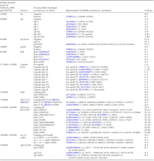
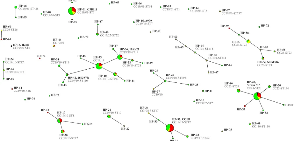
FIG 1eBURST population snapshot graph based on hybridization patterns for 11 variable marker genes. The sizes of the circles correlate with the numbers of isolates. The color codes indicate provenance: red are invasive isolates, yellow are isolates from local infections, and green are from carriage. Black indicates reference strains, and brown are isolates from local infections (mastitis) in cattle.
on May 16, 2020 by guest
http://jcm.asm.org/
[image:8.585.40.544.449.691.2]With regard to population structure, we observed that CC19
predominated among the human isolates from Germany, whereas
in Trinidad and Tobago, CC19 and CC23 were equally common.
In cattle, CC19 was also common, but another prevalent lineage is
CC103 (
40
), which is rare among human isolates. These
observa-tions might indicate both geographic- and host-specific
differ-ences in population structure. With regard to host specificity, one
virulence factor,
scpB
(encoding a complement-inactivating C5a
peptidase), might be involved (see Table S2 in the supplemental
material). It was present in 412 of 418 human isolates (98.6%) but
in only 10 of 21 cattle isolates (48%). All isolates belonging to the
bovine-associated CC103 (including two human isolates) lacked
scpB
. It was also absent in some bovine isolates belonging to
hu-man lineages (one of two HP-07, CC19/01-ST297 isolates, one
HP-56, and a CC23 isolate).
When studying the population structure, a major disadvantage
of MLST/eBURST became visible. Some previously described CCs
(CC1, CC17, and CC22) are not recognized as CCs anymore but
lumped together into CC19. Similar observations were made for
S.
aureus
, as nowadays, e.g., distinct CC5 and CC8 are merged
to-gether by eBURST (
41
). Strains linking two distinct CCs can
the-oretically be common ancestors of both, but they also might be
recent hybrids or recombinants. MLST/eBURST is not able to
visualize the difference so that distinct CCs are merged together
when linking strains are found. In
S. aureus
, for instance, the
hy-brid strain ST239 links CC8 to the unrelated CC30 (
42
). Both
lineages became linked to a third unrelated CC (CC45), with the
identification of a recombinant strain (ST2249) that emerged
from ST239 and CC45 parents (
43
). Thus, with a broadening
da-tabase, the current concept of CCs will obscure phylogenetic
pat-terns and, eventually, an entire species might be regarded as a
single CC, especially if it is a species in which horizontal gene
transfer is as common as it is in
S. agalactiae
(
44
). Hence, clusters
within CC19 (such as CC19/01 and CC19/17) were distinguished
(
Table 2
) to visualize the relationships within the large CC19.
With DNA microarrays, it is possible to genotype high
num-bers of isolates, with the method being less cumnum-bersome than
MLST and providing additional information on specific virulence
and resistance determinants.
Similarly to what has been shown for
S. aureus
, hybridization
patterns appear to correspond to STs and CCs (
29
). Compared to
S. aureus
, this correlation in GBS is poor. It is possible to predict an
ST/CC affiliation from a hybridization pattern if it is identical or
similar to a previously MLS-typed strain. However, isolates
be-longing to the same CC/ST might yield different hybridization
patterns, and it not possible to predict a hybridization pattern
from a known ST. Otherwise, we frequently observed (
Table 2
)
that similar strains differed in single features, such as capsule
genes,
alp
, or pilus genes, and that these genes vary independently
of each other. Some ubiquitous genes or gene clusters that
appar-ently always reside at the same position in the genome sequence
(
30
) show a number of distinct alleles, and the identity of an actual
allele at a given position is of a random nature. This might be
explained by a high number of past recombination events (
30
,
44
)
or, theoretically, also by convergent evolution.
In both cases, typing by array and by MLST would not reflect a
true phylogeny in terms of providing information on the
evolu-tionary history and provenance of a given strain but just create a
random pattern to be compared with other random patterns. The
identities of two isolates might then indicate, e.g., a transmission
event, but a mere similarity between two isolates would not
nec-essarily mean they were related, i.e., have a recent common
ances-tor. The resolution of a typing method might be increased with a
greater number of interrogated markers. The markers described
here could be combined with the MLST scheme, creating an
18-marker framework. This approach could be used experimentally
or, in the near future, purely
in silico
by extracting handy
infor-mation out of the abundance of data generated by next-generation
sequencing. Besides,
erm
,
cad
,
mer
, or
tet
genes could also be used
to prove the identity or nonidentity of isolates, provided that data
on the frequency of their acquisition or loss becomes available.
In order to understand the possible differences in virulence
within
S. agalactiae
, future studies should focus not only on
ST/CC affiliations but also on the alleles of relevant genes or gene
clusters, because these might vary within an ST/CC. No single
factor was identified that distinguishes between invasive and
car-riage isolates. However, it was observed that invasive isolates are
less likely to carry
abiG
-I/II and
Q8DZ34
, but they are more likely
to show the presence of the
pil1
locus,
fbsB
(515), and capsule type
III (
13
,
45
), as well as COH1-like alleles of
pil2b
,
nss
/
srr
, and
rgf
(
Table 3
). This does not necessarily mean that these factors are
involved in invasiveness. They also just might be surrogate
mark-ers being linked to invasive strains. Several of these markmark-ers
ap-pear together in CC19/17 isolates. Previously, CC19 (including
ST01, ST17, ST19, and ST28) was found in neonatal sepsis cases
from very diverse geographic origins (
30
,
46–48
), and the
CC19/17 cluster has also frequently been observed in invasive
iso-lates (
49
,
50
).
Other virulence factors include
speM
(encoding exotoxin M)
and the
cyl
operon (see Table S2 in the supplemental material).
The
speM
gene was found in only seven isolates. The
cyl
operon
was always found, and thus, its detection cannot be helpful for
predicting virulence. However,
in vivo
expression might be a very
different issue, and possibly, the key to understanding the
differ-ence between invasive and carriage isolates is not the mere
pres-ence or abspres-ence of specific genes or alleles but their expression.
[image:9.585.41.286.76.247.2]Generally, it is not possible to safely distinguish invasive from
carriage isolates and to predict an individual fate based on
avail-able typing data. Even if some strains are associated with an
in-creased risk of infection, other strains cannot be considered
harm-less. For practical purposes, this warrants screening for and
TABLE 4Prevalence of resistance genes in clinical isolates
Resistance gene(s)
Prevalences for isolates by type:
Total
Clinical, Germany
Clinical, Trinidad and Tobago
Veterinary, Germany
No. resistant %
No. resistant %
No. resistant %
No. resistant %
erm(A) 26 5.8 26 6.8 0 0.0 0 0.0
erm(B) 47 10.5 44 11.5 0 0.0 3 14.3
erm(C) 8 1.8 8 2.1 0 0.0 0 0.0
tet(M) 345 77.0 306 79.7 24 70.6 10 47.6
emrB/qacA448 100.0 384 100.0 34 100.0 21 100.0
cadC 95 21.2 86 22.4 6 17.6 0 0.0
cadD 400 89.3 352 91.7 32 94.1 10 47.6
merR⫹
merA
213 47.5 176 45.8 30 88.2 1 4.8
on May 16, 2020 by guest
http://jcm.asm.org/
eradication of any
S. agalactiae
colonization prior to delivery,
re-gardless of the genotyping data. Vaccination might become an
additional tool for preventing infection, and a vaccine covering
capsule types Ia, Ib, and III is currently being tested (
51
). Capsule
types are geographically unevenly distributed, as the comparison
between the German and Caribbean isolates and other work (
50
,
52
,
53
) indicate. Thus, the efficacy of a capsule-based vaccination
might vary in different countries and also might change over time
(
54
), emphasizing a need for typing GBS.
For the sake of statistical significance, future studies should target
more isolates from defined clinical conditions and healthy
con-trols and aim to compare
S. agalactiae
populations from different
regions and host species. The arrays described here provide a
con-venient tool for typing large numbers of clinical/veterinary
iso-lates and thus might help to obtain insights in GBS epidemiology.
ACKNOWLEDGMENTS
We thank R. Berner (Dresden University Hospital), M. van der Linden (National Reference Centre for Streptococci, Aachen, Germany), S. Weber (Sheikh Khalifa Medical City, Abu Dhabi, UAE), P. Akpaka (Uni-versity of West Indies, St. Augustine, Trinidad and Tobago), K. Kadlec (Friedrich Loeffler Institute, Neustadt-Mariensee, Germany), M. Rench (Baylor College of Medicine, Houston, TX), the Institut Pasteur, K. Hochauf, and F. Gunzer (Institute for Medical Microbiology and Hy-giene, IMMH, Dresden, Germany), and the colleagues of Dresden Uni-versity Hospital for collecting and providing the strains and isolates, W. Rudolph (IMMH Dresden) for MLST sequencing, and S. Hildebrandt (Dresden) for advice on clinical issues. We thank A. Ruppelt (IMMH Dresden), I. Engelmann, J. Sachtschal, and G. Rößler (Alere Jena) for excellent technical assistance, and we also thank E. Jacobs, K. Hochauf, and F. Gunzer (IMMH Dresden), E. Ermantraut (Alere Jena), and the Infectognostics Research Campus Consortium Jena for their support.
The study was funded by the Faculty of Medicine Carl Gustav Carus, Technische Universität Dresden (MeDDrive program).
S.M., P.S., E.M., and R.E. are employees of Alere Technologies, the company that manufactured the assays described herein. (S.M. was em-ployed by Alere Technologies after the experiments were finished but before the paper was written.) This had no influence on the study design, on the decision to publish, or on the preparation of the manuscript.
REFERENCES
1.Kowalska B, Niemiec KT, Drejewicz H, Polak K, Kubik P, Elmidaoui A, Gierowska-Bogusz B, Jaczynska R.2003. Prevalence of group B strepto-coccal colonization in pregnant women and their newborns based on the results of examination of patients in the Obstetric and Gynecology De-partment of the National Research Institute of Mother and Child–a pilot study. Ginekol. Pol.74:1223–1227. (In Polish.)
2.Kunze M, Ziegler A, Fluegge K, Hentschel R, Proempeler H, Berner R. 2011. Colonization, serotypes and transmission rates of group B strepto-cocci in pregnant women and their infants born at a single University Center in Germany. J. Perinat. Med.39:417– 422.http://dx.doi.org/10 .1515/JPM.2011.037.
3.Marconi C, Rocchetti TT, Rall VL, Carvalho LR, Borges VT, Silva MG. 2010. Detection of Streptococcus agalactiaecolonization in pregnant women by using combined swab cultures: cross-sectional prevalence study. Sao Paulo Med. J. 128:60 – 62. http://dx.doi.org/10.1590/S1516 -31802010000200003.
4.Müller-Vranjes A, Puntaric´ D, Curzik D, Sijanovic´ S, Topolovec Z, Kasac Z, Miskulin M.2011. Prevalence and significance of vaginal group B streptococcus colonization in pregnant women from Osijek, Croatia. Coll. Antropol.35:21–26.
5.Schrag SJ, Zywicki S, Farley MM, Reingold AL, Harrison LH, Lefkowitz LB, Hadler JL, Danila R, Cieslak PR, Schuchat A.2000. Group B strepto-coccal disease in the era of intrapartum antibiotic prophylaxis. N. Engl. J. Med.342:15–20.http://dx.doi.org/10.1056/NEJM200001063420103. 6.Baker CJ, Edwards MS.1995. Group B streptococcal infections, 3rd ed.
WB Saunders, Philadelphia, PA.
7.Glantz JC, Kedley KE.1998. Concepts and controversies in the manage-ment of group B streptococcus during pregnancy. Birth25:45–53.http: //dx.doi.org/10.1046/j.1523-536x.1998.00045.x.
8.Puopolo KM, Madoff LC, Eichenwald EC.2005. Early-onset group B streptococcal disease in the era of maternal screening. Pediatrics115: 1240 –1246.http://dx.doi.org/10.1542/peds.2004-2275.
9.Betriu C, Gomez M, Sanchez A, Cruceyra A, Romero J, Picazo JJ.1994. Antibiotic resistance and penicillin tolerance in clinical isolates of group B streptococci. Antimicrob. Agents Chemother.38:2183–2186.http://dx .doi.org/10.1128/AAC.38.9.2183.
10. Aracil B, Miñambres M, Oteo J, De La Rosa M, Gómez-Garcés JL, Alos AJ.2002. Susceptibility of strains ofStreptococcus agalactiaeto macrolides and lincosamides, phenotype patterns and resistance genes. Clin. Microbiol. Infect.8:745–748.http://dx.doi.org/10.1046/j.1469-0691 .2002.00450.x.
11. Betriu C, Culebras E, Gómez M, Rodríguez-Avial I, Sánchez BA, Ágreda MC, Picazo JJ.2003. Erythromycin and clindamycin resistance and telithro-mycin susceptibility inStreptococcus agalactiae. Antimicrob. Agents Chemother. 47:1112–1114. http://dx.doi.org/10.1128/AAC.47.3.1112 -1114.2003.
12. de Azavedo JC, McGavin M, Duncan C, Low DE, McGeer A.2001. Prevalence and mechanisms of macrolide resistance in invasive and non-invasive group B streptococcus isolates from Ontario, Canada. Antimi-crob. Agents Chemother.45:3504 –3508.http://dx.doi.org/10.1128/AAC .45.12.3504-3508.2001.
13. Schuchat A.1998. Epidemiology of group B streptococcal disease in the United States: shifting paradigms. Clin. Microbiol. Rev.11:497–513. 14. Uh Y, Jang IH, Hwang GY, Yoon KJ, Song W.2001. Emerging
erythromy-cin resistance among group B streptococci in Korea. Eur. J. Clin. Microbiol. Infect. Dis.20:52–54.http://dx.doi.org/10.1007/s100960000414.
15. Pereira Ude P, Rodrigues Dos Santos A, Hassan SS, Aburjaile FF, Soares Sde C, Ramos RT, Carneiro AR, Guimarães LC, Silva de Almeida S, Diniz CA, Barbosa MS, Gomes de Sá P, Ali A, Bakhtiar SM, Dorella FA, Zerlotini A, Araújo FM, Leite LR, Oliveira G, Miyoshi A, Silva A, Azevedo V, Figueiredo HC.2013. Complete genome sequence of
Strep-tococcus agalactiaestrain SA20-06, a fish pathogen associated to
meningo-encephalitis outbreaks. Stand. Genomic Sci.8:188 –197.http://dx.doi.org /10.4056/sigs.3687314.
16. Kogan G, Uhrín D, Brisson JR, Paoletti LC, Blodgett AE, Kasper DL, Jennings HJ.1996. Structural and immunochemical characterization of the type VIII group B streptococcus capsular polysaccharide. J. Biol. Chem.271:8786 – 8790.http://dx.doi.org/10.1074/jbc.271.15.8786. 17. Kong F, Lambertsen LM, Slotved HC, Ko D, Wang H, Gilbert GL.2008.
Use of phenotypic and molecular serotype identification methods to char-acterize previously nonserotypeable group B streptococci. J. Clin. Micro-biol.46:2745–2750.http://dx.doi.org/10.1128/JCM.00189-08.
18. Paoletti LJ, Bradford J, Paoletti LC.1999. A serotype VIII strain among colonizing group B streptococcal isolates in Boston, Massachusetts. J. Clin. Microbiol.37:3759 –3760.
19. von Hunolstein C, Parisi L, Tissi L, Recchia S, Alfarone G, Nicolini L, Volpe C, Wagner B, Motlovà J, Orefici G.1999. Virulence properties of type VIIStreptococcus agalactiae(group B streptococci) and immuno-chemical analysis of capsular type polysaccharide. J. Med. Microbiol.48: 983–990.http://dx.doi.org/10.1099/00222615-48-11-983.
20. Wessels MR. 1997. Biology of streptococcal capsular polysaccharides. Soc. Appl. Bacteriol. Symp. Ser.26:20S–31S.
21. Blumberg HM, Stephens DS, Licitra C, Pigott N, Facklam R, Swami-nathan B, Wachsmuth IK.1992. Molecular epidemiology of group B streptococcal infections: use of restriction endonuclease analysis of chro-mosomal DNA and DNA restriction fragment length polymorphisms of ribosomal RNA genes (ribotyping). J. Infect. Dis.166:574 –579.http://dx .doi.org/10.1093/infdis/166.3.574.
22. Huet H, Martin C, Geslin P, Grimont F, Quentin R.1993. Ribotyping of
Streptococcus agalactiaestrains isolated from vaginas of asymptomatic
women. Res. Microbiol.144:457– 465.http://dx.doi.org/10.1016/0923-2508 (93)90053-5.
23. Benson JA, Ferrieri P. 2001. Rapid pulsed-field gel electrophoresis method for group B streptococcus isolates. J. Clin. Microbiol.39:3006 – 3008.http://dx.doi.org/10.1128/JCM.39.8.3006-3008.2001.
24. Quentin R, Huet H, Wang FS, Geslin P, Goudeau A, Selander RK.1995. Characterization ofStreptococcus agalactiaestrains by multilocus enzyme genotype and serotype: identification of multiple virulent clone families that cause invasive neonatal disease. J. Clin. Microbiol.33:2576 –2581.