0095-1137/09/$08.00⫹0 doi:10.1128/JCM.00389-09
Copyright © 2009, American Society for Microbiology. All Rights Reserved.
Matrix-Assisted Laser Desorption Ionization–Time of Flight Mass
Spectrometry for Fast and Reliable Identification of Clinical
Yeast Isolates
䌤
G. Marklein,
1M. Josten,
1U. Klanke,
1E. Mu
¨ller,
1R. Horre
´,
3T. Maier,
2T. Wenzel,
2M. Kostrzewa,
2G. Bierbaum,
1A. Hoerauf,
1and H.-G. Sahl
1*
Institute of Medical Microbiology, Immunology and Parasitology, Rheinische Friedrich-Wilhelms-Universita¨t, University of Bonn, Bonn, Germany1; Bruker Daltonik GmbH, Bremen, Germany2; and
Federal Institute for Drugs and Medical Devices (BfArM), Bonn, Germany3
Received 20 February 2009/Returned for modification 9 April 2009/Accepted 22 June 2009
The clinical impact of severe infections with yeasts and yeast-like fungi has increased, especially in immunocom-promised hosts. In recent years, new antifungal agents with different and partially species-specific activity patterns have become available. Therefore, rapid and reliable species identification is essential for antifungal treatment; however, conventional biochemical methods are time-consuming and require considerable expertise. We evaluated matrix-assisted laser desorption ionization–time of flight mass spectrometry (MALDI-TOF MS) for the rapid routine identification of clinical yeast isolates. A total of 18 type collection strains and 267 recent clinical isolates ofCandida(nⴝ250),Cryptococcus,Saccharomyces,Trichosporon,Geotrichum,Pichia, andBlastoschizomycesspp. were identified by MALDI-TOF MS. The results were compared with those obtained by conventional phenotyping and biochemical tests, including the API ID 32C system (bioMe´rieux, Nu¨rtingen, Germany). Starting with cells from single colonies, accurate species identification by MALDI-TOF MS was achieved for 247 of the clinical isolates (92.5%). The remaining 20 isolates required complementation of the reference database with spectra for the appropriate reference strains which were obtained from type culture collections or identified by 26S rRNA gene sequencing. The absence of a suitable reference strain from the MALDI-TOF MS database was clearly indicated by log(score) values too low for the respective clinical isolates; i.e., no false-positive identifications occurred. After complementation of the database, all isolates were unambiguously identified. The established API ID 32C biochem-ical diagnostic system identified 244 isolates in the first round. Overall, MALDI-TOF MS proved a most rapid and reliable tool for the identification of yeasts and yeast-like fungi, with the method providing a combination of the lowest expenditure of consumables, easy interpretation of results, and a fast turnaround time.
Pathogenic fungi such asCandida,Cryptococcus, and Aspergil-lusspp. and species of other genera commonly cause life-threat-ening infections of immunocompromised hosts, including patients receiving immunosuppressive therapy. In the United States, Can-didaspecies are the fourth leading cause of nosocomial blood-stream infections. With an estimated total of 72.8 million oppor-tunisticCandidainfections per year worldwide, the case/fatality rate due toCandidaspecies is in the range of 33.9% (32). Al-thoughCandida albicansremains the leading and most important pathogenic yeast species, the incidence rates of non-C. albicans
yeast infections have been increasing in recent years. Moreover, outbreaks in perinatal intensive care and oncohematological units have been described (23). Recently, infections caused by less common yeasts and yeast-like species such asPichia,Rhodotorula,
Saccharomyces, and Trichosporon species and other rarely en-countered species have been reported (17).
At present, more than 100 yeast and yeast-like species are known to be human pathogens and have been isolated from virtually all body sites. Identification of the increasing diversity of fungal pathogens by conventional phenotypic methods is
difficult and time-consuming, and the findings may sometimes be inconclusive, especially for unusual yeasts. For treatment with the appropriate antifungal agents, particularly with drugs belonging to the azole group, fast and reliable yeast identifi-cation can be time-saving because of the species-specific sus-ceptibility patterns. Commercially available chromogenic agar media (17) and biochemical or enzymatic panels, e.g., the API ID 20C, API ID 32C, and Vitek ID YST systems (bioMe´rieux, Nu¨rtingen, Germany), are widely used, although they are oc-casionally disadvantageous due to limited databases, and misi-dentifications have been reported (8, 27).
Since the introduction of gentle ionization techniques, mass spectrometry (MS) has been developed into a standard method in the field of protein analysis (2, 3). Time of flight (TOF) instruments are state of the art for applications in the field of proteomics, particularly in combination with matrix-assisted laser desorption ionization (MALDI) procedures. MALDI-TOF MS is also used to analyze the protein compositions of complex mixtures, e.g., biological fluids or tissues, as well as microbial cells or cell components (13, 26).
The convenient and rapid identification of microbial species by the extraction of cells from single colonies and determina-tion of species-specific patterns of peptides and protein masses have been reported (7, 18, 20, 21). Moreover, the character-ization and differentiation of bacteria to the subspecies level have been described (4, 11, 13).
* Corresponding author. Mailing address: Institute of Medical Mi-crobiology, Immunology and Parasitology, Sigmund Freud Straße 25, Bonn D-53127, Germany. Phone: 49(0)228 287-16483. Fax: 49(0)228 287-14808. E-mail: sahl@microbiology-bonn.de.
䌤Published ahead of print on 1 July 2009.
2912
on May 16, 2020 by guest
http://jcm.asm.org/
In clinical and applied laboratory settings, MALDI-TOF MS has been shown to be suitable for the fast and secure identifi-cation of pathogenic microorganisms, particularly slowly grow-ing microorganisms, whose identification by current automated biochemical tests is problematic. These microorganisms in-clude, e.g., nonfermenting bacteria (10, 28), some gram-posi-tive bacteria (4, 14, 30), and fungi (34, 36).
In the study described here, we demonstrate the suitability of identification of clinical yeasts and yeast-like fungi by MALDI-TOF MS. For the first time, the technology was used for the identification of more than 250 clinical isolates, with part of the study being run under routine diagnostic conditions. The study was controlled by conventional cultural, morpholog-ical, and biochemical criteria as well as by 26S rRNA sequenc-ing of isolates with discrepant results.
MATERIALS AND METHODS
Strains and culture conditions.The study included 52 archived and 215 fresh nonrepetitive fungal isolates subcultured from the clinical diagnostic laboratory of the Institute of Medical Microbiology, Immunology, and Parasitology of the University of Bonn, Bonn, Germany, between May 2007 and July 2008. The isolates were obtained predominantly from clinical materials, such as blood, cerebrospinal fluid, bronchoalveolar lavage fluid, intra-abdominal fluid, and in-traoperative wound and tissue specimens. All isolates were cultured on diagnos-tic fungal media, such as Columbia agar with 5% sheep blood (Becton Dickinson, Heidelberg, Germany), Sabouraud 2% glucose agar (Oxoid, Wesel, Germany), and Candi4 select agar (Bio-Rad, Munich, Germany), followed by morphological identification, including determination of growth characteristics on CHROMagar Candida (Becton Dickinson, Heidelberg, Germany) and germ tube or chlamy-dospore formation (17).
Commercially available strains were obtained from the American Type Cul-ture Collection (ATCC) and from the German Collection of Microorganisms and Cell Cultures (Deutsche Sammlung von Mikroorganismen und Zellkulturen [DSMZ]) and includedCandida albicansATCC 11949, ATCC 24433, ATCC 44373, ATCC 44374, and ATCC 90028;Candida glabrataATCC 90030 and DSMZ 11950;Candida kefyrDSMZ 11954;Candida parapsilosisATCC 22019, ATCC 90018, and DSMZ 11955;Candida tropicalisATCC 750 and ATCC 90874;
Issatchenkia orientalis(Candida krusei) ATCC 6258, ATCC 90878, and DSMZ 11957;Cryptococcus neoformans ATCC 62066; andSaccharomyces cerevisiae
ATCC 9763.
Biochemical identification.For the biochemical identification of the strains, the API ID 32C identification system for yeasts (bioMe´rieux) was used according to the manufacturer’s instructions, and the recommended additional tests were employed when necessary (17). Reading and the interpretation of the results were performed after incubation times of 24 and 48 h.
Sequence data.The 26S rRNA genes of strains that yielded discrepant results were sequenced by Scanbec GmbH (Halle/Saale, Germany) (22).
MALDI-TOF MS.After incubation of the test strains for 48 h at 30°C on Sabouraud agar, cells from up to five representative single colonies were trans-ferred into a 1.5-ml screw-cap extraction tube (Eppendorf, Germany) with a pipette tip and mixed thoroughly in 0.3 ml double-distilled water. Absolute ethanol (0.9 ml) was added, the contents of the tube were carefully mixed, and the tubes were then centrifuged at 20,000⫻g(2K15 centrifuge; Sigma, Ger-many) for 2 min; the supernatant was discarded, and the pellet was air dried. Approximately 10l of the pellet was mixed thoroughly with 50l of formic acid (70%), before addition of an equivalent volume of acetonitrile. The mixture was centrifuged at 20,000⫻gfor 2 min, and 1l of the supernatant was placed onto a ground steel MALDI target plate and allowed to dry at room temperature. Subsequently, each sample was overlaid with 2l of matrix, which consisted of a saturated solution of␣-cyano-4-hydroxy-cinnamic acid in 50% acetonitrile– 2.5% trifluoroacetic acid, and air dried at room temperature.
The MALDI-TOF MS analyses of all strains were performed in parallel on a Microflex LT spectrometer in the Bruker application laboratory in Leipzig, Germany, and on a Biflex III spectrometer in the microbiological diagnostic laboratory of the Institute of Medical Microbiology, Immunology, and Para-sitology in Bonn (both apparatuses were from Bruker Daltonik GmbH, Bre-men, Germany). The spectra were recorded in the linear positive mode at a laser frequency of 20 Hz within a mass range from 2,000 to 20,000 Da. The instrument parameter settings were ion source 1 at 20 kV, ion source 2 at 18.5
kV, lens at 8.5 kV, pulsed ion extraction of 250 ns, and no gating for the Microflex LT spectrometer and IS1 at 19 kV, IS2 at 16.5 kV, lens at 8 kV, PIE of 200 ns, and maximum gating for the Biflex III spectrometer. For each spectrum, 240 laser shots in 30-shot steps from different positions of the target spot were collected and analyzed. The spectra were externally cali-brated by usingEscherichia coliribosomal proteins.
Data analysis.FlexAnalysis (version 2.0) software (Bruker Daltonik GmbH) was used for visual inspection of the mass spectra. For automated data analysis, the raw spectra for unknown fungi were processed by using version 1.1 (Bonn) and version 2.0 (Leipzig) of MALDI Biotyper software (Bruker Daltonik GmbH) with the default settings. The software performs smoothing, normaliza-tion, baseline subtracnormaliza-tion, and peak picking, thereby creating a list of the most significant peaks (m/zvalues) of the spectrum.
As an integrated part of the MALDI Biotyper software, the main spectra were used as reference libraries that contained information about the average masses, the average intensities, and the relative abundances in the measurements for all characteristic peaks. To identify unknown fungal isolates, the raw spectra were imported into the MALDI Biotyper software and analyzed by use of the standard pattern-matching algorithm, which compared the raw spectra with the spectra in the library without any user intervention by using the standard settings. For the pattern matchings, the fingerprints of unknown samples were compared to the fingerprints for all entries in the database (main spectra), and the results were listed in a ranking table. The results of the pattern-matching process were expressed as log(score) values, which ranged from 0 to 3. Log(score) values of ⬎1.7 generally indicated relationships at the genus level, and log(score) values of ⬎2.0 generally indicated relationships at the species level. The highest log(score) of a match against the score in the database was used for species identification. The spectra for unequivocally identified isolates were added to the reference database in the course of the study, resulting in a total of 241 fungal strains representing 131 species.
RESULTS
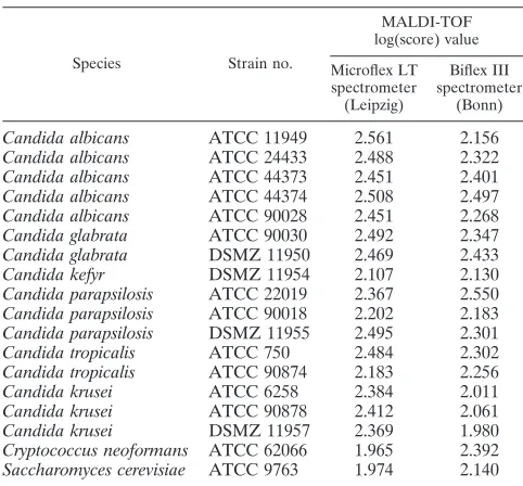
[image:2.585.300.541.96.320.2]Yeast collection strains.The cell extracts of 18 type culture collection strains representing the majority of clinically rele-vant yeast species gave suitable spectra and were included in the MALDI-TOF MS reference database. Table 1 shows the results of all log(score) values obtained in both laboratories on the Microflex LT and Biflex III spectrometers. In general, the TABLE 1. MALDI-TOF log(score) values for ATCC and DSMZ
type strains ofCandidaspecies and yeast-like organisms obtained with Microflex LT and Biflex III spectrometersa
Species Strain no.
MALDI-TOF log(score) value
Microflex LT spectrometer (Leipzig)
Biflex III spectrometer
(Bonn)
Candida albicans ATCC 11949 2.561 2.156 Candida albicans ATCC 24433 2.488 2.322 Candida albicans ATCC 44373 2.451 2.401 Candida albicans ATCC 44374 2.508 2.497 Candida albicans ATCC 90028 2.451 2.268 Candida glabrata ATCC 90030 2.492 2.347 Candida glabrata DSMZ 11950 2.469 2.433 Candida kefyr DSMZ 11954 2.107 2.130 Candida parapsilosis ATCC 22019 2.367 2.550 Candida parapsilosis ATCC 90018 2.202 2.183 Candida parapsilosis DSMZ 11955 2.495 2.301 Candida tropicalis ATCC 750 2.484 2.302 Candida tropicalis ATCC 90874 2.183 2.256 Candida krusei ATCC 6258 2.384 2.011 Candida krusei ATCC 90878 2.412 2.061 Candida krusei DSMZ 11957 2.369 1.980 Cryptococcus neoformans ATCC 62066 1.965 2.392 Saccharomyces cerevisiae ATCC 9763 1.974 2.140
a
Eighteen isolates were tested.
on May 16, 2020 by guest
http://jcm.asm.org/
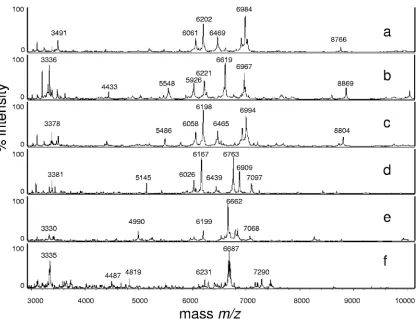
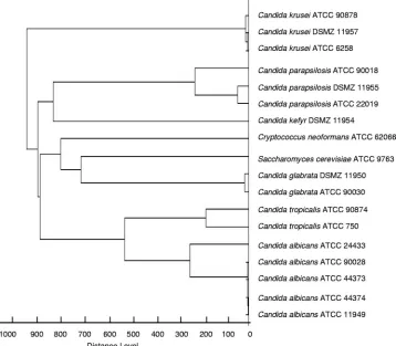
log(score) values were above 2.0; only the values forC. neo-formansandS. cerevisiae(Microflex LT spectrometer) and one of theC. kruseiisolates (Biflex III spectrometer) were slightly lower. Figure 1 shows representative MS spectra for type strains measured with the Biflex III spectrometer. The score-oriented dendrogram (Fig. 2) shows the spectral similarities of the type strains.
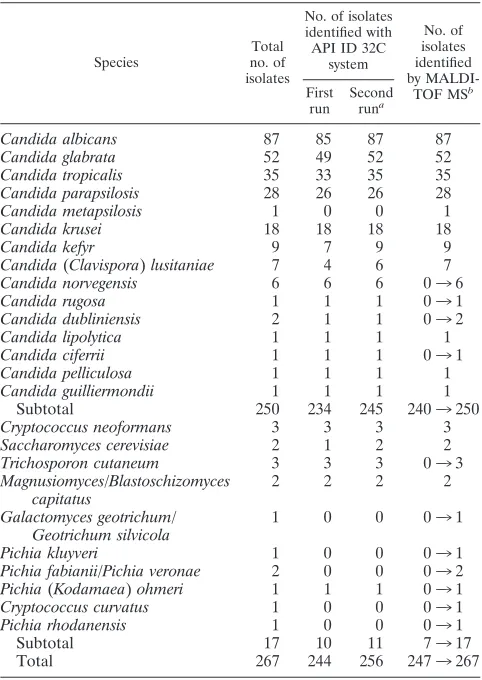
Clinical isolates ofCandidaspecies.Among the clinical iso-lates, 250Candidaspecies and 17 yeast-like fungi were tested. MALDI-TOF MS yielded the correct species identification for 240 clinical Candida isolates in the first attempt. Strains of
Candida norvegensis,Candida rugosa,Candida dubliniensis, and
Candida ciferriicould not be identified since spectra for appro-priate reference strains had not been included in the database. Inappropriate species or genus assignments were clearly in-dicated by low log(scores), generally below 1.7. With a log(score) of 1.72, the Candida dubliniensis isolate was first identified asCandidato the genus level and with a best hit of
C. albicans. Introduction of the spectra for the respective type strains into the database and subsequent comparison with the unidentified spectra resulted in the unequivocal identification of all 250Candidaisolates.
Biochemical analysis with the API ID 32C system correctly identified 234 (93.6%) of theCandida isolates (Table 2, first run). When the findings of MALDI-TOF MS and biochemical identification disagreed, the API test was repeated, starting with single colonies from pure culture, which increased the
number of correctly identifiedCandidaisolates to 245 (98%) (Table 2). The remaining strains that yielded discrepant results were subjected to 26S rRNA sequencing (Scanbec GmbH), resulting in the identification ofPichia kluyveri (99% nucleic acid sequence identity) andPichia fabianii(100% nucleic acid sequence identity) rather thanCandida kruseiorCandida pel-liculosa. Also,Debaryomyces etchellsii/D.carsonii(API ID 32C system,n⫽3) was identified asCandida parapsilosis(n⫽2) and Candida lusitaniae by 26S rRNA sequencing with 100% nucleic acid sequence identity (Table 3). One isolate, misiden-tified asCandida utilisby use of the API ID 32C system, was also examined by 26S rRNA sequencing and was identified as
Pichia fabianiiorPichia veronae, which both share 100% nu-cleic acid sequence identity. Table 3 presents a compilation of the metabolic profiles and incorrect designations of isolates by use of the API ID 32C system compared to the results of the reference methods.
In one case concerning a DNA sequencing-confirmed Can-dida dubliniensisisolate, MALDI-TOF MS yielded the correct identification with a log(score) value of 2.365, whereas the API ID 32C system identified this isolate asCandida albicans (Ta-ble 3). Another isolate which had been unambiguously identi-fied asC. parapsilosisby the API ID 32C system was identified asCandida metapsilosiswith a log(score) value of 2.103. Can-dida metapsilosiswas recently described as a new species which cannot be identified by biochemical tests (38) but which can be identified unequivocally by MALDI-TOF MS (15). The spec-FIG. 1. MALDI-TOF mass spectra (m/z3,000 to 10,000) ofCandida albicans(a),Candida glabrata(b),Candida tropicalis(c)Candida krusei (d),Candida parapsilosis(e), andCryptococcus neoformans(f) revealing differences among the species.
on May 16, 2020 by guest
http://jcm.asm.org/
[image:3.585.86.503.64.383.2]tra for all newly identified isolates and isolates whose identities were confirmed by 26S rRNA sequencing were included in the database.
Clinical isolates of yeast-like fungi. For 7 of 17 clinical isolates of yeast-like fungi, MALDI-TOF MS, biochemical testing, and morphological analysis yielded congruent re-sults in the first round of identification; oneCryptococcus neoformansisolate required subculturing on Sabouraud agar for accurate identification. The identification of the remain-ing 10 strains by MALDI-TOF-MS required the addition of the spectra for the respective reference strains to the data-base (Table 2).
One strain identified as aGeotrichumsp. by use of the API ID 32C system was identified asGalactomyces geotrichumor
Geotrichum silvicola(33) by 26S rRNA sequencing with 99% nucleic acid sequence identity (Table 3). Two isolates could not be identified by use of the API ID 32C system but were determined by 26S rRNA sequencing to beCryptococcus cur-vatus(100% sequence identity) andPichia rhodanensis(100% sequence identity) (Table 3).
DISCUSSION
The correct and fast identification of fungal pathogens from clinical specimens and from patients’ environments, especially in outbreak situations, is of major concern for optimal patient
management and the implementation of effective measures for disease control. Molecular approaches are currently being de-veloped to provide methods for the identification of yeasts that are more rapid and reliable than the classical phenotypic meth-ods. However, high-resolution DNA-based molecular tech-niques, such as 26S rRNA or internal transcribed spacer DNA sequencing (6, 29, 36) and real-time PCR assays (35), are expensive and time-consuming.
In the present study MALDI-TOF MS proved to be a rapid and reliable procedure for the accurate identification of patho-genicCandidastrains and required minimal hands-on time or time for the interpretation of the results. The test procedure was generally completed within 10 min per isolate and within 3 h for 96 samples, starting from single yeast colonies on the agar plate. In contrast, the identification of germ tube-negative
Candida species by phenotypic methods requires incubation periods of up to 72 h. Overall, MALDI-TOF MS did not produce any misidentifications, provided that spectra for the appropriate reference strains were present in the database (Table 2). Incorrect species assignments as best hits, due to the absence of reference strains, were clearly indicated by log(score) values that were too low, according to the thresholds of the MALDI Biotyper software.
[image:4.585.114.472.74.387.2]The discrepancies between the results of MALDI-TOF MS after database complementation and the phenotypic identifi-cation (Table 2, first run) might have been due to accidentally FIG. 2. Score-oriented dendrogram of ATCC and DSMZ type strains ofCandidaspecies and yeast-like fungi (n⫽18) obtained by the Microflex LT spectrometer.
on May 16, 2020 by guest
http://jcm.asm.org/
mixed or contaminated cultures; incorrect phenotypic identi-fications based on subtle alterations in yeast carbohydrate as-similation and biochemical characteristics and the subjective interpretation of the results of morphological methods may also occur.
Due to the close phylogenetic relationship,Candida dublini-ensisis often misidentified asCandida albicans(6, 23), and in addition to state-of-the-art molecular biology methods, large-scale techniques such as fatty acid analysis were applied for correct diagnosis, until recently (31). Candida parapsilosis
proved to be a heterogeneous group of yeasts with distinct DNA sequence patterns (38) and different antifungal suscep-tibilities (25). Therefore, in addition toC. parapsilosis, two new species,Candida metapsilosisand Candida orthopsilosis, have recently been proposed (38), but these two species cannot be separated by phenotypic tests.
Molecular methods for the detection and differentiation of yeasts are also able to distinguish the closely related new spe-cies Candida nivariensis(1) andCandida bracarensis (9) that were previously misidentified asCandida glabrataby conven-tional chemotaxonomy. Recently, in a global collection of 1,598C. glabratastrains received from 28 countries, Lockhart et al. found the prevalence of these cryptic species to be 0.2% (24). As ourC. glabrataisolates (n⫽52) were identified with log(score) values greater than 2.0, reference strains of these new species have so far not been introduced into the database. Uncommon yeast species are emerging as human pathogens, and their identification may pose a challenge when the type strains have not yet been included in the diagnostic databases of any identification system, leading to their misidentification as related species. Interestingly, and in agreement with our observation (Table 3), in the endocarditis case reported by Hamal et al. (16), the isolates were initially identified biochem-ically asCandida pelliculosa; however, on the basis of direct sequencing of the internal transcribed spacer region of rRNA, they were subsequently reidentified as Pichia fabianii with strong biofilm formation and voriconazole resistance.Pichia fabianiiis an example of a yeast species which requires molec-ular techniques for correct identification (5).
Two questionable strains from our study which could not be identified by standard routine methods yielded perfect DNA sequence matches for Cryptococcus curvatus and Pichia rho-danensis(Table 3). Cryptococcus curvatuscan be detected by molecular methods in submarine and terrestrial environments (37) and is known to be a rare opportunistic pathogen of animals, including humans (12).Pichia rhodanensis has been reported to be involved in the spontaneous process of fermen-TABLE 2. Identification of clinical yeast isolates by MALDI-TOF
MS and conventional tests
Species
Total no. of isolates
No. of isolates identified with API ID 32C
system
No. of isolates identified by
MALDI-TOF MSb
First run
Second runa
Candida albicans 87 85 87 87 Candida glabrata 52 49 52 52 Candida tropicalis 35 33 35 35 Candida parapsilosis 28 26 26 28 Candida metapsilosis 1 0 0 1 Candida krusei 18 18 18 18
Candida kefyr 9 7 9 9
Candida(Clavispora)lusitaniae 7 4 6 7 Candida norvegensis 6 6 6 036 Candida rugosa 1 1 1 031 Candida dubliniensis 2 1 1 032 Candida lipolytica 1 1 1 1 Candida ciferrii 1 1 1 031 Candida pelliculosa 1 1 1 1 Candida guilliermondii 1 1 1 1
Subtotal 250 234 245 2403250
Cryptococcus neoformans 3 3 3 3 Saccharomyces cerevisiae 2 1 2 2 Trichosporon cutaneum 3 3 3 033 Magnusiomyces/Blastoschizomyces
capitatus
2 2 2 2
Galactomyces geotrichum/ Geotrichum silvicola
1 0 0 031
Pichia kluyveri 1 0 0 031 Pichia fabianii/Pichia veronae 2 0 0 032 Pichia(Kodamaea)ohmeri 1 1 1 031 Cryptococcus curvatus 1 0 0 031 Pichia rhodanensis 1 0 0 031
Subtotal 17 10 11 7317
Total 267 244 256 2473267
aAfter repeating the test with the API ID 32C system for isolates with
dis-crepant results.
bMALDI-TOF MS identification was performed in parallel on a Microflex LT
[image:5.585.43.284.89.429.2]spectrometer (Leipzig) and a Biflex III spectrometer (Bonn), and there were no discrepancies in the results.3, identified after integration into the database.
TABLE 3. Phenotypic identities of clinical yeast isolates differing from those determined by MALDI-TOF MS and/or 26S rRNA sequencing
API ID 32C system MALDI-TOF MS26S rRNA sequencing
Organism identified Identification profile % Identity Organism identified % NASIa
Candida pelliculosa 4274 3501 91.6 Pichia fabianii 100 Candida utilis 4271 3501 95.6 Pichia fabianii/Pichia veronae 100 Candida parapsilosis 7147 3507 99.9 Candida metapsilosis
Candida albicans 7347 1400 99.8 Candida dubliniensis 100 Candida krusei 0300 0100 88.6 Pichia kluyveri 99 Debaryomyces etchellsii/D. carsonii 5546 3501 87 Candida parapsilosis 100 Debaryomyces etchellsii/D.carsonii 5546 3501 91.4 Candida parapsilosis 100 Debaryomyces etchellsii/D. carsonii 5547 1501 91.4 Candida lusitaniae 100 Geotrichumspecies 3000 3100 99.3 Galactomyces geotrichum/Geotrichum silvicola 99
No identification possible Cryptococcus curvatus 100
No identification possible Pichia rhodanensis 100
a
NASI, nucleic acid sequence identity.
on May 16, 2020 by guest
http://jcm.asm.org/
[image:5.585.43.542.581.716.2]tation of untreated greenArbequinatable olives in minor quan-tities (19).
The present study clearly demonstrates that MALDI-TOF MS can be used as a fast technique for the reliable identification of fungi. Moreover, the technology has been shown to be broadly applicable in different fields of clinical bacteriology. With state-of-the-art instruments and the adoption of user-friendly software, MA specialists are not needed and laboratory technicians are able to run the system after a few days of training. The investment in and maintenance of the instrument are balanced by the low costs of the consumables. Thus, MALDI-TOF MS-based microorgan-ism identification is ready to be used not only by specialized research facilities but also by clinical diagnostic laboratories, pro-vided that appropriate standard operating procedures and com-prehensive quality-controlled databases with the spectra for hu-man pathogens are available.
REFERENCES
1.Alcoba-Flo´rez, J., S. Me´ndez-A´lvarez, J. Cano, J. Guarro, E. Pe´rez-Roth, and M. del Pilar Are´valo.2005. Phenotypic and molecular characterization of
Candida nivariensissp. nov., a possible new opportunistic fungus. J. Clin. Microbiol.43:4107–4111.
2.Amiri-Eliasi, B., and C. Fenselau.2001. Characterization of protein biomarkers desorbed by MALDI from whole fungal cells. Anal. Chem.73:5228–5231. 3.Arnold, R. J., and J. P. Reilly.1998. Fingerprint matching ofE.colistrains
with matrix-assisted laser desorption/ionization time-of-flight mass spec-trometry of whole cells using a modified correlation approach. Rapid Com-mun. Mass Spectrom.12:630–636.
4.Barbuddhe, S. B., T. Maier, G. Schwarz, M. Kostrzewa, H. Hof, E. Domann, T. Chakraborty, and T. Hain.2008. Rapid identification and typing of Lis-teriaspecies by matrix-assisted laser desorption ionization-time of flight mass spectrometry. Appl. Environ. Microbiol.74:5402–5407.
5.Bhally, H. S., S. Jain, C. Shields, N. Halsey, E. Cristofalo, and W. G. Merz.
2006. Infection in a neonate caused byPichia fabianii: importance of molec-ular identification. Med. Mycol.44:185–187.
6.Boyanton, B. L., Jr., R. A. Luna, L. R. Fasciano, K. G. Menne, and J. Versalovic.2008. DNA pyrosequencing-based identification of pathogenic
Candidaspecies by using the internal transcribed spacer 2 region. Arch. Pathol. Lab. Med.132:667–674.
7.Claydon, M. A., S. N. Davey, V. Edwards-Jones, and D. B. Gordon.1996. The rapid identification of intact microorganisms using mass spectrometry. Nat. Biotechnol.14:1584–1586.
8.Coignard, C., S. F. Hurst, L. E. Benjamin, M. E. Brandt, D. W. Warnock, and C. J. Morrisson.2004. Resolution of discrepant results forCandida
species identification by using DNA probes. J. Clin. Microbiol.42:858–861. 9.Correia, A., P. Sampaio, S. James, and C. Pais.2006.Candida bracarensissp. nov., a novel anamorphic yeast species phenotypically similar toCandida glabrata. Int. J. Syst. Evol. Microbiol.56:313–317.
10.Degand, N., E. Carbonnelle, B. Dauphin, J. L. Beretti. M. Le Bourgeois, Semet-Gaudelus, C. Segonds, P. Berche, X. Nassif, and A. Ferroni.2008. Matrix-assisted laser desorption ionization-time of flight mass spectrometry for identification of nonfermenting gram-negative bacilli isolated from cystic fibrosis patients. J. Clin. Microbiol.46:3361–3367.
11.Dieckmann, R., R. Helmuth, M. Erhard, and B. Malorny.2008. Rapid classification and identification of salmonellae at the species and subspecies levels by whole-cell matrix-assisted laser desorption ionization-time of flight mass spectrometry. Appl. Environ. Microbiol.74:7767–7778.
12.Dromer, F., A. Moulignier, B. Dupont, E. Gueho, M. Baudrimont, L. Im-provisi, F. Provost, and G. Gonzalez-Canali.1995. Myeloradiculitis due to
Cryptococcus curvatusin AIDS. AIDS9:395–396.
13.Fenselau, C., and P. A. Demirev.2001. Characterization of intact microor-ganisms by MALDI mass spectrometry. Mass Spectrom. Rev.20:157–171. 14.Grosse-Herrenthey, A., T. Maier, F. Gessler, R. Schaumann, H. Bo¨hnel, M.
Kostrzewa, and M. Kru¨ger.2008. Challenging the problems of clostridial identification with matrix-assisted laser desorption and ionization-time-of-flight mass spectrometry (MALDI-TOF MS). Anaerobe14:242–249. 15.Haase, G., T. Maier, K. Ritter, and M. Kostrzewa.2008. Differentiation of
Candida metapsilosis, Candida orthopsilosis, and Candida parapsilosis using MALDI-TOF mass spectral fingerprinting. Int. J. Med. Microbiol.298S2:16, abstr. DVV08. schaft fu¨r Hygiene und Mikrobiologie.
16.Hamal, P., J. Ostransky, M. Dendis, R. Horva´th, F. Ruzicka, V. Buchta, M. Vejsova, P. Sauer, P. Hejnar, and V. Raclavsky.2008. A case of endocarditis caused by the yeastPichia fabianiiwith biofilm production and developedin vitroresistance to azoles in the course of antifungal treatment. Med. Mycol.
46:601–605.
17.Hazen, K. C., and S. A. Howell.2007.Candida,Cryptococcus, and other
yeasts of medical importance, p. 1762–1788.InP. R. Murray, E. J. Baron, J. H. Jorgensen, M. L. Landry, and M. A. Pfaller (ed.) Manual of clinical microbiology, vol. 9. ASM Press, Washington, DC.
18.Holland, R. D., J. G. Wilkes, F. Rafii, J. B. Sutherland, C. C. Persons, K. J. Voorhee, and J. O. J. Lay.1996. Rapid identification of intact whole bacteria based on spectral patterns using matrix-assisted laser desorption/ionization with time-of-flight mass spectrometry. Rapid Commun. Mass Spectrom.
10:1227–1232.
19.Hurtado, A., C. Reguant, B. Esteve-Zarzoso, A. Bordons, and N. Roze`s.2008. Microbial population dynamics during the processing ofArbequina table olives. Food Res. Int.41:738–744.
20.Jarman, K. H., D. S. Daly, C. E. Petersen, A. J. Saenz, N. B. Valentine, and K. L. Wahl.1999. Extracting and visualizing matrix-assisted laser desorption/ ionization time-of-flight mass spectral fingerprints. Rapid Commun. Mass Spectrom.13:1586–1594.
21.Krishnamurthy, T., P. L. Ross, and U. Rajamani.1996. Detection of patho-genic and nonpathopatho-genic bacteria by matrix-assisted laser desorption/ioniza-tion time-of-flight mass spectrometry. Rapid Commun. Mass Spectrom.10:
883–888.
22.Kurzmann, C. P., and C. J. Robnett.1998. Identification and phylogeny of ascomycetous yeasts from analysis of nuclear large subunit (26S) ribosomal DNA partial sequences. Antonie van Leeuwenhoek73:331–371.
23.Leaw, S. N., H. C. Chang, H. F. Sun, R. Barton, J. P. Bouchara, and T. C. Chang.2006. Identification of medically important yeast species by sequence analysis of the internal transcribed spacer regions. J. Clin. Microbiol.44:
693–699.
24.Lockhart, S. R., S. A. Messer, M. Gherna, J. A. Bishop, W. A. Merz, M. A. Pfaller, and D. J. Diekema.2009. Identification ofCandida nivariensisand
Candida bracarensisin a large global collection ofCandida glabrataisolates: comparison to the literature. J. Clin. Microbiol.47:1216–1217.
25.Lockhart, S. R., S. A. Messer, M. A. Pfaller, and D. J. Diekema.2008. Geo-graphic distribution and antifungal susceptibility of the newly described species
Candida orthopsilosisandCandida metapsilosisin comparison to the closely related speciesCandida parapsilosis. J. Clin. Microbiol.46:2659–2664. 26.Maier, T., and M. Kostrzewa.2007. Fast and reliable MALDI-TOF
MS-based microorganism identification. Chem. Today25:68–71.
27.Massonet, C., J. V. Eldere, M. Vaneechoutte, T. DeBaere, J. Verhaegen, and K. Lagrou.2004. Comparison of VITEK 2 with ITS2-fragment length poly-morphism analysis for identification of yeast species. J. Clin. Microbiol.
42:2209–2211.
28.Mellmann, A., J. Cloud, T. Maier, U. Keckevoet, I. Ramminger, P. Iwen, J. Dunn, G. Hall, D. Wilson, P. LaSala, M. Kostrzewa, and D. Harmsen.2008. Evaluation of matrix-assisted laser desorption ionization-time-of-flight mass spectrometry in comparison to 16S rRNA gene sequencing for species iden-tification of nonfermenting bacteria. J. Clin. Microbiol.46:1946–1954. 29.Montero, C. I., Y. R. Shea, P. A. Jones, S. M. Harrington, N. E. Tooke, F. G.
Witebsky, and P. R. Murray.2008. Evaluation of pyrosequencing (R) tech-nology for the identification of clinically relevant non-dematiaceous yeasts and related species. Eur. J. Clin. Microbiol. Infect. Dis.27:821–830. 30.Moura, H., A. R. Woolfitt, M. G. Calvalho, A. Pavlopulos, L. M. Teixeira,
G. A. Satten, and J. R. Barr.2008. MALDI-TOF mass spectrometry as a tool for differentiation of invasive and noninvasiveStreptococcus pyogenes iso-lates. FEMS Immunol. Med. Microbiol.53:333–342.
31.Peltroche-Llacsahuanga, H., S. Schmidt, M. Seibold, R. Lu¨tticken, and G. Haase.2000. Differentiation betweenCandida dubliniensisandCandida al-bicansby fatty acid methyl ester analysis using gas-liquid chromatography. J. Clin. Microbiol.38:3696–3704.
32.Pfaller, M. A., and D. J. Diekema.2007. Epidemiology of invasive candidi-asis: a persistent public health problem. Clin. Microbiol. Rev.20:133–163. 33.Pimenta, R. S., P. D. D. Alves, A. Correˆa, Jr., M.-A. Lachance, G. S. Prasad,
Rajaram, B. R. Sinha, and C. A. Rosa.2005.Geotrichum silvicola sp. nov., a novel asexual arthroconidial yeast species related to the genusGalactomyces. Int. J. Syst. Evol. Microbiol.55:497–501.
34.Qian, J., J. E. Cutler, R. B. Cole, and Y. Cai.2008. MALDI-TOF mass signatures for differentiation of yeast species, strain grouping and monitoring of morphogenesis markers. Anal. Bioanal. Chem.392:439–449.
35.Schabereiter-Gurtner, C., B. Selitsch, M. L. Rotter, A. M. Hirschl, and B. Willinger.2007. Development of novel real-time PCR assays for detection and differentiation of eleven medically importantAspergillusandCandida species in clinical specimens. J. Clin. Microbiol.45:906–914.
36.Seyfarth, F., M. Ziemer, H. G. Sayer, A. Burmester, M. Erhard, M. Welker, S. Schliemann, E. Straube, and U. C. Hipler.2008. The use of ITS DNA sequence analysis and MALDI-TOF mass spectrometry in diagnosing an infection withFusarium proliferatum. Exp. Dermatol.17:965–971. 37.Takishita, K., M. Tsuchiya, J. D. Reimer, and T. Maruyama.2006.
Molec-ular evidence demonstrating the basidiomycetous fungusCryptococcus cur-vatusis the dominant microbial eukaryote in sediment at the Kuroshima Knoll methane seep. Extremophiles10:165–169.
38.Tavanti, A., A. D. Davidson, N. A. R. Gow, M. C. J. Maiden, and F. C. Odds.
2005.Candida orthopsilosisandCandida metapsilosissp. nov. to replaceCandida parapsilosisgroups II and III. J. Clin. Microbiol.43:284–292.