Open Access
Research article
Inter- and intraobserver reliability of the MTM-classification for
proximal humeral fractures: A prospective study
Christian Bahrs
1, Hagen Schmal
2, Erich Lingenfelter
3, Bernd Rolauffs*
1,
Kuno Weise
1, Klaus Dietz
4and Peter Helwig
2Address: 1BG Trauma Center, Eberhard-Karls-University, Schnarrenbergstr. 95, D-72076 Tuebingen, Germany, 2Department of Orthopaedic and Trauma Surgery, Albert Ludwig University, Hugstetterstrasse 55, D-79106 Freiburg, Germany, 3Northland Bone and Joint Institute, 2750 Clay Edwards Dr. 304, Kansas City, MO 64116, USA and 4Department of Medical Biometry, Eberhard-Karls-University, Westbahnhofstr. 55, D-72070 Tübingen, Germany
Email: Christian Bahrs - [email protected]; Hagen Schmal - [email protected];
Erich Lingenfelter - [email protected]; Bernd Rolauffs* - [email protected]; Kuno Weise - [email protected]; Klaus Dietz - [email protected]; Peter Helwig - [email protected]
* Corresponding author
Abstract
Background: A precise modular topographic-morphological (MTM) classification for proximal humeral fractures may address current classification problems. The classification was developed to evaluate whether a very detailed classification exceeding the analysis of fractured parts may be a valuable tool.
Methods: Three observers classified plain radiographs of 22 fractures using both a simple version (fracture displacement, number of parts) and an extensive version (individual topographic fracture type and morphology) of the MTM classification. Kappa-statistics were used to determine reliability.
Results: An acceptable reliability was found for the simple version classifying fracture displacement and fractured main parts. Fair interobserver agreement was found for the extensive version with individual topographic fracture type and morphology.
Conclusion: Although the MTM-classification covers a wide spectrum of fracture types, our results indicate that the precise topographic and morphological description is not delivering reproducible results. Therefore, simplicity in fracture classification may be more useful than extensive approaches, which are not adequately reliable to address current classification problems.
Background
Proximal humerus fractures have a great variability and complexity. In general only two systems are used for clas-sification: the Neer classification and the classification of the Arbeitsgemeinschaft für Osteosynthesefragen/Associa-tion for the Study of Internal FixaOsteosynthesefragen/Associa-tion (AO/ASIF) [1-4].
types, which were not accurately described with either of the two classification systems [5-7]. Several studies have also shown high rates of inter- and intraobserver variabil-ity in both classifications [8-16]. Only the analysis of frac-tured parts according to Neer and the AO-main-types led to a moderate to substantial reliability. Reliability studies
Published: 17 February 2008
BMC Musculoskeletal Disorders 2008, 9:21 doi:10.1186/1471-2474-9-21
Received: 6 July 2007 Accepted: 17 February 2008 This article is available from: http://www.biomedcentral.com/1471-2474/9/21
© 2008 Bahrs et al; licensee BioMed Central Ltd.
(Neer parts and groups, AO – types and groups and sub-groups) showed only fair to slight reliability. A low relia-bility reported in these studies could be a possible reason for discrepancies when comparing outcome studies espe-cially after treatment of complex three-and four-part-frac-tures [17].
Therefore, new classification systems were introduced during the last years [18,19]. A new system based on the analysis of more than 250 humeral fractures-was devel-oped, which allows a detailed modular classification of proximal humeral fractures condensing Neer and AO with special regard to defined topographic and morphological criteria (MTM-Classification). The current study assessed the reliability of the MTM-classification and compared the results with fractured parts of the proximal humerus according to Neer-classification using plain x-rays. We also wanted to answer the question whether classification of proximal humeral fractures in this very detailed way, exceeding the analysis of fractured parts (according to Neer), may be limited by its reliability.
Methods
Presentation of the Classification
The alphanumeric classification consists of a modular
topographic and morphologic (MTM-) classification. To facilitate a more precise, reliable and reproducible fracture differentiation, initial radiological assessment, standard-ized plain x-rays in a.p. and axillary views were manda-tory.
The Topographical Basis
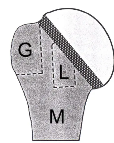
The topographic basis is the division of the proximal humerus into two segments-the articular segment (with C-fractures from C = Caput = Humeral head) and the extraarticular segment. This extraarticular segment (with A-fractures) is further divided into the three subsegments metaphysis (M), Greater tuberosity (G) and Lesser tuber-osity (L).
Both the articular and the extraarticular segments are divided by the anatomical neck (see Figure 1).
The fracture types
A-Fractures comprise fractures in the extraarticular seg-ment. Two-part-A fractures are divided in two-part meta-physeal fractures (M) that extend through the surgical neck, two-part greater tuberosity (G) and two-part lesser tuberosity fractures (L). The tuberosity fractures are defined by a complete separation of the tuberosity from the metaphysis and the anatomical neck.
Three-part A fractures (MG and ML fractures) are a meta-physeal fracture (M) with a fracture of one tuberosity (G or L).
Four-part A fractures (MT fracture) are a metaphyseal frac-ture (M) with a fracfrac-ture of both tuberosities (G+L = T) (see Figure 2a–f).
Type B: Incomplete Articular Fractures
Incomplete articular fractures show an incomplete frac-ture through the anatomic neck and extend into one of the extraarticular subsegments. This type describes a tuberos-ity or a medial wedge-shaped fragment, which is separated from the metaphysis and remains at the humeral head. Two-part B fractures are divided into three subtypes such as incomplete articular fractures with an medial metaphy-seal wedge shaped fragment at the humeral head (MB), an incomplete fracture of the anatomical neck with the greater tuberosity at the humeral head (GB) and an incomplete fracture of the anatomical neck with the lesser tuberosity connected to the humeral head (LB).
The further division of type B fractures depends on the additionally occurring tuberosity fractures. These three-part B fractures are GB fractures with a separate fracture of the lesser tuberosity (GBL), LB fractures with a separate fracture of the greater tuberosity (LBG), MB fractures with a separate fracture of the greater tuberosity (MBG) and MB fractures with a separate fracture of the lesser tuberosity (MBL).
Division of the proximal humerus into the articular segment (humeral head) and the extraarticular segment, further divided in three subsegments: M = metaphysis, G = greater tuberosity and L = lesser tuberosity
Figure 1
[image:2.612.335.518.99.328.2]B fractures with four main parts (= 4-part B fractures) are MB fractures in combination with a fracture of both tuber-osities (MBT) (see Figure 3 and 4).
Type C: Complete Articular Fractures
Type C articular fractures completely pass through the anatomic neck, but also may extend through the humeral head, which is completely separated from the extraarticu-lar segment. Type C fractures are divided into five sub-types. Two-part C fractures with just one complete fracture
through the anatomic neck are isolated articular fractures (SC).
Three-part C fractures showing a fracture of the greater tuberosity (CG), with a fracture of the lesser tuberosity (CL). Four-part C fractures show a fracture of both tuber-osities fractures (CT). Along with four-part fractures there is often a fracture of the metaphyseal subsegment. These even more complex fractures are called (CTM) (see Figure 5).
Schematic illustration of type A fractures
Figure 2
Type D-Fractures (Fracture-Dislocations)
D fractures are fracture-dislocations. D fractures are divided in type C fracture-dislocations (DC), type B
frac-ture-dislocations (DB) and type A fracfrac-ture-dislocations (DA).
[image:4.612.64.557.99.303.2]schematically shows the two-part B fractures
Figure 3
schematically shows the two-part B fractures. a) incomplete fracture of the anatomical neck with extension into the greater tuberosity (GB). b) incomplete fracture of the anatomical neck with extension into the lesser tuberosity (LB). c) incom-plete articular fractures with an medial metaphyseal wedge shaped fragment at the humeral head (MB).
The further division of type B fractures (Three-part B-fractures) depends on the additionally occurring tuberosity fractures
Figure 4
The further classification of DA fractures depends on the fractures in the extraarticular segment: disloca-tion with a metaphyseal fracture (DM), anterior fracture-dislocation fracture-dislocation and fracture of the greater tuberos-ity (DG) and posterior fracture-dislocation and fracture of the lesser tuberosity (DL).
The morphological basis
For morphological analysis, the MTM classification is based on four defined specifications, which are relevant for therapy and prognosis.
These specifications are organized by increasing fracture severity: minimally displaced and stable (S1), minimally displaced and unstable (S2), displaced (S3), displaced and comminuted (S4). In fractures with several parts, each part has to be classified individually. (see Figure 6).
S1: Minimally Displaced and Stable
The minimally displaced fractures (S 1) are defined as fractures with angulations up to 25°, a displacement of the tuberosities and the anatomical neck up to 5 mm, and a metaphyseal fracture displacement of 10 mm. Up to this extent of displacement, a real impairment of shoulder function is not to be expected.
Fracture stability is given if – through impaction of the main parts and preserved soft tissues – mobility between the main parts resulting in further displacement is unlikely. Thus, the fracture position, induced by the
trauma, is not changing by careful functional strain of the shoulder.
Therefore, minimally displaced and stable fractures are amenable to nonoperative treatment including early func-tional exercises. Regardless of fracture type and the number of main fragments, those fractures can be grouped together as S1 fractures since they are almost analogous in treatment and prognosis.
S2: Minimally Displaced and Unstable
The displacement of S2 fractures is defined similar to S1 fractures as a displacement of the tuberosities and the ana-tomical neck up to 5 mm, and a metaphyseal fracture dis-placement of 10 mm. Those fractures were defined as unstable when fractured parts were not impacted into each other resulting in instability between the fractured parts. Thus, through muscle pull and shoulder mobiliza-tion further displacements beyond the initial radiograph-ically diagnosed extent may occur.
Assessment of Stable and Unstable Fractures
If the above-defined criteria for stability and instability cannot clearly be applied radiologically, an additional fluoroscopic examination of the fracture is recommended. In this examination, a gentle abduction and rotation is applied in true a.p. view. If mobility can be visualized between fractured parts, the fracture is defined as unsta-ble.
Schematic illustration of type C fractures
Figure 5
S3: Displaced
S3 fractures always show a stronger malalignment and fragment separation, and, thus, the interfragmental soft tissue is ruptured more strongly, mostly induced by a combination of angulatory, rotatory and translatory dis-placement. Often, a compression mechanism leads to strong displacement with impaction. In displaced C frac-tures, the blood supply to the humeral head is completely destroyed. In cases with translatory and rotatory displace-ment of the humeral head, there is no integrity of the medial hinge and capsular and periosteal vessels ascend-ing intraosteally to the humeral head are ruptured[18,20].
S4: Displaced and Comminuted
In addition to the displacement, S4 fractures show com-minution of the main part complicating the therapeutic
procedure and worsening the prognosis. For example dis-placed head-splitting fractures define the fracture of the humeral head in several single fragments (C4).
The application of the classification
Due to its modularity, the classification can be applied in several ways depending on application purposes.
The short topographic version allows to classify into main types (A, B, C, D) or into the number of main parts (2, 3, 4) or into a combination of both the main-fracture-type and parts (2-p-A-fracture to 4-p-C-fracture).
A more detailed approach evaluated individual fracture types (M to CTM) or fracture types in combination with the individual specification (M to CTM and S1 to S4).
Specification scheme for proximal humeral fractures
Figure 6
Specification scheme for proximal humeral fractures.
Specification
Criteria for Specification
S1
Minimally displaced
stable
S2
Minimally displaced
unstable
S3
Displaced
S4
Displaced and
comminuted
Angulation up to 25°
Translation up to 5 mm
Metaphyseal up to 1 cm
Stability by impaction or
preserved soft
tissue connections
Instability by missing
impaction and destroyed soft
tissue connections
Displacement as S1
Angulation-Rotation > 25°
Translation > 5 mm
Metaphyseal > 1 cm
For example, a stable fracture of the anatomical neck with a displaced fracture of the greater tuberosity could be clas-sified as C1G3.
To allow the comparism with other studies we also classi-fied the fractures according to the determination between minimal-displaced and displaced fractures (S1/S2 to S3/ S4).
Methods
In a prospective study during March 2005 and August 2005 a consecutive series of 22 patients with 22 proximal humeral fractures presented at the BG Trauma Centre of the University of Tuebingen were included into the study. All patients were diagnosed with standardized plain x-rays in a.p. and axillary views with the patient supine using a shoulder splint with at least 60° abduction of the arm[21]. In addition, suspected instability in minimally displaced fractures was assessed using fluoroscopy (n = 4). All x-rays were reviewed by an experienced trauma sur-geon, who was not participating in the evaluation to ensure that the x-ray quality of all 22 patients was suffi-cient for later classification. All x-rays were collected and numerated; patient identification data was rendered anonymously. Then, the x-rays were submitted to three observers. Observer 1 was an experienced orthopaedic surgeon with fellowship training in shoulder surgery. Observer 2 was an experienced trauma surgeon with a spe-cial interest in bone and joint orthopaedic surgery. Observer 3 was a fellow for trauma surgery with a special interest in bone and joint surgery. None of the examiners were employed by the hospital, in which the fractures were seen. During routine work of the three observers only the Neer and AO classification were used. None of the observers had any experience with the MTM classifica-tion before study onset to exclude the influence of train-ing on reliability.
At the beginning of the study, the MTM classification was provided to the examiners in English and German lan-guage. In addition, the first author gave a 15-minute pres-entation of the MTM-Classification. A goniometer and a pen were given to the examiners. The data acquisition of the three examiners took place independently.
All fractures were classified by the observers according to the following guidelines:
a) Topographic analysis by individual fracture types (M to CTM) with consideration of individual specification (S1 to S4, Figures 6, Table 1).
b) Topographical analysis by individual fracture types (M to CTM) (see Table 1).
c) Analysis by a combination of both main fracture types (A, B, C, D) and number of parts (2–4) (see Table 1).
[image:7.612.53.553.633.729.2]a) Analysis by the 4 main fracture types (A, B, C or D) (Table 1).
d) Analysis by number of main parts (2-part, 3-part, and 4-part according to Neer-classification) (see Table 1).
e) Morphological distinction between minimal-displaced fracture and displaced fracture (S1/S2 or S3/S4) (see Fig-ures 6).
After initial analysis, a second evaluation of the x-rays took place after 4 months. Prior to this part of the study, none of the examiners had knowledge about this second evaluation. The classification was presented again in Eng-lish and German languages. A routine application of the MTM classification during routine work did not take place. The second evaluation was performed as described above except that radiographs were presented in reverse order. All except one fracture, that was treated conserva-tively, were stabilized by open reduction and internal fix-ation with an angular-stable plate osteosynthesis.
Statistical Methods
Percentage agreement, intraobserver reliability and inter-observer reliability were assessed with the JMP-statistical package (Version 6, SAS Campus Drive, Building S, Cary, NC, 27513, USA). For intraobserver reliability and inter-observer reliability, the kappa statistic function of the JMP-statistical package was used measuring kappa values (κ) to describe the agreement between observers while
Table 1: shows an overview of the classification of proximal humeral fractures into main parts and fracture types and subtypes.
Main fracture types
A B C D
Main Parts
2-part M G L GB LB MB SC DM DG DL
3-part MG ML GBL LBG MBG MBL CG CL
correcting for the proportion that may have occurred by chance alone.
A kappa value of 0 represented agreement by chance alone while kappa value of 1 represented a perfect agreement. Kappa values were interpreted using the guidelines pro-posed by Landis and Koch. Values between 0.81 and 1 indicated excellent or almost perfect, 0.61 and 0.80 sub-stantial, 0.41 and 0.60 moderate, 0.21 and 0.40 fair and 0 and 0.20 slight reliability [22].
Results
Intraobserver Reliability
Lowest percentages of agreement were detected for indi-vidual fracture type and morphological specification (M to CTM + S1–S4, range 54.5%–68.2%), followed by number of main parts combined with main fracture type (2–4 + A-D, range 68.2%–81.8%) and individual fracture type (M-CTM, range 68.2%–72.7%).
The highest percentage agreement between the observers was found for the parameters of fracture displacement (S1/2 – S3/4, range 90.1%–95.4%), number of main parts (2–4, range 86.4% – 90.1%) and main fracture type (A-D, range 81.8%–90.1%). Statistical analysis showed the low-est kappa values in individual fracture type and morpho-logical specification and the highest intraobserver reliability when fractures where classified according to number of main parts, fracture displacement and main fracture types including number of parts. (See Table 2).
Interobserver Reliability
The lowest percentage agreement between all observers was found in the assessment of combination of fracture type and individual specification. Followed by the classi-fication according to individual fracture type and main type and parts.
The highest percentage agreement between all observers was found in the assessment of fracture displacement fol-lowed by the classification according to main fracture types and fractured parts.
A fair level of agreement was found in the evaluation of individual fracture type in combination with specification
and individual fracture type and analysis of main fracture type and main parts. A moderate to substantial level of agreement was found in the evaluation of fracture dis-placement and number of main parts. (See Table 3).
Discussion
The MTM-Classification was developed to advance under-standing of proximal humeral fractures. Especially by introduction of incomplete articular fractures (B-frac-tures) a topographic classification could by possible. By adding morphological aspects and the possibility of a modular application of the system a precise evaluation of almost all fractures may be possible as well. However, the classification of proximal humeral fractures with the MTM-systems results in various differences in reliability depending on the short or extensive version of applica-tion. Analyzing the short version of the classification, the interobserver analysis resulted in moderate kappa values in the category 'main parts' (according to Neer-parts) and substantial kappa values for analyzing 'fracture displace-ment'. Those results were in accordance with the litera-ture[8,23]. Analyzing the extensive version of the MTM-Classification like evaluation of the main or individual fracture type with inclusion of the specification, only fair results can be expected.
[image:8.612.55.553.636.728.2]At present the classifications by Neer and the AO for prox-imal humeral fractures are widely accepted and com-monly used although both classifications have received some criticism during the last years [1,3]. Neither of the two systems achieved the general requirement of classifi-cations to adequately cover the complete spectrum of all possible fracture patterns. The Neer-system does not include 4-part valgus impacted fractures (covered by the AO-Classification as C2.1 or C2.2)[24]. Furthermore, the Neer-classification describes a large and rather inhomoge-neous group (one-part fractures/minimal-displacement) that consists of all fractures that are angulated less than 45° and displaced less than 1 cm, independently of the number of fractured parts. More so, 3 and 4-part fractures are divided by Neer in types of lesser tuberosity and greater tuberosity fractures. In these complex fractures, it remains unclear whether a fracture runs through the ana-tomical or the surgical neck. Furthermore, the AO classifi-cation includes three types (A, B and C) each divided into
Table 2: Kappa values of the intraobserver analysis of the three observers
Fracture Classification Observer 1 Kappa Observer 2 Kappa Observer 3 Kappa
Main-Type (A, B, C, D) 0.27 0.34 0.71
Main fragments (2, 3, 4) 0.73 0.82 0.76
Main Type + Main fragments (2A – 4C) 0.41 0.61 0.63
Fracture Displacement (S1/2 and S3/4) 0.76 0.46 0.64
Type of fracture (M-CTM) 0.48 0.58 0.54
3 groups and 9 subgroups. For further subgroup descrip-tion, 41 qualifications were used. Despite this detailed approach, complex injuries such as fractures of the ana-tomical neck with both tuberosities (4-part fractures according to Neer) or isolated lesser tuberosity fractures are inadequately defined.
New classifications systems such as the MTM classification were developed to address these critical points[18,19]. As a main prerequisite for correct topographic and morpho-logic fracture assessment, standardized and good-quality
[image:9.612.54.559.100.214.2]anteroposterior and axillary radiographs are required. Tamai published a series of 22 3 and 4-part fractures. He compared plain x-rays in anteroposterior and scapula lat-eral views with the intraoperative gross anatomy. Eight cases (36.4%) could neither be classified by Neer nor by the AO classification system. The authors concluded that diagnostics in complex fracture situations are difficult and that the classifications used were not as accurate as one would hope. Furthermore, Tamai attributed present diffi-culties in the choice of correct therapeutic methods to an inadequate fracture classification system[7].
Table 3: Kappa values of the interobserver analysis of the three observers
Observer 1&3 Observer 1&2 Observer 2&3 Observer 1–3
% Kappa % Kappa % Kappa % Kappa
Main-Type 77.3 0.19 84.1 0.39 79.5 0.32 80.3 0.30
Main fragments 68.2 0.41 79.5 0.60 75.0 0.55 74.2 0.52
Main Type & Main fragments 56.8 0.25 65.9 0.39 61.3 0.36 61.3 0.33
Fracture Displacement 95.4 0.73 93.2 0.63 97.7 0.85 95.4 0.74
Type of fracture 54.5 0.31 61.3 0.40 56.8 0.37 57.5 0.36
Type of fracture & specification 63.6 0.31 56.8 0.45 47.7 0.4 49.2 0.39
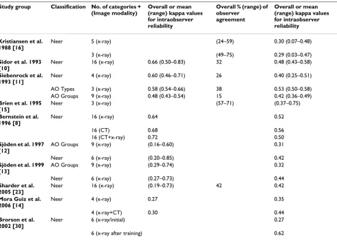
Table 4: Selected results of reliability studies of classifications for proximal humeral fractures
Study group Classification No. of categories + (Image modality)
Overall or mean (range) kappa values for intraobserver reliability
Overall % (range) of observer
agreement
Overall or mean (range) kappa values for intraobserver reliability
Kristiansen et al. 1988 [16]
Neer 5 (x-ray) (24–59) 0.30 (0.07–0.48)
3 (x-ray) (49–75) 0.29 (0.03–0.47)
Sidor et al. 1993 [10]
Neer 16 (x-ray) 0.66 (0.50–0.83) 32 0.48 (0.43–0.58)
Siebenrock et al. 1993 [11]
Neer 4 (x-ray) 0.60 (0.46–0.71) 26 0.40 (0.25–0.51)
AO Types 3 (x-ray) 0.58 (0.54–0.66) 38 0.53 (0.50–0.58) AO Groups 9 (x-ray) 0.48 (0.43–0.54) 15 0.42 (0.36–0.49)
Brien et al. 1995 [15]
Neer 3 (x-ray) (57–71) (0.37–0.75)
Bernstein et al. 1996 [8]
Neer 16 (x-ray) 0.64 0.52
16 (CT) 0.68 0.56
16 (CT+x-ray) 0.72 0.50
Sjöden et al. 1997 [12]
AO Groups 9 (x-ray) (0.16–0.60) 0.31
Neer 6 (x-ray) (0.20–0.85) 0.42
Sjöden et al. 1999 [13]
AO Groups 9 (x-ray) (0.29–0.74) 0.32
Neer 6 (x-ray) (0.27–0.73) 0.44
Sharder et al. 2005 [23]
Neer 16 (x-ray) (0.19–0.73) 42 0.42
Mora Guiz et al. 2006 [14]
Neer 4 (x-ray) 0.27 0.35
4 (x-ray+CT) 0.30 0.44
Brorson et al. 2002 [30]
Neer 6 (x-ray/initial) 0.27
[image:9.612.61.551.382.731.2]The MTM classification advanced available classifications by including the fracture model of Codman, the Neer analysis of fractured parts and the AO differentiation of fracture height, allowing for the description of almost all possible fracture types [1,3,25].
Published reliability studies differ in observer experience and diagnostic imaging methods. In addition, partly sim-plified Neer and AO classifications were used, leading only to limited study comparisons. (see Table 4).
Also, comparisons of kappa values from different studies may be problematic, as kappa values change with the prevalence of the diagnosis[26]. Despite these problem-atic aspects, the author's selected a few studies with com-parable methods for discussion.
Several authors assess the intraobserver reliability and interobserver reproducibility. Using conventional radiog-raphy and in some studies additional CT-scans, they found a fair to substantial mean intraobserver agreement as well as a fair to substantial mean interobserver agree-ment for the Neer and for the AO classification. (see Table 4).
The present study used standardized plain x-rays, enlisted experienced trauma surgeons and used the whole range of the classification spectrum. This approach led to an aver-age percentaver-age agreement of the three observers of 49.2% with a fair kappa value of 0.39 for fracture-analysis under consideration of individual fracture type and specifica-tion.
Siebenrock and Gerber also stated that the AO classifica-tion with three main types (A-C) resulted in better kappa values than the modified Neer classification with four choices of fracture types [11]. These findings were con-firmed by our study, in which we found acceptable to good agreement in 2 to 3 groups for determination of frac-ture displacement or fracfrac-tured main parts. On the other hand, Sidor found that a simplification of the Neer classi-fication from 16 into 6 categories did not result in signifi-cant changes of interobserver reliability[10]. However, a 2-choice question, for example about therapeutic man-agement or (as in the present study) about fracture dis-placement led to perfect and good reliability, indicating a dependency of interobserver reliability and the number of choices the observer has to select[8,23].
Sidor also discussed that a differentiation of single frag-ments due to multiple fractures lines is difficult. He also stresses the importance of a high-quality radiological diagnostics, which makes an overlapping free presenta-tion for the fractured region possible to avoid any classifi-cation restriction. For an excellent classificlassifi-cation of
proximal humeral fractures, a perfect radiological visuali-zation of the fractured region is mandatory[10]. In accord-ance with other experts, it is possible to increase the information with an axillary view in terms of direction and dimension of fracture dislocation, humeral head involvement, and the degree of displacement of tuberosity fractures, especially considering fractures of the lesser tuberosity [27-29].
Brorson and Shrader analyzed the importance of training in a specific classification system. They could show that kappa values for interobserver reliability were signifi-cantly improved from fair to substantial after observers had received a classification training[23,30,31].
Since the training in a specific classification system improves reliability, one weakness of the current study was, that the MTM-classification was not used in daily clinical practice. With sufficient training, the reliability of this classification could be higher.
Conclusion
In summary, some complex fracture types are inade-quately defined by classification systems such as the present Neer or AO classification. To allow a precise topo-graphic and morphological description, the MTM classifi-cation was developed for a better understanding of individual fractures and to address the question whether a very detailed classification of proximal humeral fractures may be limited by its reliability. Unfortunately, the very detailed classification approach led only to fair or unac-ceptable results and is not helpful to improve reliability.
We concluded that a detailed classification exceeding the part analysis of Neer is not a practical approach to address current problems in classification systems regarding prox-imal humerus fractures.
For future projects an evaluation of ongoing develop-ments of diagnostic imaging-technology like the CT and here specifically multiplanar visualisation of the fracture in thin-cut technique and 3 D visualisation should be undertaken.
References
1. Jakob RP KT Mayo K, Ganz R, Muller ME: Classifications and aspects of treatment of fractures of the proximal humerus.
In: Bateman JE, Welsh RP (esd): Surgery of the shoulder Philiadelphia: CV Mosby Co 1984:330-343.
2. Duparc J, Largier A: [Fracture-dislocations of the upper end of the humerus]. Rev Chir Orthop Reparatrice Appar Mot 1976,
62(1):91-110.
3. Neer CS 2nd: Displaced proximal humeral fractures. I. Classi-fication and evaluation. J Bone Joint Surg Am 1970,
52(6):1077-1089.
4. Habermeyer P: [Fracture of the head of the humerus].
Publish with BioMed Central and every scientist can read your work free of charge "BioMed Central will be the most significant development for disseminating the results of biomedical researc h in our lifetime."
Sir Paul Nurse, Cancer Research UK
Your research papers will be:
available free of charge to the entire biomedical community
peer reviewed and published immediately upon acceptance
cited in PubMed and archived on PubMed Central
yours — you keep the copyright
Submit your manuscript here: BioMedcentral
5. Meyer DC, Espinosa N, Hertel R: Combined fracture of the greater and lesser tuberosities with intact connection of the humeral head to the shaft. J Trauma 2006, 61(1):206-208. 6. Ackermann C, Lam Q, Linder P, Kull C, Regazzoni: [Problems in
classification of fractures of the proximal humerus]. Z Unfallchir Versicherungsmed Berufskr 1986, 79(4):209-215.
7. Tamai K, Hamada J, Ohno W, Saotome K: Surgical anatomy of multipart fractures of the proximal humerus. J Shoulder Elbow Surg 2002, 11(5):421-427.
8. Bernstein J, Adler LM, Blank JE, Dalsey RM, Williams GR, Iannotti JP:
Evaluation of the Neer system of classification of proximal humeral fractures with computerized tomographic scans and plain radiographs. J Bone Joint Surg Am 1996,
78(9):1371-1375.
9. Sallay PI, Pedowitz RA, Mallon WJ, Vandemark RM, Dalton JD, Speer KP: Reliability and reproducibility of radiographic interpreta-tion of proximal humeral fracture pathoanatomy. J Shoulder Elbow Surg 1997, 6(1):60-69.
10. Sidor ML, Zuckerman JD, Lyon T, Koval K, Cuomo F, Schoenberg N:
The Neer classification system for proximal humeral frac-tures. An assessment of interobserver reliability and intraobserver reproducibility. J Bone Joint Surg Am 1993,
75(12):1745-1750.
11. Siebenrock KA, Gerber C: The reproducibility of classification of fractures of the proximal end of the humerus. J Bone Joint Surg Am 1993, 75(12):1751-1755.
12. Sjoden GO, Movin T, Guntner P, Aspelin P, Ahrengart L, Ersmark H, Sperber A: Poor reproducibility of classification of proximal humeral fractures. Additional CT of minor value. Acta Orthop Scand 1997, 68(3):239-242.
13. Sjoden GO, Movin T, Aspelin P, Guntner P, Shalabi A: 3D-radio-graphic analysis does not improve the Neer and AO classifi-cations of proximal humeral fractures. Acta Orthop Scand 1999,
70(4):325-328.
14. Mora Guix JM, Gonzalez AS, Brugalla JV, Carril EC, Banos FG: Pro-posed protocol for reading images of humeral head frac-tures. Clin Orthop Relat Res 2006, 448:225-233.
15. Brien H, Noftall F, MacMaster S, Cummings T, Landells C, Rockwood P: Neer's classification system: a critical appraisal. J Trauma 1995, 38(2):257-260.
16. Kristiansen B, Andersen UL, Olsen CA, Varmarken JE: The Neer classification of fractures of the proximal humerus. An assessment of interobserver variation. Skeletal Radiol 1988,
17(6):420-422.
17. Rees J, Hicks J, Ribbans W: Assessment and management of three-and four-part proximal humeral fractures. Clin Orthop 1998:18-29.
18. Hertel R: Fractures of the proximal humerus in osteoporotic bone. Osteoporos Int 2005, 16 Suppl 2:S65-72.
19. Edelson G, Kelly I, Vigder F, Reis ND: A three-dimensional classi-fication for fractures of the proximal humerus. J Bone Joint Surg Br 2004, 86(3):413-425.
20. Hertel R, Hempfing A, Stiehler M, Leunig M: Predictors of humeral head ischemia after intracapsular fracture of the proximal humerus. J Shoulder Elbow Surg 2004, 13(4):427-433.
21. Schnabel M, Bahrs C, Walthers E, Gotzen L: [Marburg shoulder radiography splint (MSR splint) for standardized and high quality plain film radiography in fractures of the proximal humerus]. Unfallchirurg 2004, 107(11):1099-1102.
22. Landis JR, Koch GG: The measurement of observer agreement for categorical data. Biometrics 1977, 33(1):159-174.
23. Shrader MW, Sanchez-Sotelo J, Sperling JW, Rowland CM, Cofield RH: Understanding proximal humerus fractures: image anal-ysis, classification, and treatment. J Shoulder Elbow Surg 2005,
14(5):497-505.
24. Jakob RP, Miniaci A, Anson PS, Jaberg H, Osterwalder A, Ganz R:
Four-part valgus impacted fractures of the proximal humerus. J Bone Joint Surg Br 1991, 73(2):295-298.
25. Codman EA: Fractures in relation to the subacromial bursa. In In: Codman EA, ed The Shoulder: Rupture of the Supraspinatus Tendon and other Lesions in or about the subacromial Bursa Boston, MA , Thomas Todd; 1934:313-333.
26. Gjorup T: The kappa coefficient and the prevalence of a diag-nosis. Methods Inf Med 1988, 27(4):184-186.
27. Leibman MI, Zuckerman JD: Proximal Humeral Fractures:Clini-cal Evaluation and Classification. in Shoulder Fractures-The
Practi-cal Guide to Management, Zuckerman JD and Koval KJ eds, Thieme New York, Stuttgart 2005, 1:37-49.
28. Ballmer FT, Hertel R: [Indications and results of shoulder pros-thetics in complex proximal humerus fractures]. Ther Umsch 1998, 55(3):197-202.
29. Resch H: [Fractures of the humeral head]. Unfallchirurg 2003,
106(8):602-617.
30. Brorson S, Bagger J, Sylvest A, Hrobjartsson A: Improved interob-server variation after training of doctors in the Neer system. A randomised trial. J Bone Joint Surg Br 2002, 84(7):950-954. 31. Brorson S, Bagger J, Sylvest A, Hrobjartsson A: Low agreement
among 24 doctors using the Neer-classification; only moder-ate agreement on displacement, even between specialists.
Int Orthop 2002, 26(5):271-273.
Pre-publication history
The pre-publication history for this paper can be accessed here: