What Is a Sentinel Node? Re-Evaluating the 10% Rule
for Sentinel Lymph Node Biopsy in Melanoma
HIDDE M. KROON,MD,1LORI LOWE,MD,2SANDRA WONG,MD,1DOUG FULLEN,MD,2LYNDON SU,MD,2 VINCENT CIMMINO,MD,1ALFRED E. CHANG,MD,1TIMOTHY JOHNSON,MD,1,2,3ANDMICHAEL S. SABEL,MD1*
1Department of Surgery, University of Michigan Comprehensive Cancer Center, Ann Arbor, Michigan 2Department of Pathology, University of Michigan Comprehensive Cancer Center, Ann Arbor, Michigan 3Department of Dermatology, University of Michigan Comprehensive Cancer Center, Ann Arbor, Michigan
Introduction:Many surgeons use the ‘‘10% rule’’ to define whether a lymph node is a sentinel node (SLN) when staging malignant melanoma. However, this increases the number of SLN removed and the time and cost of the procedure. We examined the impact of raising this threshold on the accuracy of the procedure.
Methods:We reviewed the records of 561 patients with melanoma (624 basins) who underwent SLN with technetium Tc99 labeled sulfur colloid using a definition of a SLN as 10% of that of the node with the highest counts per minute (CPM).
Results:Of the 624 basins, 154 (25%) were positive for metastases. An average of 1.9 nodes per basin were removed (range 1–6). Metastases were found in the hottest node in 137 cases (89% of positive basins, 97% of basins overall). Increasing the threshold above 10% decreased the number of nodes excised and the costs involved, but incrementally raised the number of false negative cases above baseline (a 4% increase for a ‘‘20% rule,’’ 5% for a ‘‘30% rule,’’ 6% for a ‘‘40% rule,’’ and 7% for a ‘‘50% rule’’). Taking only the hottest node would raise the false negative rate by 11%. Conclusions:Although using thresholds higher than 10% for the definition of a SLN will minimize the extent of surgery and decrease the costs associated with the procedure, it will compromise the accuracy of the procedure and is not recommended.
J. Surg. Oncol. 2007;95:623–628. ß2007 Wiley-Liss, Inc.
KEYWORDS: melanoma; sentinel lymph node biopsy; costs and cost analysis
INTRODUCTION
Lymphatic mapping and sentinel lymph node (SLN) biopsy have emerged as the standard method of evaluating the tumor status of the regional lymph nodes in patients with malignant melanoma. There is little doubt that SLN biopsy provides important prognostic information with relatively low morbidity. The fact that the SLN accurately reflects the status of the entire regional node field in the vast majority of cases has been well validated [1–3]. Knowledge of the SLN status provides the patient with a more reliable estimate of prognosis, and allows more accurate stratification for entry into adjuvant therapy trials. It also identifies those patients who may benefit from a completion lymphade-nectomy.
Using both isosulfan blue dye and radioactive colloid to identify the SLN has improved both the success rate and accuracy of the procedure, but often results in the excision of a large number of lymph nodes per basin.
Many surgical oncologists use the ‘‘10% rule’’ to define whether a lymph node is a ‘‘sentinel’’ node or not [2,4–8]. This rule dictates that any lymph node that has at least 10% of the counts per minute (CPM) of the hottest SLN ex vivo should be removed and labeled as a SLN. If the hottest node has CPM of 5,000 for example, then any remaining node in the basin that has CPM of 500 or more should be removed. Support for this rule comes from the results of the Sunbelt Melanoma Trial, where thresholds of higher than 10% would have resulted in higher rates of missed positive nodes [9]. In addition, the interim
*Correspondence to: Dr. Michael S. Sabel, University of Michigan Comprehensive Cancer Center, 3304 Cancer Center, 1500 East Medical Center Drive, Ann Arbor, MI 48105. Fax: (734)-647-9647.
E-mail: [email protected]
Received 14 July 2006; Accepted 16 October 2006 DOI 10.1002/jso.20729
Published online 7 March 2007 in Wiley InterScience (www.interscience.wiley.com).
findings of the Multicenter Selective Lymphadenectomy Trial-I (MSLT-I) demonstrated that use of the 10% rule will identify almost the same number of patients with micrometastases as will eventually develop clinically evident regional nodal metastases [10,11].
However, the more SLNs removed, the higher the cost of the procedure, due to added time during surgery and increased pathology charges for the serial sectioning, hematoxylin and eosin (H&E) staining, and immunohis-tochemistry (IHC) of each sentinel node. We therefore examined in our patient group what the impact would be on both the accuracy and the number of lymph nodes excised when a threshold higher than 10% would be used to define a lymph node a sentinel node.
MATERIALS AND METHODS
Approval of this study was granted by the University of Michigan Institutional Review Board for Medicine. From our prospective melanoma database we identified 1,274 patients who underwent lymphatic mapping and sentinel node biopsy between August, 1997 and December, 2004. Of these patients, 561 had the CPM recorded for each individual sentinel node (we began prospectively record-ing the CPM and presence of blue dye for each lymph node in late 2001) from 624 basins.
SLN biopsy was performed in patients with melanoma
1.0 mm in Breslow thickness, or in patients with melanoma<1.0 mm if there were other adverse clinical or histopathologic features (young age, high mitotic rate, ulceration, significant dermal regression). All patients underwent same-day preoperative injection of techne-tium 99 m sulfur colloid (CIS-US, Inc., Bedford, MA) intradermally around the primary lesion or biopsy site, followed by lymphoscintigraphy 2 hr later. Isosulfan blue dye (Lymphazurin 1%; Hirsch Industries, Inc., Richmond, VA) was injected intradermally around the primary lesion or biopsy site, and the area massaged for 5 min to promote lymphatic flow. A handheld gamma probe (Navigator GPS; US Surgical, Norwalk, CT) was used to identify ‘‘hot’’ spot(s) over the SLN(s). A small incision was made directly over the ‘‘hot’’ spot(s) and carried down through the skin and subcutaneous tissue into the node bearing fat.
Lymph nodes were removed and labeled as sentinel nodes if they were clinically suspicious, blue, had blue stained afferent lymphatic vessels, or were ‘‘hot,’’ defined as the hottest node plus any lymph node with at least 10% of the CPM of the hottest node. The impact of the use of isosulfan blue dye was not evaluated for this study. Each SLN was sent for careful histopathologic analysis. SLNs were serially sectioned and stained with H&E. Sentinel nodes that were negative on H&E were then stained with a battery of IHC stains, consisting of S-100, Melan-A,
and/or HMB-45 as described previously [12]. Patients with drainage to multiple basins had the same rules applied to each individual basin.
RESULTS
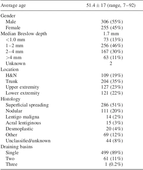
The clinical and pathologic characteristics of the patients included in this analysis are presented in Table I. The 561 patients who had the ex vivo CMP recorded from the Navigator probe recorded for each lymph node had a demographic profile similar to the full dataset of 1,274 patients. Among these 561 patients there were 624 nodal basins explored. Sixty-two patients (11%) had drainage to more than one basin; 61 with drainage to two lymphatic basins and 1 patient with drainage to 3 basins. An average of 1.9 nodes per basin were removed as SLN (range 1–6). In 39% of cases, only 1 SLN was removed from the basin. In 37%, two lymph nodes were removed and in 24% of the cases, three or more lymph nodes were removed (Fig. 1).
[image:2.594.304.544.88.373.2]We sought to determine how often metastases are detected in lymph nodes other than the ‘‘hottest’’ node. Of the 624 nodal basins with evidence of lymphatic drainage on preoperative lymphoscintigraphy, 470 basins (75%) were negative for evidence of melanoma metas-tases. Among the 154 lymph node positive basins (25%), there were 46 basins in which the positive lymph node was the only lymph node removed (Table II). In 89 TABLE I. Clinical and Pathologic Features of Study Population
Average age 51.417 (range, 7–92)
Gender
Male 306 (55%) Female 255 (45%) Median Breslow depth 1.7 mm
<1.0 mm 73 (13%) 1–2 mm 256 (46%) 2–4 mm 167 (30%) >4 mm 63 (11%) Unknown 2 Location
H&N 109 (19%) Trunk 204 (35%) Upper extremity 127 (23%) Lower extremity 121 (22%) Histology
Superficial spreading 286 (51%) Nodular 111 (20%) Lentigo maligna 14 (2%) Acral lentiginous 15 (3%) Desmoplastic 20 (4%) Other 69 (12%) Unclassified/unknown 44 (8%) Draining basins
basins, there were more than one lymph node labeled as sentinel, however, the metastases were detected in the hottest node. There were two cases where metastases were detected in a lymph node that was cold (no significant CPM) but was labeled as a sentinel node because it was either blue or clinically suspicious. Overall, there were 17 of 624 basins (2.7%) where the hottest node did not have evidence of metastases, but melanoma was detected in a less radioactive node. Of these 17 cases, 11 (65%) had the melanoma detected in the 2nd hottest node, 5 (29%) were in the 3rd hottest and 1 case where metastases were detected in the 4th hottest node (6%). Stopping after the 3rd or 4th lymph node was excised would have minimal to no impact on the accuracy of the procedure; however, it was rare that more than 3 or
4 nodes needed to be excised, and this would not significantly decrease the number of nodes that need to be examined.
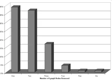
We then examined how many instances we would have missed metastases if we followed a rule other than the 10% rule (Fig. 2). As stated, there were 17 cases where the metastases were not in the hottest lymph node. Therefore, had we removed only the hottest node on a regular basis, 11% of the positive basins would have been falsely interpreted as negative. Using a ‘‘50% rule,’’ meaning we would have left behind any lymph node that was not at least 50% of the hottest node, would have resulted in a false negative finding in 11 (7%) cases. This drops to 9 cases (6%) for a ‘‘40% rule,’’ 7 cases (5%) for a ‘‘30% rule,’’ and 6 cases (4%) for a ‘‘20% rule.’’
[image:3.594.104.492.67.346.2]Using this data, we examined what would be the change in the false negative rate and the accuracy of the procedure had we used a different threshold for how we define a SLN. We based these on the assumption that there is a baseline false negative rate of 18%, as was found in the MSLT-I [11]. In addition, using all patients (lymph node positive and negative), we calculated for each basin how many lymph nodes would have been removed for each threshold. For example, if the hottest node had counts of 5,000, and a node of 500 was removed using the 10% rule, then under the 20% rule this node would not have been removed. Looking at all 624 basins, moving from a 10% rule to a 20% rule would have meant examining 1,051 nodes instead of 1,190. The change in Fig. 1. Percentage of numbers (one to six) of sentinel lymph nodes (SLN) removed per basin.
TABLE II. Number of Metastasis Detected in the ‘‘Hottest’’ Node and in Subsequent Nodes Among Positive Basins
Number of basins
% Total
% of Positive basins
Node positive 154 25
[image:3.594.50.290.605.719.2]the accuracy of the procedure and the number of lymph nodes not excised is presented in Table III.
DISCUSSION
After the landmark report in 1992 by Morton et al. [13] from the John Wayne Cancer Institute on lymphatic mapping and SLN biopsy in 1993 and subsequent confirmation by other centers of its feasibility and accuracy [1,3], this procedure is increasingly used in the management of primary melanoma. Its use, however, is not without controversy. Although the sentinel node procedure provides accurate prognostic information, its true impact on survival remains unclear. Even if the early
surgical eradication of micrometastatic disease in the nodes (as opposed to waiting until metastasis become clinically apparent) is associated with improved survival, this benefit will be limited only to the approximately 20% of patients who harbor micrometastases in the nodes.
The controversy surrounding the use of SLN biopsy centers on the increased costs of a procedure that benefits a small subset of patients. For patients deemed appropriate candidates, surgical therapy shifts from an office-based procedure, which can be done for approxi-mately $1,000–$1,750, to one where IV sedation or more commonly general anesthesia is utilized, nuclear medicine is involved for the injection of Technitium 99 colloid sulfur and the performance of lymphoscintigra-phy, and a more time-consuming pathologic evaluation of the sentinel nodes is necessary, with thin-sectioning and IHC staining. This raises the cost of treating melanoma to between $7,150 and $15,223 [14,15].
[image:4.594.103.495.65.334.2]The benefit of serial sectioning of the SLNs, and the use of IHC when no metastases are detected by routine H&E, has been well described in the literature [16,17]. More (micro-)metastases are detected by this meticulous approach and these have clinical significance in mela-noma. However, this more in-depth histologic evaluation increases costs compared to routine histology, and each lymph node the surgeon labels for the pathologist as ‘‘sentinel’’ makes the procedure more expensive. Mini-mizing the number of lymph nodes that need to be Fig. 2. Percentage of cases in which an increase of the ‘‘10% rule’’ for the definition of a sentinel node, based on isotope counts per minute, would have resulted in missed positive sentinel nodes in the regional basin.
TABLE III. Change in False Negative Rate, Accuracy, and Nodes Not Examined With Incremental Increases of the ‘‘10% Rule’’
Rule applied
False negative ratea
(%)
Accuracy (%)
Nodes excised
Nodes not excised
10% rule 18 95 1,190 0 20% rule 22 94 1,051 139 30% rule 22 94 951 239 40% rule 23 93 885 305 50% rule 25 93 833 357 Hottest node only 28 92 624 566
[image:4.594.55.293.595.697.2]examined may improve the cost-effectiveness of the procedure. Several authors have examined the most appropriate threshold where the nodes most likely to harbor disease have been excised. Porter et al. [18] suggested it may be reasonable to increase this threshold based on their experience with 633 patients in whom removal of lymph nodes with CPM lower than two thirds (66%) of the hottest node affected patient management in less than 0.2% of the cases. Using data from the Sunbelt Melanoma Trial, McMasters et al. [9] looked at 1,184 patients who had melanomas 1.0 mm and concluded that removal of the nodes with blue dye or with radioactive counts greater than 10% of the ‘‘hottest’’ node yields excellent results, with in their study only one false-negative result. Jacobs et al. [19] also adopted the 10% rule based on their experience with 134 SLN biopsies. Our data also suggest that raising the threshold above 10% will compromise the accuracy of the procedure.
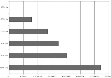
However, maintaining this threshold is not without cost. To illustrate the economic impact of decreasing the number of SLNs excised, we estimated the savings per lymph node based on the additional time in the operating room and the pathology costs (Fig. 3). We assumed an additional 5 min of operating time per additional sentinel node removed, at a cost of $10 per minute. Pathology costs were based on the Medicare RVU payments in Michigan for the specific CPT codes (H&E evaluation per sentinel node, assuming one cassette, $178.06; two
immunohisto-chemical stains, $172.98). Because IHC is no longer used once metastases are detected on H&E, the pathology cost savings were calculated with the following formula. Cost savings¼{(nodes not examinednode negative rate)cost H&Eþ2 IHC stains}þ{(nodes not exam-inednode positive rate)cost H&E only}.
These figures demonstrate the relative cost savings with incremental increases in the 10% rule, but represent the absolute minimum in cost savings by decreasing the number of lymph nodes excised. The true cost savings will vary among institutions depending on the average reimbursement rate for the hospital’s operating room and pathology services, which depends upon the payor mix. The Medicare reimbursement rates used in the calcula-tions are generally lower than those of private insurance companies or private paying patients. There are also hidden costs that are difficult to quantify and not included in the calculations such as the cost of managing post-operative complications. It is not unreasonable to believe that less perturbation of the basin might result in lower complications (infection, bleeding, seroma, numbness, and rarely lymphedema) with a decrease of the indirect costs associated with the procedure.
[image:5.594.106.481.439.705.2]If we had found (as we had hoped) that these potential cost savings could be obtained with no or minimal compromise to the goal of the procedure, namely identifying those patients who harbor micrometastases, then the conclusion would be obvious. This was not the case, however. While raising the threshold to 30% would
decrease the costs of the procedure, it would falsely label an additional 5 out of 100 positive patients as node-negative, denying them the potential benefits of comple-tion node disseccomple-tion or adjuvant high-dose interferon [20]. While a more formal cost analysis would require a detailed quantification of these benefits, at this time these savings do not appear to justify routinely raising the threshold at which we terminate the procedure.
While raising the 10% threshold for labeling a lymph node as ‘‘sentinel’’ does not appear justified, this does not alleviate our responsibility as surgeons to minimize the costs and morbidity of the procedure. What these data do suggest is we are in need of better tracers to identify the true SLN without migrating to multiple second-tier nodes. Large-particle radiocolloids, such as unfiltered
99m
Tc-sulfur colloid, do not illustrate well the lymphatic collecting vessels on imaging, and only a small fraction of the dose reaches the nodes [21]. Thus surgeons are dependent upon count ratios such as the 10% rule, and the excision of multiple lymph nodes, to maintain the accuracy of the procedure. 99mTc-antimony sulfide colloid is a small-particle radiocolloid, not available in the United States, that gathers in the lymph nodes in a much higher fraction [21]. Using this tracer, surgeons from the Sydney Melanoma Unit report that the sentinel node is often the only node identified on imaging, and the visualization of the lymphatic vessels (with careful imaging techniques) provide further guidance to the surgeon in identifying the true sentinel node [22]. A new radiopharmaceutical agent under investigation, Lympho-seek, binds to a receptor on the surface of macrophage cells, so as to limit drainage to distal nodes that are not anatomically sentinel [23,24]. Use of these tracers may significantly reduce the cost of the procedure without compromising accuracy.
In conclusion, despite potential cost savings to using a higher threshold than 10% for excising and labeling regional lymph nodes as ‘‘sentinel,’’ the accuracy of the procedure will be adversely affected. Our data suggest that surgeons using99mTc-sulfur colloid, who routinely terminate the procedure after identifying only the hottest node, or when counts drop below just half or one-third the CPM of the hottest node, will experience higher false-negative rates. These results support the continued use of the 10% rule as a guideline for the performance of SLN biopsy in melanoma.
REFERENCES
1. Thompson JF, McCarthy WH, Bosch CM, et al.: Sentinel lymph node status as an indicator of the presence of metastatic melanoma in regional lymph nodes. Melanoma Res 1995;5:255–260. 2. Morton DL, Thompson JF, Essner R, et al.: Validation of the
accuracy of intraoperative lymphatic mapping and sentinel lymphadenectomy for early-stage melanoma: A multicenter trial.
Multicenter selective lymphadenectomy trial group. Annals of Surgery 1999;230:453–463.
3. Reintgen DS, Cruse CW, Wells K, et al.: The orderly progression of melanoma nodal metastases. Ann Surg 1994;220:759–767. 4. Sabel MS, Gibbs JF, Cheney R, et al.: Evolution of sentinel lymph
node biopsy for melanoma at a National Cancer Institute-designated cancer center. Surgery 2000;128:556–563.
5. Krag DN, Weaver D, Ashikaga T, et al.: The sentinel node in breast cancer—A multicenter validation study. N Engl J Med 1998;339:941–946.
6. Nathanson SD: Will the true sentinel node please stand? Ann Surg Oncol 1999;6:514–516.
7. Spillance A: Sentinel node biopsy in breast cancer and melanoma requires adequate self-audit. ANZ J Surg 2004;74:308–313. 8. Quan ML, McCready D, Temple WJ, et al.: Biology of lymphatic
metastasis in breast cancer: Lessons learned from sentinel node biopsy. Ann Surg Oncol 2002;9:467–471.
9. McMasters KM, Reintgen DS, Ross MI, et al.: Sentinel lymph node biopsy for melanoma: How many radioactive nodes should be removed? Ann Surg Oncol 2001;8:192–197.
10. Morton DL: Interim results of the Multicenter Selective Lymphadenectomy Trial (MSLT-I) in clinical stage I melanoma. Presented at the 41st Annual Meeting of the American Society of Clinical Oncology. Annual Meeting of the American Society of Clinical Oncology. Orlando, Florida, United States, 2005. 11. Morton DL, Cochran AJ, Thompson JF, et al.: Sentinel node
biopsy for early-stage melanoma: Accuracy and morbidity in MSLT-I, an international multicenter trial. Ann Surg 2005;242: 302–313.
12. Karimipour DJ, Lowe L, Su L, et al.: Standard immunostains for melanoma in sentinel lymph node specimens: Which ones are most useful? J Am Acad Dermatol 2003;50:759–764.
13. Morton DL, Wen DR, Wong JH, et al.: Technical details of intraoperative lymphatic mapping for early stage melanoma. Arch Surg 1992;127:392–399.
14. Essner R, Stern SL, Bostick PJ, et al.: Results of lymphatic mapping in melanoma. In: Neiweg OE, Essner R, Reintgen DS, Thompson JF, editors. Lymphatic mapping and probe applications in oncology. New York: Marcel Dekker; 2000. pp 101–124.
15. Agnese DM, Abdessalam SF, Burak WE, et al.: Cost-effective-ness of sentinel lymph node biopsy in thin melanomas. Surgery 2003;134:542–548.
16. Spanknebel K, Coit DG, Bieligk SC, et al.: Characterization of micrometastatic disease in melanoma sentinel lymph nodes by enhanced pathology: Recommendations for standardizing patho-logic analysis. Am J Surg 2005;29:305–317.
17. Abrahamsen HN, Hamilton-Dutoit SJ, Larsen J, et al.: Sentinel lymph nodes in malignant melanoma: Extended histopathologic evaluation improves diagnostic precision. Cancer 2004;100: 1683–1691.
18. Porter GA, Ross MI, Berman RS, et al.: How many lymph nodes are enough during sentinel lymphadenectomy for primary melanoma? Surgery 2000;128:306–311.
19. Jacobs IA, Chang CK, Das Gupta TK, et al.: High isotope counts and sentinel node positivity in patients with melanoma. Arch Surg 2003;138:63–66.
20. Sabel MS, Sondak VK: Pros and cons of adjuvant interferon in the treatment of melanoma. Oncologist 2003;8:451–458.
21. Bergqvist L, Strand S-E, Persson BRR: Particle sizing and biokinetics of interstitial lymphoscintigraphic agents. Semin Nucl Med 1983;8:9–19.
22. Uren RF, Howman-Giles RB, Thompson JF: Patterns of lymphatic drainage from the skin in patients with melanoma. J Nucl Med 2003;44:570–582.
23. Wallace AM, Hoh CK, Vera DR, et al.: Lymphoseek: A molecular radiopharmaceutical for sentinel node detection. Ann Surg Oncol 2003;10:531–538.