John Wiley & Sons. Printed in Singapore
IMP-3 expression in melanocytic
lesions
Background:Insulin-like growth factor-II mRNA-binding protein 3 (IMP-3), a member of the insulin-like growth factor mRNA-binding protein family, is expressed in several human malignancies, including melanomas. However, the expression of IMP-3 has not been explored in melanomain situ, various histologic subtypes of invasive melanomas and atypical Spitz tumors.
Methods:IMP-3 immunostain was performed in 157 melanocytic lesions.
Results:Nearly all benign (8/8), dysplastic (8/8) and Spitz nevi (8/9) were negative for IMP-3. Focal IMP-3 positivity was observed in 5/12 melanomain situand 4/15 superficial melanomas (Breslow depth
≤1 mm). Half (10/20) of deep melanomas (Breslow depth>1 mm) and 25/52 metastatic melanomas demonstrated strong IMP-3 staining. IMP-3 expression differs significantly between non-desmoplastic melanomas (superficial and deep) and benign or dysplastic or Spitz nevi (p=0.0427, respectively). Four of 23 desmoplastic melanomas
expressed IMP-3, which was significantly different from deep
melanomas (p=0.0109). IMP-3 stained 7 of 10 atypical Spitz tumors. The difference between atypical Spitz tumors and Spitz nevi was statistically significant (p=0.0256).
Conclusion:A malignant circumstance, such as non-desmoplastic melanoma or atypical Spitz tumor, can be inferred when IMP-3 is expressed, suggesting potential diagnostic value of IMP-3 in melanocytic lesions.
Yu L, Xu H, Wasco MJ, Bourne PA, Ma L. IMP-3 expression in melanocytic lesions.
J Cutan Pathol 2010; 37: 316–322.©2009 John Wiley & Sons A/S.
Limin Yu1, Haodong Xu3,
Matthew J. Wasco1, Patricia A.
Bourne3and Linglei Ma1,2
1Department of Pathology, and
2Department of Dermatology, University of
Michigan Medical Center, Ann Arbor, MI, USA
3Department of Pathology, University of
Rochester Medical Center, Rochester, NY, USA
Linglei Ma, Department of Pathology, Dermatopathology Division, University of Michigan, M3260, Medical Sciences 1, 1301 Catherine Street, Ann Arbor, MI
48109-0602, USA Tel: +1 734 764 4460 Fax: +1 734 764 4690 e-mail: [email protected]
Accepted for publication August 12, 2009
Malignant melanoma is a life-threatening cutaneous cancer. Its incidence and mortality has risen worldwide.1 The diagnosis of melanocytic lesions is mainly based on a number of well-defined histology criteria, but remains one of the most challenging areas in dermatopathology. Interobserver differences are not infrequently encountered for difficult lesions.2 Despite advances in melanoma research, no reliable diagnostic and/or prognostic markers have yet been identified.
Insulin-like growth factor-II (IGF-II) mRNA-binding protein 3 (IMP-3), also known as K homology domain-containing protein overexpressed in cancer (KOC) or L523S, is a member of the
Data from in vitro IMP-3 knockdown study and from IMP-3 administration in patients with lung cancer indicate that IMP-3 may be a promising therapeutic target for human malignancies.8,9 A recent study demonstrated that IMP-3 was expressed in malignant melanomas but not in nevi.10However, the expression of IMP-3 in melanomain situ(MIS), desmoplastic melanoma and atypical Spitz tumor (AST) has not been examined in the literature. Interestingly, IMP-1, another member of IMP family protein, was recently found to be elevated in primary human melanomas and melanoma cell lines.11
In this study, we investigated the expression of IMP-3 in a spectrum of cutaneous melanocytic lesions, including benign/dysplastic/Spitz nevus, MIS, primary melanoma (non-desmoplastic and desmoplastic melanoma), AST and metastatic melanoma. We further assessed the diagnostic and prognostic utility of IMP-3 in melanocytic lesions.
Materials and methods Case selection
This study was approved by the Institutional Review Board at the University of Michigan Health System. A broad range of melanocytic lesions was identified through a search of the pathology database from the Department of Pathology at the University of Michigan. As shown in Table 1, the study group included 8 benign nevi, 8 dysplastic nevi, 9 Spitz nevi, 12 MIS, 58 primary melanomas (35 non-desmoplastic and 23 desmoplastic melanomas), 52 metastatic melanomas and 10 ASTs. The histologic subtypes of non-desmoplastic melanomas were superficial spreading (21), lentigo maligna (5), nodular (4), nevoid (1), Spitzoid (1) and acral lentiginous types (3). Based on the Breslow depth, non-desmoplastic melanomas were further separated into superficial (depth≤1.0 mm) and deep melanomas (depth>1.0 mm). Sentinel lymph node (SLN) biopsies were performed on 19 of 20 patients
with deep melanomas. Five showed nodal metastases, while one patient with negative SLNs had bone metastasis (Table 4). All desmoplastic melanomas had a Breslow depth greater than 1.0 mm. SLN biopsies were performed on 21 cases, 3 of which were positive. All ASTs had a depth greater than 1 mm. Among eight cases with SLN biopsies available, six showed microscopic tumor deposits (minute foci), one had <5% surface area involved and one was negative.
The hematoxylin and eosin-stained slides were reviewed to confirm the diagnoses. Important prognostic information, such as Breslow depth, mitotic rate and SLN status, was collected for primary invasive melanomas (see Table 4). The pathologic and clinical stages were determined using the American Joint Committee on Cancer (AJCC) staging system.12 Unstained whole tissue sections were available for all nevi, all MIS, most non-desmoplastic melanomas, five desmoplastic melanomas and six metastatic melanomas. Three non-desmoplastic melanomas and the rest of desmoplastic and metastatic melanomas were derived from tissue microarray sections.
Immunohistochemistry
[image:2.595.54.536.601.715.2]Formalin-fixed paraffin-embedded 4-μm tissue sections were deparaffinized and pretreated with 3% H2O2 and Tris buffer saline (TBS). After antigen retrieval, the sections were incubated with a monoclonal mouse anti- IMP-3/L523S antibody (1:100 dilution; clone 69.1; Dako, Carpinteria, CA, USA) at room temperature for 40–60 min and then with EnVision+ System horseradish peroxidase (HRP) labeled polymer conjugated to goat anti-mouse (Dako) for 30 min. Staining was achieved with 3-amino-9-ethylcarbazole (AEC)+Substrate-Chromogen (Dako) for 10 min. Tissue from large cell neuroendocrine carcinoma of the lung was used as positive control.
Table 1. Clinical characteristics of the patients
N Average age (range) M : F (%)
Benign nevus 8 34 (14 – 46) 25/75
Dysplastic nevus 8 39 (20 – 71) 50/50
Spitz nevus 9 20 (5 – 61) 56/44
Melanomain situ 12 67 (22 – 85) 58/42
Non-desmoplastic melanoma 35 57 (29 – 94) 54/46
Desmoplastic melanoma 23 62 (23 – 92) 70/30
Metastatic melanoma 52 54 (20 – 83) 67/33
Atypical Spitz tumor 10 15 (6 – 41) 30/70
Table 2. The percentage of tumor cells stained with IMP-3 in melanocytic lesions
N Negative <10% 10 – 50% >50% ≥10%
Nevus 25 24 (96.0%) 1 (4.0%) 0 (0%) 0 (0%) 0 (0%)∗
benign nevus 8 8 (100%) 0 (0%) 0 (0%) 0 (0%) 0 (0%)
dysplastic nevus 8 8 (100%) 0 (0%) 0 (0%) 0 (0%) 0 (0%)
Spitz nevus 9 8 (88.9%) 1 (11.1%) 0 (0%) 0 (0%) 0 (0%)
Melanomain situ 12 7 (58.3%) 5 (41.7%) 0 (0%) 0 (0%) 0 (0%)
Non-desmoplastic melanoma 35 21 (60.0%) 1 (2.9%) 7 (20.0%) 6 (17.1%) 13 (37.1%)
melanoma≤1 mm 15 11 (73.3%) 1 (6.7%) 3 (20.0%) 0 (0%) 3 (20.0%)
melanoma>1 mm 20 10 (50.0%) 0 (0%) 4 (20.0%) 6 (30.0%) 10 (50.0%)
Desmoplastic melanoma 23 19 (82.6%) 1 (4.3%) 2 (8.7%) 1 (4.3%) 3 (13.0%)†
Metastatic melanoma 52 25 (48.1%) 2 (3.8%) 2 (3.8%) 23 (44.2%) 25 (48.1%)
Atypical Spitz tumor 10 3 (30.0%) 2 (20.0%) 2 (20.0%) 3 (30.0%) 5 (50%)‡
∗p=0.0251 when compared with non-desmoplastic melanoma. †p=0.0109 when compared with melanoma>1 mm.
‡p=0.0256 when compared with Spitz nevus.
Cytoplasmic staining was considered positive for IMP-3. The percentage of positive cells was recorded as negative (0),<10% (1+), 10– 50% (2+) or>50% (3+). The staining intensity was graded as weak (1+), moderate (2+) or strong (3+). Two pathologists (LY and LM) independently recorded the staining results. The two had complete concordance in scoring a case positive or negative and reached general consensus as regard to the percentage and intensity of positive staining.
Statistical analysis
Statistical analysis was carried out using SAS 8.2 software (SAS Institute Inc., Cary, NC). The Fisher’s exact test was used to assess the differences in IMP-3 expression among various melanocytic lesions. A p value<0.05 was considered statistically significant.
Results
In normal skin, IMP-3 did not label any epidermal or dermal structures (data not shown).
The clinical characteristics of the 157 melanocytic lesions are listed in Table 1. Table 2 and 3 summarize IMP-3 immunostain results. One Spitz nevus displayed IMP-3 positivity in less than 10% of the lesional cells. All other benign, dysplastic and Spitz nevi were negative for IMP-3 (Fig. 1A–C).
Five of 12 (41.7%) cases of MIS demonstrated moderate IMP-3 staining in less than 10% of the tumor cells. The positive cells were arranged as iso-lated single cells or rare small aggregates (Fig. 2A). Similarly, focal IMP-3 positivity was detected in a subset (4/15) of superficial melanomas (depth
≤1 mm) with most cases (3/4) demonstrating strong intensity in at least 10% of the tumor cells (Fig. 2B). Compared to MIS, thein situ component of super-ficial melanomas above the dermal invasion was marked as continuous linear arrays rather than dis-crete single cells (Fig. 2B). Half (10/20) of deep melanomas (depth >1.0 mm) expressed IMP-3 in at least 10% of the tumor cells with most (8/10) having moderate to strong intensity (Fig. 2C). The expression of IMP-3 in non-desmoplastic melanomas (superficial and deep) was significantly different from that in benign nevi, dysplastic nevi or Spitz nevi (p=0.0427 individually; p=0.0251 when all nevi combined). The difference between superficial and deep melanomas was not significant (p=0.0916). As shown in Table 4, the expression of IMP-3 in non-desmoplastic melanomas appeared to be inde-pendent of age, sex, histologic subtype and other important prognostic factors. Interestingly, only a small subset of desmoplastic melanomas (4/23; 17.4%) expressed IMP-3 with moderate to strong intensity (Fig. 2D). The staining was significantly dif-ferent between desmoplastic and non-desmoplastic deep melanomas (p=0.0109).
[image:3.595.245.547.655.730.2]Fig. 2. (A) A case of MIS demonstrated weak IMP-3 positivity with single cell pattern. (B) Focal and strong IMP-3 staining in superficial melanoma (Breslow depth≤1 mm). Thein situcomponent of superficial melanomas was stained mostly as confluent tumor nests. (C) Deep melanoma (Breslow depth>1.0 mm) showing diffuse and strong IMP-3 staining. (D) A case of desmoplastic melanoma with moderate IMP-3 positivity. (E) Strong IMP-3 staining in metastatic melanoma. (F) Moderate IMP-3 labeling in a case of AST (×400).
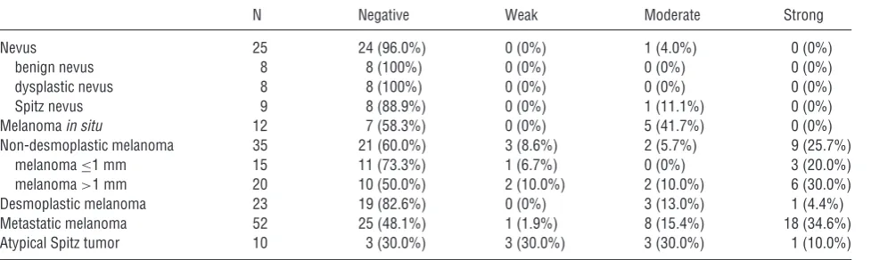
Table 3. IMP-3 staining intensity in melanocytic lesions
N Negative Weak Moderate Strong
Nevus 25 24 (96.0%) 0 (0%) 1 (4.0%) 0 (0%)
benign nevus 8 8 (100%) 0 (0%) 0 (0%) 0 (0%)
dysplastic nevus 8 8 (100%) 0 (0%) 0 (0%) 0 (0%)
Spitz nevus 9 8 (88.9%) 0 (0%) 1 (11.1%) 0 (0%)
Melanomain situ 12 7 (58.3%) 0 (0%) 5 (41.7%) 0 (0%)
Non-desmoplastic melanoma 35 21 (60.0%) 3 (8.6%) 2 (5.7%) 9 (25.7%)
melanoma≤1 mm 15 11 (73.3%) 1 (6.7%) 0 (0%) 3 (20.0%)
melanoma>1 mm 20 10 (50.0%) 2 (10.0%) 2 (10.0%) 6 (30.0%)
Desmoplastic melanoma 23 19 (82.6%) 0 (0%) 3 (13.0%) 1 (4.4%)
Metastatic melanoma 52 25 (48.1%) 1 (1.9%) 8 (15.4%) 18 (34.6%)
Atypical Spitz tumor 10 3 (30.0%) 3 (30.0%) 3 (30.0%) 1 (10.0%)
Similar to non-desmoplastic deep melanomas, 27/52 (51.9%) metastatic melanomas were positive for IMP-3 (Fig. 2E). Nearly all (26/27) showed moderate to strong staining and most (23/27) stained diffusely. No significant difference was attained between deep melanomas and metastatic melanomas.
Unlike Spitz nevus, most ASTs (7/10; 70%) expressed IMP-3, with five showing staining in at least 10% of tumor cells and six having weak to moderate intensity (Fig. 2F). There was a statistical difference between AST and Spitz nevus (p=0.0256). But IMP-3 expression between AST and deep melanoma was not significantly different (p=0.686).
Discussion
Our study investigated for the first time the expression of IMP-3 in MIS, desmoplastic melanoma and AST. The expression of IMP-3 was significantly differ-ent between primary non-desmoplastic melanomas
and benign/dysplastic/Spitz nevi (p=0.0427 indi-vidually; p=0.0251 when all nevi combined). Our findings confirm Pryor’s observation10 and sug-gest that a diagnosis of melanomas as opposed to melanocytic nevi may be inferred when IMP-3 is strongly expressed. Interestingly, Pryor et al.10 observed that 2 of 10 Spitz nevi showed diffuse IMP-3 staining, while only 1 Spitz nevus in our study showed positivity in<10% of tumor cells. It is unclear what may have attributed to this difference. Future studies with more Spitz nevi included would be helpful to address the issue.
We observed discrete and isolated single IMP-3-positive cells in MIS. However, because the staining was limited to <10% of tumor cells, IMP-3 is less likely to be of any use in separating MIS from dysplastic nevus. Conventional histology remains the most important tool in diagnosing MIS.
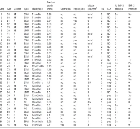
[image:4.595.54.543.303.449.2]Table 4. Clinical characteristics and IMP-3 staining in primary non-desmoplastic melanomas
Case Age Gender Type TNM stage
Breslow depth
(mm) Ulceration Regression
Mitotic
rate/mm2 TIL SLN
% IMP-3 staining
IMP-3 intensity
1 74 M SSM T1aNxMx 0.26 no no insuf 2 ND 2+ 3+
2 55 M SSM T1aNxMx 0.27 no yes insuf 2 ND 0 0
3 81 F SSM T1aNxMx 0.34 no yes 0 2 ND 2+ 3+
4 52 M SSM T1aNxMx 0.35 no no 0 1 ND 0 0
5 73 M LMM T1aNxMx 0.41 no no 4 1 ND 2+ 3+
6 70 M LMM T1aNxMx 0.43 no no 0 1 ND 0 0
7 41 F SSM T1aNxMx 0.44 no no insuf 2 ND 0 0
8 45 F SSM T1aNxMx 0.48 no no 0 2 ND 0 0
9 63 F SSM T1aNxMx 0.55 no yes insuf 1 ND 0 0
10 59 M SSM T1bNxMx 0.55 no yes insuf 2 ND 0 0
11 61 F SSM T1aNxMx 0.56 no yes 0 2 ND 0 0
12 40 M SSM T1aNxMx 0.60 no no insuf 1 ND 0 0
13 48 M SSM T1aNxMx 0.63 no yes 0 2 ND 0 0
14 57 M SSM T1aNxMx 0.64 no yes insuf 1 ND 0 0
15 53 M LMM T1bNxMx 0.82 no no 0 2 ND 1+ 1+
16 74 F SSM T2aN0Mx 1.01 no no 1 2 neg 2+ 2+
17 43 F ALM T2bN2bM1a 1.15 yes no 0 1 pos 0 0
18 73 M SSM T2aN0Mx 1.16 no no 0 1 neg 0 0
19 66 M SSM T2aN0Mx 1.16 no no 0 1 neg 3+ 3+
20 64 M SSM T2aN0Mx 1.46 no no 0 1 neg 2+ 2+
21 85 M LMM T2aN0Mx 1.57 no no 3 2 neg 3+ 3+
22 51 F SSM T3aN0M1c 1.7 no no 2.5 2 neg∗ 0 0
23 76 M NO T3aN0Mx 2.4 no no 6 1 neg 0 0
24 44 M SSM T3aN0Mx 2.4 no yes 0 1 neg 0 0
25 94 F LMM T3aNxMx 2.5 no no 3 1 ND 3+ 3+
26 39 F SSM T3aN2bMx 2.5 no no 2 1 pos 3+ 3+
27 72 M NO T3aN2bMx 2.9 no yes 11 2 pos 3+ 3+
28 40 F NE T3aN3Mx 3.05 no no 3.5 1 pos 0 0
29 51 F SSM T3aN0Mx 3.6 no no 2 1 neg 2+ 1+
30 29 M NO T3aN0Mx 3.65 no no 1.5 1 neg 0 0
31 50 F SSM T3aN0Mx 3.75 no no 1.5 1 neg 3+ 3+
32 51 F ALM T4bN0Mx 4.1 yes no 3.5 1 neg 0 0
33 34 F NO T4aN0Mx 4.5 no no 1 2 neg 0 0
34 38 F SPM T4aN1bMx 5.25 no no 1 2 pos 2+ 1+
35 46 M ALM T4bN0Mx 7.1 yes no 3 1 neg 0 0
∗Bone metastasis identified.
SSM, superficial spreading melanoma; LMM, lentigo maligna melanoma; ALM, acral lentiginous melanoma; NE, nevoid melanoma; NO, nodular melanoma; SPM, Spitzoid melanoma; TNM, Pathologic T (tumor) stage, N (nodal), M (metastasis); insuf, insufficient tumor volume for count; TIL, tumor infiltrating lymphocytes (1, non-brisk; 2, brisk); ND, not done.
et al.10 found that IMP-3 was expressed at lower levels in superficial melanomas than deep melanomas, but the difference between the two was not statistically significant. In concordance with their findings, we also noted stronger and more diffuse IMP-3 staining in deep melanomas than in superficial melanomas. However, there was no statistical difference between superficial and deep melanomas (p=0.0916). In addition, when deep melanomas were compared at incremental Breslow depth (Table 4), no significant correlation was detected between IMP-3 levels and tumor depths, other melanoma prognostic factors or clinical stages.
Our findings suggest that IMP-3 is probably of no value in predicting melanomas with greater risk to metastasize and/or shorter survival intervals. IMP-3 does not seem to play a role in melanoma tumor progression. Future studies with additional cases are necessary to substantiate our observation.
melanoma patients are similar to those for patients with other types of melanomas at comparable thickness.15 – 17It was recently shown thatγ–H2AX, a marker of activated DNA damage, was over-expressed in most primary cutaneous melanomas except desmoplastic melanomas.18 Our observa-tion of lower IMP-3 expression in desmoplastic melanomas compared to other deep melanomas (p=0.0109) is interesting. We suspect that the unique desmoplastic stromal response in desmoplas-tic melanoma may be related to the limited expression of many common molecules that are upregulated in other melanomas.
AST is a borderline melanocytic lesion with uncertain biologic potential.19 It shows overlapping histologic features seen in both Spitz nevus and Spitzoid melanoma. AST generally does not meet the diagnostic threshold for melanoma, but demonstrates concerning features, such as large size, asymmetry, aberrant dermal growth pattern, incomplete maturation, cytologic atypia, increased dermal mitotic rates and deep dermal mitoses.19 – 21 Unlike conventional melanoma, patients with AST carry a favorable prognosis, despite a high incidence of microscopic SLN deposits.22 Occasionally, differentiating AST from melanoma or Spitz nevus can be problematic. Among a number of tests (i.e. p53, Ki-67 index and fatty acid synthase) studied in the past, comparative genomic hybridization (CGH) appears to hold promise in determining the malignant potential of some controversial melanocytic lesions, including AST.23 – 26 In this study, we found that the expression of IMP-3 was significantly different between Spitz nevus and AST (p=0.0256), indicating IMP-3 may be helpful in differentiating AST from Spitz nevus. Although deep melanoma showed stronger IMP-3 staining intensity than AST, the lack of statistical difference argues against its diagnostic value in differentiating the two. In summary, this is the first study to demonstrate the expression of IMP-3 in MIS, desmoplastic melanoma and AST. Our findings confirm the potential diagnostic utility of IMP-3 in differentiating melanocytic nevus from melanoma and in separating Spitz nevus from AST, when a positive IMP-3 expression is observed. As IMP-3 was expressed at high levels in deep melanoma and metastatic melanoma, it may serve as an intriguing therapeutic target for malignant melanomas.
References
1. Ries LMD, Krapcho M, Stinchcomb DG, et al. SEER cancer statistics review, 1975 – 2005. Bethesda, MD: National Cancer Institute, 2005.
2. Corona R, Mele A, Amini M, et al. Interobserver variability on the histopathologic diagnosis of cutaneous melanoma and other pigmented skin lesions. J Clin Oncol 1996; 14: 1218.
3. Nielsen FC, Nielsen J, Christiansen J. A family of IGF-II mRNA binding proteins (IMP) involved in RNA trafficking. Scand J Clin Lab Invest Suppl 2001; 234: 93.
4. Mueller-Pillasch F, Pohl B, Wilda M, et al. Expression of the highly conserved RNA binding protein KOC in embryogenesis. Mech Dev 1999; 88: 95.
5. Jiang Z, Chu PG, Woda BA, et al. Analysis of RNA-binding protein IMP3 to predict metastasis and prognosis of renal-cell carcinoma: a retrospective study. Lancet Oncol 2006; 7: 556.
6. Li C, Rock KL, Woda BA, Jiang Z, Fraire AE, Dresser K. IMP3 is a novel biomarker for adenocarcinoma in situ of the uterine cervix: an immunohistochemical study in comparison with p16(INK4a) expression. Mod Pathol 2007; 20: 242.
7. Sitnikova L, Mendese G, Liu Q, et al. IMP3 predicts aggressive superficial urothelial carcinoma of the bladder. Clin Cancer Res 2008; 14: 1701.
8. Liao B, Hu Y, Herrick DJ, Brewer G. The RNA-binding protein IMP-3 is a translational activator of insulin-like growth factor II leader-3 mRNA during proliferation of human K562 leukemia cells. J Biol Chem 2005; 280: 18517.
9. Wang T, Fan L, Watanabe Y, et al. L523S, an RNA-binding protein as a potential therapeutic target for lung cancer. Br J Cancer 2003; 88: 887.
10. Pryor JG, Bourne PA, Yang Q, Spaulding BO, Scott GA, Xu H. IMP-3 is a novel progression marker in malignant melanoma. Mod Pathol 2008; 21: 431.
11. Thomas AJ, Erickson CA. The making of a melanocyte: the specification of melanoblasts from the neural crest. Pigment Cell Melanoma Res 2008; 21: 598.
12. Greene FLPD, Fleming ID, Fritz A, Balch CM, Haller DG, Morrow M, eds. AJCC cancer staging manual, 6th ed. New York: Springer, 2002.
13. Jiang Z, Lohse CM, Chu PG, et al. Oncofetal protein IMP3: a novel molecular marker that predicts metastasis of papillary and chromophobe renal cell carcinomas. Cancer 2008; 112: 2676. 14. Busam KJ. Cutaneous desmoplastic melanoma. Adv Anat
Pathol 2005; 12: 92.
15. Quinn MJ, Crotty KA, Thompson JF, Coates AS, O’Brien CJ, McCarthy WH. Desmoplastic and desmoplastic neurotropic melanoma: experience with 280 patients. Cancer 1998; 83: 1128.
16. Smithers BM, McLeod GR, Little JH. Desmoplastic, neural transforming and neurotropic melanoma: a review of 45 cases. Aust N Z J Surg 1990; 60: 967.
17. Jain S, Allen PW. Desmoplastic malignant melanoma and its variants. A study of 45 cases. Am J Surg Pathol 1989; 13: 358. 18. Wasco MJ, Pu RT, Yu L, Su L, Ma L. Expression of
gamma-H2AX in melanocytic lesions. Hum Pathol 2008; 39: 1614. 19. Barnhill RL. The Spitzoid lesion: rethinking Spitz tumors,
atypical variants, ‘Spitzoid melanoma’ and risk assessment. Mod Pathol 2006; 19: S21.
20. Barnhill RL, Argenyi ZB, From L, et al. Atypical Spitz nevi/tumors: lack of consensus for diagnosis, discrimination from melanoma, and prediction of outcome. Hum Pathol 1999; 30: 513.
22. Ludgate MW, Fullen DR, Lee J, et al. The atypical Spitz tumor of uncertain biologic potential: a series of 67 patients from a single institution. Cancer 2009; 115: 631.
23. Kapur P, Selim MA, Roy LC, Yegappan M, Weinberg AG, Hoang MP. Spitz nevi and atypical Spitz nevi/tumors: a histologic and immunohistochemical analysis. Mod Pathol 2005; 18: 197.
24. Kanter-Lewensohn L, Hedblad MA, Wejde J, Larsson O. Immunohistochemical markers for distinguishing Spitz nevi from malignant melanomas. Mod Pathol 1997; 10: 917.
25. Bastian BC, LeBoit PE, Hamm H, Brocker EB, Pinkel D. Chromosomal gains and losses in primary cutaneous melanomas detected by comparative genomic hybridization. Cancer Res 1998; 58: 2170.