0095-1137/06/$08.00⫹0 doi:10.1128/JCM.02618-05
Copyright © 2006, American Society for Microbiology. All Rights Reserved.
Efficient Tracing of Global Isolates of
Yersinia pestis
by Restriction
Fragment Length Polymorphism Analysis Using Three
Insertion Sequences as Probes†
Gabriela Torrea, Viviane Chenal-Francisque, Alexandre Leclercq, and Elisabeth Carniel*
Yersinia Research Unit, National Reference Laboratory and WHO Collaborating Center for Yersinia, Institut Pasteur, 75724 Paris Cedex 15, France
Received 16 December 2005/Returned for modification 1 February 2006/Accepted 24 March 2006
Yersinia pestisis the etiologic agent of plague, a disease that is transmitted from rodent to rodent and from
rodent to humans by fleabites. Multiple copies of three insertion sequences (IS100, IS285, and IS1541) are
scattered over theY. pestisgenome. The genomic instability generated by these insertion sequences (IS) creates
a polymorphism of the hybridizing restriction fragments (restriction fragment length polymorphism [RFLP]) which can be used to subtype this relatively clonal species. The aim of this work was to evaluate and compare the potential of the three IS-RFLP techniques, individually or in combination, to define clusters of strains
according to their focus of origin. The analysis of 61Y. pestisisolates of worldwide origin indicated that no
satisfactory strain clustering was observed with each IS-RFLP used individually. In contrast, the combination
of the three IS-RFLP data (3IS-RFLP) resulted in both an efficient strain discrimination (Dⴝ0.999) and a
robust clustering of the isolates according to their biovar and geographical origin. This geographical clustering was observed even within the Orientalis group, although these strains had only a short period of time (one century) to diverge from the original clone that spread globally. Therefore, 3IS-RFLP is a technique that may be useful for addressing epidemiological problems and forensic issues. When plague reemerges after several decades of silence in a quiescent focus, it may help in determining whether the disease was reimported or reactivated. It may also be of value to identify the origin of a strain when plague cases appear in a previously
plague-free region. Finally, this technique could be useful for the tracing of aY. pestisisolate that has been used
as a biological terrorism threat.
Yersinia pestisis a gram-negative bacillus which belongs to the familyEnterobacteriaceae. This bacterium is the etiologic agent of plague, a disease that is transmitted from rodent to rodent by fleabites. Most often, humans become infected after an infectious fleabite and develop a bubonic form of plague. If the bacillus secondarily reaches the lungs of the patient, it causes a pneumonia which allows a direct human-to-human transmission through the spread of infected aerosols. Plague foci still persist that are endemic to Africa (mainly the South-ern part), Central and East Asia, and North and South Amer-ica (31).
Plague is considered a reemerging disease (27). This is due to the rise in human plague cases since the beginning of the 1990s and the reappearance of the disease in countries where no cases where reported for several decades, such as in the Northern seaport of Majunga in Madagascar in 1991 (26), in India (25) and Mozambique (4) in 1994, in Zambia in 1996 (30), and recently, in Algeria in 2003 (32). Whether these outbreaks were due to the reactivation of quiescent autoch-thon plague foci or were imported from distant countries by modern means of transportation is a question of key impor-tance for the implementation of appropriate and efficient
con-trol measures. Having a molecular tool which could clusterY. pestis strains based on their geographical focus, even when isolated at intervals of several years, would be of great help to answer this question. Furthermore,Y. pestishas been catego-rized as one of the major bacterial agents of bioterrorism (category A) (17). Having the possibility to trace an isolate that has been used as a biological weapon would also be critical.
Y. pestishas been shown to be a clone ofYersinia pseudotu-berculosis that emerged less than 20,000 years ago (2). This recent clonal expansion accounts for the limited phenotypic and genetic diversity observed in this species. Phenotypically, the ability to ferment glycerol and to reduce nitrates to nitrites led to the subdivision ofY. pestisinto three biovars: Antiqua, Medievalis, and Orientalis (10). Today, most of the strains isolated worldwide belong to biovar Orientalis, the biovar that spread globally from Hong Kong in 1894 during the third plague pandemic. The two other biovars have a geographically restricted distribution: Medievalis in Asia and Antiqua in some parts of Africa and in Central Asia.Y. pestisalso displays a very low degree of genetic polymorphism. An analysis of six house-keeping genes by multilocus sequence typing demonstrated a complete lack of nucleotide polymorphism among 36 strains belonging to the three biovars and isolated from various coun-tries at different times (2). A recent comparison of the three availableY. pestisgenome sequences confirmed the very low genetic diversity of this species: as few as 80 synonymous sin-gle-nucleotide polymorphisms were detected among 3,250 or-thologous coding sequences (1).
Despite the very high degree of conservation at the gene * Corresponding author. Mailing address: Institut Pasteur, National
Reference Laboratory and WHO Collaborating Center forYersinia, 28, rue du Dr. Roux, 75724 Paris Cedex 15, France. Phone: 33 1 45 68 83 26. Fax: 33 1 40 61 30 01. E-mail: [email protected].
† Supplemental material for this article may be found at http://jcm .asm.org/.
2084
on May 16, 2020 by guest
http://jcm.asm.org/
level, the genotypic subdivision of the speciesY. pestishas been possible. Techniques using variations in the number of re-peated sequences, usually located in noncoding chromosomal regions, such as variable-number tandem repeats (3), clustered regularly interspaced short palindromic repeats (24), or inter-genic spacer sequences (11), have successfully allowed the dis-crimination of strains within the same biovar. Another group of techniques, including ribotyping (13) and pulsed-field gel electrophoresis (PFGE) (19), uses the restriction fragment length polymorphism (RFLP) generated by the high genome instability ofY. pestis(13, 22) to subdivide the species.
This genome instability is in large part attributable to the presence of multiple copies of insertion sequences (IS) scat-tered over theY. pestisgenome (9, 22, 29). Insertion sequences are simple genetic elements which can insert at multiple sites in a bacterial genome. Recombination between these IS may lead to chromosomal macro-rearrangements (20). The most numer-ous ISs are IS100(30 to 44 chromosomal copies on the three Y. pestissequenced genomes), IS1541(43 to 62 copies), and IS285(18 to 20 copies) (9, 22, 29). Variations in the chromo-somal location of IS100have been used to study the microevo-lution ofY. pestis (1, 21). Furthermore, RFLP analyses using one of these IS as a probe (IS-RFLP) have been applied to subtypeY. pestis(6, 15, 28). IS100-RFLP discriminated strains isolated from the same country (United States) more efficiently than IS285-RFLP, but both techniques were found to be infe-rior to PFGE for this purpose (15).
The aim of this work was to evaluate and compare the potential of each of the three IS-RFLP techniques (IS100-, IS285-, and IS1541-RFLP), individually or in combination, to define clusters of strains according to their focus of origin.
MATERIALS AND METHODS
Bacterial strains and growth conditions.Sixty-one strains ofY. pestisisolated between 1908 and 1994 from 17 countries on four continents (Table 1) were taken from the strain collection of theYersiniaResearch Unit (Institut Pasteur) and analyzed. These strains belonged to the three classical biovars ofY. pestis
(10): Antiqua (ferment glycerol [G⫹] and reduce nitrates to nitrites [N⫹]), Medievalis (G⫹, N⫺), and Orientalis (G⫺, N⫹). Bacterial suspensions were prepared from stock cultures kept at⫺80°C and streaked onto Luria Bertani agar plates containing 0.002% hemin or grown in peptone broth with shaking for 24 to 48 h at 28°C.
DNA extraction, restriction, and transfer to nylon membrane.Total DNA extraction from eachY. pestisstrain was performed as described previously (7). Plasmid extracts were obtained by the method of Birnboim and Doly (5). Five micrograms of each sample was digested overnight at 37°C with various restric-tion enzymes before being loaded onto 0.8% agarose gels and subjected to electrophoresis (50 V in 1⫻Tris-Borate-EDTA buffer) for 24 h (IS285- and IS1541-RFLP) or 26 h (IS100-RFLP). The DNA of strain IP304 was systemat-ically loaded on the flanking and middle lanes of each gel to serve for intra- and intergel normalization. DNA bands were stained with ethidium bromide. Alka-line denaturation, neutralization, and transfer of total DNA onto nylon filters (Hybond N⫹; Amersham, England) with a VacuGene apparatus (Pharmacia LKB Biotechnology, Uppsala, Sweden) were performed as previously de-scribed (13).
Preparation of IS probes and hybridization.For IS fingerprinting, three sets of primers were designed and used to amplify a portion of the insertion sequences: IS100-F, 5⬘-AAAACGTTCGAAGAGTATGA-3⬘; IS100-R, 5⬘-GATGAGCAG GCGGGGGGCCA-3⬘ (255 bp); IS1541-F, 5⬘-AAAGCTTTCAGCTTTGGGT C-3⬘; IS1541-R, 5⬘-TCTTTCCCTTCAGGTACCCC-3⬘(319 bp); IS285-F, 5⬘-A GCTTACCGAACACCTCGGG-3⬘; IS285-R, 5⬘-GTTGATGCCCAGCGCTAG GA-3⬘ (406 bp). The PCR amplification reactions were performed on the genomic DNA ofY. pestisstrain IP304 as a template in a final volume of 50l with 1.25 U ofTaqDNA polymerase (Roche/Cetus) used with the supplier’s buffer, 2 mM MgCl2, and 200 M concentrations of each of the four
de-oxynucleoside triphosphates. All primers were used at a final concentration of 1
M. Each reaction involved a denaturing step at 94°C for 3 min, 25 cycles of amplification consisting of three steps each of 1 min at 94°C, 55°C, and 72°C, and a final extension step of 10 min at 72°C. PCR products were subjected to electrophoresis in 0.7% agarose gels and stained with ethidium bromide. The three IS probes were peroxidase labeled using the ECL direct nucleic acid labeling and detection system (Amersham) and used for Southern hybridization overnight at 42°C.
Bioinformatic analysis of IS-RFLP patterns.The hybridization patterns ob-tained with each IS were scanned, and the computerized data were analyzed using the BioNumerics software, version 4.0 (Applied Maths, Kortrijk, Belgium). Bands automatically assigned by the computer were checked visually and cor-rected manually when necessary. A position tolerance of 1.8 was selected for each of the three IS. Cluster analysis of the individual or combined IS-RFLP patterns was done by the unweighted pair group method with average linkages (UPGMA), using the Dice coefficient to analyze the similarities of the banding patterns. The discriminatory power of each IS-RFLP was determined by calcu-lating the discrimination index (D) based on Simpson’s index of diversity (16).D
depends on the number of types defined by the test method and the relative frequencies of these types:D⫽1⫺{[(1/(N⫻(N⫺1)))]⫻[⌺nj(nj⫺1)]}, whereNis the total number of unrelated isolates, andnjis the number of strains that belong to thejth type. Two IS profiles were considered identical when their percent similarity was⬎98%.
RESULTS
Optimization of IS-RFLP conditions. The three insertion
sequences IS100, IS285, and IS1541were chosen because they are present in the highest copy numbers on the genome ofY. pestis(9, 22, 29). Primers internal to each IS were designed and used to generate probes. To obtain the most distinct banding patterns, various restriction enzymes (EcoRI, HindIII, EcoRV, BamHI, PvuI, XbaI, XhoI, and XmaI) were tested on the DNA of a set ofY. pestisstrains which were hybridized with each of the three IS probes. The best resolution was obtained when the genomic DNAs were digested with EcoRI for IS100-RFLP and with HindIII for IS285- and IS1541-RFLP. The duration of DNA electrophoresis was also varied to optimize band sepa-ration for each IS. Optimal sepasepa-ration of the hybridizing bands was obtained after 26 h of migration for IS100-RFLP and 24 h for IS285- and IS1541-RFLP. Because some hybridizing frag-ments with the largest or smallest sizes were not sufficiently resolved, a size window containing bands that were clearly and systematically separated on all Southern blots was defined for each IS (Fig. 1).
Since the presence of IS copies on the threeY. pestis plas-mids, pFra (101 kb), pYV (70.5 kb), and pPla (9.6 kb), may artificially introduce a variation in the hybridization profile due to the loss in vitro of one or several plasmids upon subcultures, the expected sizes of the plasmid-borne hybridizing fragments were determined based on the Y. pestis plasmid sequences available in the databases (9, 14, 18, 22). The plasmid bands were further identified by extracting the DNA of 11 differentY. pestisstrains containing either one, two, or three of theY. pestis plasmids and hybridizing them with each IS. For IS100-RFLP, a 2.75-kb pYV hybridizing fragment was present within the size window and was thus manually removed for cluster analysis (Fig. 1). Similarly, a 2.36-kb pFra fragment present in the IS285-RFLP size window was withdrawn before performing the UPGMA analysis. All other plasmid-borne IS sequences were located on fragments outside the size windows selected. The in vitro instability of the 102-kbpgmlocus was another potential source of artificial profile variation (8, 12). The in silico analysis of the IS positions on theY. pestis sequenced
VOL. 44, 2006 TRACING OF Y. PESTIS BY IS-RFLP 2085
on May 16, 2020 by guest
http://jcm.asm.org/
Institut Pasteur no. (n)
Original designation
Geographical origina
Yr of
isolation Biotype
IS100
type
IS285
type
IS1541
type
Asia (19)
IP772 Nhatrang 63-127 Vietnam 1963 Orientalis 17 11 22
IP513 Nhatrang 64-65 Vietnam 1964 Orientalis 13 11 24
IP820 Nhatrang 64-260 Vietnam 1964 Orientalis 14 12 23
IP940 Nhatrang 65-30 Vietnam 1965 Orientalis 19 13 22
IP507 Saigon 55-720 Vietnam 1955 Orientalis 21 11 25
IP532 Saigon 55-801 Vietnam 1955 Orientalis 21 16 20
IP989 Dalat 131 Vietnam 1964 Orientalis 15 11 22
IP613 548 Burma 1970 Orientalis 30 11 22
IP612 637 Burma 1970 Orientalis 30 19 26
IP519 PKH4 Kurdistan 1951 Medievalis 33 22 14
IP562 PKR6 Kurdistan 1947 Medievalis 32 22 14
IP516 PKR18 Kurdistan 1948 Medievalis 33 25 14
IP564 PKR25 Kurdistan 1948 Medievalis 32 23 13
IP557 PKR292 Kurdistan 1963 Medievalis 34 24 14
IP565 10/5 Turkey 1952 Medievalis 35 26 14
IP521 10/1 Turkey UNb Orientalis 1 1 19
IP283 Surat 9/95 India 1994 Orientalis 4 15 10
IP1595 Surat 4/95 India 1994 Orientalis 4 15 10
IP579 Bombay 195 India 1908 Orientalis 28 15 12
America (8)
IP567 Exu 21 Brazil 1967 Orientalis 24 5 12
IP568 Exu 53 Brazil 1967 Orientalis 24 5 18
IP569 Exu 56 Brazil 1967 Orientalis 24 5 15
IP571 Exu 184 Brazil 1967 Orientalis 24 5 11
IP1747 CO92 United States 1992 Orientalis 22 15 13
IP573 7793 United States 1948 Orientalis 18 15 13
IP574 193 United States 1950 Orientalis 8 15 13
IP610 Cuis 14 Argentina 1946 Orientalis 16 6 13
Europe (3)
IP695 Hamburg 9 Germany 1952 Orientalis 9 10 2
IP685 Hamburg 10 Germany 1952 Orientalis 9 5 13
IP696 Hamburg 26 Germany 1952 Orientalis 9 5 13
Africa (31)
IP577 243 Morocco 1940 Orientalis 13 15 1
IP578 48 Morocco 1940 Orientalis 10 15 21
IP1867 Arn Algeria 1945 Orientalis 23 20 27
IP523 Th Senegal 1944 Orientalis 12 17 3
IP524 Fa Senegal 1944 Orientalis 9 3 31
IP550 Ed Kenya UN Antiqua 41 27 30
IP544 Mi Kenya UN Antiqua 43 30 29
IP677 Be´a Kenya UN Antiqua 42 33 28
IP554 Ros Kenya UN Antiqua 47 29 28
IP545 Da Kenya UN Antiqua 44 31 29
IP552 Ma Kenya UN Antiqua 46 28 28
IP553 Ky Kenya UN Antiqua 45 32 29
IP542 144 Kenya 1952 Antiqua 40 36 7
IP537 147 Kenya 1952 Antiqua 36 38 7
IP539 164 Kenya 1952 Antiqua 38 34 8
IP549 logo 1/53 Belgian Congo 1953 Antiqua 39 37 5
IP678 Eli Belgian Congo 1950 Orientalis 2 4 6
IP543 Lita Belgian Congo 1953 Antiqua 37 35 7
IP1537 254 Namibia 1984 Orientalis 27 18 32
IP1538 272 Namibia 1984 Orientalis 26 14 34
IP1540 292 Namibia 1985 Orientalis 26 18 35
IP1541 340 Namibia 1986 Orientalis 25 14 33
IP1542 346 Namibia 1986 Orientalis 29 2 32
IP1535 164 South Africa 1982 Orientalis 5 16 9
IP304 6/69 Madagascar 1969 Orientalis 9 15 10
IP528 62 Madagascar 1946 Orientalis 20 8 10
IP529 112 Madagascar 1951 Orientalis 11 9 4
IP530 Ga Madagascar ⬎1939 Orientalis 3 21 10
IP241 3/89 Madagascar 1989 Orientalis 6 7 15
IP644 6/89 Madagascar 1989 Orientalis 31 7 17
IP666 100/92 Madagascar 1992 Orientalis 7 7 16
aName of the country at time of strain isolation. bUN, unknown.
2086
on May 16, 2020 by guest
http://jcm.asm.org/
[image:3.585.45.539.65.705.2]genomes indicated that deletion of the pgm locus does not modify the IS285- and IS1541-RFLP profiles. In contrast, de-letion of this locus was predicted to result in the loss of a 1.5-kb EcoRI fragment on the IS100-RFLP pattern. This was con-firmed by comparing the IS100 hybridization profiles of the wild-type strain 6/69 and its⌬pgmderivative (data not shown). However, since this fragment is not in the size window selected, the instability of the pgmlocus had no effect on the IS100 -RFLP pattern.
The reproducibility of the clustering was evaluated by com-paring the position on the UPGMA dendrograms of 12 Y. pestis strains studied independently (DNA extraction, diges-tion, migradiges-tion, preparation of probes, and hybridizations) by two different investigators at intervals of several months. The same strains studied independently always fell into the same clusters with each IS-RFLP method.
IS100-RFLP profiles.When IS100-RFLP was applied to the
61Y. pestisstrains analyzed, 47 profiles were identified (Table 1), leading to a discrimination index of 0.987. No dominant IS100profile was noted. As previously observed (2), the three biovars formed distinct clusters in the UPGMA dendrogram (Fig. 2). The weight of the biovar was superior to that of the geographic origin in the clustering analysis. For instance, the three strains from Belgian Congo were separated into the Anti-qua and Orientalis branches, although they were isolated from the same country. The same held true for the two strains from Turkey (biovar Medievalis and Orientalis) (Fig. 2). For the Orientalis strains, which are found worldwide because of their recent global spread, no strong geographical clustering was achieved by IS100-RFLP (Fig. 2). Nonetheless, a tendency of the strains from the same country to be grouped into some subbranches was sometimes observed. This was the case for all the strains from Brazil and Namibia, which were grouped in
cluster a1, while cluster a2 contained all the isolates from Germany (Fig. 2). In several instances, strains isolated the same year from the same region, such as those from Burma in 1970, Hamburg (Germany) in 1952, Saigon (Vietnam) in 1955, Exu (Brazil) in 1967, and Surat (India) in 1994 displayed highly similar or identical IS100types (Table 1 and Fig. 2).
IS285-RFLP profiles.Thirty-eight IS285-RFLP types were
identified among the 61Y. pestisstrains analyzed (D⫽0.963). Three IS285types represented 33% of the strains: IS285type 15 (9 strains), IS285 type 5 (6 strains), and IS285type 11 (5 strains) (Table 1). The UPGMA dendrogram delineated three main branches (Fig. 3). One branch was entirely composed of Antiqua strains. The second branch was split into two sub-branches, one corresponding to biovar Antiqua and the other to biovar Medievalis strains (Fig. 3). The Orientalis strains were all found in a third branch (Fig. 3). Within the Orientalis group, no clear clustering of the strains according to their focus of origin was noted. Some strains isolated from the same coun-try were found in different subbranches. Grouping of strains from the same geographic origin could nonetheless be ob-served, as for the strains from Brazil which were all found in cluster b1 (Fig. 3). Clusters b2, b3, and b4 each contained only strains from Madagascar, Southeast Asia, and Namibia, re-spectively (Fig. 3). Of note was the fact that identical IS285 types were found for the two strains from Morocco. This was also the case for the three strains from India and for the three strains from the United States (Table 1 and Fig. 3).
IS1541-RFLP profiles. IS1541-RFLP analysis allowed the
delineation of 35 profiles (D⫽0.968). Three of them, IS1541 type 13 (7 strains), IS1541type 14 (5 strains), and IS1541type FIG. 1. Examples of IS-RFLP profiles obtained after digestion and
hybridization of the genomic DNA of variousY. pestisstrains with each IS probe. Lanes: 1, IP304 (used as an intra- and intergel standard); 2, IP696; 3, IP685; 4, IP677; 5, IP666. Tick marks on the left indicate the sizes of the molecular mass markers (lambda DNA HindIII digest) in kilobases. Rectangles correspond to the size windows chosen for the analysis of the hybridization profiles for each IS. Arrows on the right indicate a hybridizing plasmid band (from pYV for IS100and pFra for
IS285). the 61FIG. 2. Dendrogram generated from the ISY. pestisstrains studied using the UPGMA clustering analysis100-RFLP patterns of with the BioNumerics software. A position tolerance of 1.8% was chosen. Dotted rectangles outline clusters of strains of interest. A, Antiqua branch; M, Medievalis branch; O, Orientalis branch.
VOL. 44, 2006 TRACING OF Y. PESTIS BY IS-RFLP 2087
on May 16, 2020 by guest
http://jcm.asm.org/
[image:4.585.56.271.70.250.2]10 (5 strains), represented 28% of theY. pestis isolates ana-lyzed (Table 1). The UPGMA dendrogram did not exhibit biovar-specific branches (Fig. 4). Most of the Antiqua strains were grouped in two clusters and the Medievalis strains were grouped in one cluster, but these clusters were interspersed among Orientalis clusters. Furthermore, one Antiqua strain (IP549) and one Medievalis strain (IP564) were found within Orientalis branches (Fig. 4). In contrast to the two other IS-RFLPs, the two strains from Belgian Congo, although of dif-ferent biovars, were found in the same cluster. Among the Orientalis strains, three clusters exhibiting some geographic specificity were identified. Cluster c1 was composed of strains from Africa (except one strain from Germany), cluster c2 was exclusively constituted of strains from Southeast Asia (Vietnam and Burma), and cluster c3 contained all strains from Namibia (Fig. 4).
Combination of two IS-RFLP profiles. Although each
IS-RFLP technique allowed, to some extent, a geographical grouping of strains, this clustering remained limited and varied
with each IS. To determine whether a combination of two IS would improve the geographically based Y. pestisclustering, the IS-RFLP data were combined two by two and analyzed. The discrimination indexes increased for the three combina-tions: IS100/IS285-RFLP,D ⫽0.995; IS1541/IS285-RFLP, D
[image:5.585.132.455.67.456.2]⫽ 0.993; and IS100/IS1541-RFLP, D ⫽ 0.998. While IS100/ IS285-RFLP and IS100/IS1541-RFLP allowed a clear delinea-tion of the three biovars into distinct main branches, this dis-tinction was less pronounced with IS1541/IS285-RFLP (see Fig. S1, S2, and S3 in the supplemental material). Within the Orientalis group, combining two IS-RFLPs improved strain grouping. With each of the three IS-RFLP combinations, all isolates from Namibia and all those from the United States fell into specific clusters. This was also true for the isolates from Burma, but only IS1541/IS285-RFLP grouped these strains with those from the neighboring Vietnam. IS100/IS285-RFLP and IS100/IS1541-RFLP, but not IS1541/IS285-RFLP, allowed the clustering of Brazilian isolates. Interestingly, the strains from Germany fell into the same cluster as the isolate from FIG. 3. Dendrogram generated from the IS285-RFLP patterns of the 61Y. pestisstrains studied using the UPGMA clustering analysis with the BioNumerics software. A position tolerance of 1.8% was chosen. Dotted rectangles outline clusters of strains of interest. A, Antiqua branch; M, Medievalis branch; O, Orientalis branch.
on May 16, 2020 by guest
http://jcm.asm.org/
Argentina with each of the three combined IS-RFLPs. IS100/ IS285-RFLP was the only combination which grouped the three strains from India into the same cluster (which also contained isolates from other geographical origins). In con-trast, strains from Madagascar fell into different clusters with each of the three IS-RFLP combinations (see Fig. S1, S2, and S3 in the supplemental material).
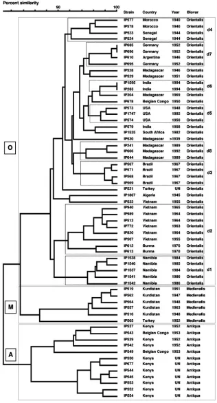
Combination of three IS-RFLP profiles.When the data
ob-tained with the three IS-RFLP techniques were combined (3IS-RFLP), strain discrimination became extremely efficient (D ⫽ 0.999). Only two pairs of strains displayed identical 3IS-RFLP patterns (Table 1 and Fig. 5). The three biovars formed three distinct main branches on the UPGMA dendro-gram. The Antiqua branch split into two subbranches, and interestingly, the two isolates from Belgian Congo clustered into the same Antiqua subbranch (Fig. 5). Similarly, in the Medievalis branch, the five isolates from Kurdistan were much more closely related to each other than to the Turkish strain. Most importantly, within the Orientalis branch, the majority of the subbranches typified the geographic origin of the strains: all Namibian isolates were in cluster d1, all but one (IP532) strain from Southeast Asia were in cluster d2 (with a subdivi-sion of this cluster into two subbranches corresponding to the Vietnamese and Burmese isolates), all Brazilian isolates were in cluster d3, all strains from West Africa were in cluster d4 (with the two strains from Morocco and the two from Senegal
being into distinct subbranches), and all strains from the United States were in cluster d5 (Fig. 5). Furthermore, cluster d6 contained the two strains from Surat (India), and cluster d7 contained the three strains from Germany. Single Orientalis strains from a given country (Turkey, Belgian Congo, Algeria, and South Africa) were scattered in the dendrogram. Mada-gascar was the only focus for which no marked strain clustering was observed. The three most recent isolates from this country were grouped in cluster d8, while the four others were in different subbranches (Fig. 5).
DISCUSSION
IS-RFLP and molecular typing.Two extremes may be found
[image:6.585.45.286.69.372.2]among the molecular typing methods used to analyzeY. pestis isolates. One corresponds to ribotyping, which provides a global view of the circulating strains but has a low discrimina-tory power, especially within the Orientalis group. The other extreme is PFGE which, because of the high number of re-striction fragments generated and the complexity of the pat-terns, has a high discriminatory power but is hardly applicable toY. pestisstrains of worldwide origin (unpublished results). IS-RFLP may represent an intermediate between these two techniques because the number of fragments to be analyzed is higher than with ribotyping, but the bands are better resolved than with PFGE, allowing a more reliable strain-to-strain com-parison. Of the three IS-RFLP techniques used in this study, we found that IS100-RFLP had the highest discriminatory power and IS285-RFLP had the lowest discriminatory power. Similar results were obtained whenY. pestisisolates from the United States were analyzed by IS100- and IS285-RFLP (15). This may not be entirely attributable to the difference in IS copy numbers on the Y. pestis chromosome because a size window was defined for each IS pattern, and the number of bands analyzed within these windows was comparable for the three IS. This suggests that variations in the position of the IS on the chromosome are more frequent for IS100 and that IS285 is the most stable. Combination of two IS-RFLPs in-creased the discriminatory power of the technique. The highest discrimination index (D⫽0.998) was obtained with the IS100/ IS1541-RFLP combination, which allowed the delineation of 58 IS types among the 61 strains analyzed. Combination of the three IS-RFLP patterns further increased the discriminatory power of the technique (D⫽0.999). Although accurate com-parison of the discriminatory power of different techniques would need them to be applied to the same set of strains, it appears from this study that 3S-RFLP can discriminate Y. pestisstrains of worldwide origin at least as efficiently as two other highly sensitive typing methods such as variable-number tandem repeats and PFGE. From a practical viewpoint, since the gain in sensitivity was relatively limited when 3IS-RFLP was used instead of IS100/IS1541-RFLP (59 IS types instead of 58), it may be simpler and faster to simply use IS100/IS1541 -RFLP for the purpose of discriminating global isolates ofY. pestis. Remarkably, IS100/IS1541-RFLP could differentiate most strains having the same biovar and country of origin and sometimes even having been isolated the same year. This rel-atively easy to perform technique may thus be a valuable tool for molecular typing of globalY. pestisstrains.
FIG. 4. Dendrogram generated from the IS1541-RFLP patterns of the 61Y. pestisstrains studied using the UPGMA clustering analysis with the BioNumerics software. A position tolerance of 1.8% was chosen. Dotted rectangles outline clusters of strains of interest. A, Antiqua branch; M, Medievalis branch; O, Orientalis branch.
VOL. 44, 2006 TRACING OF Y. PESTIS BY IS-RFLP 2089
on May 16, 2020 by guest
http://jcm.asm.org/
FIG. 5. 3IS-RFLP dendrogram. This dendrogram was generated after combination of the three individual RFLP patterns (IS100, IS285, and IS1541). Dotted rectangles indicate clusters of strains of interest. A, Antiqua branch; M, Medievalis branch; O, Orientalis branch.
on May 16, 2020 by guest
http://jcm.asm.org/
IS-RFLP and Y. pestis microevolution. The Antiqua and Medievalis biovars have a specific geographic distribution. The Medievalis plague focus is situated in Central Asia. Two main Antiqua plague foci exist, one in Central Asia and the other one in Central Africa. It has been recently shown that these two Antiqua foci belong to two distinct evolutionary branches termed 1.ANT and 2.ANT, respectively (1). Only the African branch (1.ANT) was studied here because almost all of the Antiqua isolates in our strain collection belong to this branch. We found that, as previously observed (2), IS100-RFLP grouped Y. pestis strains based on their biovar. This study further shows that IS285-RFLP allowed the clustering of the three classical Y. pestis biovars in independent parts of the dendrogram. In contrast, this grouping was less effective with IS1541-RFLP. The biovar-based clustering became even more pronounced when the IS100/IS1541-RFLP or 3IS-RFLP com-binations were used. Therefore, IS-RFLP techniques may be useful for the analysis ofY. pestismicroevolution.
IS-RFLP andY. pestistracing.The primary and major aim of
this study was to determine whether various IS-RFLP tech-niques, used individually or in combination, could efficiently groupY. pestis strains according to their geographical focus. For the Antiqua and Medievalis strains, since they have a specific and precise geographical distribution, the simple de-termination of their biovar may be sufficient to identify their main focus of origin. This is clearly different for biovar Orien-talis strains which are now the most commonly isolatedY. pestis strains worldwide because of their global spread during the third pandemic. The major challenge was thus to identify a technique which could cluster strains within the Orientalis group according to their geographical foci. When the three IS-RFLP techniques were used individually, some degree of geographical clustering was observed with each technique, but none gave reliable and satisfactory results. IS100-RFLP dis-played nonetheless a slightly better clustering capacity than the other two RFLPs. A two-by-two combination of the IS-RFLP techniques significantly improved the geographical clus-tering of the Orientalis strains. The least effective combination was IS1541/IS285-RFLP, while the two others were compara-ble, with a slight advantage for IS100/IS1541-RFLP. With a combination of the three IS-RFLP methods, a clear clustering of most of the Orientalis strains according to their geographic origin was obtained. Indeed, strains from the Namibian, South-east Asian (Burma and Vietnam), U.S., Brazilian, and North East African (Morocco and Senegal) foci each formed distinct clusters. One exception was the strains from Madagascar, which were found in different clusters. However, it is notewor-thy that the three strains that were grouped in a single cluster (d8) were recent isolates (1989 to 1992) from the same region (Ambositra). The other Malagasy strains were more ancient (1946 to 1969) and were isolated from different regions of the island. This indicates that the clustering potential of 3IS-RFLP may not be limited to distantly related foci but may also be applicable at a regional or local scale. One possible reason for the greatest IS-RFLP diversity of the Malagasy isolates, com-pared to the other isolates worldwide, may be that Madagascar is one of the most active plague foci in the world, and there-fore, the higher rate of bacterial generations has allowed a faster accumulation of genetic diversity in the strains from this country.
The 3IS-RFLP dendrogram displayed a clustering of the strains by country or geographical focus but not by continent. For instance, the Namibian and Southeast Asian clusters were closer to each other than to the other clusters from Africa or Asia. This can be explained by the fact that a singleY. pestis clone (biovar Orientalis and ribotype B) spread over the world in a short period of time during the third pandemic. Most of the current Orientalis plague foci worldwide result from the establishment and local spread of the original strain imported by steamships at the end of the 19th century (23). However, no large extension of these foci over a continent has been re-ported. Each focus evolved independently from the others regardless of the continent where they became established.
Tracing ofY. pestismay be important to implement appro-priate public health measures when a plague outbreak occurs in a country after several decades of silence. Indeed, if the epidemic clone has been imported, a better control at the frontiers may be necessary. In contrast, if the outbreak results from the resurgence of a local and transiently dormant focus, the national surveillance and control system needs to be reac-tivated. For instance, when plague reemerged in India in 1994 after almost 30 years of silence, it was not clear whether the causative agent was of local or foreign origin. The ribotype (S) of the strains isolated during the 1994 pneumonic outbreak in Surat was different from the classical ribotype B found in an older strain (Bombay 195) isolated in India in 1908 (25). The recent and older Indian strains also had different pulsotypes (unpublished data). The results of the 3IS-RFLP analysis per-formed here demonstrate that the two strains recently isolated in Surat have identical 3IS types, confirming that they both derive from the epidemic clone which caused the pneumonic outbreak of 1994. These results also confirm that the Bombay and Surat strains are different clones. However, the fact that the three Indian strains are found in the same higher-order branch (which includes clusters d5 and d6 and additional strains) may suggest that, despite the 88 years which separated their isolation, the strains from Bombay and Surat are related. Although the plague arrived with steamships in numerous harbors over the world during the third pandemic, it did not systematically form permanent foci. In the seaport of Ham-burg, for instance, the disease was imported during the 20th century but did not become established. The 3IS-RFLP anal-ysis of the isolates from Hamburg grouped them into the same cluster, supporting the hypothesis that all three originated from the same focus. Furthermore, the fact that these strains clustered with a strain from Argentina suggests either that this country was the source of the imported strains in Germany or that a same clone of unknown origin was imported simulta-neously in both countries.
The application of IS-RFLP techniques to the analysis of global isolates ofY. pestishas shown that this tool has a high discriminatory power and can thus be used for molecular typ-ing of this species. For this purpose, IS100/IS1541-RFLP and 3IS-RFLP are the most appropriate techniques. This study also indicates that the IS100and IS285fingerprints reflect the di-vergence of the species into the three classical biovars and, therefore, that not only IS100 but also IS285could be useful for the study of the microevolution ofY. pestis. But the major goal of this work was to determine whether IS-RFLP, individ-ually or in combination, would be applicable to the
identifica-VOL. 44, 2006 TRACING OF Y. PESTIS BY IS-RFLP 2091
on May 16, 2020 by guest
http://jcm.asm.org/
tion of the origin of an unknown isolate. Our results indicate that 3IS-RFLP may indeed be a powerful tool to traceY. pestis strains, even within the Orientalis group, which had only a short period of time (one century) to diverge from the original clone. We now dispose of a database of globalY. pestisisolates in which any new strain may be incorporated and compared with those of known geographic origin. In the future, this database will need to be enlarged and enriched with isolates as varied as possible (including pestoides/microtus strains [branch 0] and Antiqua strains from Asia [branch 2.ANT]) to reflect the most accurately the global diversity of the species. Alto-gether, the results of this study show that 3IS-RFLP may be of great utility when plague reemerges in an ancient focus to help determine whether the disease has been imported or results from the reactivation of the ancient local focus. It may also be valuable when plague cases appear in a previously plague-free region, to identify the source of importation. Finally, this tech-nique may also prove useful for the tracing of aY. pestisisolate used as a biological terrorism threat.
ACKNOWLEDGMENTS
This work was supported in part by grants 97/25200/DCE/CEB (Centre d’Etude du Bouchet) and 93811-77/A000/DRET/DS (Direc-tion de la Recherche et de la Technologie, Ministe`re de la De´fense, France).
We thank Mark Achtman for critical reading of the manuscript.
REFERENCES
1.Achtman, M., G. Morelli, P. Zhu, T. Wirth, I. Diehl, B. Kusecek, A. J. Vogler, D. M. Wagner, C. J. Allender, W. R. Easterday, V. Chenal-Francisque, P. Worsham, N. R. Thomson, J. Parkhill, L. E. Lindler, E. Carniel, and P. Keim.2004. Microevolution and history of the plague bacillus,Yersinia pestis. Proc. Natl. Acad. Sci. USA101:17837–17842.
2.Achtman, M., K. Zurth, C. Morelli, G. Torrea, A. Guiyoule, and E. Carniel.
1999.Yersinia pestis, the cause of plague, is a recently emerged clone of
Yersinia pseudotuberculosis. Proc. Natl. Acad. Sci. USA96:14043–14048. 3.Adair, D. M., P. L. Worsham, K. K. Hill, A. M. Klevytska, P. J. Jackson,
A. M. Friedlander, and P. Keim.2000. Diversity in a variable-number tan-dem repeat fromYersinia pestis. J. Clin. Microbiol.38:1516–1519. 4.Barreto, A., M. Aragon, and P. R. Epstein.1995. Bubonic plague outbreak in
Mozambique, 1994. Lancet345:983–984.
5.Birnboim, H. C., and J. Doly.1979. A rapid alkaline extraction procedure for screening recombinant plasmid DNA. Nucleic Acids Res.7:1513–1523. 6.Bobrov, A. G., and A. A. Filippov.1997. Prevalence of IS285and IS100in
Yersinia pestisandYersinia pseudotuberculosisgenomes. Mol. Genet. Mikro-biol. Virusol.2:36–40.
7.Carniel, E., O. Mercereau-Puijalon, and S. Bonnefoy.1989. The gene coding for the 190,000-dalton iron-regulated protein ofYersiniaspecies is present only in the highly pathogenic strains. Infect. Immun.57:1211–1217. 8.De Almeida, A. M. P., A. Guiyoule, I. Guilvout, I. Iteman, G. Baranton, and
E. Carniel.1993. Chromosomalirp2gene inYersinia: distribution, expres-sion, deletion and impact on virulence. Microb. Pathog.14:9–21. 9.Deng, W., V. Burland, G. Plunkett, A. Boutin, G. F. Mayhew, P. Liss, N. T.
Perna, D. J. Rose, B. Mau, S. G. Zhou, D. C. Schwartz, J. D. Fetherston, L. E. Lindler, R. R. Brubaker, G. V. Plano, S. C. Straley, K. A. McDonough, M. L. Nilles, J. S. Matson, F. R. Blattner, and R. D. Perry.2002. Genome sequence ofYersinia pestisKIM. J. Bacteriol.184:4601–4611.
10.Devignat, R.1951. Varie´te´s de l’espe`cePasteurella pestis. Nouvelle hypoth-e
`se. Bull. W. H. O.4:247–263.
11.Drancourt, M., W. Roux, L. V. Dang, L. Tran-Hung, D. Castex, V.
Chenal-Francisque, H. Ogata, P. E. Fournier, E. Crubezy, and D. Raoult.2004. Genotyping, orientalis-likeYersinia pestis, and plague pandemics. Emerg. Infect. Dis.10:1585–1592.
12.Fetherston, J. D., P. Schuetze, and R. D. Perry.1992. Loss of the pigmen-tation phenotype inYersinia pestisis due to the spontaneous deletion of 102 kb of chromosomal DNA which is flanked by a repetitive element. Mol. Microbiol.6:2693–2704.
13.Guiyoule, A., F. Grimont, I. Iteman, P. A. D. Grimont, M. Lefevre, and E. Carniel.1994. Plague pandemics investigated by ribotyping ofYersinia pestis
strains. J. Clin. Microbiol.32:634–641.
14.Hu, P., J. Elliott, P. McCready, E. Skowronski, J. Garnes, A. Kobayashi, R. R. Brubaker, and E. Garcia.1998. Structural organization of virulence-associated plasmids ofYersinia pestis. J. Bacteriol.180:5192–5202. 15.Huang, X. Z., M. C. Chu, D. M. Engelthaler, and L. E. Lindler.2002.
Genotyping of a homogeneous group ofYersinia pestisstrains isolated in the United States. J. Clin. Microbiol.40:1164–1173.
16.Hunter, P. R., and M. A. Gaston.1988. Numerical index of the discrimina-tory ability of typing systems: an application of Simpson’s index of diversity. J. Clin. Microbiol.26:2465–2466.
17.Kortepeter, M. G., and G. W. Parker.1999. Potential biological weapons threats. Emerg. Infect. Dis.5:523–527.
18.Lindler, L. E., G. V. Plano, V. Burland, G. F. Mayhew, and F. R. Blattner.
1998. Complete DNA sequence and detailed analysis of theYersinia pestis
KIM5 plasmid encoding murine toxin and capsular antigen. Infect. Immun.
66:5731–5742.
19.Lucier, T. S., and R. R. Brubaker.1992. Determination of genome size, macrorestriction pattern polymorphism, and nonpigmentation-specific dele-tion inYersinia pestisby pulsed-field gel electrophoresis. J. Bacteriol.174:
2078–2086.
20.Mahillon, J., C. Leonard, and M. Chandler.1999. IS elements as constitu-ents of bacterial genomes. Res. Microbiol.150:675–687.
21.Motin, V. L., A. M. Georgescu, J. M. Elliott, P. Hu, P. L. Worsham, L. L. Ott, T. R. Slezak, B. A. Sokhansanj, W. M. Regala, R. R. Brubaker, and E. Garcia.2002. Genetic variability ofYersinia pestisisolates as predicted by PCR-based IS100genotyping and analysis of structural genes encoding glyc-erol-3-phosphate dehydrogenase (glpD). J. Bacteriol.184:1019–1027. 22.Parkhill, J., B. W. Wren, N. R. Thomson, R. W. Titball, M. T. G. Holden,
M. B. Prentice, M. Sebaihia, K. D. James, C. Churcher, K. L. Mungall, S. Baker, D. Basham, S. D. Bentley, K. Brooks, A. M. Cerden˜o-Ta´rraga, T. Chillingworth, A. Cronin, R. M. Davies, P. Davis, G. Dougan, T. Feltwell, N. Hamlin, S. Holroyd, K. Jagels, A. V. Karlyshev, S. Leather, S. Moule, P. C. F. Oyston, M. Quail, K. Rutherford, M. Simmonds, J. Skelton, K. Stevens, S. Whitehead, and B. G. Barrell.2001. Genome sequence of Yer-sinia pestis, the causative agent of plague. Nature413:523–527.
23.Pollitzer, R.1954. Plague. W.H.O. Monograph Series 22. World Health Organization, Geneva, Switzerland.
24.Pourcel, C., G. Salvignol, and G. Vergnaud. 2005. CRISPR elements in
Yersinia pestisacquire new repeats by preferential uptake of bacteriophage DNA, and provide additional tools for evolutionary studies. Microbiology
151:653–663.
25.Ramalingaswami, V.1995. Plague in India. Nat. Med.1:1237–1239. 26.Rasolomaharo, M., B. Rasoamanana, Z. Andrianirina, P. Buchy, N.
Rakotoarimanana, and S. Chanteau.1995. Plague in Majunga, Madagas-car. Lancet346:1234.
27.Schrag, S. J., and P. Wiener.1995. Emerging infectious diseases: what are the relative roles of ecology and evolution? Trends Evol. Ecol.10:319–324. 28.Simonet, M., B. Riot, N. Fortineau, and P. Berche.1996. Invasin production byYersinia pestisis abolished by insertion of an IS200-like element within the
invgene. Infect. Immun.64:375–379.
29.Song, Y., Z. Tong, J. Wang, L. Wang, Z. Guo, Y. Han, J. Zhang, D. Pei, D. Zhou, H. Qin, X. Pang, J. Zhai, M. Li, B. Cui, Z. Qi, L. Jin, R. Dai, F. Chen, S. Li, C. Ye, Z. Du, W. Lin, J. Yu, H. Yang, P. Huang, and R. Yang.2004. Complete genome sequence ofYersinia pestisstrain 91001, an isolate aviru-lent to humans. DNA Res.11:179–197.
30.World Health Organization.2003. Human plague in 2000 and 2001. Wkly. Epidemiol. Rec.78:130–135.
31.World Health Organization.2004. Human plague in 2002 and 2003. Wkly. Epidemiol. Rec.79:301–306.
32.World Health Organization.2003. Plague, Algeria. Wkly. Epidemiol. Rec.
78:253.