Copyright © 2004, American Society for Microbiology. All Rights Reserved.
Comparison of Conventional and Molecular Methods
for Identification of Aerobic Catalase-Negative
Gram-Positive Cocci in the Clinical Laboratory
P. P. Bosshard,* S. Abels, M. Altwegg, E. C. Bo¨ttger, and R. Zbinden
Institute of Medical Microbiology, University of Zu¨rich, 8028 Zu¨rich, Switzerland Received 8 October 2003/Returned for modification 2 December 2003/Accepted 11 January 2004
Over a period of 18 months we have evaluated the use of 16S ribosomal DNA (rDNA) sequence analysis as a means of identifying aerobic catalase-negative gram-positive cocci in the clinical laboratory. A total of 171 clinically relevant strains were studied. The results of molecular analyses were compared with those obtained with a commercially available phenotypic identification system (API 20 Strep system; bioMe´rieux sa, Marcy l’Etoile, France). Phenotypic characterization identified 67 (39%) isolates to the species level and 32 (19%) to the genus level. Seventy-two (42%) isolates could not be discriminated at any taxonomic level. In comparison, 16S rDNA sequencing identified 138 (81%) isolates to the species level and 33 (19%) to the genus level. For 42 of 67 isolates assigned to a species with the API 20 Strep system, molecular analyses yielded discrepant results. Upon further analysis it was concluded that among the 42 isolates with discrepant results, 16S rDNA sequencing was correct for 32 isolates, the phenotypic identification was correct for 2 isolates, and the results for 8 isolates remained unresolved. We conclude that 16S rDNA sequencing is an effective means for the identification of aerobic catalase-negative gram-positive cocci. With the exception ofStreptococcus pneumoniae and beta-hemolytic streptococci, we propose the use of 16S rDNA sequence analysis if adequate species identification is of concern.
In clinical laboratories the present means of identification of aerobic catalase-negative gram-positive cocci mainly rely on phenotypic tests. These tests have been miniaturized and semi-automated, leading to major progress in diagnostic accuracy (16). Among the commercially available test systems, the API 20 Strep system (bioMe´rieux sa, Marcy l’Etoile, France) is widely used and is generally accepted as a reliable identifica-tion system (2, 37). However, phenotypic tests are character-ized by potential inherent problems; e.g., (i) not all strains within a given species may exhibit a common characteristic (3, 17), (ii) the same strain may give different results upon re-peated testing (36), (iii) the corresponding database does not enclose newly or not yet described species, and (iv) the test result relies on individual interpretation and expertise. More-over, small alterations in the execution of an assay may give false test results. Consequently, identification based on pheno-typic tests does not always allow an unequivocal identification (24).
Small-subunit (16S) rRNA gene sequencing is a widely ac-cepted tool for identifying bacterial isolates (4, 18, 21) and for diagnosing microbial infections (26, 27, 38, 40). rRNA mole-cules comprise several functionally different regions. Some of these are characterized by highly conserved sequences, i.e., sequences that can be found among a wide range of bacteria. Other regions show highly variable sequences, i.e., nucleic acid sequences that are specific for a species or a genus. Thus, the 16S rRNA sequence of a species is a genotypic feature which allows the identification of microbes at the genus or the species
level (4). In addition, molecular identification offers the pos-sibility of recognizing yet undescribed taxa, because ribosomal DNA (rDNA) similarity reflects phylogenetic relationships (41).
Despite the broad acceptance of 16S rDNA sequencing as a tool for identification of bacterial pathogens, few studies so far have systematically compared molecular and phenotypic iden-tification procedures to determine their usefulness for the di-agnostic laboratory (5, 8, 10, 11, 22, 32, 34, 35). The available studies focused on mycobacteria (8, 22, 32), gram-negative bacilli (11, 34), and gram-positive rods (35). In the prospective study described here, we have evaluated the suitability of 16S rDNA sequencing for the identification of aerobic catalase-negative gram-positive cocci under routine conditions in a clin-ical microbiology laboratory.
MATERIALS AND METHODS
Clinical isolates.From October 2000 to April 2002, a total of 171 isolates of
gram-positive cocci were analyzed. Except for enterococci and beta-hemolytic streptococci, all clinically relevant aerobic catalase-negative gram-positive cocci were included in this study. For enterococci, only those isolates that were iden-tified as unusual clinical species and those that were not clearly ideniden-tified by the commercial API 20 Strep system (bioMe´rieux sa), i.e., isolates with only a genus-level identification or an equivocal species-level identification, were in-cluded. The isolates investigated were from cultures of blood or specimens from other normally sterile body sites.
Identification with the API 20 Strep system.Identification with the API 20
Strep was performed according to the instructions of the manufacturer (bio-Me´rieux sa). Fermentations were read after 4 and 24 h. Identification was achieved after 24 h by using the corresponding identification software (version V6.0). According to these results, all strains were classified into one of the following three groups: (i) strains identified to the species level, (ii) strains identified to the genus level, and (iii) strains not identified (i.e., strains with a low level of discrimination). According to the manufacturer’s instructions, strain identification to the species level was divided into four subgroups: (i) excellent
species identification, %id ofⱖ99.9% and aTvalue ofⱖ0.75; (ii) very good
* Corresponding author. Mailing address: Institute of Medical Mi-crobiology, University of Zu¨rich, Gloriastrasse 30, CH-8028 Zu¨rich, Switzerland. Phone: 41 1 634 27 00. Fax: 41 1 634 49 06. E-mail: philboss@immv.unizh.ch.
2065
on May 15, 2020 by guest
http://jcm.asm.org/
species identification, %id ofⱖ99.0% and aTvalue ofⱖ0.5; (iii) good species
identification, %id ofⱖ90.0% and aTvalue ofⱖ0.25; and (iv) acceptable species
identification, %id of ⱖ80.0% and aTvalueⱖ0.0 (with %id andTbeing
manufacturer-defined variables).
Sequencing of 16S rDNA.DNA was extracted by enzymatic lysis and alkaline
hydrolysis. A loopful of bacterial cells was lysed in 200l of lysis buffer (0.05 M
Tris-HCl, 1 mM EDTA [pH 7.5]) containing 0.5 mg of lysozyme (Sigma-Aldrich Chemie GmbH, Schnelldorf, Germany) by incubation for 1 h at 37°C. After
addition of 10l each of 1 M NaOH and 10% sodium dodecyl sulfate, the
mixture was incubated at 95°C for 10 min and neutralized with 10l of 1 M HCl.
Nucleic acids were then purified with a QIAamp DNA blood mini kit (Qiagen
AG, Basel, Switzerland), resulting in a sample volume of 100l.
An 800-bp 16S rDNA fragment, corresponding toEscherichia colipositions 10
to 806 (7), was amplified with primers BAK11w [5⬘-AGTTTGATC(A/C)TGGC
TCAG] and BAK2 [5⬘-GGACTAC(C/T/A)AGGGTATCTAAT] (6). Cycling
pa-rameters included an initial denaturation for 5 min at 95°C; 40 cycles of 1 min at 94°C, 1 min at 48°C, and 1 min at 72°C; and a final extension for 10 min at 72°C. Five microliters of the DNA extract was used for amplification in a total volume
of 50l containing 1.25 U of AmpliTaq DNA polymerase LD (Applied
Biosys-tems, Rotkreuz, Switzerland) and the appropriate buffer. Amplicons were puri-fied with a QIAquick PCR purification kit (Qiagen AG) and were sequenced with forward primer BAK11w by use of the BigDye kit and an automatic DNA sequencer (ABI Prism 310 Genetic Analyzer; Applied Biosystems).
Sequence analysis.The 16S rDNA sequences were compared with those
avail-able in the GenBank, EMBL, and DDBJ databases by a two-step procedure. A first search was performed with the FASTA algorithm of the Wisconsin Genetics Computer Group program package (9). All positions showing differences from the best-scoring reference sequence were visually inspected in the electrophero-gram, and the sequence was corrected if adequate, i.e., when obvious sequencing software errors occurred, such as when false spacing occurred or when undeter-mined nucleotides in the sequence could be deterundeter-mined according to the elec-tropherogram. Thereafter, a second search was done with the BLASTN algo-rithm. Undetermined nucleotides (designated by an N) in either the sequence determined or the reference sequence were counted as matches. The mean
length of the sequences after manual editing was 429⫾68 nucleotides, with 1.1
⫾1.7 undetermined (N) positions.
Criteria for identification.The following criteria were used for identification
to the genus or species level: (i) when the comparison of the sequence deter-mined with a reference sequence (i.e., a public database sequence) of a classified
species yielded a similarity scoreⱖ99%, the unknown isolate was assigned to that
species; (ii) when the score was⬍99% andⱖ95%, the unknown isolate was
assigned to the corresponding genus; and (iii) when the score was⬍95%, the
unknown isolate was not identified to any taxonomic level. If the unknown isolate was assigned to a species and the second classified species in the scoring list showed less than 0.5% additional sequence divergence, the unknown isolate was categorized as a “species with a low level of demarcation to the next species.”
Discrepant analysis.If the results of sequencing were different from the
results obtained with the API 20 Strep system or the species revealed was not in the database of the API 20 Strep system, testing with the API 20 Strep system was
repeated with the isolate, which had been kept frozen at⫺70°C (except in the
case in which API 20 Strep revealedStreptococcus acidominimusand sequencing
resulted inAerococcus urinae; see Results). In some cases, additional reactions,
e.g., motility, were used for analysis.
RESULTS
Isolate identification with the API 20 Strep system.A total of 171 aerobic catalase-negative gram-positive isolates that included nine different genera comprising 29 different species were investigated. The API 20 Strep system identified 67 iso-lates to the species level and yielded excellent, very good, good, and acceptable species identifications for 6, 19, 30, and 12 isolates, respectively; identification to the genus level was achieved for 32 cases; and 72 isolates could not be identified (Table 1).
Isolate identification by rDNA sequencing. By use of the criteria defined for sequence analysis, 16S rDNA sequencing resulted in the identification of 138 isolates to the species level and 33 isolates to the genus level (Table 1). For 24 of the 138
isolates identified to the species level, comparisons of the se-quences with those available in public databases resulted in the retrieval of two sequences for different species with identical similarity scores; thus, the isolate was not assigned to a single taxon but was reported to belong to either of the two species. Twenty-six of the 138 isolates identified to the species level were identified as a species with a low level of demarcation to the next species, i.e., less than 0.5% additional sequence dif-ference from another sequence entry.
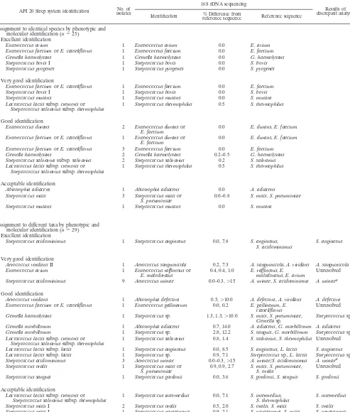
Sequencing of isolates identified to the species level with the API 20 Strep system.For 25 of the 67 strains identified to the species level with the API 20 Strep system, molecular identi-fication assigned the isolate to the same species. Discrepant results were found for 42 isolates (Tables 2 and 3).
Analysis of discrepant results and assignment to different species.For 29 of 42 isolates with discrepant results (Table 2), 16S rDNA sequencing assigned the strains to a species differ-ent from that to which the strain was assigned by the API 20 Strep system. The results for 20 of the 29 isolates were re-garded as major discrepancies; i.e., the isolate was assigned either to a different genus or to a different group within the streptococci (15).
For 22 of the 29 isolates, the 16S rDNA sequence deter-mined exhibited less than 97% similarity to the 16S rDNA sequence of the species to which it was assigned by the API 20 Strep system (for 21 isolates the sequence similarity was even less than 93%). According to Stackebrandt and Goebel (33), 16S rDNA similarities of less than 97% indicate that isolates belong to different species. Although only partial sequences were used here, it was thus concluded that these isolates do not belong to the species identified by the API system. For exam-ple, 12 strains were identified as Streptococcus acidominimus
with the API 20 Strep system, whereas sequencing resulted in 99.7 to 100.0% similarity withAerococcus urinaeand less than 85% similarity withS. acidominimus. These isolates clearly do not belong toS. acidominimusbut belong toA. urinae. Of note,
[image:2.603.300.542.90.160.2]A. urinaeis not included in the API 20 Strep system database. It has been shown previously that an unknown isolate that shows a profile forS. acidominimusin the API 20 Strep system and that is positive for-glucuronidase and leucine arylami-dase should be reported asA. urinae(42). If this rule is applied (which would result in the assignment of 12 isolates to A. urinae on the basis of the results obtained with the API 20 Strep system), molecular identification and phenotypic identi-fication would assign an isolate to the same species for 37 of the 67 isolates for which species assignment was achieved with the API 20 Strep system (5 of 6, 13 of 19, 14 of 30, and 5 of 12 isolates with excellent, very good, good, and acceptable species identifications by the API 20 Strep system, respectively).
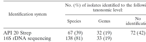
TABLE 1. Molecular versus phenotypic identification for 171 isolates (unresolved data)
Identification system
No. (%) of isolates identified to the following taxonomic level:
Species Genus identificationNo
API 20 Strep 67 (39) 32 (19) 72 (42)
16S rDNA sequencing 138 (81) 33 (19)
on May 15, 2020 by guest
http://jcm.asm.org/
TABLE 2. Molecular identification versus phenotypic identification for 67 isolates identified to the species level with the API 20 Strep system
API 20 Strep system identification isolatesNo. of
16S rDNA sequencing
Results of discrepant analysis Identification % Difference fromreference sequence Reference sequence
Assignment to identical species by phenotypic and
molecular identification (n⫽25)
Excellent identification
Enterococcus avium 1 Enterococcus avium 0.0 E. avium Enterococcus faeciumorE. casseliflavus 1 Enterococeus faecium 0.0 E. faecium Gemella haemolysans 1 Gemella haemolysans 0.0 G. haemolysans Streptococcus bovisI 1 Streptococcus bovis 0.0 S. bovis Streptococcus pyogenes 1 Streptococcus pyogenes 0.0 S. pyogenes
Very good identification
Enterococcus faeciumorE. casseliflavus 1 Enterococcus faecium 0.0 E. faecium Streptococcus bovisI 1 Streptococcus bovis 0.0 S. bovis Streptococcus mutans 1 Streptococcus mutans 0.0 S. mutans Lactococcus lactissubsp.cremorisor
Streptococcus salivariussubsp.thermophilus 1 Streptococcus thermophilus 0.5 S. thermophilus
Good identification
Enterococcus durans 2 Enterococcus duransor
E. faecium 0.0 E. durans,E. faecium Enterococcus faeciumorE. casseliflavus 1 Enterococcus duransor
E. faecium 0.0 E. durans,E. faecium Enterococcus faeciumorE. casseliflavus 3 Enterococcus faecium 0.0 E. faecium Gemella haemolysans 2 Gemella haemolysans 0.2–0.5 G. haemolysans Streptococcus salivariussubsp.salivarius 2 Streptococcus salivarius 0.2 S. salivarius Lactococcus lactissubsp.cremorisor 1 Streptococcus thermophilus 0.5 S. thermophilus
Streptococcus salivariussubsp.thermophilus
Acceptable identification
Abiotrophia adiacens 1 Abiotrophia adiacens 0.0 A. adiacens Streptococcus mitis 3 Streptococcus mitisor
S. pneumoniae 0.0–0.8 S. mitis,S. pneumoniae Streptococcus mutans 1 Streptococcus mutans 0.0 S. mutans
Assignment to different taxa by phenotypic and
molecular identification (n⫽29)
Excellent identification
Streptococcus acidominimus 1 Streptococcus anginosus 0.0, 7.8 S. anginosus,
S. acidominimus S. anginosus
Very good identification
Aerococcus viridansII 1 Aerococcus sanguinicola 0.2, 7.3 A. sanguinicola,A. viridans A. sanguinicola Enterococcus avium 1 Enterococcus raffinosusor
E. malodoratus 0.4, 0.4, 1.0 E. raffinosusmalodoratus,E.,E. avium Unresolved Streptococcus acidominimus 9 Aerococcus urinae 0.0–0.3,⬎15 A. urinae,S. acidominimus A. urinaea
Good identification
Aerococcus viridans 1 Abiotrophia defectiva 0.5,⬎10.0 A. defectiva,A. viridans A. defectiva Enterococcus faeciumorE. casseliflavus 1 Enterococcus gallinarum 0.0, 0.2 E. gallinarum, E.
casseliflavus Unresolved Gemella haemolysans 1 Streptococcussp. 1.3, 1.3,⬎10.0 S. mitis,S. pneumoniae,
Gemellasp. Streptococcussp. Gemella morbillorum 1 Abiotrophia adiacens 0.7, 14.0 A. adiacens,G. morbillorum A. adiacens Gemella morbillorum 1 Streptococcussp. 2.8, 12.2 S. sanguis,G. morbillorum Streptococcussp. Lactococcus lactissubsp.cremorisor
Streptococcus salivariussubsp.thermophilus 1 Streptococcus salivarius 0.8, 1.4 S. salivarius,S. thermophilus Unresolved Lactococcus lactissubsp.lactis 1 Streptococcus anginosus 0.0, 8.5 S. anginosus,L. lactis S. anginosus Lactococcus lactissubsp.lactis 1 Streptococcussp. 0.9, 7.1 Streptococcussp.,L. lactis Streptococcussp. Streptococcus acidominimus 3 Aerococcus urinae 0.0–0.3,⬎15 A. urinae/S. acidominimus A. urinaea
Streptococcus oralis 1 Streptococcus mitisor
S. pneumoniae 0.9, 0.9, 2.7 S. mitisS. oralis,S. pneumoniae, Unresolved Streptococcus sanguis 1 Streptococcus gordonii 0.0, 3.6 S. gordonii,S. sanguis S. gordonii
Acceptable identification
Lactococcus lactissubsp.cremorisor
Streptococcus salivariussubsp.thermophilus 1 Streptococcus intermedius 0.0, 7.1 S. intermediusS. thermophilus, S. intermedius Streptococcus mitisI 2 Streptococcus oralis 0.5, 2.0 S. oralis,S. mitis S. oralis Streptococcus mitisI 1 Streptococcus parasanguis 0.9, 2.1 S. parasanguis,S. mitis S. parasanguis
Continued on following page
on May 15, 2020 by guest
http://jcm.asm.org/
For 7 of the 29 isolates with discrepant results, the 16S rDNA sequence of the isolate showedⱖ97% sequence simi-larity to the 16S rDNA sequence of the species to which the isolate was assigned by the API 20 Strep system. It thus cannot be excluded that the strains belong to the species identified by the API 20 Strep system. For three of these seven isolates, however, a repeat of the test with the API 20 Strep system did not confirm the primary result obtained with the system. For these isolates it is thus assumed that the molecular approach correctly identified the species (99.5% sequence similarity with
Streptococcus oralis[two isolates] and 99.1% sequence similar-ity withStreptococcus parasanguis, which is not included in the API 20 Strep system database). The results for four isolates remained unresolved.
Analysis of discrepant results and assignment to the genus level by molecular analysis.For 13 of 42 isolates with discrep-ant results (Table 2), the isolates were identified to the genus level by sequencing; i.e., these isolates showed less than 99.0% similarity (our defined threshold value for species-level iden-tification) to the best-scoring reference sequence. For 7 of these 13 isolates, the similarity of the sequence to that of the species identified by the API 20 Strep system was below 97%, leading us to conclude that these isolates do not belong to the species identified by the API 20 Strep system. For example, phenotypic identification resulted in Aerococcus viridans II (five isolates); the 16S rDNA sequences determined showed, however, that the isolates had between 95.5 and 95.8% se-quence similarity withA. urinaeand between 93.3 and 93.7% sequence similarity withA. viridans. It is likely that these five isolates represent anAerococcusspecies that has yet not been described.
For 6 of the 13 isolates, the nucleic acid sequences deter-mined showed 97% or more similarity with the sequences of the species to which the isolates were assigned by the API 20 Strep system. For two of these isolates, the species determined with the API 20 Strep system was identical to the best-scoring species, as determined by sequence analysis. It is thus assumed that the biochemical system correctly assigned the two isolates. For four of the six isolates, the API 20 Strep system assigned the isolate to a species different from the best-scoring species
from the molecular investigation. For example, for three iso-lates identified as Streptococcus sanguisby the API 20 Strep system, the 16S rDNA sequences determined showed between 98.7 and 98.8% sequence similarity withStreptococcus gordonii
and between 97.3 and 97.7% sequence similarity withS. san-guis; the three isolates were reported to belonging to the genus
Streptococcus. Thus, for these four isolates, the species identity could not be determined conclusively (unresolved data).
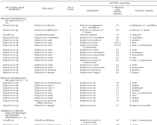
Sequencing of isolates identified to the genus level with the API 20 Strep system.With the API 20 Strep system, 32 of 171 gram-positive cocci investigated were identified to the genus level. For 23 of them, 16S rDNA sequencing allowed assign-ment to a species (Tables 3 and 4). For all but two isolates, the species assignment did not contradict the genus assignment determined conventionally: for one strain, the strain was iden-tified as a Streptococcus sp. with the API 20 Strep system, whereas molecular methods resulted in a sequence that was identical to that ofA. urinae; in the other case, the strain was identified as a Gemella sp. with the API 20 Strep system, whereas sequence analysis resulted inStreptococcus mitisorS. pneumoniae.
[image:4.603.43.546.80.250.2]For 9 of 32 strains identified to the genus level with the API 20 Strep system, 16S rDNA sequencing did not yield more
TABLE 2—Continued
API 20 Strep system identification isolatesNo. of
16S rDNA sequencing
Results of discrepant analysis Identification % Difference fromreference sequence Reference sequence
Assignment to genus level by molecular analysis
(n⫽13)
Very good identification
Aerococcus viridansII 3 Aerococcussp. 4.2–4.3, 6.3–6.7 A. urinae,A. viridans Aerococcussp. Streptococcus sanguis 1 Streptococcussp. 2.6, 3.8 Sreptococcus peroris,S.
sanguis Streptococcussp.
Good identification
Aerococcus viridansII 2 Aerococcussp. 4.2–4.5, 6.3–6.6 A. urinae,A. viridans Aerococcussp. Streptococcus mitisII 1 Streptococcussp. 1.5, 3.2 S. oralis,S. mitis Streptococcussp. Streptococcus oralis 1 Streptococcussp. 2.8 S. oralis S. oralis Streptococcus sanguis 2 Streptococcussp. 1.2, 2.3–2.7 S. gordonii,S. sanguis Unresolved Acceptable identification
Streptococcus mitisI 1 Streptococcussp. 1.9, 3.0 S. oralis,S. mitis Unresolved Streptococcus sanguis 1 Streptococcussp. 1.3, 2.6 S. gordonii,S. sanguis Unresolved Streptococcus sanguis 1 Streptococcussp. 2.1 S. sanguis S. sanguis
aA. urinaeis not included in the API 20 Strep system database and is identified asS. acidominimusby the API 20 Strep system (42).
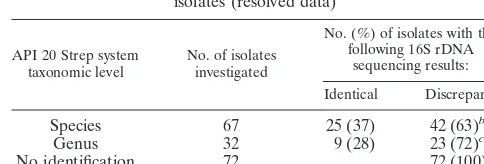
TABLE 3. Molecular versus phenotypic identification for 171 isolates (resolved data)
API 20 Strep system
taxonomic level No. of isolatesinvestigated
No. (%) of isolates with the following 16S rDNA
sequencing results:
Identical Discrepanta
Species 67 25 (37) 42 (63)b
Genus 32 9 (28) 23 (72)c
No identification 72 72 (100)d
aSee Tables 2, 4, and 5 for detailed analysis.
bFor 32 isolates, sequencing yielded a more reliable result. For two isolates,
the conventional method yielded a more reliable result. The results for eight isolates remained unresolved.
c16S rDNA sequencing allowed species identification for all 23 isolates.
dBy 16S rDNA sequencing 64 isolates were assigned to a species and 8 isolates
were assigned to a genus.
on May 15, 2020 by guest
http://jcm.asm.org/
[image:4.603.300.542.588.670.2]discriminative results; i.e., the isolate was assigned to the same genus without further species assignment.
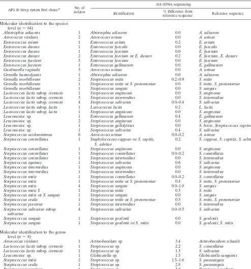
Sequencing of isolates not identified with the API 20 Strep system. Molecular methods allowed identification of all 72 strains which could not be assigned to a genus by the API 20 Strep system identification procedure (Tables 3 and 5); 63 strains were identified to the species level, and 9 strains were identified to the genus level.
DISCUSSION
This prospective study was performed under routine diag-nostic conditions. A collection of clinically relevant strains (n
⫽ 171) of aerobic catalase-negative gram-positive cocci iso-lated in the diagnostic laboratory was investigated over a pe-riod of 18 months. Accurate identification of these strains, mostly obtained from normally sterile body sites, was at-tempted with the commercially available API 20 Strep system. rDNA sequencing was performed in parallel.
We demonstrate that 16S rDNA sequence analysis has an
improved ability to identify aerobic gram-positive cocci com-pared to that of the API 20 Strep system: (i) 81% (138 of 171) of isolates were identified to the species level by sequence analysis, whereas 39% (67 of 171) were identified to the species level with the API 20 Strep system; (ii) for 72% (23 of 32) of the isolates which could be identified only to the genus level with API 20 Strep system, sequence analysis allowed identifi-cation to the species level; and (iii) among the strains that could not be discriminated at any taxonomic level biochemi-cally (72 of 171), all of the isolates could be assigned to a species (89%) or a genus (11%) level by molecular analysis.
Molecular analysis yielded discrepant results for 42 of the 67 strains which were assigned to the species level by the API 20 Strep system. For 32 of the 42 isolates with discrepant results, it was concluded that 16S rDNA sequencing correctly identi-fied the isolates (or at least had more discriminative power, as sequence analysis revealed that the isolate did not belong to a classified species; e.g., the sequence similarity to a reference sequence was less than 97% [33]). For two isolates with
dis-TABLE 4. Molecular identification versus phenotypic identification for 32 isolates identified to the genus level with the API 20 Strep system
API 20 Strep system
identification First choicea isolatesNo. of
16S rDNA sequencing
Identification
% Difference from reference sequence
Reference sequence
Molecular identification to
the species level (n⫽
21)
Enterococcussp. Enterococcus faecium 1 Enterococcus gallinarum
orE. casseliflavus 0.3 E. gallinarum,E. casseliflavus Enterococcussp. Enterococcus gallinarum 1 Enterococcus faeciumor
E. durans 0.4 E. faecium,E. durans Gemellasp. Gemella haemolysans 1 Gemella sanguinis 0.3 G. sanguinis Streptococcussp. Streptococcus constellatus 1 Streptococcus constellatus 0.0 S. constellatus Streptococcussp. Streptococcus mitis 1 Streptococcus gordonii 0.6 S. gordonii Streptococcussp. Streptococcus mitis 2 Streptococcus mitis 0.0–0.5 S. mitis Streptococcussp. Streptococcus mitis 4 Streptococcus mitis,
S. pneumoniae 0.2–0.7 S. mitis,S. pneumoniae Streptococcussp. Streptococcus mitis 1 Streptococcus oralis 0.2 S. oralis
Streptococcussp. Streptococcus mitis 1 Streptococcus parasanguis 0.6 S. parasanguis Streptococcussp. Streptococcus mitis 1 Streptococcus pneumoniae 0.2 S. pneumoniae Streptococcussp. Streptococcus oralis 1 Streptococcus mitis 0.3 S. mitis Streptococcussp. Streptococcus oralis 2 Streptococcus mitisor
S. pneumoniae 0.5 S. mitis,S. pneumoniae Streptococcussp. Streptococcus oralis 1 Streptococcus oralis 0.2 S. oralis
Streptococcussp. Streptococcus oralis 1 Streptococcus pneumoniae 0.7 S. pneumoniae Streptococcussp. Streptococcus salivarius 1 Streptococcus gordonii 0.0 S. gordonii Streptococcussp. Streptococcus sanguis 1 Streptococcus sanguis 0.4 S. sanguis
Molecular identification to
the genus level (n⫽9)
Streptococcussp. Streptococcus acidominimus 1 Streptococcussp. 3.0 S. oralis Streptococcussp. Streptococcus mitis1 1 Streptococcussp. 2.6 S. sanguis Streptococcussp. Streptococcus mitis2 1 Streptococcussp. 1.5 S. parasanguis Streptococcussp. Streptococcus mitis 1 Streptococcussp. 1.2 S. gordonii
Streptococcussp. Streptococcus mitis 1 Streptococcussp. 1.1 S. mitis,S. pneumoniae Streptococcussp. Streptococcus oralis 1 Streptococcussp. 1.1 S. mitis
Streptococcussp. Streptococcus oralis 1 Streptococcussp. 2.3 S. sanguis Streptococcussp. Streptococcus salivarius
subsp.salivarius 1 Streptococcussp. 2.9 S. sanguis
Streptococcussp. Streptococcus sanguis 1 Streptococcussp. 1.1 Streptococcus australis
Assignment to different genera by phenotypic and molecular
identification (n⫽2)
Gemellasp. Gemella morbillorum 1 Streptococcus mitisor
S. pneumoniae 0.4 S. mitis,S. pneumoniae Streptococcussp. Streptococcus acidominimus 1 Aerococcus urinae 0.0 A. urinae
aThe species best corresponding to the profile in the API 20 Strep system, with identity of⬍80.0%.
on May 15, 2020 by guest
http://jcm.asm.org/
[image:5.603.42.542.79.477.2]crepant results, it was assumed that the API 20 Strep system yielded a correct species assignment. For eight isolates with discrepant results, further investigations such as DNA-DNA hybridization or sequencing of other targets (e.g., the manga-nese-dependent superoxide dismutase [24]) would be neces-sary to resolve the discrepancies. For 12 isolates with major
discrepancies, the phenotypic system misidentifiedA. urinaeas
[image:6.603.45.539.81.611.2]S. acidominimus, a finding that has been reported previously (42). In the future, gram-positive cocci in tetrads that are identified as S. acidominimus with the API 20 Strep system (and which are positive for-glucuronidase and leucine aryl-amidase) should be reported to probably beA. urinae.
TABLE 5. Molecular identification versus conventional methods for 72 isolates not identified by the API 20 Strep system
API 20 Strep system first choicea No. of
isolates
16S rDNA sequencing
Identification reference sequence% Difference from Reference sequence
Molecular identification to the species level (n⫽64)
Abiotrophia adiacens 1 Abiotrophia adiacens 0.0 A. adiacens Aerococcus viridans 2 Aerococcus urinae 0.0 A. urinae Enterococcus avium 1 Enterococcus avium 0.2 E. avium Enterococcus durans 1 Enterococcus faecalis 0.0 E. faecalis Enterococcus durans 1 Enterococcus faecium 0.0 E. faecium
Enterococcus durans 2 Enterococcus faeciumorE. durans 0.2 E. faecium,E. durans Enterococcus faecium 3 Enterococcus faecium 0.0 E. faecium
Enterococcus faecium 1 Enterococcus gallinarum 0.0 E. gallinarum Gardnarella vaginalis 3 Aerococcus urinae 0.0 A. urinae Gemella haemolysans 1 Abiotrophia adiacens 0.0 A. adiacens Gemella morbillorum 2 Streptococcus mitis 0.2–0.9 S. mitis
Gemella morbillorum 1 Streptococcus mitisorS. pneumoniae 0.0 S. mitis,S. pneumoniae Gemella morbillorum 1 Streptococcus sanguis 0.0 S. sanguis
Lactococcus lactissubsp.cremoris 1 Streptococcus anginosus 0.0 S. anginosus Lactococcus lactissubsp.cremoris 3 Streptococcus intermedius 0.0 S. intermedius Lactococcus lactissubsp.cremoris 4 Streptococcus salivarius 0.0–0.4 S. salivarius Lactococcus lactissubsp.lactis 1 Lactococcus lactis 0.2 L. lactis Lactococcus lactissubsp.lactis 1 Streptococcus anginosus 0.0 S. anginosus Leuconostocsp. 1 Enterococcus gallinarum 0.4 E. gallinarum Leuconostocsp. 2 Streptococcus anginosus 0.0 S. anginosus
Leuconostocsp. 1 Streptococcus gallolyticus 0.0 S. bovis,Streptococcus caprinusb Leuconostocsp. 1 Streptococcus salivarius 0.4 S. salivarius
Streptococcus acidominimus 6 Aerococcus urinae 0.0–0.2 A. urinae Streptococcus constellatus 1 Staphylococcus cappraeorS. capitis,
S. arlettae 0.2 S. capprae,S. captitis,S. arlettae Streptococcus constellatus 1 Streptococcus anginosus 0.0 S. anginosus
Streptococcus constellatus 1 Streptococcus constellatus 0.0–0.2 S. constellatus Streptococcus constellatus 1 Streptococcus intermedius 0.0 S. intermedius Streptococcus equinus 1 Streptococcus salivarius 0.6 S. salivarius Streptococcus intermedius 1 Streptococcus anginosus 0.0 S. anginosus Streptococcus intermedius 1 Streptococcus intermedius 0.0 S. intermedius Streptococcus mitis 1 Streptococcus constellatus 0.0–0.2 S. constellatus Streptococcus mitis 1 Streptococcus mitisorS. pneumoniae 0.4 S. mitis,S. pneumoniae Streptococcus mitis 4 Streptococcus sanguis 0.0–1.0 S. sanguis
Streptococcus mitisI 1 Streptococcus mitis 0.3 S. mitis Streptococcus mitisorS. sanguis 1 Streptococcus sanguis 0.0 S. sanguis
Streptococcus oralis 1 Streptococcus mitisorS. pneumoniae 0.5 S. mitis,S. pneumoniae Streptococcus porcinus 1 Streptococcus intermedius 0.0 S. intermedius Streptococcus salivariussubsp.
salivarius 4 Streptococcus salivarius 0.0–0.5 S. salivarius Streptococcus sanguis 1 Streptococcus gordonii 0.0 S. gordonii Streptococcus sanguis 1 Streptococcus gordoniiorS. mitis 0.0 S. gordonii,S. mitis
Molecular identification to the genus level (n⫽8)
Aerococcus viridans 1 Actinobaculumsp. 3.4 Actinobaculum schaalii Lactococcus lactissubsp.cremoris 1 Streptococcussp. 2.2 S. constellatus Lactococcus lactissubsp.cremoris 1 Streptococcussp. 1.3 S. salivarius Leuconostocsp. 1 Globicatellasp. 1.5 Globicatella sanguinis Streptococcus mitis 2 Streptococcussp. 1.5–1.6 S. parasanguis Streptococcus oralis 1 Streptococcussp. 2.8 S. parasanguis Streptococcus porcinus 1 Streptococcussp. 3.6 Streptococcus uberis
aThe species best corresponding to the profile in the API 20 Strep system, with identity of⬍80.0%.
bS. caprinus,S. gallolyticus, and some strains ofS. bovismay belong to the same species (30).
on May 15, 2020 by guest
http://jcm.asm.org/
It is concluded that under routine conditions in a clinical laboratory the API 20 Strep system frequently does not provide accurate identifications. The possible reasons for misidentifi-cations are that (i) the species is not included in the API 20 Strep system database (e.g.,A. urinae,Aerococcus sanguinicola, and S. gordonii); (ii) the strain presumably belongs to a new, not yet described species (sequence similarity to a classified species,⬍97%); (iii) the reactions of the API 20 Strep system are misinterpreted; and (iv) biochemical variability exists within a species. It has been shown previously that commercial phenotypic identification systems, such as the API 20 Strep system or the Rapid ID 32 Strep system, are not entirely satisfactory for accurate identification of a strain to the species level (13, 14, 16, 29, 39). Supplementary manual tests are often needed, which somewhat impairs the usefulness of commercial kits.
It has been proposed that molecular methods such as PCR-restriction fragment length polymorphism analysis (20, 28, 31), DNA sequencing (1, 15, 23, 24), and other PCR-based proto-cols (12, 25) accurately identify aerobic catalase-negative gram-positive cocci. However, those studies exhibited several drawbacks that limit the routine use of these methods in a
clinical laboratory: (i) they were restricted to certain groups of bacteria and did not cover the whole range of aerobic catalase-negative gram-positive cocci (1, 12, 14–16, 20, 23, 24, 25, 28, 31, 39); (ii) they cannot be applied to other bacteria unless the corresponding databases (i.e., restriction patterns and se-quences of genes other than 16S rDNA) are enlarged (12, 15, 20, 24, 28, 31); (iii) they have not been tested under routine conditions (12, 15, 23, 24, 25, 28, 31); and (iv) their use is limited to reference laboratories (20, 28, 31).
[image:7.603.94.496.71.437.2]Therefore, we decided to evaluate the use of 16S rDNA sequencing for the identification of aerobic catalase-negative gram-positive cocci under routine conditions. 16S rDNA se-quencing for identification is not restricted to a specific group of bacteria and can readily be implemented in the laboratory. The procedure for sequence analysis (i.e., database search and manual editing of the sequence) in combination with the cri-teria for species and genus assignment (i.e., ⱖ99% sequence similarity for species assignment andⱖ95% sequence similar-ity for genus assignment) proved to be helpful for the accurate identification of the isolates. If the sequence can be assigned to a species but the second-scoring reference species shows less than 0.5% additional sequence divergence, this should be
FIG. 1. Algorithm for the identification of aerobic catalase-negative gram-positive cocci. Pos, positive; Neg, negative; VP, Voges-Proskauer test.
on May 15, 2020 by guest
http://jcm.asm.org/
noted (as was noted in our category of species with a low level of demarcation to the next species). It has been shown previ-ously that this approach allows accurate species identification for gram-positive rods (5).
The part of the 16S rRNA gene chosen for analysis covers the most discriminating regions within the 16S rDNA and is therefore suitable for identification purposes (19). In general, 16S rDNA analysis has low phylogenetic resolving power at levels of close relatedness (above 97% similarity [33]); in the extreme, two species may share identical 16S rDNA gene se-quences. It has been shown previously thatS. mitis, S. pneu-moniae, andS. oralisexhibit more than 99% sequence homol-ogy to each other (15). Similar findings have been reported for some enterococci (23). In the present study, the 16S rDNA sequences of some isolates (n⫽24) were identical to those of different species. This was true in particular forS. mitisandS. pneumoniae,S. gordoniiandS. mitis;Enterococcus faeciumand
Enterococcus durans(and in some cases, additionally, Entero-coccus faecalis),Enterococcus raffinosusandEnterococcus mal-odoratus, and Enterococcus gallinarumand Enterococcus cas-seliflavus. These organisms can readily be distinguished by additional phenotypic tests, such as the bile solubility test (which differentiates S. pneumoniae from S. mitis) and stan-dard biochemical tests (which are also part of the API 20 Strep system). For 21 of these 24 isolates with equivocal results, a definite species assignment was achieved by additional pheno-typic tests.
Another problem arises from the quality of the public data-bases, such as the GenBank, EMBL, and DDBJ databases. Sequences can be deposited in these databases largely inde-pendently of their quality, e.g., regardless of the number of ambiguous nucleotides, the length of the sequence, or the correct assignment of the strain investigated. However, such situations should normally not lead to false identifications but, rather, should lead to problems assigning a strain to a partic-ular species (a low level of demarcation), at least if the correct species is also contained in the database. This in turn would induce further investigations (e.g., biochemical tests or phylo-genetic analysis of the sequences).
In this study, sequence-based identification was compared to the identification based on the widely used commercial API 20 Strep system. As a limitation, we did not consider other com-mercially available identification systems, such as the Rapid ID 32 Strep system (bioMe´rieux), the VITEK 2 system (bio-Me´rieux), the BD Phoenix automated microbiology system (BD diagnostic systems), the BBL Crystal system (BD Diag-nostic Systems), or the MicroLog system (Biolog Inc.). How-ever, as discussed for the API 20 Strep system, most pheno-typic systems have general drawbacks, such as the quality and the quantity of the underlying database and phenotypic vari-ability within a species. This demonstrates that identification by molecular analysis is superior to that with the API 20 Strep system and is ready to be implemented in the clinical labora-tory.
In our study, the majority (96%) of strains were not reliably identified to the species level by the API 20 Strep system or the species assignment was doubtful (6 of 19, 15 of 30, and 6 of 12 isolates with very good, good, and acceptable qualities of iden-tification were falsely identified). A species assignment in the API 20 Strep system may be considered reliable only when an
excellent species identification according to the criteria of the system is achieved. However, this was the case for only 6 of 171 isolates. We thus conclude that the API 20 Strep system is not an effective system for the identification of gram-positive cata-lase-negative cocci. Consequently, corresponding isolates, with the exception of S. pneumoniae and beta-hemolytic strepto-cocci, should be subjected to 16S rDNA sequence analysis if adequate species identification is of concern (see the algorithm in Fig. 1). Phenotypic tests may be used for definite species assignment only for those few strains for which the sequencing result is equivocal.
ACKNOWLEDGMENTS
We thank the technicians of the Institute of Medical Microbiology for excellent technical assistance.
This study was supported by the University of Zu¨rich.
REFERENCES
1. Angeletti, S., G. Lorino, G. Gherardi, F. Battistoni, M. De Cesaris, and G.
Dicuonzo.2001. Routine molecular identification of enterococci by
gene-specific PCR and 16S ribosomal DNA sequencing. J. Clin. Microbiol.39:
794–797.
2. Appelbaum, P. C., P. S. Chaurushiya, M. R. Jacobs, and A. Duffett.1984.
Evaluation of the Rapid Strep system for species identification of
strepto-cocci. J. Clin. Microbiol.19:588–591.
3. Beighton, D., J. M. Hardie, and R. A. Whiley.1991. A scheme for the
identification of viridans streptococci. J. Med. Microbiol.35:367–372.
4. Boettger, E. C.1996. Approaches for identification of microorganisms. ASM
News62:247–250.
5. Bosshard, P. P., S. Abels, R. Zbinden, E. C. Bo¨ttger, and M. Altwegg.2003.
Ribosomal DNA sequencing for identification of aerobic gram-positive rods
in the clinical laboratory (an 18-month evaluation). J. Clin. Microbiol.41:
4134–4240.
6. Bosshard, P. P., A. Kronenberg, R. Zbinden, C. Ruef, E. C. Boettger, and M.
Altwegg.2003. Etiologic diagnosis of infective endocarditis by broad-range
PCR: a 3-year experience. Clin. Infect. Dis.37:167–172.
7. Brosius, J., M. L. Palmer, P. J. Kennedy, and H. F. Noller.1978. Complete
nucleotide sequence of a 16S ribosomal RNA gene fromEscherichia coli.
Proc. Natl. Acad. Sci. USA75:4801–4805.
8. Cloud, J. L., H. Neal, R. Rosenberry, C. Y. Turenne, M. Jama, D. R. Hillyard,
and K. C. Carroll.2002. Identification ofMycobacteriumspp. by using a
commercial 16S ribosomal DNA sequencing kit and additional sequencing
libraries. J. Clin. Microbiol.40:400–406.
9. Devereux, J., P. Haeberli, and O. Smithies.1984. A comprehensive set of
sequence analysis programs for the VAX. Nucleic Acids Res.12:387–395.
10. Drancourt, M., C. Bollet, A. Carlioz, R. Martelin, J. P. Gayral, and D.
Raoult.2000. 16S ribosomal DNA sequence analysis of a large collection of
environmental and clinical unidentifiable bacterial isolates. J. Clin.
Micro-biol.38:3623–3630.
11. Ferroni, A., I. Sermet-Gaudelus, E. Abachin, G. Quesne, G. Lenoir, P.
Berche, and J. L. Gaillard.2002. Use of 16S rRNA gene sequencing for
identification of nonfermenting gram-negative bacilli recovered from
pa-tients attending a single cystic fibrosis center. J. Clin. Microbiol.40:3793–
3797.
12. Garnier, F., G. Gerbaud, P. Courvalin, and M. Galimand.1997.
Identifica-tion of clinically relevant viridans group streptococci to the species level by
PCR. J. Clin. Microbiol.35:2337–2341.
13. Hamilton-Miller, J. M., and S. Shah.1999. Identification of clinically
iso-lated vancomycin-resistant enterococci: comparison of API and BBL Crystal
systems. J. Med. Microbiol.48:695–696.
14. Hinnebusch, C. J., D. M. Nikolai, and D. A. Bruckner.1991. Comparison of
API Rapid Strep, Baxter MicroScan Rapid Pos ID Panel, BBL Minitek Differential Identification system, IDS RapID STR system, and Vitek GPI to conventional biochemical tests for identification of viridans streptococci.
Am. J. Clin. Pathol.96:459–463.
15. Kawamura, Y., X. G. Hou, F. Sultana, H. Miura, and T. Ezaki.1995.
De-termination of 16S rRNA sequences ofStreptococcus mitisandStreptococcus
gordoniiand phylogenetic relationships among members of the genus Strep-tococcus. Int. J. Syst. Bacteriol.45:406–408.
16. Kikuchi, K., T. Enari, K. Totsuka, and K. Shimizu.1995. Comparison of
phenotypic characteristics, DNA-DNA hybridization results, and results with a commercial rapid biochemical and enzymatic reaction system for
identifi-cation of viridans group streptococci. J. Clin. Microbiol.33:1215–1222.
17. Kilian, M., L. Mikkelsen, and J. Henrichsen.1989. Taxonomic study of
viridans streptococci: description of Streptococcus gordonii sp. nov. and
emended description ofStreptococcus sanguis(White and Niven 1946),
on May 15, 2020 by guest
http://jcm.asm.org/
tococcus oralis(Bridge and Sneath 1982), andStreptococcus mitis(Andrews
and Horder 1906). Int. J. Syst. Bacteriol.39:471–484.
18. Kolbert, C. P., and D. H. Persing.1999. Ribosomal DNA sequencing as a
tool for identification of bacterial pathogens. Curr. Opin. Microbiol.2:299–
305.
19. Ludwig, W., O. Strunk, S. Klugbauer, N. Klugbauer, M. Weizenegger,
J. Neumaier, M. Bachleitner, and K. H. Schleifer.1998. Bacterial phylogeny
based on comparative sequence analysis. Electrophoresis19:554–568.
20. Ohara-Nemoto, Y., S. Tajika, M. Sasaki, and M. Kaneko.1997.
Identifica-tion ofAbiotrophia adiacensandAbiotrophia defectivaby 16S rRNA gene
PCR and restriction fragment length polymorphism analysis. J. Clin.
Micro-biol.35:2458–2463.
21. Patel, J. B.2001. 16S rRNA gene sequencing for bacterial pathogen
identi-fication in the clinical laboratory. Mol. Diagn.6:313–321.
22. Patel, J. B., D. G. Leonard, X. Pan, J. M. Musser, R. E. Berman, and I.
Nachamkin.2000. Sequence-based identification ofMycobacteriumspecies
using the MicroSeq 500 16S rDNA bacterial identification system. J. Clin.
Microbiol.38:246–251.
23. Patel, R., K. E. Piper, M. S. Rouse, J. M. Steckelberg, J. R. Uhl, P. Kohner,
M. K. Hopkins, F. R. Cockerill III, and B. C. Kline.1998. Determination of
16S rRNA sequences of enterococci and application to species identification
of nonmotileEnterococcus gallinarumisolates. J. Clin. Microbiol.36:3399–
3407.
24. Poyart, C., G. Quesne, S. Coulon, P. Berche, and P. Trieu-Cuot.1998.
Identification of streptococci to species level by sequencing the gene
encod-ing the manganese-dependent superoxide dismutase. J. Clin. Microbiol.36:
41–47.
25. Reed, R. P., V. G. Sinickas, C. Lewis, and K. A. Byron.1999. A comparison
of polymerase chain reaction and phenotyping for rapid speciation of
en-terococci and detection of vancomycin resistance. Pathology31:127–132.
26. Relman, D. A.1998. Detection and identification of previously unrecognized
microbial pathogens. Emerg. Infect. Dis.4:382–389.
27. Relman, D. A., J. S. Loutit, T. M. Schmidt, S. Falkow, and L. S. Tompkins.
1990. The agent of bacillary angiomatosis. An approach to the identification
of uncultured pathogens. N. Engl. J. Med.323:1573–1580.
28. Rudney, J. D., and C. J. Larson.1993. Species identification of oral viridans
streptococci by restriction fragment polymorphism analysis of rRNA genes.
J. Clin. Microbiol.31:2467–2473.
29. Sader, H. S., D. Biedenbach, and R. N. Jones.1995. Evaluation of Vitek and
API 20S for species identification of enterococci. Diagn. Microbiol. Infect.
Dis.22:315–319.
30. Schlegel, L., F. Grimont, E. Ageron, P. A. Grimont, and A. Bouvet.2003.
Reappraisal of the taxonomy of theStreptococcus bovis/Streptococcus equinus
complex and related species: description ofStreptococcus gallolyticussubsp.
gallolyticussubsp. nov.,S. gallolyticussubsp.macedonicussubsp. nov. andS. gallolyticussubsp.pasteurianussubsp. nov. Int. J. Syst. Evol. Microbiol.53: 631–645.
31. Schlegel, L., F. Grimont, P. A. Grimont, and A. Bouvet.2003. Identification
of major streptococcal species byrrn-amplified ribosomal DNA restriction
analysis. J. Clin. Microbiol.41:657–666.
32. Springer, B., L. Stockman, K. Teschner, G. D. Roberts, and E. C. Boettger.
1996. Two-laboratory collaborative study on identification of mycobacteria:
molecular versus phenotypic methods. J. Clin. Microbiol.34:296–303.
33. Stackebrandt, E., and B. M. Goebel.1994. A place for DNA-DNA
reasso-ciation and 16S rRNA sequence analysis in the present species definition in
bacteriology. Int. J. Syst. Bacteriol.44:846–849.
34. Tang, Y. W., N. M. Ellis, M. K. Hopkins, D. H. Smith, D. E. Dodge, and D. H.
Persing.1998. Comparison of phenotypic and genotypic techniques for
iden-tification of unusual aerobic pathogenic gram-negative bacilli. J. Clin.
Mi-crobiol.36:3674–3679.
35. Tang, Y. W., A. Von Graevenitz, M. G. Waddington, M. K. Hopkins, D. H.
Smith, H. Li, C. P. Kolbert, S. O. Montgomery, and D. H. Persing.2000.
Identification of coryneform bacterial isolates by ribosomal DNA sequence
analysis. J. Clin. Microbiol.38:1676–1678.
36. Tardif, G., M. C. Sulavik, G. W. Jones, and D. B. Clewell.1989. Spontaneous
switching of the sucrose-promoted colony phenotype inStreptococcus
san-guis. Infect. Immun.57:3945–3948.
37. Tillotson, G. S.1982. An evaluation of the API-20 STREP system. J. Clin.
Pathol.35:468–472.
38. Trotha, R., T. Hanck, W. Konig, and B. Konig.2001. Rapid
ribosequenc-ing—an effective diagnostic tool for detecting microbial infection. Infection 29:12–16.
39. von Baum, H., F. R. Klemme, H. K. Geiss, and H. G. Sonntag.1998.
Comparative evaluation of a commercial system for identification of
gram-positive cocci. Eur. J. Clin. Microbiol. Infect. Dis.17:849–852.
40. Wilson, K. H., R. Blitchington, R. Frothingham, and J. A. Wilson.1991.
Phylogeny of the Whipple’s-disease-associated bacterium. Lancet338:474–
475.
41. Woese, C. R., and G. E. Fox.1977. Phylogenetic structure of the prokaryotic
domain: the primary kingdoms. Proc. Natl. Acad. Sci. USA74:5088–5090.
42. Zbinden, R., P. Santanam, L. Hunziker, B. Leuzinger, and A. Von
Graeve-nitz.1999. Endocarditis due toAerococcus urinae: diagnostic tests, fatty acid
composition and killing kinetics. Infection27:122–124.