Copyright © 2000, American Society for Microbiology. All Rights Reserved.
Rapid, Efficient Detection and Drug Susceptibility Testing of
Mycobacterium tuberculosis
in Sputum by Microscopic
Observation of Broth Cultures
LUZ CAVIEDES,
1TIEN-SHUN LEE,
2ROBERT H. GILMAN,
1,2,3* PATRICIA SHEEN,
1EMILY SPELLMAN,
2ELLEN H. LEE,
4DOUGLAS E. BERG,
5SONIA MONTENEGRO-JAMES,
6AND
THE TUBERCULOSIS WORKING GROUP IN PERU
1†
Department of Pathology, Universidad Peruana Cayetano Heredia,
1and Asociacio´n Benefica Proyectos en Informatica,
Salud, Medicina y Agricultura (AB PRISMA),
2Lima, Peru; Johns Hopkins School of Public Health and Hygiene,
Baltimore, Maryland
3; State University of New York—Stony Brook School of Medicine, Stony Brook, New York
4;
Washington University, St. Louis, Missouri
5; and Ochsner Medical Foundation, New Orleans, Louisiana
6Received 28 July 1999/Returned for modification 7 October 1999/Accepted 16 December 1999
Inexpensive, rapid, and reliable methods of detecting infection by and drug susceptibility of
Mycobacterium
tuberculosis
(MTB) are crucial to the control of tuberculosis. The novel microscopic observation broth-drug
susceptibility assay (MODS) detects early growth of MTB in liquid medium, allowing more timely diagnosis
and drug susceptibility testing. Sputum samples from hospitalized patients in Peru were analyzed by using
stains, culture, and PCR. Sensitivity of MODS (92%) compared favorably with the most sensitive of the other
culture methods (93%). Sputum samples positive for tuberculosis were tested for susceptibility to isoniazid and
rifampin with the microwell alamar blue assay (MABA) and MODS. In 89% of cases, there was concordance
between MODS and MABA. Of the diagnostic and susceptibility testing methods used, MODS yielded results
most rapidly (median, 9.0 and 9.5 days, respectively). MODS is a rapid, inexpensive, sensitive, and specific
method for MTB detection and susceptibility testing; it is particularly appropriate for use in developing
countries burdened by significant infection rates and increasing numbers of multiple-drug-resistant cases.
In the last decade, there has been a dramatic resurgence in
the incidence of tuberculosis (TB) throughout the world.
Con-tributing factors include population growth, the human
immu-nodeficiency virus epidemic, and poorly implemented public
health measures (19). In developing countries, efforts to curb
TB are further hindered by overcrowding, poverty, poor
infec-tion control in hospitals, widespread AIDS, and severe lack of
financial and technical resources for effective control
pro-grams. The number of TB patients infected with
multiple-drug-resistant strains is also on the rise, particularly in developing
countries; this is due in large part to the failure of control
programs to provide adequate treatment and to the marked
increase in high-risk groups, such as patients with AIDS.
To successfully control the spread of TB, cases must be
detected and treated in a timely, effective manner.
Well-funded clinical laboratories can detect TB cases within 7 to
14 days using sophisticated liquid culturing systems such as
BACTEC or Mycobacteria Growth Indicator tubes (MGITs)
(15, 24). TB laboratories in most developing countries lack the
sophisticated and costly equipment required by these systems
but generally have a centrifuge, a microscope, and an
incuba-tor. If cultures are attempted at all, most laboratories in
de-veloping countries use solid media such as Lowenstein-Jensen
(LJ) and Ogawa because of their lower cost. With these
tradi-tional culture methods,
Mycobacterium tuberculosis
(MTB
de-tection takes an average of 3 weeks under optimal conditions,
and drug susceptibility testing takes an additional 3 to 4 weeks
(9). Consequently, TB control programs in developing
coun-tries rely upon results of stained sputum smears for diagnosis,
and treatment regimens are usually prescribed without
assess-ing the drug susceptibility profiles of infectassess-ing strains (4, 5).
MTB grows more rapidly on liquid medium, where it grows
as strings and tangles, than on solid medium (1). Based on this,
we developed a new, efficient, reliable, and inexpensive
meth-od that permits simultaneous MTB detection and
determina-tion of drug susceptibility in less than 2 weeks. Sputum samples
are first digested to reduce viscosity and kill other bacteria and
then concentrated by centrifugation. They are diluted into 7H9
broth and distributed to wells of a 24-well plastic culture tray
with and without antimicrobial agents. MTB growth is
ob-served as the formation of characteristic strings and tangles by
simple light microscopy. This microscopic observation broth
drug susceptibility (MODS) assay, unlike other rapid detection
systems, does not require either radioactive isotopes or
fluo-rescent indicators. It is particularly well suited for use in
de-veloping countries, as it is rapid, reliable, and inexpensive.
MATERIALS AND METHODS
This study was carried out in a level P2 biosafety tuberculosis laboratory in the Department of Pathology at the Universidad Peruana Cayetano Heredia in Lima, Peru. In the United States and Western Europe, it is customary to handle MTB at a P3 level; in Peru, however, as in much of the developing world, the majority of MTB is handled in P1 containment facilities due to resource con-straints.
* Corresponding author. Mailing address: Department of
Interna-tional Health, Johns Hopkins University, School of Public Health and
Hygiene, 615 North Wolfe St., Room 3501, Baltimore, MD 21205.
Phone: (410) 614-3958. Fax: (410) 614-6060. E-mail: rgilman@jhsph
.edu.
† Members of the Tuberculosis Working Group in Peru are Patricia
Fuentes, Teresa Valencia, Rosa Chumpitaz, Inex Salas, and S.
Reca-varren (Universidad Peruana Cayetano Heredia, Lima, Peru); Rosa
Cama and Lilia Cabrera (AB PRISMA, Lima, Peru); L. M. Franchi
(Pediatric Hospital, Lima, Peru); E. Ticona, L. Ticona, and V. Chavez
(Dos de Mayo Hospital, Lima, Peru); A. Vivar (Loayza Hospital,
Lima, Peru); S. Rodriguez (Maria Auxiliadora Hospital, Lima, Peru);
W. Checkley and R. Black (Johns Hopkins University, Baltimore,
Md.); S. Franzblau (Hansens Disease Center, Baton Rouge, La.);
J. Friedland (Imperial College, London, United Kingdom); and C.
Evans (University of Cambridge, Cambridge, United Kingdom).
1203
on May 15, 2020 by guest
http://jcm.asm.org/
Detection of mycobacteria. (i) Specimen processing.Six to 8 ml of sputum was collected from 172 samples from patients who were admitted to six hospitals in Lima, Peru. Most patients were suspected of having tuberculosis based on clin-ical symptoms and/or a positive stain for acid-fast bacilli (AFB). Sputum samples collected first thing in the morning from 10 subjects with mild chronic bronchitis who were otherwise healthy (laboratory personnel) were assayed as negative controls. All specimens were digested and decontaminated of other bacteria by the standardN-acetyl-L-cysteine–NaOH–Na citrate method (4). After
centrifu-gation for 15 min at 3,000⫻g, the pellet was then resuspended into 2 ml of 7H9 broth.
(ii) Smear test.A total of 100l of each decontaminated sample was placed
on a microscope slide and stained with 0.1% auramine O by standard procedures (9). Slides were examined microscopically at⫻1000 magnification. A stain was considered positive if it contained at least five bacilli per 300 fields.
(iii) MTB culture techniques.Five hundred microliters of each
decontami-nated sample was inoculated into an MGIT (Becton Dickinson, Sparks, Md.) containing both 10% OADC (oleic acid, albumin, dextrose, and catalase; Becton Dickinson) and 100l of PANTA antimicrobial supplement (polymyxin B, am-photericin B, nalidixic acid, trimethoprim, and azlocillin; Becton Dickinson), as per manufacturer’s instructions. Two hundred-fifty microliters of sample was used to inoculate one LJ slant (Difco, Detroit, Mich.), and another 250l was used to inoculate one plate of Middlebrook’s 7H11 medium (Difco). MGITs were incubated at 37°C and examined for fluorescence every day for up to 40 days with a box containing two longwave UV light sources. LJ slants were incubated at 37°C and examined twice a week, from week 2 to 8, following inoculation. Microagar 7H11 plates were incubated at 37°C in an atmosphere of 5% CO2in humidified air and then examined under an inverted light microscope. The plates were examined once in the first week and then 2 to 3 times per week, every week, for up to 6 weeks following inoculation (8). Plates and slants were interpreted according to criteria developed by the National Centers for Disease Control and Prevention (9). A culture was considered positive if it contained at least one colony. All positive MGITs were checked with an auramine O stain to confirm the presence of AFB. Any suspicious colonies observed as growing on LJ or 7H11 were also stained to check for acid fastness. The identities of all cultures were blinded to laboratory workers.
(iv) Growth detection in MODS.Eight hundred microliters of each
decontam-inated sample was inoculated into 7.2 ml of Middlebrook 7H9 broth (Difco) containing 5.9 g of Middlebrook 7H9 broth base per liter, 0.31% glycerol, 1.25 g of Bacto Casitone (Difco) per liter, 10% OADC, and 160l of PANTA Anti-microbic Supplement Stock (20l/ml). For each sputum-medium mixture, ali-quots of 1.2 ml (each) were distributed to 6 wells of a sterile 24-well plate (Falcon, Franklin Lakes, N.J.). A 24-well plate rather than a 96-well plate was used to provide increased growth medium and also to improve microscopic visualization of the bacteria. When three specimens had been run, the fourth set of 6 wells was filled with broth only to control for cross-contamination. Each run also included a susceptible control of MTB strain H37Rv in which a 1/50 dilution of 0.5 McFarland standard equivalent in broth was added to one well. This was examined daily for growth between 6 and 8 days. Plates were sealed with Scotch polyethylene tape (Fisher, Springfield, N.J.) and incubated at 37°C for up to 40 days. Aliquots were examined daily under an inverted light microscope at⫻40 magnification. Each well was examined for approximately 30 to 45 s each. The identities of the cultures were blinded to laboratory workers.
Cultures ofM. aviumATCC 35713 and 35718,Mycobacterium chelonaeATCC 35752,Mycobacterium smegmatisATCC 19420,Mycobacterium kansasiiATCC 12478,Mycobacterium bovisATCC 19210 (obtained from American Type Cul-ture Collection, Washington, D.C.), and MTB strain H37Rv (ATCC 27294) were inoculated into coded wells containing 7H9 medium. The cultures were exam-ined, and species morphology was described by laboratory personnel who did not know the identity of the cultures. The study was done to determine the ability of the microscopist to identify MTB accurately in comparison with the other strains.
(v) PCR.PCR was performed using separate rooms and separate pipettes to
avoid cross-contamination. Before DNA extraction, 500l of each decontami-nated sample was incubated with 2 volumes of absolute ethanol for 2 h at room temperature. Samples were then centrifuged at 13,000⫻gfor 10 min; superna-tants were discarded and pellets were submitted to a simple heat–and–Triton X-100 detergent treatment for DNA extraction (3). A heminested PCR was performed on each DNA sample with three primers specific for insertion se-quence IS6110from the MTB complex genome. Outer primers Pt 8 (5⬘-GTGC GGATGGTCGCAGAGAT-3⬘) (10) and Pt 9 (5⬘-GTGCGGATGGTCGCAGA GAT-3⬘) were used for the first amplification. The inner primer TB 290 (5⬘-G GCGGGACAACGCCGAATGCGAA-3⬘) (21) plus Pt 9 were used to amplify products of the first PCR (Pt 8-Pt 9). The first amplification was carried out according to the following program: hold at 94°C for 4 min; 20 cycles at 94°C for 20 s, 65°C for 35 s, and 72°C for 35 s. The program of the second amplification was similar to the first except that 35 cycles were used instead of 20. A sample was considered positive if this second PCR yielded a 337-bp product. DNA was extracted from the isolates, and the above procedure was used to confirm that these isolates were MTB.
(vi) Susceptibility testing.In a separate study, 88 auramine O-positive sputum
samples were selected randomly from specimens sent from various hospitals in Lima for laboratory analysis at Hospital Cayetano Heredia, Lima, Peru. Two methods were used to determine drug susceptibility to isoniazid (INH) and
rifampin (RIF): the microwell alamar blue assay (MABA), an indirect suscepti-bility assay (2, 7, 28), and MODS, which tests susceptisuscepti-bility directly on 2 ml of sputum sample. With MABA, MTB isolates are grown and then inoculated into a series of wells of a 96-well microtiter plate containing twofold increments of the drug of interest; MTB growth is evaluated by adding alamar blue color indicator (TREK, Westlake, Ohio) (2, 7, 28). MABA provides a mean MIC. MODS uses only two antimicrobial concentrations, similar to the BACTEC format; INH concentrations were 0.4g/ml and 0.1g/ml and the RIF concentrations was 1.0
g/ml; we added an additional concentration of RIF of 0.5g/ml (1, 7). H37Rv was used as a sensitive control each time susceptibilities were run.
(vii) Indirect testing on strain isolates by MABA.Three hundred microliters
of each concentrated sample was inoculated onto Middlebrook 7H11 agar (Bec-ton Dickinson, Cockeysville, Md.), incubated at 37°C in an atmosphere of 5% CO2in humidified air until colonies appeared, and then refrigerated. Bacterial suspensions were prepared and tested for susceptibility to INH and RIF by a standardized modification of the colorimetric method (7). Wells were tested for MTB growth by adding 50l of freshly prepared 1:1 mixture of 10⫻alamar blue reagent and 10% Tween 80. A change in color from blue to pink in medium with characteristic growth was interpreted as MTB growth. Medium that appeared turbid was considered to be contaminated (7). The MIC for each sample was defined as the lowest drug concentration that prevented a color change from blue to pink.
(viii) direct sputum testing by MODS.Sputum samples obtained from patients
at the seven hospitals were decontaminated using the procedure described above, except pellets were resuspended in 4.5 ml of Middlebrook 7H9 broth containing a final concentration of 20l of PANTA per ml of broth and 10% OADC. The four antimicrobial drug stock solutions were prepared at the fol-lowing concentrations: INH, 4.0 and 1.0g/ml; RIF, 10.0 and 5.0g/ml. Five hundred-forty microliters of each sputum-medium solution was placed onto each of 8 microwells of a sterile 24-well plate; each plate was used to process three samples. A 24-well plate rather than a 96-well plate was used to minimize cross-contamination. For each sample, a 60-l aliquot of each of the four drug stock solutions was added to a well containing sputum-medium solution. Sixty microliters of 7H9 media was added to each of the remaining four wells con-taining the sputum-media mixture to serve as antimicrobial agent-free controls. Plates were sealed with Scotch polyethylene tape, incubated at 37°C, and moved to the microscope without agitation. Wells were examined for the presence of mycobacteria every 1 or 2 days under an inverted light microscope at 40⫻
magnification. A sample was considered susceptible if growth was observed in the drug-free control wells but not in wells containing antimicrobial agents. A sample was considered resistant if growth was observed in both control wells and wells containing antimicrobial agents. The final drug concentrations were 0.4 and 0.1
g/ml for INH and 1.0 and 0.5g/ml for RIF. Concordance was determined by comparing MODS results at final high- and low-drug concentrations with MABA results at MICs comparable to the final MODS drug concentrations. MODS was repeated twice in 14 randomly selected specimens to determine reproducibility.
(ix) SSCP:In order to determine whether results of rifampin antimicrobial
susceptibility tests were accurate, we used a method that determines whether a mutation has occurred in therpoBgene. Previous studies have reported encour-aging results by using the single-stranded conformation polymorphism (SSCP) method to detect mutations on MTB cultures resistant to RIF (23). Ninety-five percent of RIF-resistant isolates have a mutation in a 69-bp region of therpoB
gene, making this a good target for molecular genotypic diagnostic methods (25, 26, 27) such as the SSCP or heteroduplex test. DNA from M. tuberculosis
RIF-susceptible reference strain H37Rv and 14M. tuberculosiscultured strains was extracted by proteinase K digestion, and phenol-chloroform purification was performed. The RIF-resistant region of therpoBgene was amplified by primers TR9 (5⬘-TCGCCGCGATCAAGGAGT-3⬘) and TR8 (5⬘-TGCACGTCGCGGA CCTCCA-3⬘) (23). The PCR (50l) contained 50 mM KCl, 10 mM Tris-HCl (pH 8.3), 1.5 mM MgCl2, 0.2 mM deoxynucleoside triphosphates, 0.5M (each) primers, 1.25 U ofTaqpolymerase, and 5 ng of DNA. After a 4-min denaturation at 94°C, the reaction was subjected to 35 cycles of amplification (30 s at 94°C, 45 s at 55°C, and 45 s at 72°C), followed by a 4-min extension at 72°C. This reaction generates a 157-bp PCR product. After amplification, 20l of PCR product was mixed with 20l of SSCP dilution solution (95% formamide, 20 mM EDTA, 10 mM NaCl, 0.05% bromophenol blue), boiled for 10 min, cooled on ice, and loaded onto nondenaturing sequencing-format MDE gel (Hydrolink; AT Bio-chem, Inc., Malvern, Pa.) composed of 0.5⫻MDE, 0.6⫻Tris-borate-EDTA, 0.045% (vol/vol)N,N,N⬘,N⬘-tetramethylenediamine, and 0.40% (wt/vol) ammo-nium persulfate. Electrophoresis was performed at room temperature at the following settings: 1 W for 1 h, 3 W for 2 h, and 2.5 W for 3 h, using a 16- by 18-by 0.3-cm gel. Gels were stained 18-by silver nitrate (20) and analyzed.
Statistical analysis.All data were compiled and analyzed using SPSS (SPSS,
Inc., Chicago, Ill.).Pvalues for the difference in speeds of various methods were calculated using Wilcoxon’s nonparametric test.
Cost.All costs were calculated using recent purchase records or catalogue
prices from biotechnology companies. Costs for decontamination of samples, shipping and handling, customs taxes, and labor were not included.
on May 15, 2020 by guest
http://jcm.asm.org/
RESULTS
Detection of mycobacteria.
Ninety-eight (57%) of 172
spu-tum samples were positive for MTB by at least one standard
culture method (MGIT, LJ, or microagar 7H11). The study
was performed on 100 specimens from 84 patients from six
Lima hospitals suspected clinically of having tuberculosis, of
which 33 specimens from 27 patients were positive on culture.
An enriched sample was obtained from a tuberculosis program
which evaluated smear-positive patients from the community.
This program provided 72 specimens from 64 patients, of
which 65 specimens from 56 patients were positive. All positive
specimens were confirmed to be MTB by PCR. In determining
sensitivities of various methods (Table 1), we considered true
positives to be samples which were positive by one or more
standard culture methods. Of the 172 sputum samples
col-lected, 4 (2%) of MODS, 7 (4%) of MGIT, 2 (1%) of LJ, and
2 (1%) of microagar 7H11 cultures were contaminated and
therefore excluded from analysis. PCR was not possible in 13
(8%) cases due to insufficient patient sample. Auramine O
smears were successfully performed on all samples. In eight
cases, the speed of the MGIT test could not be determined due
to a temporary failure of our UV light box.
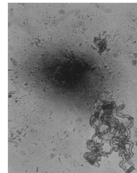
Figure 1 shows a microphotograph of MTB in the 7H9 liquid
culture medium used for MODS. Small colonies have a
char-acteristic string-and-tangles appearance that is easily
distin-guishable from the noncorded colonies of
M. avium
and
M.
kansasii
. The MODS assay had a sensitivity of 92% (89 of 97),
while MGIT had a sensitivity of 93% (88 of 95). Results for
MODS and MGIT were concordant in all cases except for one,
which was positive by MGIT but negative by MODS. Solid
media were less sensitive with detection rates of 76% (73 of 96)
and 78% (75 of 96) for LJ and microagar 7H11 methods,
respectively. Using all culture methods collectively as the “gold
standard,” PCR had a sensitivity of 90%. Eighteen samples
were positive by PCR but negative by all culture methods.
Specificity was high for both PCR and MODS, as none of the
10 control sputum samples were positive by either method. The
auramine O staining method detected TB in 78% (76 of 98) of
culture-positive samples. All samples that had positive
aura-mine O stains were also positive by at least one culture
method. A summary of the results obtained using various MTB
detection methods is shown in Table 1.
MODS detected MTB in sputum samples within a median
time of 9.0 days after inoculation (range, 4 to 31 days). This
was significantly faster than MGIT, which had a median of 10.0
days (
P
⬍
0.05) (range, 4 to 39 days). In contrast, 7H11 and LJ
agar cultures took a median of 14 and 24 days, respectively, to
detect MTB, as shown in Table 1.
Susceptibility testing.
Susceptibility results using MODS
were available between 5 and 20 days (median, 9.5 days) after
the inoculation of patient sputum samples directly into 7H9
broth containing antimicrobials. Susceptibility assays such as
MABA require a pure MTB isolate (which typically takes 3
weeks to obtain); this is much slower than MODS.
[image:3.612.313.549.71.369.2]Thirty-three of the 88 patients analyzed for susceptibility
were taking anti-TB therapy, 22 for less than 1 week and 11 for
over 1 week. By MABA, there were 64 patient isolates sensitive
to both INH and RIF, 8 resistant to INH alone, 2 resistant to
RIF alone, and 14 resistant to both. The results obtained with
MODS were concordant in 78 of these 88 cases (89%) and
discordant in 10 cases (11%). Discordant results occurred in
[image:3.612.53.294.92.190.2]FIG. 1. Appearance ofM. tuberculosisat 100⫻magnification after 25 days of culture in broth. Note the serpentine, ropy aspect of the microcolonies.
TABLE 1. MODS sensitivity and time to detection of
MTB versus standard culture methods
Method No. of samplespositive by method
No. of samples positive by at
least one culturea
Sensitivity (%)b
Median detection time (range)c
Auramine O stain 76 98 78
MODS 89 97 92 9 (4–31)
MGIT 88 95 93 10 (4–39)
LJ 73 96 76 24 (6–59)
microagar 7H11 75 96 78 14.5 (4–28)
PCR 81 90 90
aOnly samples which were positive by at least one culture (MODS, MGIT, LJ, or 7H11) and interpretable by the given method were considered in determining sensitivity. Of the 172 samples, 98 were positive by at least one culture. Denom-inators differ for different methods because not all results were interpretable due to contamination or (in the case of PCR) insufficient patient sample.
bSensitivity is based on the gold standard of detection by any culture. cIncludes only interpretable results. Values shown are in days.
TABLE 2. Comparison of MODS and MABA in evaluating
MTB sensitivity to INH and RIF
Method
INH RIF
Total (%) specimens No.
sensitive (%)
No. resistant
(%)
No. sensitive
(%)
No. resistant
(%)
MODS 65 (74) 23 (26)a 63 (72) 25 (28)b 88 (100)
MABA 66 (75) 22 (25) 72 (82) 16 (18) 88 (100)
aResistant at both low (0.1g/ml) and high (0.4g/ml) concentrations of INH.
bSeven specimens were resistant at low (0.5g/ml) but susceptible at high (1.00g/ml) concentrations of RIF; two specimens were resistant at both low and high concentrations.
on May 15, 2020 by guest
http://jcm.asm.org/
[image:3.612.312.553.617.687.2]both the treated and untreated groups. MODS classified all
resistant strains correctly but misclassified 10 isolates as
resis-tant that MABA classified as susceptible. Seventy-two
RIF-sensitive strains were detected by MABA. Of these, MODS
classified nine (13%) strains resistant to RIF; seven samples
were scored resistant to RIF at 0.5
g/ml but sensitive at 1.0
g/ml, and two samples were resistant to RIF at both low and
high concentrations by MODS. Only 1 of the 66 (2%) strains
sensitive to INH by MABA was misclassified as INH resistant
by MODS at both high and low concentrations (Table 2).
MODS yielded the same results in all 14 specimens that were
repeated.
SSCP.
Fourteen of the MTB strains for which rifampin
susceptibility was previously tested by MABA and MODS of
MTB were also evaluated by SSCP.
As seen on Table 3, over 90% of the strains that were
resistant to RIF by MABA had a mutant found in the
rpoB
gene by SSCP. Three sensitive isolates were also studied by
SSCP. MODS did not misclassify any SSCP-resistant strains
but classified three SSCP-sensitive strains as resistant when
tested with a 0.5
g/ml cutoff. Two of these strains were
cor-rectly classified when the cutoff was increased to 1.00
g/ml. It
appears that MODS at a 0.5
g/ml cutoff will falsely classify
sensitive strains as RIF resistant.
Cost.
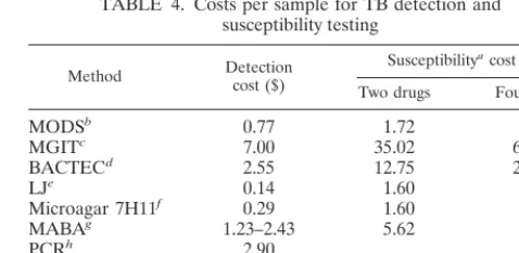
The current estimated cost per sample with MODS is
$0.77 for detection and $1.72 for direct susceptibility testing
with two drugs at two different concentrations (Table 4). This
is substantially cheaper than MGIT, which costs $7.00 for
de-tection testing and $35.02 for susceptibility testing with two
drugs. It is also substantially less expensive than the BACTEC,
which costs $2.55 per sample for detection and $12.75 per
sample for susceptibility testing with two drugs (not including
the costly investment of a BACTEC machine). Table 4 shows
the estimated costs per sample for TB detection and
suscepti-bility testing. Susceptisuscepti-bility costs were calculated for two drugs
(INH and RIF) at two concentrations (high and low) each, and
four drugs (INH, RIF, ethambutol, and streptomycin) at two
concentrations each. Items included in cost analysis are noted
below the table.
Identification.
Three strains,
M. chelonae
,
M. smegmatis
, and
M. bovis
, had a rope-type growth pattern similar to that of
MTB.
M. chelonae
and
M. smegmatis
, both rapid growers, were
observed on the second day of growth; this contrasts with
MTB, which was never detected earlier than 4 days after
in-oculation.
M. bovis
was not distinguishable from MTB, but all
other strains were correctly identified and easily separated
from MTB and from each other by their colony morphology.
DISCUSSION
Each year there are an estimated 8 million new cases of
clinical tuberculosis, and 3 million deaths caused by TB (17,
19). Most cases occur in developing, resource-poor countries,
where the conventional MGIT and BACTEC rapid detection
and susceptibility testing techniques are simply not feasible
because of their high costs and equipment requirements. The
present study demonstrated that MODS is a sensitive and
specific new method for detecting and determining drug
sus-ceptibility of MTB. MODS uses microscopy to detect early
growth of MTB as strings and tangles of bacterial cells in
Middlebrook 7H9 broth medium with or without antimicrobial
agents of interest. Given its simplicity, low cost, speed (under
2 weeks in most cases), and accuracy, MODS would be an
excellent method for routine tuberculosis testing in developing
nations such as Peru, where tuberculosis is highly prevalent
(12, 22).
At present, the only affordable method for the definitive
diagnosis of MTB in most resource-poor nations is in vitro
culture on solid medium. However, MTB grows better in liquid
than on agar; diagnosis using solid medium culture is both less
sensitive and slower than liquid medium culture (1). MODS
was developed as a simple adaptation of the microagar 7H11
test, which uses microscopic observation to detect MTB on
agar. In MODS, liquid medium is used to replace agar. Our
results confirm the advantage of using liquid medium: both
liquid culture methods used, MODS and MGIT, were at least
14% more sensitive and 4 to 5 days faster than either the
microagar 7H11 or LJ culture.
To our knowledge, microscopic observation of MTB
grow-TABLE 3. Comparison of MODS, MABA, and SSCP in
evaluating MTB sensitivity to RIF
Response
No. (%) of specimens showing RIF susceptibility by
MABA MODS SSCP
Resistance
11 (79)
14 (100)
a10 (71)
Sensitivity
3 (21)
0 (0)
4 (29)
Total
14 (100)
14 (100)
14 (100)
[image:4.612.53.294.92.172.2]aThe cutoff level used to determine RIF resistance was 0.5g/ml. When a concentration of 1.00g/ml was used, two of the three strains initially classified by MODS as resistant were reclassified as sensitive.
TABLE 4. Costs per sample for TB detection and
susceptibility testing
Method Detectioncost ($) Susceptibility acost ($)
Two drugs Four drugs
MODS
b0.77
1.72
1.80
MGIT
c7.00
35.02
63.03
BACTEC
d2.55
12.75
23.00
LJ
e0.14
1.60
1.57
Microagar 7H11
f0.29
1.60
2.92
MABA
g1.23–2.43
5.62
6.87
PCR
h2.90
Acid-fast stain
0.10
Smear
iaCalculated for two different concentrations of each drug. Two drugs, RIF and INH; four drugs, RIF, INH, ethambutol, and streptomycin.
bIncludes costs of Middlebrook 7H9 medium, glycerol, Bacto Casitone, OADC, PANTA, 24-well plate, Scotch polyethylene tape, pipette tips, and an-timicrobial drugs.
cIncludes costs of MGIT (prepared tubes), OADC, PANTA, pipette tips, and antimicrobial drugs.
dIncludes costs of BACTEC vials and antimicrobial drugs; does not include the cost of syringes and reconstituting fluid.
eIncludes costs of LJ medium, glycerol, eggs, Scotch polyethylene tape, pipette tips, and antimicrobial drugs.
fIncludes costs of Middlebrook 7H11 medium, glycerol, OADC, cyclohexam-ide, Scotch polyethylene tape, pipette tips, and antimicrobial drugs.
gIncludes costs of Middlebrook 7H9 medium, glycerol, Bacto Casitone, OADC, PANTA, Tween-80, alamar blue reagent, 24- or 96-well plates, Scotch polyethylene tape, pipette tips, Combitips, and antimicrobial drugs.
hIncludes costs of one DNA extraction (Tris, EDTA, ethanol, 1.5-ml micro-centrifuge tubes, and pipette tips), and two regular PCR reactions (Taq poly-merase, 10⫻PCR buffer, deoxynucleoside triphosphates, MgCl, distilled water, 0.5-ml microcentrifuge tubes, pipette tips, primers, and agarose) and one nested PCR reaction (equal in cost to two regular PCR reactions).
iIncludes costs of microscope slides, auramine O or Ziehl-Neelsen stain, phenol, HCl, and ethanol.
on May 15, 2020 by guest
http://jcm.asm.org/
[image:4.612.312.551.425.541.2]ing in broth culture has not been evaluated as a diagnostic
method, although it has served as a research tool (6). The
appearance of MTB in liquid medium is distinct (16). Our
blinded controls indicate that contaminating bacteria or fungi
are not mistaken for
Mycobacteria
species in our test. We
readily distinguished the characteristic nonropy growth of
M.
avium
complex and
M. kansasii
from MTB with the MODS
assay. Rapid growers such as
M. chelonae
and
M. smegmatis
also grew as stringy tangles, similar to MTB, but were
distin-guished by speed of appearance (
⬍
3 versus
ⱖ
4 days for MTB).
The minimum major equipment required for performing a
simple test such as MODS (as should be required for any TB
work in a developed country) include a P2 biosafety cabinet, a
37°C incubator, and a light microscope (preferably inverted).
Although the techniques we have used, such as PCR and
SSCP, are not applicable to many developing country
labora-tories, they were useful to demonstrate the validity of our
results.
The auramine O and Ziehl-Neelsen acid-fast staining
tech-niques are cheap and rapid but not very sensitive and not
suited for drug susceptibility testing. Various studies have
re-ported that a single sputum smear test has a sensitivity ranging
from 22 to 78% (11). In this study, the auramine O-stained
smear test had a sensitivity of 78%, compared to 93% of
MGIT, the most sensitive method. PCR is a rapid, sensitive,
and relatively inexpensive method that could be used for the
detection of MTB in midlevel developing countries. However,
in practice, positive PCR results are often discordant with
results obtained from in vitro cultures, both in our laboratory
and in laboratories with extensive MTB and PCR experience
(18).
MODS offers a superb alternative for susceptibility testing in
developing countries. In these countries, drug resistance was
relatively low, and susceptibility tests were mainly performed
for surveillance than for patient management. Now, with the
rise in multiple-drug-resistant tuberculosis, timely drug
suscep-tibility test results are a must for proper patient management.
Yet presently, susceptibility tests are not performed in many
areas where TB is endemic, often because rapid testing systems
are expensive, and the time needed to obtain results via the
less-costly solid culture tests often renders the results clinically
irrelevant. Susceptibility results with solid medium testing are
generally unavailable until 4 to 5 weeks after inoculation, even
when done directly from sputum concentrates (13). With
MODS, the patient sample is used directly for susceptibility
testing, and results are generally obtained within 2 weeks.
Based on our results, we suggest that isolates scored by
MODS as resistant to RIF alone be retested by a more
stan-dard technique, such as MABA. Moreover, we suggest that a
concentration of 1.0
g/ml be the lowest concentration of RIF
used when testing drug susceptibility with MODS, to improve
concordance with MABA. Also, the agreement between SSCP
and MABA RIF susceptibility results were considerably higher
than that obtained between the SSCP and MODS. This is
evidence that MODS is less reliable for determinating RIF
resistance. There was no discordance between MODS and
MABA for the 14 multiply resistant strains. The high
agree-ment between the MABA and SSCP also supports the accuracy
of the MABA test. We now perform all susceptibility testing in
our laboratory using MABA because of its simplicity and
speed. Subjects for this study were considered to be at high risk
for MTB, and many had tested positive by AFB stain before
being referred to us. Further testing of MODS should be
performed in an unselected patient population; such a study
will assist in determining the true sensitivity of MODS.
It is well recognized that the resurgence of TB calls for
second-generation laboratory methods for more rapid
detec-tion, identificadetec-tion, and drug susceptibility testing (14).
Unfor-tunately, all such second-generation methods developed to
date are for use in wealthy developed countries and are
inap-propriate for settings with limited resources. Our MODS
method provides the first rapid, sensitive, and specific method
for TB detection and drug susceptibility testing that is clinically
relevant and appropriate for use in developing countries.
ACKNOWLEDGMENTS
We are grateful to R. Black, J. Friedland, and C. Evans for their
comments and to J. B. Phu and D. Sara for their assistance.
REFERENCES
1.Cheng, A. F., M. S. Li, C. Y. Chan, D. Lyon, R. Wise, and J. C. Lee.1994.
Evaluation of three culture media and their combinations for the isolation of
Mycobacterium tuberculosisfrom pleural aspirates of patients with tubercu-lous pleurisy. J. Trop. Med. Hyg.97:249–253.
2.Collins, L., and S. G. Franzblau.1997. Microplate alamar blue assay versus
BACTEC 460 system for high-throughput screening of compounds against
Mycobacterium tuberculosisandMycobacterium avium. Antimicrob. Agents Chemother.41:1004–1009.
3.Dalovisio, J. R., S. Montenegro-James, S. A. Kemmerly, C. F. Genre, R.
Chambers, D. Greer, G. A. Pankey, D. M. Failla, K. G. Haydel, L. Hutchin-son, M. F. Lindley, B. M. Nunez, A. Praba, K. D. Eisenach, and E. S. Cooper.
1996. Comparison of the amplifiedMycobacterium tuberculosis(MTB) direct test, Amplicor MTB PCR, and IS6110-PCR for detection of MTB in respi-ratory specimens. Clin. Infect. Dis.23:1099–1106, 1107–1108.
4.De Cock, K. M., and D. Wilkinson.1995. Tuberculosis control in
resource-poor countries: alternative approaches in the era of HIV. Lancet346:675– 677.
5.Enarson, D. A., H. L. Rieder, and T. Arnadottir.1994. Tuberculosis guide for
low income countries, 3rd ed. International Union Against Tuberculosis and Lung Disease, Paris, France.
6.Fazal, N., R. Bartlett, D. A. Lammas, and D. S. Kumararatne.1992. A
comparison of the different methods available for determining BCG-macro-phage interactions in vitro, including a new method of colony counting in broth. FEMS Microbiol. Immunol.5:355–362.
7.Franzblau, S. G., R. S. Witzig, J. C. McLaughlin, P. Torres, G. Madico, A.
Hernandez, M. T. Degnan, M. B. Cook, V. K. Quenzer, R. M. Ferguson, and
R. H. Gilman.1998. Rapid, low-technology MIC determination with clinical
Mycobacterium tuberculosisby using the Microplate Alamar Blue Assay. J. Clin. Microbiol.36:362–366.
8.Idigoras, P., E. Perez-Trallero, M. Alcorta, C. Gutierrez, and I.
Munoz-Baroja.1995. Rapid detection of tuberculosis and non-tuberculosis
myco-bacteria by microscopic observation of growth on Middlebrook 7H11 agar. Eur J. Clin. Microbiol. Infect. Dis.14:6–10.
9.Kent, B. D., and G. P. Kubica.1985. Public health mycobacteriology. A guide
for the level III laboratory p. 36–39, 47–69, and 185–187. U.S. Department of Health and Human Services, Centers for Disease Control, Atlanta, Ga.
10. Kolk, A. H., L. F. Kox, S. Kuijper, and C. Richter.1994. Detection of
Mycobacterium tuberculosisin peripheral blood. Lancet344:694.
11. Lipsky, B. A., J. Gates, F. C. Tenover, and J. J. Plorde.1984. Factors
affecting the clinical value of microscopy for acid-fast bacilli. Rev. Infect. Dis.
6:214–222.
12. Madico, G., R. Gilman, W. Checkley, L. Cabrera, I. Kohlstadt, K. Kacena,
J. F. Diaz, and R. Black.1995. Community infection ratio as an indicator for
tuberculosis control. Lancet345:416–419.
13. Matthew, S., C. N. Paramasivan, F. Rehman, R. Balambal, K. Rajaram, and
R. Prabhakar.1995. A direct rifampicin sensitivity test for tubercle bacilli.
Indian J. Med. Res.102:99–103.
14. McGowan, J. E., Jr., B. Mechock, and F. S. Nolte.1995. Laboratory diagnosis
of tuberculosis: past, present and future. J. Med. Assoc. Ga.84:215–220.
15. Middlebrook, G., Z. Reggiardo, and W. D. Tigertt.1977. Automatable
ra-diometric detection of growth ofMycobacterium tuberculosis in selective media. Am. Rev. Respir. Dis.115:1066–1069.
16. Morris, A. J., and L. B. Reller.1993. Reliability of cord formation in
BACTEC media for presumptive identification of mycobacteria. J. Clin. Microbiol.31:2533–2534.
17. Murray, C. J. L., and A. D. Lopez.1997. Mortality by cause for eight regions
of the world: global burden of disease study. Lancet349:1269–1276.
18. Noordhoek, G. T., J. D. van Embden, and A. H. J. Kolk.1996. Reliability of
nucleic acid amplification for detection ofMycobacterium tuberculosis: an international collaborative quality control study among 30 laboratories. J. Clin. Microbiol.34:2522–2525.
19. Raviglione, M. C., D. E. Snider, and A. Kochi.1995. Global epidemiology of
tuberculosis: morbidity and mortality of a worldwide epidemic. JAMA273:
220–226.
on May 15, 2020 by guest
http://jcm.asm.org/
20.Sambrook, J., E. F. Fritsch, and T. Maniatis.1989. Molecular cloning: a laboratory manual, 2nd ed. Cold Spring Harbor Laboratory, Cold Spring Harbor, N.Y.
21.Sechi, L. A., M. P. Pinna, A. Sanna, P. Pirina, F. Ginesu, F. Saba, A. Aceti,
F. Turrini, S. Zanetti, and G. Fadda.1997. Detection ofMycobacterium
tuberculosisby PCR analysis of urine and other clinical samples from AIDS and non-HIV-infected patients. Mol. Cell Probes11:281–285.
22. Shangavi, D., R. Gilman, G. Lescano-Guevara, W. Checkley, L. Cabrera, and
V. Cardenas.1998. Hyperendemic pulmonary tuberculosis in a Peruvian
shantytown. Am. J. Epidemiol.148:384–389.
23. Telenti, A., P. Imboden, F. Marchesi, T. Schmidheini, and T. Bodmer.1993.
Direct, automated detection of rifampin-resistantMycobacterium tuberculo-sisby polymerase chain reaction and single-strand conformation polymor-phism analysis. Antimicrob. Agents Chemother.37:2054–2058.
24. Walters, S. B., and B. A. Hanna.1996. Testing of susceptibility of
Mycobac-terium tuberculosisto isoniazid and rifampin by mycobacterium growth
indi-cator tube method. J. Clin. Microbiol.34:1565–1567.
25. Watterson, S., S. Wilson, M. Yates, and F. Drobniewski.1998. Comparison
of three molecular assay for rapid detection of rifampin resistance in Myco-bacterium tuberculosis. J. Clin. Microbiol.36:1969–1973.
26. Williams, D. L., C. Waguespack, K. Eisenach, J. T. Crawford, F. Portaels, M.
Salfinger, C. M. Nolan, C. Abe, V. Sticht-Groh, and T. P. Gillis.1994.
Characterization of rifampin resistance in pathogenic Mycobacteria. Anti-microb. Agents Chemother.38:2380–2386.
27. Williams, D. L., L. Spring, T. P. Gillis, M. Salfinger, and D. H. Persing.1998.
Evaluation of a polymerase chain reaction-based universal heteroduplex generator assay for direct detection of rifampin susceptibility of Mycobacte-rium tuberculosisfrom sputum specimens. Clin. Infect. Dis.26:446–450.
28. Yajko, D. M., J. J. Madej, M. V. Lancaster, C. A. Sanders, V. L. Cawthon, B.
Gee, A. Babst, and W. K. Hadley.1995. Colorimetric method for determining
MICs of antimicrobial agents forMycobacterium tuberculosis. J. Clin. Micro-biol.33:2324–2327.