—Original—
Comparison of the Effectiveness of Ovulation Synchronization
Protocol in Anestrous and Cycling Beef Cows
Takeshi OSAWA
1), Daisaku MORISHIGE
1), Daisaku OHTA
1),
Yusuke KIMURA
1), Toh-Ichi HIRATA
1)and Yoh-Ichi MIYAKE
1)1)
Faculty of Agriculture, Iwate University, Ueda 3–18–8, Morioka 020–8550, Japan
Abstract. Applicability of ovulation synchronization protocol using GnRH and PGF2α (PGF) injection to anestrous beef cows remains controversial. We compared the effectiveness of the protocol in the anestrous stage of the beef cow with that in the cycling stage using the same animals. Ovaries of five Japanese Black and three Japanese Shorthorn cows were ultrasonographically examined, and blood samples were collected daily for hormonal analyses. Each animal received the protocol twice (Day –6 to –8: GnRH, Day 0: PGF, Day 2: GnRH). Additional blood samples were taken before and after GnRH injection for LH and FSH measurements to evaluate the pituitary function. For the ovarian status at the onset of the protocol cows were divided into anestrous (n=8) and cycling (n=8) stages. There was no significant difference in size of the dominant follicle at the first and second GnRH injections, and in the magnitude of the pituitary response to GnRH between the two stages. However, the size of the corpus luteum and progesterone concentrations at the PGF injection in the anestrous stage were significantly smaller and lower (P<0.01), respectively, and ovulation synchronization rate in the anestrous stage was significantly lower (P<0.05) than in the cycling stage. In conclusion, ovulation synchronization protocol in anestrous beef cows has limited effectiveness.
Key words: Anestrous beef cows, Ovulation synchronization, LH, GnRH, Ultrasonography
(J. Reprod. Dev. 49: 513–521, 2003)
nestrus is the major component of postpartum infertility, and suckling is the major factor that causes anestrus in postpartum beef cows [1]. Thus, treatment of anestrus is one of the crucial measures to be taken to improve reproductive performance in po stp art um s u ckle d be ef cow s. V ar io us procedures have been developed to either induce or synchronize ovulation in suckled beef cows [2, 3]. As an option among several different breeding programs, ovulation synchronization protocol using two injections of GnRH and prostaglandin F2α (P GF ) and time d artificial ins emination (Ovsynch protocol) has been applied worldwide in beef cows [4–7] as well as in dairy cows [8–11], and acceptable conception rates have been obtained. A
first dose of GnRH curtails follicular development and induces the emergence of a new follicular wave. PGF treatment 7 days after the first GnRH induces the regression of the luteal phase and a second dose of GnRH two days after the PGF treatment synchronizes the LH surge and ovulation of a dominant follicle of the new follicular wave. A series of these hormonal treatments enables artificial insemination at a fixed time. Moreover, a modified Ovsynch protocol in combination with progestins has been successfully used to prevent estrus prior to PGF treatment [12, 13]. Geary et al. [5] reported that Ovsynch protocol was capable of inducing fertile ovulation in anestrous beef cows as well as cycling animals. On the other hand, the pregnancy rate by Ovsynch protocol for anestrous dairy cows was reported to be lower than that for
Accepted for publication: September 8, 2003
cycling cows [14]. However, the mechanism behind the low pregnancy rate in anestrous cows inseminated by timed artificial insemination (TAI) remains unclear, and the effectiveness of Ovsynch protocol in anestrous beef cows has not been well documented to date.
We p reviou sly demonstrated that the TAI protocol was applicable to grazing Japanese Black cattle [7]. However, more detailed observations of ovaries during the protocol period is required to e va lu at e t he e f fe ct ive ne ss o f t he o vu la tio n synchronization protocol in anestrous beef cattle. In addition, a direct comparison of the responses to the protocol between the two ovarian statuses, i.e. anestrous and cycling statuses, using the same animals wou ld g ive reliab le da ta for fu ture application of the technique in the field.
The objective of the present study, therefore, was to investigate the effectiveness of ovulation synchronization protocols in postpartum anestrous and cycling beef cows by assessing the size of ovulatory follicles and corpus luteum, the pituitary responsiveness to the exogenous GnRH, and the ovulation rate at a targeted range of time.
Materials and Methods
Animals
Eight cows (five Japanese Black and three Japanese Shorthorn cows), weighing between 550– 650 kg, were used in this study. The cows were being suckled by their own calves during the experimental period, with the exception of one cow that was not suckled due to a stillbirth and was cycling at the onset of treatment. Six of the seven suckled cows were anestrous (not cycling), and the
other one was cycling. The average (± SD) age, pa rity and da ys post partu m a t the onset of ovulation synchronization protocol were 4.3 (± 1.3) years old, 1.6 (± 0.7) and 70.4 (± 5.7, range: 64–76) days, respectively.
Synchronization of ovulation and timed AI
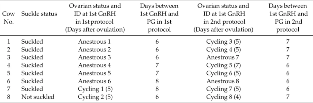
Prior to starting the ovulation synchronization protocol, the ovaries of the cows were scanned daily by ultrasonography for 14 to 24 days to determine the ovarian status (either anestrous or c y c l in g) . E a c h a n im a l r e ce iv e d o v u l a t io n synchronization protocol twice during the study period. In the first ovulation synchronization protocol, the eight cows were randomly assigned to receive GnRH analog (10 µg buserelin, Hoechst Roussel Vet, Wiesbaden, Germany) i.m. on either Day –8, –7 or –6. The animals, then, received PGF analog (25 mg dinoprost, Pharmacia & Upjohn Inc., USA) intramuscularly on Day 0. The animals received another dose of GnRH 48 h after the PGF injection. Six to eight days after the second GnRH injection of the first protocol (4 to 7 days after ovulation in cases of cycling animals), the cows were treated again with ovulation synchronization protocol (second protocol), which was the same as the first protocol except for the interval between the first GnRH and PGF2α (Table 1). Anestrous and cycling stages were defined as stages without and with, respectively, a history of formation of functional corpus luteum after the last calving at the onset of the ovulation synchronization protocol. Consequently, the total number of trials was 16. Under the experimental design, the 16 trials were divided into two groups, that is, the anestrous stage (n=8) and the cycling stage (n=8). Artificial insemination (AI) was conducted 16 h after the Table 1. Experimental design of ovulation synchronization protocol in anestrous and cycling beef cows
Ovarian status and Days between Ovarian status and Days between Cow Suckle status ID at 1st GnRH 1st GnRH and ID at 1st GnRH 1st GnRH and No. in 1st protocol PG in 1st in 2nd protocol PG in 2nd
(Days after ovulation) protocol (Days after ovulation) protocol
1 Suckled Anestrous 1 6 Cycling 3 (5) 7
2 Suckled Anestrous 2 6 Cycling 4 (5) 7
3 Suckled Anestrous 3 6 Anestrous 7 7
4 Suckled Anestrous 4 7 Cycling 5 (7) 6
5 Suckled Anestrous 5 7 Cycling 6 (5) 6
6 Suckled Anestrous 6 8 Anestrous 8 6
7 Suckled Cycling 1 (5) 8 Cycling 7 (5) 6
second GnRH injection of the second protocol in eight cows (six anestrous and two cycling cows, Table 1). Pregnancy diagnosis by rectal palpation was performed 58 to 63 days after AI.
Ultrasonography and blood sampling
The ovarian status of each cow was monitored daily by ultrasonography (SSD-500, Aloka Co., Ltd., Tokyo) to identify anestrous and cycling cows f o r 1 4 t o 2 4 da y s b e f o re t he t re a t m e nt f o r synchronization of ovulation. Ovaries were monitored in each cow at the time of drug injection and every six to 24 hours after the first and second GnRH injections to estimate the time of ovulation by ultrasonography. Ovarian dynamics were further scanned by ultrasonography after the ovulation. A dominant follicle was defined as that d es crib e d b y M u r ph y et al . [ 15 ] wit h s o me modifications, namely, the largest follicle present in the ovaries in the absence of any other large (≥ 10 mm) follicle. Blood samples were collected daily from the coccygeal vein during the experimental period. Institutional approval was obtained for this e x p e ri me nt a n d a nim a l c a re co n f o r m e d to international guidelines. Plasma samples were stored at –30 C until hormone assays.
Pituitary response to the injection of GnRH
Additional blood samples were taken five minutes before and two hours after the injection of GnRH to evaluate the pituitary responsiveness. The pituitary responsiveness was evaluated on the basis of the luteinizing hormone (LH) and follicular stimulating hormone (FSH) responses following the first and second GnRH injections, and it was defined as the amplitude of LH or FSH release two hours after the GnRH injection (the difference between the concentrations two hours after and those five minutes before the GnRH injection).
Hormone assays
Plasma progesterone, estradiol-17β, LH and FSH c o n c e n t r a t i o n s w e r e d e t e r m i n e d b y radioimmunoassay [16–18]. The intra- and inter-assay coefficients of variation were, 9.3% and 7.6% for progesterone, 7.0% and 12.7% for estradiol-17β, 7.1% and 12.2% for LH, and 5.9% and 10.7% for FSH, respectively. Plasma concentrations of progesterone and estradiol-17β were used to confirm ultrasonographic observations of the reproductive status of the cows.
Statistical analyses
The data obtained in this study were analyzed by Chi-square test or Fisher’s exact probability test w h e r e a p p r o p r i a t e f o r t h e c o m p a r i s o n o f p r o p o r t i o n s , a n d S t u d e n t ’ s t- t e s t f o r t h e comparison of average values between two groups.
Results
Ovarian dynamics before and during ovulation synchronization protocol
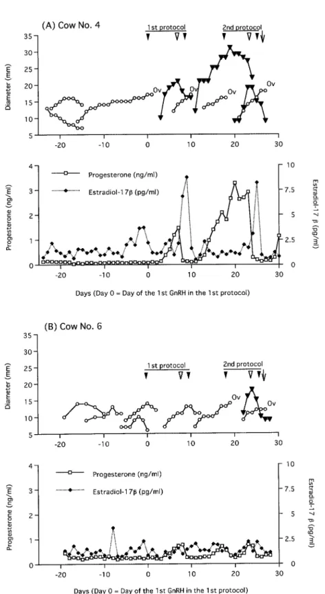
Data from two representative cows showing the pattern of growth of dominant follicles (DF) and formation of corpus luteum (CL) as well as the p r of ile s o f p ro g e s te r o ne a nd e s t ra di ol -1 7β concentrations, before and during ovulation synchronization protocol, are presented in Fig. 1.
T he o v a rie s o f c ow N o . 4 ( F i g . 1 A ) we r e monitored by ultrasonography for 23 days before starting the protocol, and the cow was diagnosed as be ing anestru s (Anestrous cow No. 4). The diameter of the dominant follicle was 17 mm at the first GnRH administration in the first protocol. Then, the dominant follicle ovulated and CL subsequently formed. The CL was 22 mm in diameter and plasma concentration of progesterone was 1.5 ng/ml at the PGF injection. A new dominant follicle 15 mm in diameter formed and plasma concentration of estradiol-17β was 8.1 pg/ ml at the second GnRH administration. The DF ovulated 22 to 28 h after GnRH injection and CL reached its maximum size in 9 days. Seven days after the ovulation, the second protocol was initiated (Cycling cow No. 5) and the size of DF was 16 mm in diameter at the first GnRH of the second protocol. The DF ovulated, a new CL formed, and a new follicular wave emerged. The diameters of the o l d a n d ne w C L w e r e 2 7 m m a n d 2 0 m m , r e s p e c t iv e ly , a n d p la s m a c o n ce n t ra t io n o f progesterone was 2.9 ng/ml at the PGF injection. A DF, 15 mm in diameter, was detected and plasma concentration of estradiol-17β was 8.2 pg/ml at the second GnRH injection. The DF ovulated 28 to 40 h after the second GnRH (12 to 24 h after AI). The cow was confirmed to be pregnant.
The ovaries of cow No.6 (Fig. 1B) were monitored b y u lt ra so no g ra p hy f or 1 9 d ay s b e fo r e t he ovulation synchronization protocol, and the cow was diagnosed as being anestrus (Anestrous cow No. 6). The largest follicle was 14 mm in diameter
Fig. 1. Representative examples of dynamics of the dominant follicles and the corpora lutea and profiles of progesterone and estradiol-17β concentrations during ovulation synchronization protocol in postpartum beef cows. Cow No. 4 was in the anestrous stage at the onset of the first ovulation synchronization protocol and was in the cycling stage at the onset of the second protocol (A). Cow No. 6 was in the anestrous stage at the onset of the first protocol as well as the second protocol (B). Ov: ovulation. : GnRH injection. : PGF2α injection. ↓: AI.
at the first GnRH administration of the first protocol. It did not ovulate and, therefore, no CL was present at the PGF injection. A new follicular wave emerged and the diameter of the largest follicle was 13 mm at the second GnRH injection (plasma concentration of estradiol-17β was 1.8 pg/ ml). The follicle did not ovulate.
First GnRH of the second protocol (Anestrous cow No. 8) was administered seven days after the second GnRH of the first protocol, and the DF of a newly emerging wave that was 13 mm in diameter at that time ovulated. Subsequently CL was formed and it was 18 mm in diameter at the PGF injection. The DF was 11 mm in diameter at the second GnRH injection and it ovulated 28 to 40 h after the injection (12 to 24 h after AI).
Sizes of the largest follicle and corpus luteum during ovulation synchronization protocol
Diameters of the DF at the time of the first and second GnRH injections and those of the CL at the PGF injection in the ovulation synchronization protocol at anestrous and cycling stages are compared in Table 2. Plasma concentrations of progesterone at the PGF injection were also compared between the two stages. While there was no significant difference in the size of the DF at the f ir s t a nd s e co nd G nR H inj e c tio n s b e t w e e n
anestrous and cycling stages, the size of CL and the plasma progesterone level at the PGF injection in the cycling stage were greater (P<0.01) than those in the anestrous stage (24.0 ± 3.1 vs 17.2 ± 3.3 mm and 2.8 ± 1.5 vs 0.6 ± 0.4 ng/ml, respectively). In addition, all of the six cows in the cycling stage that ovulated after the first GnRH injection had two CL, namely, a spontaneous CL that had existed at the time of first GnRH injection and an accessory CL induced by the injection of the first GnRH.
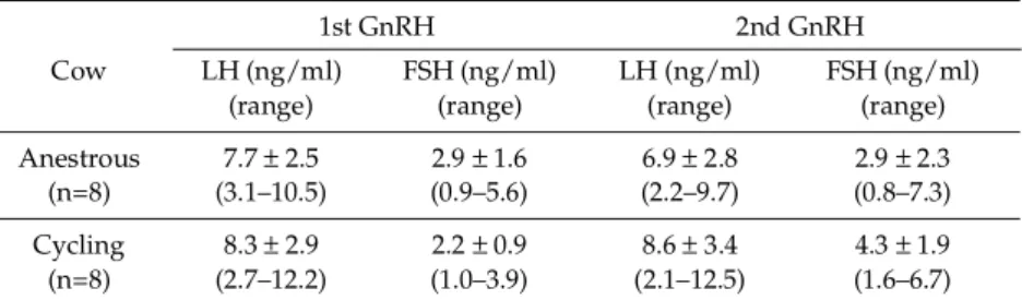
LH and FSH responses to GnRH injections
The pituitary responsiveness to exogenous GnRH was compared between the anestrous and cycling stages in terms of LH and FSH secretions two hours after the GnRH injection (Table 3). There was no significant difference in the pituitary responsiveness to GnRH injections between the two groups.
Ovulation synchronization rate and conception rate
After the first and second GnRH injections, the time when ovulation was confirmed was recorded at the anestrous and/or cycling stage of each animal (Table 4). Ovulation rate (75%) after the first GnRH injection in the anestrous stage was the same as the cycling stage. While all the cycling cows ovulated between 22 to 40 h after the second Table 2. Comparison of sizes of the dominant follicle (DF) at GnRH administrations and sizes of
the corpus luteum (CL) and progesterone (P4) level at prostaglandin F2α administration
between the anestrous and cycling stages during ovulation synchronization protocol Cow DF size at CL size at P4 at PGF2α DF size at
1st GnRH (mm) PGF2α (mm) (ng/ml) 2nd GnRH (mm)
Anestrous 1 10 14 0.6 13
Anestrous 2 14 18 0.2 8
Anestrous 3 10 13 0.3 11
Anestrous 4 17 22 1.5 15
Anestrous 5 10 18 0.6 15
Anestrous 6 14 NA 0.8 13
Anestrous 7 19 NA 0.1 16
Anestrous 8 13 18 0.7 11
(Subtotal; mean ± SD) 13.4 ± 3.4 17.2 ± 3.3a 0.6 ± 0.4a 12.8 ± 2.7
Cycling 1 10 29 3.1 9
Cycling 2 14 26 2.8 16
Cycling 3 16 23, 17 2.1 14
Cycling 4 13 26, 16 5.6 12
Cycling 5 16 27, 20 2.9 15
Cycling 6 12 19, 13 0.9 14
Cycling 7 12 24, 16 1.3 11
Cycling 8 16 24, 20 3.9 11
(Subtotal; mean ± SD) 13.6 ± 2.3 24.8 ± 3.0*b 2.8 ± 1.5b 12.8 ± 2.4
GnRH injection, only half of the anestrous cows ovulated 22 to 40 h after the second GnRH injection. The percentage of the animals that ovulated between 22 to 40 h after the second GnRH injection, that is, the rate of ovulation synchronization, was significantly higher in the cycling stage than in the anestrous stage (P<0.05). While neither of the two cows at the anestrous stage when they were inseminated became pregnant, 83.3% of the cycling
cows (five out of six) conceived after AI, although the difference was not statistically significant (P= 0.11) due to the small number of the animals.
Discussion
We compared the effectiveness of the ovulation synchronization protocol in the anestrous stage Table 3. Comparison of luteinizing hormone (LH) and follicular stimulating
hormone (FSH) responses* (mean ± SD; range) to GnRH administrations during ovulation synchronization protocol between cows at anestrous and cycling stages
1st GnRH 2nd GnRH
Cow LH (ng/ml) FSH (ng/ml) LH (ng/ml) FSH (ng/ml) (range) (range) (range) (range) Anestrous 7.7 ± 2.5 2.9 ± 1.6 6.9 ± 2.8 2.9 ± 2.3 (n=8) (3.1–10.5) (0.9–5.6) (2.2–9.7) (0.8–7.3) Cycling 8.3 ± 2.9 2.2 ± 0.9 8.6 ± 3.4 4.3 ± 1.9 (n=8) (2.7–12.2) (1.0–3.9) (2.1–12.5) (1.6–6.7) * Response was defined as the amplitude of LH or FSH release two hours after GnRH administration.
Table 4. Comparison of ovulation synchronization rate and conception rate between anestrous and cycling cows
Time from 1st Time from 2nd
Cow GnRH injection GnRH injection Consequence of AI until ovulation until ovulation
Anestrous 1 24 to 48 h 22 to 28 h NI*** Anestrous 2 0 to 24 h (Not ovulated)** NI Anestrous 3 24 to 48 h (Not ovulated) NI Anestrous 4 24 to 48 h 22 to 28 h Not pregnant Anestrous 5 24 to 48 h 22 to 28 h NI Anestrous 6 (Not ovulated) (Not ovulated) NI Anestrous 7 (Not ovulated) (Not ovulated) Not pregnant Anestrous 8 24 to 48 h 28 to 40 h NI Ovulation rate*
or Conception rate 75% (6/8) 50% (4/8)a 0% (0/2)
(right column)
Cycling 1 (Not ovulated) 28 to 40 h NI Cycling 2 (Not ovulated) 28 to 40 h Pregnant Cycling 3 24 to 48 h 22 to 28 h Pregnant Cycling 4 48 to 72 h 28 to 40 h Not pregnant Cycling 5 24 to 48 h 28 to 40 h Pregnant Cycling 6 24 to 48 h 28 to 40 h Pregnant Cycling 7 24 to 48 h 28 to 40 h Pregnant
Cycling 8 24 to 48 h 28 to 40 h NI
Ovulation rate*
or Conception rate 75% (6/8) 100% (8/8)b 83.3% (5/6)
(right column)
* Percentage of cows ovulated between 22 to 40 h after injection of second GnRH. ab: P<0.05. ** Ovulated earlier (at PGF2α injection). *** Not inseminated.
with that in the cycling stage of eight postpartum beef cows, and had four major findings. First, sizes of the largest follicles at the first and second GnRH injections in the anestrous stage were similar to those in the cycling stage. Second, there was no difference in the pituitary responsiveness to GnRH between the two stages. Third, the size of the corpus luteum was smaller and progesterone concentrations were lower at the time of the PGF injection in the anestrous stage than those in the cycling stag e. Finally, the rate of ovulation synchronization in the anestrous stage was lower than the synchronization rate in the cycling stage.
Seven out of the eight cows were suckled cows, and six out of the seven were anestrous at the onset of the first ovulation synchronization protocol. Suckling can affect the resumption of follicular growth in cows after calving [19]. Murphy et al. [15] observed that the incidence of ovulation of the first postpartum dominant follicle in beef cows was much lower than that in dairy cows, and that the interval from calving to first ovulation was about 36 days (range 20 to 61 days) in suckled beef cows. After day 30 postpartum through first ovulation, the GnRH pulse-g enerator escap es from the suppressive effect of suckling [20]. Average length of the postpartum period in the cows of the present study was 70 days (range 64 to 76 days). Therefore, the anestrus condition in these cows had lasted longer than the average length. In spite of this anestrous condition, all of the cows at the anestrous stage had a follicle of 10 mm or larger in diameter, the same as cows at the cycling stage in our study, and the size of dominant follicles (or largest follicles) at the first and second GnRH injections in the anestrous stage were similar to those in the cycling stage. Moreover, ovulation was induced af te r the first GnRH injection in 7 5% of the anestrous cows, although no functional luteal tissue was formed after ovulation except for one of these animals. This figure coincides with the figure of 75% in beef cows [12], and with that of 80% in dairy cows [8]. These results indicate that even cows at the anestrous stage could have a follicle that is large enough to respond to exogenous GnRH.
Most of the cows in our study were anestrous at day 70 postpartum. It has been demonstrated that concentrations of LH in the anterior pituitary accumulate and increase gradually from Day 15 to 20, reaching concentrations observed in cycling
cows by Day 30 postpartum [20]. Therefore, we conjectured at the start of this study that it would be worthwhile to evaluate the gonadotropin store in the pituitary by measuring the response to exogenous GnRH. We found that the magnitude of the pituitary response in the anestrous stage was similar to that in the cycling stage. This result indicates that the pituitary function in these anestrous cows had been fully restored to the level of cycling cows, and it is consistent with the finding by McDougall et al. [21], who observed sufficient releasable pituitary stores of LH in primiparous cows at three weeks postpartum at the time of treatment with GnRH and large follicles that were able to ovulate.
The average size of the corpus luteum at the PGF injection in the anestrous stage was significantly smaller than that in the cycling stage. Lower plasma progesterone concentrations at the PGF injection in the anestrous stage support this morphological observation. These findings suggest that the follicles in the anestrous stage, which are capable of ovulating in response to exogenous GnRH, may not form a CL that is equivalent to that formed in the cycling stage, even though the follicular size at the GnRH injection is equivalent to those seen in the cycling stage. In other words, a large follicle in an anestrous cow may have the potential to respond to exogenous GnRH, but the ovulatory follicle, regardless of its size, may be converted into a small corpus luteum that might last a shorter period than the functional corpus luteum of a cycling cow. It has been reported that a short (less than 10 days) luteal phase follows when GnRH is injected in anestrous dairy cows [21].
In our study, all the cycling cows ovulated between 22 and 40 h after treatment. This result is in accordance with a previous finding that GnRH caused ovulation between 24 and 32 h af ter ovulation synchronization protocol, whe n a dominant follicle was present [8]. We observed that all cows but one had a large follicle (10 mm or larger in diameter) at the second GnRH injection in c y c li n g c o w s , a n d s o d id a n e s t r o u s c o w s . However, ovulation was not synchronized after the protocol in half of the anestrous cows. The influence of the difference in the interval between the first GnRH and PGF injections was unlikely because it has been reported that six- and seven-day GnRH-PGF injection intervals resulted in similar synchronized reproductive performance in
postpartum beef cows [22], and also because there was no relationship between the length of the interval and whether ovulation was synchronized or not in the present study. The dose of GnRH injection we used, 10 µg buserelin, has been reported to be sufficient for the induction of LH surge, which is equivalent to a physiological LH surge [23]. One of the anestrous cows had no large follicle at the second GnRH injection, and therefore it is understandable the cow did not ovulate. However, the other three anestrous cows had a large follicle that apparently ovulated in response to t he Gn RH inj e ct ion . S ince L H a nd F S H responses to exogenous GnRH in anestrous cows were similar to those in cycling cows in the present study, the reason why some anestrous cows did not ovulate following GnRH administration may be because the follicle had lost its dominance, that is, had lost sufficient volume of LH receptors on the granulosa layer; LH receptors decrease as the dominant follicle develops from growth into the plateau and regression phases [24].
The results in the present study suggest that although the first GnRH injection may induce ovulation in anestrous beef cows, the rate of ovulation synchronization after the second GnRH injection may not be as high as that in cycling cows
partly because subsequently formed CL may not be fully functional at the time of the PGF injection, and partly because the largest follicle may have lost its dominance by the time of the second GnRH injection. In conclusion, ovulation synchronization protocol using GnRH and PGF in anestrous beef cows has limited effectiveness while the protocol in cycling beef cows has the potential to result in a satisfactory synchronization rate. These findings lend su pport to the view that the ovulation synchronization protocol for anestrous beef cows should be combined with the use of progesterone prior to or during the GnRH- PGF regimen to improve the ovulation synchronization rate and subsequently to improve the conception rate.
Acknowledgements
Thanks are due to Mitaka pharmaceutical Co., Ltd., Tokyo, for the provision of the GnRH product used in this study. The authors also wish to thank the staff of Field Science Center and Veterinary Teaching Hospital, Faculty of Agriculture, Iwate University, for their help in the management of the animals.
References
1. Short RE, Bellow RA, Staigmiller RB, Berardinelli JG, Custer EE. Physiological mechanisms controlling anestrus and infertility in postpartum beef cattle. J Anim Sci 1990; 68: 799–816.
2. Stevenson JS, Hoffman DP, Nichols DA, McKee RM, Krehbiel CL. Fertility in estrus-cycling and noncycling virgin heifers and suckled beef cows after induced ovulation. J Anim Sci 1997; 75: 1343– 1350.
3. Grimard B, Humblot P, Mialot JP, Jeanguyot N, Sauvant D, Thibier M. Absence of response to oestrus induction and synchronization treatments related to lipid mobilization in suckled beef cows. Repro Nutr Dev 1997; 37: 129–140.
4. Twagiramungu H, Guilbault LA, Proulx J, Dufour JJ. Synchronization of estrus and fertility in beef cattle with two injections of buserelin and prostaglandin. Theriogenology 1992; 38: 1131–1144. 5. Geary TW, Whittier JC, Downing ER, LeFever DG,
Silcox RW, Holland MD, Nett TM, Niswender GD. Pregnancy rates of postpartum beef cows that were synchronized using Syncro-Mate-B or the Ovsynch
protocol. J Anim Sci 1998; 76: 1523–1527.
6. Geary TW, Whittier JC, Hallford DM, MacNeil MD. Calf removal improves conception rates to the Ovsynch and CO-Synch protocols. J Anim Sci 2001; 79: 1–4.
7. Osawa T, Morishige D, Ohta D, Kimura Y, Miyake Y-I. Application of timed artificial insemination protocols to grazing Japanese Black cattle with long open period. J Vet Med Sci 2003; 65: 459–464. 8. Pursley JR, Mee MO, Wiltbank MC.
Synchronization of ovulation in dairy cows using PGF2α and GnRH. Theriogenology 1995; 44: 915–923.
9. Moreira F, de la Sota RL, Diaz T, Thatcher WW. Effect of day of the estrous cycle at the initiation of a timed artificial insemination protocol on reproductive response in dairy heifers. J Anim Sci 2000; 78: 1568–1576.
10. Vasconcelos JL, Silcox RW, Rosa GJ, Pursley JR, Wiltbank MC. Synchronization rate, size of the ovulatory follicle, and pregnancy rate after synchronization of ovulation beginning on different days of the estrous cycle in lactating dairy cows.
Theriogenology 1999; 52: 1067–1078.
11. Yamada K, Nakao T, Nakada K, Matsuda G. Influence of GnRH analogue (fertirelin acetate) doses on synchronization of ovulation and fixed-time artificial insemination in lactating dairy cows. Anim Reprod Sci 2002; 74: 27–34.
12. Thompson KE, Stevenson JS, Lamb GC, Grieger DM, Loest CA. Follicular, hormonal, and pregnancy responses of early postpartum suckled beef cows to GnRH, norgestomet, and prostaglandin F2α. J Anim
Sci 1999; 77: 1823–1832.
13. Martinez MF, Kastelic JP, Adams GP, Cook B, Olson WO, Mapletoft RJ. The use of progestins in regimens for fixed-time artificial insemination in beef cattle. Theriogenology 2002; 57: 1049–1059. 14. Cartmill JA, El-Zarkouny SZ, Hensley BA, Lamb
GC, Stevenson JS. Stage of cycle, incidence, and timing of ovulation, and pregnancy rates in dairy cattle after three timed breeding protocols. J Dairy Sci 2001; 84: 1051–1059.
15. Murphy MG, Boland MP, Roche JF. Pattern of follicular growth and resumption of ovarian activity in post-partum beef suckler cows. J Reprod Fertil 1990; 90: 523–533.
16. Taya K, Watanabe G, Sasamoto S.
Radioimmunoassay for progesterone, testosterone and estradiol-17β using 125I-iodohistamine radioligands. Jpn J Anim Reprod 1985; 31: 189–197. 17. Echternkamp SE, Bolt DJ, Hawk HW. Ovarian and
pituitary hormones in blood of progesterone-treated
ewes. J Anim Sci 1976; 42: 893–900.
18. Bolt DJ, Rollins R. Development and application of a radioimmunoassay for bovine follicle-stimulating hormone. J Anim Sci 1983; 56: 146–154.
19. Peters AR, Lamming GE, Fisher MW. A comparison of plasma LH concentrations in milked and suckling post-partum cows. J Reprod Fertil 1981; 62: 567–573.
20. Yavas Y, Walton JS. Postpartum acyclicity in suckled beef cows: a review. Theriogenology 2000; 54: 25–55.
21. McDougall S, Williamson NB, Macmillan KL. GnRH induces ovulation of a dominant follicle in primiparous dairy cows undergoing anovulatory follicle turnover. Anim Reprod Sci 1995; 39: 205–214. 22. Dejarnette JM, Wallace RW, House RB, Salverson
RR, Marshall CE. Attenuation of premature estrous behavior in postpartum beef cows synchronized to estrus using GnRH and PGF2α. Theriogenology 2001; 56: 493–501.
23. Chenault JR, Kratzer, DD, Rzepkowski RA, Goodwin MC. LH and FSH response of Holstein heifers to fertirelin acetate, gonadorelin and buserelin. Theriogenology 1990; 34: 81–98.
24. Rollosson MM, Crim JW, Silcox RW, Kiser TE. Density of [125I] hCG binding to the dominant follicle of the first wave of the estrous cycle in cows. J Anim Sci 72 (Suppl. 1) / J Dairy Sci 77 (Suppl. 1) 1994; 231 (Abstr.).