Original Research Article
Significance of relationship between anatomical variants of middle
turbinate and nasal septum in recurrent acute rhinosinusitis patients
Satya Sundar G. Mohapatra1
, Niranjan Sahu
1, Siba N. Rath
2*,
Mahesh C. Sahu
3, Rabindra N. Padhy
2INTRODUCTION
Acute bacterial rhinosinusitis (ABRS) is diagnosed when signs and symptoms of acute rhinosinusitis fail to improve within 10 days from the day of onset, or worsening of symptoms occur within 10 days after an initial improvement period, called as ‘double worsening’.
Moreover, recurrent attacks of ABRS occurring at
frequent intervals called as, recurrent acute rhinosinusitis (RARS), when 4 or more episodes of ABRS are
presented within a year with no symptoms persisting in between the acute episodes.1 During this asymptomatic interval phase of the disease, a majority of RARS patients demonstrated subtle findings of isolated ethmoid sinusitis, which is revealed while patients had undergone computed tomography (CT) scan of brain for any reasons other than sinonasal symptoms. Nevertheless, this subtle isolated infection of ethmoid sinuses acts as a potential nidus, which is responsible for the recurrent nature of the RARS disease.2 RARS is recently considered as a mild
ABSTRACT
Background: Aim of the study was to evaluate the significance of relationship between anatomical variants of middle
turbinate and nasal septum, and role for the causation of recurrent acute rhinosinusitis (RARS) in a tertiary care teaching hospital.
Methods: Retrospectiveanalysisofsinonasal computerized tomography (CT) images of 160 patients with history of
RARS during August 2015 to December 2016 was done for evaluation of concha bullosa (CB) and paradoxical middle turbinate (PMT) associated with deviated nasal septum (DNS). Measurements of CB, PMT and septal deviation angles were recorded. Patients with acute and expansile paranasal sinus diseases were excluded.
Results: Of the total 160 RARS cases with DNS, 120 cases had unilateral (contralateral and ipsilateral) and bilateral
(dominant and similar sized) CB, and were distributed into group I (contralateral and dominant) and group II (ipsilateral and similar sized bilateral) basing on the direction of septal convexity. Maximum transverse diameter of CB (MTDCB) and septal deviation angle values were highly significant (p =0.0001) in group I CB cases. Direction of septal convexity had no significance (58 to right and 62 to left) in relation to CB pneumatization. Septal deviation severity in group I CB cases was highly significant (p =0.0001). The fraction of 40 unilateral and bilateral PMT cases revealed no significance (p =0.45) with severity of DNS.
Conclusions: Anatomical variants of middle turbinate associated with DNS caused increased prevalence of OMC
pattern RARS. Contralateral and dominant CBs had shown relationship in direct proportion to the severity of septal deviation contributing to recurrent rhinosinusitis.
Keywords: Concha bullosa, Deviated nasal septum, Paradoxical middle turbinate, Deviation angle, Recurrent acute
rhinosinusitis
1Department of Radiology, 2Central Research Laboratory, 3Directorate of Medical Research, IMS and SUM hospital,
Siksha ‘O’ Anusandhan University, K8, Kalinga Nagar,Bhubaneswar, Odisha, India
Received: 20 April 2017
Accepted: 06 May 2017
*Correspondence: Dr. Siba N. Rath,
E-mail: snrath19@gmail.com
Copyright: © the author(s), publisher and licensee Medip Academy. This is an open-access article distributed under
the terms of the Creative Commons Attribution Non-Commercial License, which permits unrestricted non-commercial use, distribution, and reproduction in any medium, provided the original work is properly cited.
form of chronic rhinosinusitis disease, which remains a challenging problem nowadays due to under evaluation of the disease pattern.
Nasal airways including paranasal sinuses play an important role in filtering and conditioning the inspired air, thus protect lungs by capturing particulate materials.3 In presence of inflammation, nasal airway obstruction usually occurs in the osteomeatal complex (OMC) region, the key drainage site of paranasal sinuses. Patients having recurrent attacks of inflammatory sinusitis present 5 patterns of acute rhinosinusitis based on CT appearance: OMC, sinonasal polyposis, spheno-ethmoidal (SE)
recess, isolated infundibular type and sporadic
distribution patterns. Among these patterns, majority of RARS patients present OMC pattern of rhinosinusitis due to preponderance of anatomic variants in this region. Sinonasal anatomical variants of middle turbinate and nasal septum, being more common in the OMC region, are responsible for predisposing to recurrent attacks of rhinosinusitis.4 In the presence of deviated nasal septum (DNS), common variants of middle turbinate such as, concha bullosa (CB) and paradoxical middle turbinate (PMT) are responsible for frequent OMC obstruction, eventually increasing the prevalence of OMC pattern RARS many folds.
Bone and cartilaginous constituents of the nasal septum are the anterior quadrangular cartilage and the posterior vomer bone joined with perpendicular plate of the ethmoid bone. Prevalence of DNS is reported to be 14-58% in the literature.5,6 The major etiological factors of septal deviation are deformities caused by pressure and expansion during the growth, trauma, particularly including injuries in infancy and childhood.5 Along with genetic and environmental factors, other determinative etiological factors include irregularity in growth of the maxilla, thumb-sucking and the tongue-pressure habits causing shifts in the alveolar ridge.6
CB, especially involving the middle turbinate is one of the common anatomical variations of sinonasal structures with a reported prevalence range of 12 - 78% in earlier studies.7-9 Etiological factors of CB include trauma, developmental defects, growth anomalies of maxilla and nasal septal deviation.8 As compared to superior and inferior nasal concha, the middle nasal concha is the most frequent site of pneumatisation.10,11 Upon evaluation, it was revealed that most patients with headache were found to have varying degrees of CB. Several patients of CB with coexistent DNS had frequent episodes of
rhinosinusitis.12 CBs were classified based on
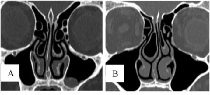
pneumatisation of different portions of the middle turbinate as follows: lamellar CB, pneumatisation of the stem or vertical lamella (Figure 1A); bulbous CB, pneumatisation of the distal bulbous segment (Figure 1B); and extensive ‘true CB’, pneumatisation of both lamellar and bulbous portions (Figures 2 and 3).13
Figure 1: Coronal CT scan shows concha bullosa (CB), defined as pneumatization of more than 50% of
vertical height of the middle turbinate.
A= Pneumatization of stem or vertical lamella of bilateral middle turbinates (Lamellar CB), B= Pneumatization of caudal bulbous portion of right middle turbinate (Bulbous CB) with mild septal deviation to left.
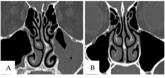
Figure 2: Coronal CT scan.
A= Bilateral similar sized CB with mild septal deviation to right, B= Bilateral CB with moderate septal deviation to left showing dominant CB on right. Note, there is preservation of the air channel between CB and the nasal septum.
Figure 3: Coronal CT scan.
A= Mild septal deviation to right with contralateral CB on left. B= Moderate septal deviation to right with contralateral CB on left. Note, there is preservation of the air channel between the CB and the nasal septum.
Normally, the convexity of middle turbinate is directed medially towards nasal septum. When the convexity is directed laterally, it is termed as PMT. In this study, PMT had presented as either unilateral (Figure 4A) or bilateral (Figure 4B) in presence of DNS. Most authors state that PMT when associated with DNS can be a contributing factor towards OMC obstruction.14
Coronal sinonasal CT scan study is the imaging modality of choice for detailed evaluation of both anatomy and pathology of nasal cavity and paranasal sinuses including
A B
A B
the anatomical variants in OMC region, as CT demonstrates air, bone and soft tissue components with awesome clarity. OMC is the complex anatomical region, where normal mucociliary drainage of paranasal sinuses such as, frontal, anterior ethmoid and maxillary sinuses
occur.5 Moreover, both DNS and CB are usually
associated with each other, but the cause of this association is still not clear.9 Evaluation of prevalence of CB and PMT in RARS patients having DNS and significance of this relationship are the main goals of the present study. It is anticipated that the current analysis may help optimal endoscopic surgical management of nasal obstruction.
Figure 4: Coronal CT scan.
A= Paradoxical middle turbinate (PMT) on right with moderate septal deviation to left. Note, bony septal spur and maxillary sinusitis on left, B= Bilateral PMT with negligible septal deviation. Note bilateral ethmoid bulla.
Figure 5: Coronal CT scan.
A= Septal deviation to left with the deviation angle measuring 19.2o, showing intranasal mucosal adhesion or synechia along contiguous left inferior nasal turbinate. Note contralateral CB on right and bilateral prominent ethmoid bulla. Inferior orbital groove (IOG, ) B= Size of CB measured as the maximum transverse diameter (MTD). The transverse diameter of the IOG () can be measured at the same coronal scan for comparison. Note nasal septum is deviated to left with contralateral CB on right showing preservation of air channels between the CB and the nasal septum.
METHODS
In this retrospective study, sinonasal CT scan images during August 2015 to December 2016 of 160 patients with history of RARS were evaluated for DNS, CB and PMT. All the data of the patients and their sinonasal CT scan images previously done at the radiology department were collected and analyzed. Total 160 cases having DNS were divided into two groups; 120 cases were having CB and rest 40 cases were having PMT. On the
basis of subjective assessment of size and severity of CB pneumatization, 120 cases with CB were further subdivided into two groups. Based on presence of moderate and large sized CB, Group I comprised of contralateral (Figure 3) and dominant variety of bilateral CB (Figures 2B and 5A). And according to the presence of small sized CB, group II cases comprised of ipsilateral and equal sized bilateral CB cases (Figures 1A and 2A). Similarly total PMT cases whether unilateral (Figure 4A) or bilateral PMT (Figure 4B) associated with DNS were analyzed with Chi-square test.
CT scans were performed by 128 multi-slice CT scanner of GE Healthcare Systems. Patients were positioned in supine position and scanning was done with contiguous thin slices from superior margin of frontal sinuses to inferior margin of maxillary sinuses. Reformatted coronal images obtained from the axial data are mostly preferred to direct coronal images obtained with patients in less comfort prone position, due to high quality resolution from high end multi-slice scanner. Scanning parameters were 3 mm table incrementation, 3 mm slice thickness, 2 seconds scanning time, 120 kVp and 180 mAs tube current. The field of view was confined to the sinonasal area for optimal visualization. Bone and soft tissues were best visualized at a window width of 1500-2000 HU and window level of 200-300 HU.
Inclusion criteria were at least 18 years of age at the time of imaging and that the sinus CT images were of diagnostic quality. Patients with features of acute rhinosinusitis, expansile sinonasal lesions such as polyp or tumor and history of prior surgery or craniofacial trauma were excluded. As other inclusion criterion, pneumatization of at least half of the turbinate volume is selected as CB for this study, thereby small lamellar CB with negligible pneumatization is excluded. Subjects with bidirectional septal deformities including S-shaped DNS were excluded. Because a perfectly straight nasal septum is uncommon, we also ignored minor deviations. We considered a deviation angle of less than 4 degrees as non-significant.
Common parameters like deviation angles of DNS and maximum transverse diameters of the CB (MTDCB) and the PMT (MTDPMT) were calculated. Deviation angles were calculated according to the angle between the planes along the crista galli and the most prominent point of septal deviation (Figure 5A). The MTDCB, MTDPMT and transverse diameter of the inferior orbital groove (IOG) were calculated; IOG considered to be an internal reference as it is visible in almost all patients in the same coronal plane where CB and PMT were visible (Figure 5B). Unilateral CB was classified as contralateral or ipsilateral according to the direction of septal deviation. When unequal sized CBs are present on either side of DNS, larger one was designated as the dominant CB (Figure 2B), usually seen contralateral to the direction of septal deviation.
B A
RESULTS
Total 160 RARS cases associated with DNS were selected for this study; 84 cases were males and 76 cases were females; age of patients ranged between 18 to 70 years (mean age of 34.6 years. Of total 120 cases having CB; as per direction of septal curvature, 80 cases were unilateral (67 contralateral and 13 ipsilateral CB) and rest 40 cases were bilateral (30 dominant and 10 similar sized CB). Among 80 unilateral CB, 41 were found on right
and 39 on left side of the nasal cavity. Among 40 bilateral CB, similar sized CB were seen in 10 cases and unequal CB in 30 cases; among which right sided dominant CB in 14 cases and left sided dominant CB in 16 cases (Table 1). With Chisquare test, total cases of contralateral and dominant variety of bilateral CB (group 1) were compared with ipsilateral and similar sized bilateral CB (group II). The association of severity of DNS with group I (contralateral and dominant) CB cases was found statistically highly significant (p=0.0001, Table 2).
Table 1: Prevalence of concha bullosa (CB) in relation to severity of nasal septal deviation.
Severity of
nasal septal deviation
Unilateral CB Bilateral CB
Total (%)
Contralateral Ipsilateral Dominant on one side Similar size on both
sides
Right Left Right Left Right Left
Significant (moderate
/ severe DNS) 32 28 2 3 14 16 2 97 (80.8%)
Negligible (mild /
absent DNS) 4 3 3 5 0 0 8 23 (19.2%)
Total (%) 80 (66.66%) 40 (33.33%) 120 (100%)
Table 2: Comparative analysis between severity of CB pneumatization and septal deviation.
Severity of
nasal septal deviation
Group I cases (relatively larger)
Group II cases (relatively smaller)
Total p- value* Contralateral
CB
Bilateral CB (Dominant on one side)
Ipsilateral CB
Bilateral CB (both of similar size)
Significant (moderate
/ severe DNS) 60 30 5 2 97
0.0001 Negligible (mild /
absent DNS) 7 0 8 8 23
Total 97 23 120
* Chi-square test; highly significant.
Table 3: Direction of septal curvature as per location of concha bullosa (CB).
Number of CB cases as per location (n) Septal deviation to right*
Septal deviation
to left* Total
Unilateral CB (80)
Contralateral (67)
Right (36) 0 36 36
Left (31) 31 0 31
Ipsilateral (13)
Right (5) 5 0 5
Left (8) 0 8 8
Bilateral CB (40) Dominant (30)
Right(14) 0 14 14
Left (16) 16 0 16
Similar Sized (10) 6 4 10
Total 58 62 120
* Student’s t test, p=0.9, no significance.
Of total 120 CB cases, leftward deviation of septal convexity (62 cases) was found slightly more than rightward septal deviation (58 cases), without any significance (Table 3). The CB cases included all varieties, bulbous CB in 26 (22%), lamellar CB in 12 (10%) and true CB in 82 (68%) cases. Deviation angles of the patients with contralateral CB, ipsilateral CB, dominant and similar sized bilateral CB were 12.2±2.4 degree (range, 8 to 17), 6.4±1.9 degree (range, 3 to 12),
to12), 8.6±1.9 mm (range, 5 to 14), and 4.6±1.3 mm (range, 2 to 11) respectively. MTDCB of patients with
group I were found significantly higher (p=0.0001, Table 4) than patients with group II CB cases.
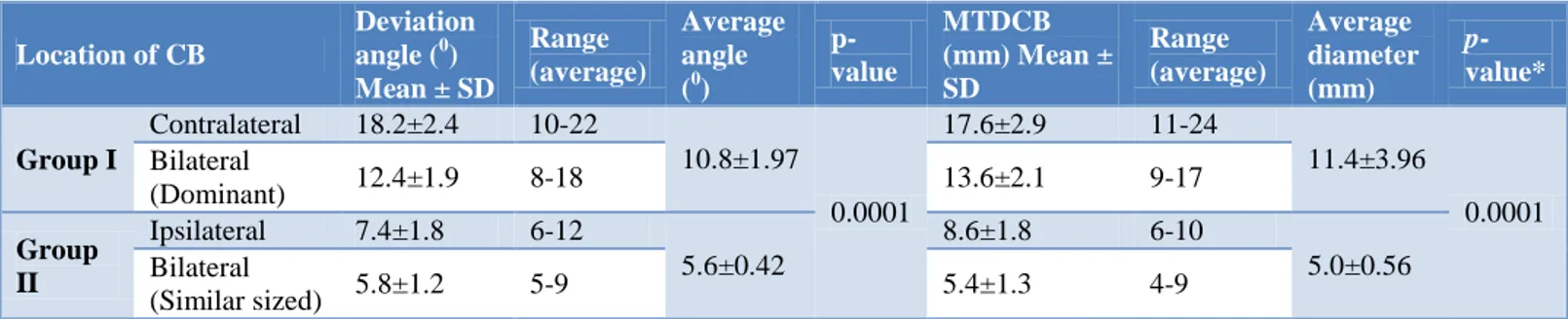
Table 4: Nasal septal deviation angle and maximum transverse diameter of CB (MTDCB).
Location of CB
Deviation angle (0) Mean ± SD
Range (average)
Average angle (0)
p-value
MTDCB (mm) Mean ± SD
Range (average)
Average diameter (mm)
p -value*
Group I
Contralateral 18.2±2.4 10-22
10.8±1.97
0.0001
17.6±2.9 11-24
11.4±3.96
0.0001 Bilateral
(Dominant) 12.4±1.9 8-18 13.6±2.1 9-17
Group II
Ipsilateral 7.4±1.8 6-12
5.6±0.42
8.6±1.8 6-10
5.0±0.56 Bilateral
(Similar sized) 5.8±1.2 5-9 5.4±1.3 4-9
* Chi-square test; highly significant.
Table 5: Prevalence of paradoxical middle turbinate (PMT) as per severity of septal deviation.
Severity of
nasal septal deviation
Unilateral PMT
Bilateral PMT
Total cases
of PMT p- value*
Contralateral Ipsilateral
Right Left Right Left
Significant 8 10 2 2 6 28 (70%)
0.692
Negligible 2 3 2 1 4 12 (30%)
Total/ MTD 23 (57.5 %) 7 (17.5%) 10 (25 %) 40 (100%)
* Chi-square test; no significance.
Similar calculation followed in case of measurement of PMT. MTD of contralateral, ipsilateral and bilateral CB were calculated and found to be 11.2±2.1 mm (range, 8 to 15), 4.6±1.4 mm (range, 3 to 9) and 5.1 ±1.6 mm (range, 4 to 10) respectively. Of total 40 cases of PMT, 14 cases were found in right and 16 in left nasal cavity while 10 cases were bilateral. As per severity of DNS, unilateral and bilateral PMT revealed no significance (p =0.69) (Table 5).
DISCUSSION
The role of sinonasal anatomical variants in the development of chronic sinus diseases is debatable till date. In order to define the relationship between the CB and DNS with recurrent sinus diseases, more detailed investigations are needed.5 Here in, anatomic variations in relation to the OMC region, though do not represent disease states per se, but compromise drainage pathways and produce significant obstruction in the presence of inflammation predisposing to RARS. As CB may predispose to sinusitis, proper knowledge regarding sinonasal anatomical variations is very much essential before planning for functional endoscopic sinus surgery (FESS) to avoid any dreadful mishap.12 Few reports emphasized no specific association of anatomic variations with rhinosinusitis and rather claimed the role of local, systemic, environmental factors or intrinsic mucosal disease in its pathogenesis.15,16 While Ameri et al. reported that anatomical variants of paranasal sinuses may be considered as predictors for the recurrence of rhinosinusitis.17 This study established a ‘cause and effect’ relationship between CB and PMT with septal deviations, in causing recurrent episodes of rhinosinusitis.
In the present study, common anatomic variations in RARS patients were DNS followed by CB and PMT. The significance of CB and PMT lies in increasing the probability of obstruction of the middle meatus and OMC in presence of a moderate to severe septal deviation. CB cases presented as unilateral in 66.66% cases; Zandi et al reported a very high incidence of CB presenting as 39.8% bilateral and 60.2% unilateral showing significant correlation with rhinosinusitis, which corroborated with present findings.18 As reported in a Turkish study, the incidence of CB was higher in individuals with DNS, while in this study, there was a strong association between the presence of a CB and contralateral DNS, similar to another work.19,20 However, septal deviation away from the dominant concha with preserved adjacent air channels suggests that the deviation is not a direct result of mass effect from the CB, thus excluding the etiological role of CB in DNS.20 Severe septal deviation had been noted as a contributing factor for sinusitis.21 In this work, among three kinds of septal deviations, cartilaginous deviation, bony deviation and high septal deviation, the later had a significant relationship with recurrent rhinosinusitis, which was corroborated with another study.22
variation and leads to susceptibility to rhinosinusitis.13,25 As compared to PMT, significant relationship of OMC pattern of RARS with CB was recorded in this study.
CONCLUSION
CB and PMT are the most common variants of middle turbinate. Incidence of RARS often seen in patients of CB associated with DNS. Contralateral variety of unilateral CB and dominant variety of bilateral CB showed significant relationship with severity of septal deviations. Preserved adjacent air channels suggest that CB, though coexistent, develops independent of DNS. While much attention received for the ongoing debate over optimal endoscopic surgical management of nasal obstruction, it is important to assess reasons of the development of CB and PMT in presence of DNS particularly in RARS patients. Preoperative sinonasal CT scan is a much essential tool for evaluating various coexisting variants in the OMC region before planning for the coveted FESS in RARS cases.
ACKNOWLEDGMENTS
Authors are thankful to Prof. Dr. Gangadhara Sahoo, Dean, IMS and SUM Hospital, Siksha ‘O’ Anusandhan University, for extended facilities.
Funding: No funding sources Conflict of interest: None declared
Ethical approval: The study was approved by the Institutional Ethics Committee
REFERENCES
1. Bhattacharyya N, Lee KH. Chronic recurrent
rhinosinusitis: disease severity and clinical
characterization. Laryngoscope. 2005;115:306-10.
2. Sahu N, Mohapatra SSG, Rath SN, Padhy RN.
Radiological significance of isolated Ethmoid sinus infections in asymptomatic patients of recurrent
acute rhinosinusitis. Int J Res Med Sci.
2017;5(5):1781-4.
3. Farid MM, Metwalli N. Computed tomographic
evaluation of mouth breathers among paediatric patients. Dentomax Radiol. 2010;39:1-10.
4. Moghadas H, Abouali O, Faramarzi A, Ahmadi G.
Numerical investigation of septal deviation effect on deposition of nano/microparticles in human nasal passage. Resp Physiol Neurobiol. 2011;177:9-18.
5. Aktas D, Kalcioglu MT, Kutlu R, Ozturan O, Oncel
S. The relationship between the concha bullosa, nasal septal deviation and sinusitis. Rhinology. 2003;41:103-6.
6. Keles B, Öztürk K, Ünaldı D, Arbag H, Özer B. Is
There Any Relationship Between Nasal Septal Deviation And Concha Bullosa? Euro J Gen Med. 2010;7(4):359-64.
7. Hatipoglu HG, Cetin MA, Yuksel E. Concha bullosa
types: their relationship with sinusitis, ostiomeatal
and frontal recess disease. Diagn Intervent Radiol. 2005;11:145-9.
8. Paksoy M, Sanli A, Evren C, Kayhan FT, Bozkurt Z, Aydin S, et al. The role of concha bullosa in nasal pathologies. Kulak burun bogaz ihtisas dergisi: KBB. J Ear, Nose, Throat. 2007;18(4):238-41.
9. Al-Qudah M. The relationship between anatomical
variations of the sino-nasal region and chronic
sinusitis extension in children. I J Ped
Torhinolaryngol. 2008;72(6):817-21.
10. Yang BT, Chong VF, Wang ZC, Xian JF, Chen QH.
CT appearance of pneumatized inferior turbinate. Clin Radiol. 2008;63:901-5.
11. Uygur K, Tuz M, Dogru H. The correlation between
septal deviation and concha bullosa. Otolaryngol Head Neck Surg. 2003;129(1):33-6.
12. Bhandary SK. Study of relationship of concha
bullosa to nasal septal deviation and sinusitis. I J Otolaryngol Head Neck Surg. 2009;61(3):227-9.
13. Bolger WE, Clifford AB, Parson DS. Paranasal
sinus bony anatomic variations and mucosal abnormalities: CT analysis for endoscopic sinus surgery. Laryngoscope. 1991;101:56-64.
14. El-Shazly AE, Poirrier AL, Cabay J, Lefebvre PP. Anatomical Variations of the Lateral Nasal Wall: The Secondary and Accessory Middle Turbinates. Cli An. 2012;25:340-6.
15. Fadda GL, Rosso S, Aversa S, Petrelli A, Ondolo C,
Succo G. Multiparametric statistical correlations between paranasal sinus anatomic variations and chronic rhinosinusitis. Acta Otorhinolaryngol Ital. 2012;32:244-51.
16. Tantilipikorn P. Accuracy of limited CT with the full CT as the standard evaluation for inflammatory disease of PNS and the identification of anatomical variations Department of Otorhinolaryngology. Siriraj Med J. 2009;61:3-7.
17. Ameri AA, Eslambolchi A, Bakshandeh H.
Anatomical Variations of Paranasal Sinuses & Chronic Sinusitis. Iran J Radiol. 2005;2:3-4.
18. Zandi B, Davoodi MR, Mirgholami AR. Concha
Bullosa and Other Sino-Nasal Variants: Clinical and CT Correlation. Iran J Radiol. 2003;1(1-2):33-6.
19. Sevinc O, Barut C, Kacar D, Is M. Evaluation of the
lateral wall of the nasal cavity in relation to septal deviation. Int J Morpho. 2013;31:438-43.
20. Stallman JS, Lobo JN, Som PM. The incidence of concha bullosa and its relationship to nasal septal deviation and paranasal sinus disease. Am J Neuroradiol. 2004;25(9):1613-8.
21. Sarna A, Hayman LA, Laine FJ, Taber KH. Coronal
imaging of the osteomeatal unit: anatomy of 24 variants. J Comput Assist Tomogr. 2002;26(1):153-7.
22. Homsioglou E, Balatsouras DG, Alexopoulos G,
Kaberos A, Katotomichelakis M, Danielides V. Pneumatized superior turbinate as a cause of headache. Head Face Med. 2007;3:1-5.
23. Ural A, Kanmaz A, İnançli HM, İmamoğlu M.
concha bullosa and nasal valve collapse with the convexity of septal deviation. Acta Oto-Laryngolog. 2010;130(2):271-4.
24. Keles B, Öztürk K, Ünaldı D, Arbag H, Özer B. Is
There Any Relationship Between Nasal Septal Deviation And Concha Bullosa? Euro J Gen Med. 2010;7(4):359-64.
25. Roozbahany NA, Nasri S. Nasal and paranasal sinus
anatomical variations in patients with rhinogenic
contact point headache. Auris Nasus Larynx. 2013;40(2):177-83.
Cite this article as: Mohapatra SSG, Sahu N, Rath