Achiraya Teyateeti, M.D., Pawana Pusuwan, M.D., Ajalaya Teyateeti, M.D. Department of Radiology, Faculty of Medicine Siriraj Hospital, Mahidol Unversity, Bangkok 10700, Thailand.
Impact of
18
F-FDG PET/CT Scan on Management of
Non-small Cell Lung Cancer Patients in Contexts
of University-Based Hospital in Thailand
ABSTRACT
Objective: To determine the impact of Fluorine-18 fluorodeoxy glucose (18F-FDG) positron emission tomography
with computed tomography (PET/CT scan) on management of non-small cell lung cancer (NSCLC) in contexts of university-based hospital in Thailand where most patients were in advanced disease and conventional imaging (CI) had previously been done.
Methods: Between December 2006 to December 2014, PET/CT scan and CI - e.g. chest and upper abdominal CT scan
and bone scintigraphy of 120 pathologically proven NSCLC patients were independently reviewed. Stagings based on CI and PET/CT scan were evaluated. Proposed treatment plan based on CI and given treatment based on PET/ CT scan were compared in patients whose medical records were available. Impact of PET/CT scan on management was categorized into inter-modality and intra-modality treatment change – i.e. change in radiation fields.
Results: Of 120 NSCLC patients, PET/CT scans were performed for initial staging (INS) in 60 patients,
post-treatment evaluation (PTE) in 41 patients and restaging at time of recurrence or progression (RR) in 19 patients. PET/CT scan changed the disease status in 38 patients (31.7%). Changes to more advanced and less advanced disease were found in 26 patients (11 INS, 8 PTE and 7 RR patients) and 12 patients (3 INS, 6 PTE and 3 RR patients), respectively. Among 83 patients with available proposed treatment plan, inter-modality treatment changes were observed in 19 patients (22.9%). Changes were more frequent in setting of PTE and RR (14/19 patients; 73.7%). The most common changes were toward palliative chemotherapy (15/19 patients, 78.9%) owing to unexpected distant metastasis. Among 43 patients who received radiation therapy, changes in radiation approach were found in 18 patients (41.9%), mostly due to positive or negative lymph node (LN) metastasis.
Conclusion: PET/CT scan has considerable impact on staging and both inter-modality and intra-modality treatment
change in NSCLC patients in all settings – INS, PTE and RR even with the presence of CI. Therefore, PET/CT scan should be considered for staging and guiding the multidisciplinary treatment.
Keywords: PET/CT; non-small cell lung cancer; NSCLC; staging; management; treatment (Siriraj Med J 2018;70:
169-174)
Correspondence to: Ajalaya Teyateeti E-mail: littlecreammy@gmail.com
Received 22 February 2017 Revised 22 May 2017 Accepted 30 May 2017 doi:10.14456/smj.2018.27
INTRODUCTION
In non-small cell lung cancer (NSCLC), conventional imaging (CI) – e.g. computed tomography (CT), magnetic resonance imaging (MRI) and bone scintigraphy generally provides adequate information for staging and subsequent management. However, patients have to undergo multiple imaging modalities causing delayed treatment.
management of NSCLC from staging to detection of recurrence.1-14 Nevertheless, lack of effective screening
program and unawareness of malignancy have resulted in late presentation of many lung cancer patients in Thailand. Moreover, to be eligible for reimbursement, CT scan of chest including upper abdomen and bone scintigraphy must be done prior to PET/CT scan. We hypothesized that benefit of PET/CT scan may be altered in patients with advanced disease and who previously received the CI. Therefore, this study was elicited to evaluate the impact of PET/CT scan on NSCLC treatment in the contexts of our university-based hospital in Thailand.
MATERIALS AND METHODS
During December 2006 to December 2014, 120 pathologically proven NSCLC patients who underwent PET/CT scan for 1) initial staging or evaluation of disease extension at time of diagnosis (INS), 2) post-treatment evaluation (PTE), 3) evaluation of suspected residual or recurrent disease or restaging at time of recurrent disease (RR) were included into the study. Patients with other co-existing malignancy were excluded. Medical records and all diagnostic imaging studies were retrospectively reviewed to collect patient, tumor and treatment characteristics following approval from the Institute Ethical Committee (Si 190/2015).
Conventional imaging in this study included 1) CT scan performed as part of PET/CT scan, 2) CT scan of chest with or without upper abdomen, 3)99mTc-MDP whole body bone scintigraphy and 4) contrast-enhanced CT scan or MRI of brain. Apart from CT scans which were simultaneously performed as part of PET/CT scans, other imaging must be performed within 2 months prior to PET/CT scan.
The PET/CT scan required fasting for at least 6 hours with blood sugar less than 200 mg/dL. 18F-FDG of 5.2-7.4 MBq/kg was intravenously injected and followed by uptake period of 60 minutes. Standard whole body imaging - base of skull to mid-thigh or further extension as indicated was performed. CT acquisition parameter was 120 kV, auto mA (30-300 mA) and slice thickness of 3.75 mm. Contrast media was used in all cases, if patient had no contraindication. PET/CT scan and CI of each patient were separately reviewed with at least 1 week apart by consensus of two reviewers.
Primary purpose of the study was to evaluate the impact of PET/CT scan on inter-modality treatment changes - i.e. a surgical candidate received chemotherapy (CMT) instead due to presence of unexpected distant metastasis on PET/CT scan. Proposed treatment plans were based on clinical data including available CI and
derived from opinion of referring physicians stated in medical records at time of requesting for PET/CT scans. Proposed treatment was compared to given treatment after performing PET/CT scan in all patients whose medical records were available.
Secondary purpose was whether PET/CT scan triggered intra-modality change - i.e. change in target delineation or radiation (RT) field due to detection of additional metastatic mediastinal LN. Contouring based on CI was compared to contouring based on PET/CT scan in order to assess the change in target coverage.
Other secondary purpose was if PET/CT scan led to upstage and downstage in setting of INS and changed disease status to more advanced (equivalent to upstage) or less advanced (equivalent to downstage) in PTE and RR. The 7th edition AJCC staging system15 was applied
for staging in all settings – INS, PTE and RR. Therefore, patients without detectable disease after treatment were categorized as stage 0. Staging based on findings on all available CI was compared to that on PET/CT scan and reported in term of upstage, unchange or downstage.
RESULTS
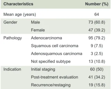
Of 120 patients, 73 patients were male (60.8%). Age range was 35-92 years with mean age of 64 years. Major histopathology was adenocarcinoma in 95 patients (79.2%). CT scan of chest with or without upper abdomen was available in all patients. Bone scintigraphy was done in 49 patients (40.8%) and CT scan or MRI of brain was done in 3 patients (2.5%). Most common indication was INS in 60 patients (50%), followed by PTE in 41 patients (34.2%). (Table 1)
TABLE 1. Patients and tumors’ characteristics.
Characteristics Number (%)
Mean age (years) 64
Gender Male 73 (60.8)
Female 47 (39.2)
Pathology Adenocarcinoma 95 (79.2) Squamous cell carcinoma 9 (7.5) Adenosquamous carcinoma 3 (2.5) Not specified subtype 13 (10.8) Indication Initial staging 60 (50)
PET/CT scan led to staging change
In comparison to CI, PET/CT scan led to more advanced disease in 26 patients and less advanced disease in 12 patients. Among these 38 out of 120 patients (31.7%), there were 14 INS patients, 14 PTE patients and 10 RR patients.
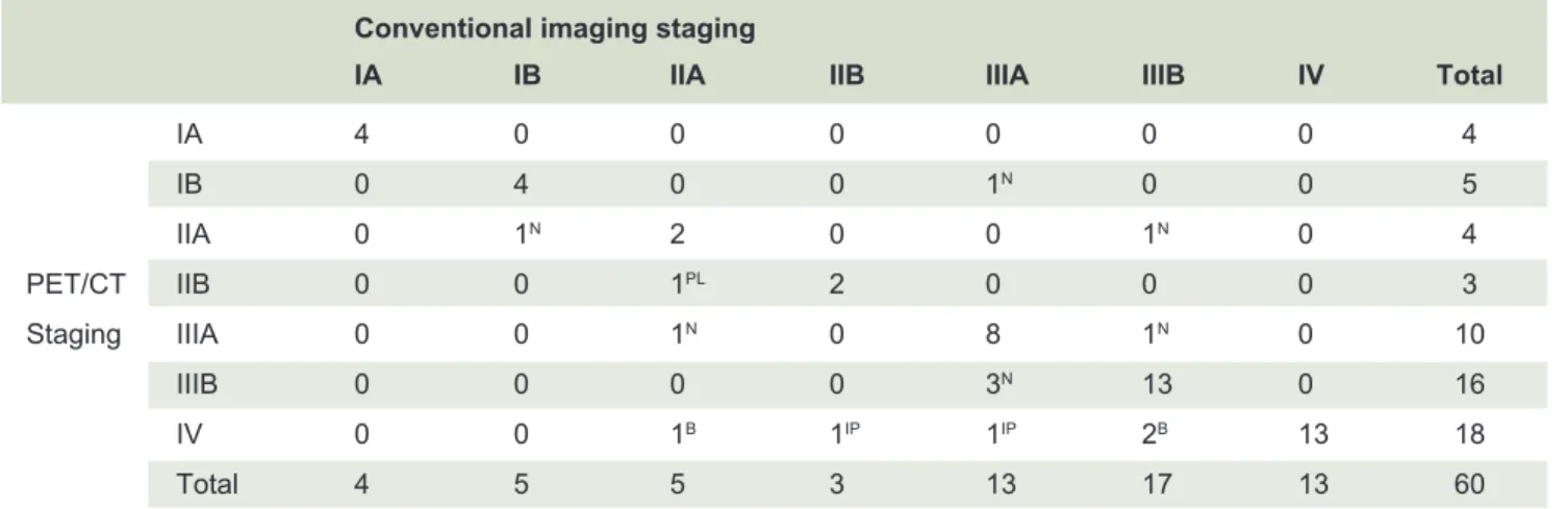
PET/CT scan changed initial staging in 14 of 60 patients (23.3%) - upstage in 11 patients and downstage in 3 patients (Table 2). T-staging was upstage in 2 patients and none was downstage. N-staging was changed in 12 patients – upstage in 7 patients and downstage in 5 patients. M-staging was more advanced in 6 patients and none was downstage. Most common additional findings detected on PET/CT scan, which affected staging were mediastinal LN in 8 patients (13.3%) and bone in 3 patients (5%).
Regarding PTE setting, PET/CT scans were resulted in staging change in 14 patients (34.1%) – upstage in 8 patients and downstage in 6 patients (Table 3). T-staging was upstage in none and downstage in 1 patient. N-staging was upstage in 4 patients and downstage in 5 patients. M-staging was upstage in 7 patients and downstage in 1 patient. Most common additional findings detected on PET/CT scan which affected staging were bone in 6 patients and mediastinal LN in 5 patients.
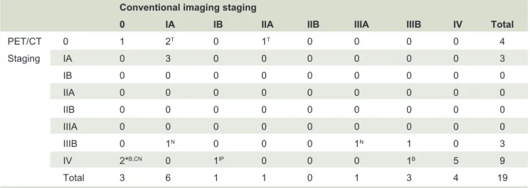
PET/CT scan led to staging change in 10 out of 19 RR patients (52.6%) – upstage in 7 patients and downstage in 3 patients (Table 4). T-staging was upstage in none and downstage in 3 patients. N-staging was upstage in 3 patients and downstage in 1 patient. M-staging was upstage in 4 patients and downstage in none. Most common additional findings detected on PET/CT scan
TABLE 2. Impact of PET/CT on change of initial staging.
Conventional imaging staging
IA IB IIA IIB IIIA IIIB IV Total
IA 4 0 0 0 0 0 0 4
IB 0 4 0 0 1N 0 0 5
IIA 0 1N 2 0 0 1N 0 4
PET/CT IIB 0 0 1PL 2 0 0 0 3
Staging IIIA 0 0 1N 0 8 1N 0 10
IIIB 0 0 0 0 3N 13 0 16
IV 0 0 1B 1IP 1IP 2B 13 18
Total 4 5 5 3 13 17 13 60
Staging on PET/CT scan was different from CI due to visceral pleural involvement (PL), intrapulmonary metastasis (IP), bone metastasis (B) and mediastinal LN (N)
TABLE 3. Impact of PET/CT on change of staging in post-treatment evaluation.
Conventional imaging staging
0 IA IB IIA IIB IIIA IIIB IV Total
PET/CT 0 3 1T 0 0 0 2N 0 1PL 7
Staging IA 0 0 0 0 0 0 0 0 0
IB 0 0 0 0 0 0 0 0 0 IIA 0 0 0 2 0 0 0 0 2
IIB 0 0 0 0 0 0 1N 0 1
IIIA 0 0 0 0 0 3 1N 0 4
IIIB 0 0 0 0 0 1N 7 0 8
IV 2*PL,B 0 0 0 0 3B 2B 12 19
Total 5 1 0 2 0 9 11 13 41
TABLE 4. Impact of PET/CT on change of staging in residual or recurrent disease.
Conventional imaging staging
0 IA IB IIA IIB IIIA IIIB IV Total
PET/CT 0 1 2T 0 1T 0 0 0 0 4
Staging IA 0 3 0 0 0 0 0 0 3
IB 0 0 0 0 0 0 0 0 0 IIA 0 0 0 0 0 0 0 0 0 IIB 0 0 0 0 0 0 0 0 0 IIIA 0 0 0 0 0 0 0 0 0
IIIB 0 1N 0 0 0 1N 1 0 3
IV 2*B,CN 0 1IP 0 0 0 1B 5 9
Total 3 6 1 1 0 1 3 4 19
Staging on PET/CT scan was different from CI due to primary tumor or tumor bed (T), intrapulmonary metastasis (IP), bone metastasis (B), mediastinal LN (N) and cervical LN (CN), *Staging change due to bone metastasis in one patient and both cervical LN and bone metastasis in other.
which affected staging were tumor bed or primary tumor in 3 patients and bone in 3 patients.
Of 49 patients who underwent bone scintigraphy, change of M-staging due to newly diagnosed bone metastasis on PET/CT scan were seen in 4 patients (8.16%) - 3 INS patients and 1 PTE patient. As for 71 patients without bone scintigraphy, first diagnosis of bone metastasis on PET/CT scan were observed in 8 patients (11.3%) – 5 PTE patients and 3 RR patients.
Impact on management
Consequent management resulted from PET/CT scan were analyzed in 2 aspects as follows;
1) PET/CT scan triggered inter-modality treatment change; Out of 83 patients with available proposed
treatment plan prior to PET/CT scan (38 INS patients, 33 PTE patients, 12 RR patients), given treatment were inter-modality change in 19 patients (22.9%). Treatment modality changed from surgery or RT to CMT due to distant metastasis in 15 patients (18.1%) and from surgery or radiation to observation due to negative PET/CT scan in 4 patients (4.8%) (Table 5).
2) PET/CT scan triggered intra-modality treatment change; Out of 43 patients who received RT as a locoregional
treatment for intra-thoracic disease, findings of PET/CT scan, particularly FDG avid subcentimeter intrathoracic LN, led to changes in radiation approach i.e. target delineation in 18 patients (41.9%).
DISCUSSION
According to previous studies conducted in developed countries, PET and PET/CT scan has been proven to
provide higher diagnostic performance than CI and consequently affected the management.1-14,16-19 However,
benefit of PET/CT scan may be questionable in different contexts – i.e. more advanced disease and presence of other CI prior to PET/CT scan. Consequently, this study was aimed to assess the impact of PET/CT scan on management of NSCLC in the context of a university-based hospital in Thailand.
While most studies evaluated the effect of PET/CT scan on stage migration in INS, in this study we evaluated the effect in all settings - INS, PTE and RR. We found that PET/CT scans benefit in all settings as 31.7% of patients were changed to more advanced/upstage or less advanced/downstage after performing PET/CT scan.
Regarding PET/CT scan for INS, stage changing in current study was 23.3% corresponding to previous studies which reported changes ranging from 22-42%.4,6-8 These
figures varied among the studies due to several reasons.
TABLE 5. Inter-modality treatment change according
to indication and given treatment.
Sx/RT to CMT Sx/RT to OBS Total
INS (n=38) 5 0 5
PTE (n=33) 8 3 11
RR (n=12) 2 1 3
Total 15 4 19
First, settings were different – i.e. evaluation of solitary pulmonary nodule, staging of newly diagnosed NSCLC and staging in surgical candidate. Our study included patients in all aforementioned settings and we found that change in staging was consistent to other studies. Second, CI in each study included different imaging modalities – e.g. the study integrating bone scintigraphy as part of CI may detect more bone metastatic lesions. Therefore, upstage to stage IV disease following PET/CT scan might be less frequent in a study which included bone scintigraphy as part of CI. Third, interpretation of equivocal lesions on CI had a considerable impact on stage shifting. For example, an adrenal nodule on CT scan could be considered as either benign adenoma or metastasis. FDG avid adrenal nodule on subsequent PET/CT scan would lead to upstage or unchanged, respectively. Lastly, time interval between CI and PET/ CT scan may lead to disease progression giving the false impression of upstaging by PET/CT scan. With this reason, we compared findings on PET/CT scan and CI which included CT imaging of PET/CT scans themselves to diminish the upstage from tumor progression during waiting time. Therefore, additional benefit of PET/CT scan was more accurately assessed.
Among TNM-staging of INS, change of M-staging was the most common one.6-7 However, changes of
M-staging in our study was less frequent than N-staging. The possible explanation was 41% of patients underwent bone scintigraphy prior to PET/CT scan. CT scan from base of skull to mid-thigh as part of PET/CT scan was also incorporated as part of CI. Therefore, M-staging change due to unexpected bony metastasis was decreased. Additionally, over 70% of patients in our study were in advanced stage. These patients carried high disease burden and high potential of overt distant metastasis which might be easily detected even on CI. As a result, unexpected distant metastasis discovered on PET/CT scan was less frequent.
Change of M-staging was more common in PTE and RR. We postulated that after receiving treatment these patients carried lower disease burden and risk of distant metastasis. Consequently, occult or small metastasis might not be detected easily on CI. Moreover, some CI as a metastatic screening might be omitted by referring physician. As a result, detection of unexpected distant metastasis was increased on PET/CT scan.
Noticeably, most of inter-modality changes in our study were toward CMT owing to presence of distant metastasis (15 of 19 patients; 78.9%). Moreover, majority of changes were observed in PTE and RR (11 of 33 patients; 33.3% and 3 of 12 patients; 25%, respectively). Regarding
inter-modality treatment change in INS patients, it was only 13.2% (5 of 38 patients) and all of them were given CMT. This result was quite different from that of 19.2-39% in the previous studies.4-6 We hypothesized that it
might relate to advanced stage of disease and practices of our institute. Although, stage III patients had several treatment options – i.e. preoperative CMT, preoperative CMT and RT and definitive CCRT, definitive CCRT tended be chosen in our institute. Therefore, PET/CT scan seemed to have only modest impact on treatment.
PET/CT scan has been proven to provide benefit in evaluation of post-treatment residual disease20 and
prediction of treatment outcome by monitoring change of metabolic activity parameters.1,21 Even so PET/CT scan
in our institute was infrequently used to differentiate between fibrosis and residual disease. Majority of PET/ CT scans in PTE setting were to guide further adjuvant treatment. Remarkably, among 12 PTE patients who received curative surgery prior to PET/CT scan, in 5 of them unexpected metastasis were found on PET/CT scan. In much the same way, there were 2 of 12 RR patients whose PET/CT scan showed unexpected metastasis leading to exclusion of futile surgery/radiation. Consequently, performing of PET/CT scan before starting treatment might assist to prevent futile definitive treatment.
In the study containing early stage NSCLC patients, intra-modality treatment change following PET scan was observed in 15% of patients, and change in surgical approach was the most common one (7.3%).5 However,
there were too few patients who underwent surgery in our study. Our study had high proportion of N2 and N3 staging on CI staging (63%). Main treatment modalities were radiation and change of radiation approach resulting from exclusion or detection of metastatic LN which was 42%.
limitation was lack of pathological confirmation and follow-up imaging which prevented us from evaluation of accuracy of PET/CT scan.
In conclusion, PET/CT scan had considerable impact on TNM-staging and management of NSCLC in all settings – INS, PTE and RR even with the presence of previous CI. However, It seems like the benefits of PET/CT were more obvious in M-staging for patients with low disease burden or in early stage as detection of distant metastasis might help to avoid futile definitive treatment. In contrast, patients with high disease burden or in advanced stage might gain more benefit in N-staging reflecting in change in RT treatment planning.
ACKNOWLEDGMENTS
Authors would like to thank all of our colleagues in Division of Nuclear Medicine and Division of Radiation Oncology, Department of Radiology. This study was supported by a grant from Academic Affairs of Faculty of Medicine Siriraj Hospital and International Atomic Energy Agency (IAEA) for poster presentation at International conference on Clinical PET/CT and molecular imaging (IPET 2015). Research authors also received a Chalermprakiat grant from the Faculty of Medicine Siriraj Hospital, Mahidol University.
REFERENCES
1. Truong MT, Viswanathan C, Erasmus JJ. Positron emission tomography/computed tomography in lung cancer staging, prognosis, and assessment of therapeutic response. J Thorac Imaging. 2011;26:132-46.
2. Sharma P, Singh H, Basu S, Kumar R. Positron emission tomography-computed tomography in the management of lung cancer: An update. South Asian J Cancer. 2013;2:171-8.
3. Bury T, Dowlati A, Paulus P, Corhay JL, Hustinx R, Ghaye B, Radermecker M, Rigo P. Whole-body 18FDG positron emission tomography in the staging of non-small cell lung cancer. Eur Respir J. 1997;10:2529-34.
4. Hicks RJ, Kalff V, MacManus MP, Ware RE, Hogg A, McKenzie AF, Matthews JP, Ball DL. (18)F-FDG PET provides high-impact and powerful prognostic stratification in staging newly diagnosed non-small cell lung cancer. J Nucl Med. 2001;42:1596-604.
5. Seltzer MA, Yap CS, Silverman DH, Meta J, Schiepers C, Phelps ME, et al. The impact of PET on the management of lung cancer: the referring physician's perspective. J Nucl Med. 2002;43: 752-6.
6. Sachs S, Bilfinger TV. The impact of positron emission tomography on clinical decision making in a university-based multidisciplinary lung cancer practice. Chest. 2005;128:698-703.
7. Saunders CA, Dussek JE, O'Doherty MJ, Maisey MN. Evaluation of fluorine-18-fluorodeoxyglucose whole body positron emission tomography imaging in the staging of lung cancer. Ann Thorac Surg. 1999;67:790-7.
8. Taus Á, Aguiló R, Curull V, Suárez-Piñera M, Rodríguez-
Fuster A, Rodríguez de Dios N, et al. Impact of 18F-FDG PET/ CT in the treatment of patients with non-small cell lung cancer. Arch Bronconeumol. 2014;50:99-104.
9. Subedi N, Scarsbrook A, Darby M, Korde K, Mc Shane P, Muers MF. The clinical impact of integrated FDG PET-CT on management decisions in patients with lung cancer. Lung Cancer. 2009;64:301-7.
10. Kalff V, Hicks RJ, MacManus MP, Binns DS, McKenzie AF, Ware RE, et al. Clinical impact of (18)F fluorodeoxyglucose positron emission tomography in patients with non-small cell lung cancer: a prospective study. J Clin Oncol. 2001;19:111-8.
11. Schrevens L, Lorent N, Dooms C, Vansteenkiste J. The role of PET scan in diagnosis, staging, and management of non-small cell lung cancer. Oncologist. 2004;9:633-43.
12. Hoekstra CJ, Stroobants SG, Hoekstra OS, Vansteenkiste J, Biesma B, Schramel FJ, et al. The value of [18F]fluoro-2- deoxy-D-glucose positron emission tomography in the selection of patients with stage IIIA-N2 non-small cell lung cancer for combined modality treatment. Lung Cancer. 2003;39:151-7.
13. Hicks RJ, Kalff V, MacManus MP, Ware RE, McKenzie AF, Matthews JP, et al. The utility of (18)F-FDG PET for suspected recurrent non-small cell lung cancer after potentially curative therapy: impact on management and prognostic stratification. J Nucl Med. 2001;42:1605-13.
14. Bury T, Corhay JL, Duysinx B, Daenen F, Ghaye B, Barthelemy N, et al. Value of FDG-PET in detecting residual or recurrent non-small cell lung cancer. Eur Respir J. 1999;14:1376-80.
15. Edge SB, Byrd D, Compton C, Fritz A, Greene F, Trotti A, editors. AJCC cancer staging manual. 7th ed. New York:
Springer; 2010.
16. De Wever W, Stroobants S, Coolen J, Verschakelen JA. Integrated PET/CT in the staging of nonsmall cell lung cancer: technical aspects and clinical integration. Eur Respir J. 2009;33:201-12.
17. Lv YL, Yuan DM, Wang K, Miao XH, Qian Q, Wei SZ, et al. Diagnostic performance of integrated positron emission tomography/computed tomography for mediastinal lymph node staging in non-small cell lung cancer: a bivariate systematic review and meta-analysis. J Thorac Oncol. 2011;6:1350-8
18. Qu X, Huang X, Yan W, Wu L, Dai K. A meta-analysis of ¹⁸FDG- PET-CT, ¹⁸FDG-PET, MRI and bone scintigraphy for diagnosis of bone metastases in patients with lung cancer. Eur J Radiol. 2012;81:1007-15.
19. Gould MK, Kuschner WG, Rydzak CE, Maclean CC, Demas AN, Shigemitsu H et al. Test performance of positron emission tomography and computed tomography for mediastinal staging in patients with non-small-cell lung cancer: a meta-analysis. Ann Intern Med. 2003;139:879-92.
20. De Leyn P, Stroobants S, De Wever W, Lerut T, Coosemans W, Decker G, et al. Prospective comparative study of integrated positron emission tomography-computed tomography scan compared with remediastinoscopy in the assessment of residual mediastinal lymph node disease after induction chemotherapy for mediastinoscopy-proven stage IIIA-N2 Non-small-cell lung cancer: a Leuven Lung Cancer Group Study. J Clin Oncol. 2006;24:3333-9.