Original Research Article
Usefulness of High Resolution Computed Tomography scans in
depicting the status of the middle ear structures
Ponnam Bharath Kumar
1, Kiran Mai
2, Santosh Karpur
3*
INTRODUCTION
There are many causes of deafness, but atticoantral disease is the leading one. In this disease, there is inflammation of the middle ear cleft. Inside the ear, cholesteatoma is formed in this disease. This disease is associated with complications which can even lead to fatality. Cholesteatoma are cyst-like, expansible lesions of the temporal bone lined by stratified squamous
epithelium that contain desquamated keratin. There is perforation of the tympanic membrane in atticoantral disease.
This perforation occurs in the attic region. There is formation of cholesteatoma also in atticoantral disease. Cholesteatoma is associated with a higher risk of major complications, e.g. brain abscess and other intracranial infection.1
1Department of Radio Diagnosis, Malla Reddy Medical College for Women, Quthbullapur Municipality, Hyderabad,
Telangana, India
2Consultant Radiologist, Clarity Diagnostics, Raichur, Karnataka, India
3Department of Radio Diagnosis, Malla Reddy Institute of Medical Sciences, Suraram, Hyderabad, Telangana, India
Received: 09 January 2019
Accepted: 05 March 2019
*Correspondence:
Dr. Santosh Karpur,
E-mail: [email protected]
Copyright: © the author(s), publisher and licensee Medip Academy. This is an open-access article distributed under the terms of the Creative Commons Attribution Non-Commercial License, which permits unrestricted non-commercial use, distribution, and reproduction in any medium, provided the original work is properly cited.
ABSTRACT
Background: Prior to surgery it is very important that the surgeon is fully aware, and he should have the clear picture as to the extent of the disease and the nature of the disease to give satisfactory surgical outcomes for the patient. HRCT (High resolution computed tomography) is one such guiding tool for the surgeon. The objective was to study usefulness of HRCT scan in attico-antral disease in depicting the status of the middle ear structures.
Methods: A total of 30 patients were studied. HRCT temporal bone was performed by using SIEMENS EMOTION 16 slice CT machine in axial plane and coronal images were reformatted. Findings of HRCT temporal bone were recorded. Findings of mastoid exploration surgery were recorded. Report of HRCT of temporal bone was correlated with surgical findings and tabulated using percentages.
Results: Surgery showed cholesteatoma in 26 (86.6%) patients. Epitympanum was involved in 29 (96.6%) patients in HRCT and 30 (100%) patients at surgery. Extension beyond middle ear cleft was seen in 4 (13.3%) patients in HRCT and 5 (16.6%) patients at surgery. Tympanic segment of facial canal was the most commonly involved, showing erosion in 10 (33.3%) patients in HRCT and 12 (40%) patients at surgery. Lateral SCC was the most commonly involved in bony labyrinth seen in 4 (13.3%) patients in both HRCT and surgery. Erosion of dural plate was seen in 6 (20%) patients in HRCT whereas 9 (30%) patients showed dural plate erosion at surgery.
Conclusions: HRCT of temporal bone plays a promising role in pre-operative assessment of cholesteatoma as it depicts the extent of the disease and integrity of most of the middle ear structures.
Keywords: Atticoantral disease, Deafness, HRCT, Middle ear disease
Examination by otoscope can detect cholesteatoma traditionally. It can be managed by doing the explorative surgery. The problem with traditional methods of radiography is that it is difficult to study the ear structures and hence diagnosis accuracy is difficult to get. Hence now days “High resolution computed tomography (HRCT)” is being used which gives accurate results. HRCT has high resolution, multiplanar reconstructions and 3D reconstructions provide global information of middle ear structures. This gives surgeon better information and plays a major role in pre-operative evaluation and post-operative follow up of the patients with attico-antral disease.2
Surgery is the only method of choice to treat cholesteatoma. Surgery may be contraindicated under certain conditions. Tympanomastoidectomy is usually performed to eradicate the disease. HRCT is useful in finding the location and the spread of the lesions in the ear. There exists an apparent resistance among otologists universally to HRCT scanning prior to mastoid surgery. Prior to surgery it is very important that the surgeon is fully aware, and he should have the clear picture as to the extent of the disease and the nature of the disease to give satisfactory surgical outcomes for the patient. HRCT is one such guiding tool for the surgeon.3
Hence present study was undertaken to study usefulness of HRCT scan in atticoantral disease in depicting the status of the middle ear structures.
METHODS
Source and method of collection of data
Over a period of 18 months (from January 2011 to June 2012), patients with clinical diagnosis of atticoantral disease in the department of Otorhinolaryngology of Sri RL Jalappa Hospital and Research Center who were referred to Department of Radio-diagnosis were examined by HRCT of temporal bone.
A total of 30 patients were studied. HRCT temporal bone was performed by using SIEMENS EMOTION 16 slice CT machine in axial plane and coronal images were reformatted. Findings of HRCT temporal bone were recorded. Findings of mastoid exploration surgery were recorded. Report of HRCT of temporal bone was correlated with surgical findings and tabulated using percentages.
Inclusion criteria
• All patients who are diagnosed clinically with atticoantral disease.
• Age-15 years and above.
Exclusion criteria
• Past history of any ear surgery
• Patients with history of RTA
• Pregnant women
Technique for HRCT study
Routine lateral to program of the skull base was initially taken in all patients in the supine position. The axial view was performed with the patients in the supine position and the coronal images were reformatted. Contiguous axial 0.6 mm thickness images were obtained from top of the petrous apex to the inferior tip of the mastoid, and coronal images were obtained from the anterior margin of the petrous apex to the posterior margin of the mastoid.
The axial CT examinations were performed at 120 Kv and 240 mAs, the field of view was approximately 18 cm with an imaging matrix of 512 X 512 pixels and the slice increment was the same as slice thickness. Images were evaluated in both soft tissue and bone algorithm.
HRCT findings were noted according to the proforma. Intraoperative findings of mastoid exploration surgery were recorded and were taken as standard for determination of sensitivity and specificity of HRCT scan for various study variables. The data were analyzed using descriptive statistic tools like proportions.
RESULTS
Table 1 shows gender distribution of study population. There were 16 males and 14 females. The difference in the number of males and females studied was not much different.
Table 1: Gender distribution of study population.
Gender Number %
Male 16 53.3
Female 14 46.7
Total 30 100
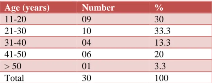
Table 2 shows age distribution of study population. Majority of the patients were in the age group of 21-30 years i.e. 10 (33.3%) followed by the age group of 11-20 years i.e. 9 (30%). There was only one case above the age of 50 years and six cases in the age group of 41-50 years. Thus, the younger age group was commonly affected as per the findings of the present study.
Table 2: Age distribution of study population.
Age (years) Number %
11-20 09 30
21-30 10 33.3
31-40 04 13.3
41-50 06 20
> 50 01 3.3
Table 3: Frequency of clinical symptoms of study population.
Clinical features Number %
Ear discharge 27 90
Ear ache 25 83.33
Hearing loss 20 66.67
Facial weakness 6 20
Giddiness, vomiting, others 5 16.66
Table 3 shows frequency of clinical symptoms of study population. Ear discharge was the most common presenting symptoms in 90% of the cases followed by pain in the ear in 83.3% of the cases. Hearing loss frequency was seen in 20 (66.7%) of the cases while in 20% of the cases the facial nerve was affected and it
resulted in facial weakness. Five cases have shown constitutional symptoms.
Table 4 shows distribution of cholesteatoma with respect to side. Right side was found to be more commonly affected than the left side i.e. 19 (63.3%) cases were affected with right side and 11 (36.7%) of the cases were affected on left side.
Table 4: Distribution of cholesteatoma with respect to side.
Side Number %
Right 19 63.3
Left 11 36.7
Total 30 100
Table 5: Sensitivity and specificity of HRCT with surgical findings with reference to extent of cholesteatoma.
Extent of cholesteatoma Sensitivity Specificity Positive predictive value Negative predictive value
Epitympanum 100 100 100 100
Auditus 96.1 75 96.1 75
Mastoid antrum 96 100 100 93.3
Facial recess 92.8 68.7 72.2 91.6
Sinus tympani 80 70 57.1 87.5
Hypotympanum 50 100 100 92.8
Eustachian tube 66.6 100 100 96.4
Beyond middle ear cleft 80 100 100 96.1
Table 6: Sensitivity and specificity of HRCT with surgical findings with reference to status of ossicles.
Status of ossicles Sensitivity Specificity Positive predictive value Negative predictive value
Malleus 84 20 84 20
Incus 80 90 94 69
Stapes 66 100 100 75
Table 7: Sensitivity and specificity of HRCT with surgical findings with reference to integrity of facial canal.
Integrity of facial canal Sensitivity Specificity Positive predictive value Negative predictive value
Labyrinthine segment 66.67 92.59 50 50
Tympanic segment 66.6 88.8 80 80
Mastoid segment 25 100 100 89.6
Table 5 shows sensitivity and specificity of HRCT with surgical findings with reference to extent of cholesteatoma. Of the 30 cases studied, 27 (90%) patients had imaging findings of cholesteatoma whereas 3 (10%) patients had no findings suggestive of cholesteatoma. Surgery showed cholesteatoma in 26 (86.6%) patients and no cholesteatoma in 4 (13.3%) patients. Epitympanum was involved in 29 (96.6%) patients in HRCT and 30 (100%) patients at surgery. Auditus was involved in 26 (86.6%) patients at both HRCT and at surgery. Mastoid antrum was involved in 24 (80%) patients in HRCT and 25 (83.3%) patients at surgery.
Facial recess was involved in 18 (60%) patients in HRCT and 14 (46.6%) patients at surgery. Sinus tympani were involved in 14 (46.6%) patients in HRCT and 10 (33.3%) patients at surgery. Extension beyond middle ear cleft was seen in 4 (13.3%) patients in HRCT and 5 (16.6%) patients at surgery. Hypotympanum was involved in 2 (6.6%) patients in HRCT and 4 (13.3%) patients at surgery.
HRCT and surgery. Incus erosion was seen in 17 (56.6%) patients in HRCT and 20 (66.6%) patients at surgery. Stapes erosion was seen in 10 (33.3%) patients in HRCT and 15 (50%) patients at surgery.
Table 7 shows sensitivity and specificity of HRCT with surgical findings with reference to Integrity of facial canal. Tympanic segment of facial canal was the most commonly involved, showing erosion in 10 (33.3%) patients in HRCT and 12 (40%) patients at surgery. Labyrinthine segment of facial canal showed erosion in 4 (13.3%) patients in HRCT and 3 (10%) patients at
surgery. Mastoid segment of facial canal showed erosion in 1 (3.3%) patient in HRCT and 4 (13.3%) patients at surgery.
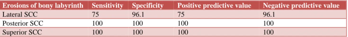
Table 8 shows sensitivity and specificity of HRCT with surgical findings with reference to erosions of bony labyrinth. Lateral SCC was the most commonly involved in bony labyrinth seen in 4 (13.3%) patients in both HRCT and surgery. Posterior SCC erosion was seen in 1(33.3%) patient in both HRCT and surgery. Superior SCC erosion was not seen in any of the patients in this study.
Table 8: Sensitivity and specificity of HRCT with surgical findings with reference to erosions of bony labyrinth.
Erosions of bony labyrinth Sensitivity Specificity Positive predictive value Negative predictive value
Lateral SCC 75 96.1 75 96.1
Posterior SCC 100 100 100 100
Superior SCC 100 100 100 100
Table 9: Sensitivity and specificity of HRCT with surgical findings with reference to Erosions of dural/sinus plate.
Erosions of dural/sinus plate Sensitivity Specificity Positive predictive value Negative predictive value
Dural plate 66.6 100 100 87.5
Sinus plate 100 96.2 75 100
Table 9 shows sensitivity and specificity of HRCT with surgical findings with reference to Erosions of dural/sinus plate. Erosion of dural plate was seen in 6 (20%) patients in HRCT whereas 9 (30%) patients showed dural plate erosion at surgery. Erosion of sinus plate was seen in 4 (13.3%) patients in HRCT and 3 (10%) patients at surgery
DISCUSSION
As per the findings of the present study, the atticoantral disease was found to be affecting the younger age groups below 50 years of age. Other studies also reported that attico-antral disease affected the age groups of 20-50 years of age.4-6
In the present study males were more compared to the females i.e. 53.3% vs. 35%. This finding was supported by other studies also.7 Ear discharge was the most
common clinical presentation. This was followed by otalgia.
The common clinical features were ear discharge, otalgia and hearing loss which is in agreement with other studies. Left ear was most commonly involved (63.3%) than the right (36.6%) in this study which is variable in literature.4,5
In this study, a moderate radio-surgical correlation was noted for differentiating cholesteatoma and granulation
tissue in the middle ear cavity using soft tissue mass and bony erosions as the radiologic criteria. At surgery, cholesteatoma was present in 26 out of 30 patients (86.6%) whereas it was reported in 27 of the 30 CT scans (90%) thereby giving a sensitivity of 86.6%, specificity of 92.3%, positive predictive value of 88.8% and negative predictive value of 33.3% for HRCT in detecting cholesteatoma preoperatively. Similar findings were also noted in the study conducted by other authors.6,7
Leighton et al,reported that cholesteatoma can be better diagnosed by taking temporal bone CT scan.8 Gaurano et
al, reported sensitivity of the CT scan taken before surgery was 97%.5 Chee NWC et al, noted that HRCT
scan is useful in the diagnosis of the cholesteatoma.7
They suggested using the radiographic criteria like location, mass of the tissue, and erosion of the bone and finding that any two out of these three can give the diagnostic accuracy of 94.4%. HRCT is very specific also due to its ability detect the mass of the soft tissue as well as erosion of the bone. But the authors also cautioned that HRCT may not be able to differentiate between the fluid and the mass of the soft tissue in few cases.
Firas Q et al, who found that HRCT had a 48% of the specificity and 80% of the sensitivity for making differentiation between chronic mucosal disease and the cholesteatoma.9 Jackler et al, found in their study that
bone was seen, the likelihood of cholesteatoma was about 30%.10
Gaurano and Joharjy showed that in cases of chronic suppurative otitis media, erosion was commonly present in the facial canal and scutum and then it can be seen in incus and tegmen.5 Berry S et al,in their study noted that
erosion of the bone was commonly seen on the ossicular chain and incus was also mostly involved. Sigmoid sinus plate was not that much affected.11
In this study, for the assessment of status of various middle and inner ear structures, radio surgical correlation was excellent for erosions of malleus and semicircular canals, moderate too good for erosions of incus, scutum, sigmoid plate, tegmen tympani, labyrinthine and tympanic segments of facial canal and poor for stapes and dehiscence of mastoid segment of facial canal.
It is necessary to take axial image as well as coronal image so that one can accurately detect the fistula in the labyrinthine. This is useful to look for the semicircular canal erosions. Lateral side of the semicircular canal is the most commonly affected. There is chance that half of the cases may be wrongly diagnosed if only coronal section image is taken.10
In this study all cases of lateral SCC erosion were accurately diagnosed. The discrepancy between our study and Gerami et al study for labyrinthine fistula may be because of the difference in the slice thickness employed. Cuts of 2 mm slice were employed in Gerami et al, study whereas our study was done using 0.65 mm slice thickness.12
Erosion of the tegmen type are classically seen when we take coronal image. But in this there is chance of misinterpretations. 10 The author reported three such
cases when on scanning it was found to be normal but on opening during operation, it was found to be breached. Sometimes it may be possible that on scanning it may look to be abnormal but actually it may be normal. 6 We
exactly found such situation in three cases in the present study. Discrepancy with Gerami et al, study may be because of the reason described above.12
Our study shows high sensitivity for HRCT in detecting erosions of epitympanum (100%), aditus (100%), mastoid antrum (96%), lateral SCC (100%), malleus (83.3%) whereas sensitivity is low in depicting erosions of incus (66.6%), stapes (50%). Similar results were also noted in Gerami et al, study with high sensitivity for ossicular erosions and low sensitivity for tegmen, semicircular canal and facial canal erosions.
HRCT showed 100% specificity for erosions of
epitympanum, mastoid antrum, hypo tympanum,
extension beyond middle ear cleft whereas it is relatively less specific for erosions of facial recess and sinus tympani.
CONCLUSION
Authors have found that HRCT scan was valuable in assessing the patients before surgery for cholesteatoma. It was useful in assessing the not only the extent to which the disease was present but also helped to know whether the middle ear structures were integral or not. However certain drawbacks of CT such as partial volume averaging does lead to false interpretation of the disease process. Partial volume artifacts can best be avoided by using a thin acquisition section width. In spite of this, HRCT is useful.
Funding: No funding sources Conflict of interest: None declared
Ethical approval: The study was approved by the Institutional Ethics Committee
REFERENCES
1. Valvassori GE. Laminagraphy of the ear: Normal Roentgenographic anatomy. Am J Roentgenol Radium Ther Nucl Med. 1963;89:1155-67.
2. Buckingham RA, Valvassori GE. Tomographic
anatomy of the temporal bone. Otolaryngol Clin North Am. 1973;6(2):337-62.
3. Pimontel-Appel B, Ettore GC. Poschl positioning and the radiology of Meniere’s disease. J Belge Radiol. 1980;63(2-3):359-67.
4. Barath K, Huber AM, Stampfli P, Varga Z, Kollias S. Neuroradiology of cholesteatomas. Am J Neuroradiol. 2010;10.3174:1-9.
5. Gaurano LJ, Joharjy AI. Middle ear cholesteatoma: characteristic findings in 64 patients. Ann Saudi Med (online). 2004;24(6):442-7.
6. Mafee FM, Levin CB, Applebaum LE, Campos M, James FC. Cholesteatoma of the middle ear and mastoid: A comparison of CT scan and operative
findings. Otolaryngol Clin North Am.
1998;21(2):265-92.
7. Chee NWC, Tan TY. The value of pre-operative high resolution CT scans in cholesteatoma surgery. Singapore Med J. 2001;42(4):155-9.
8. Leighton SE, Robson AK, Anslow P, Milford CA. The role of CT imaging in the management of chronic suppurative otitis media. Clin Otolaryngol Allied Sci. 1993;18:23-9.
9. Alzoubi FQ, Odat HA, Al-balas HA, Saeed SR. The role of preoperative CT scan in patients with chronic otitis media. Eur Arch Otorhinolaryngol. 2009;266:807-9.
10. Jackler RK, Dillon WP, Schindler RA. Computed tomography in suppurative ear disease: A correlation of surgical and radiographic findings. Laryngoscope. 1984;94:746-52.
12. Gerami H, Naghavi E, Moghadam MW, Forghanparast K, Akbar MH. Comparison of preoperative computerized tomography scans imaging of temporal bone with the intra-operative
findings in patients undergoing mastoidectomy. Saudi Med J. 2009;30(1):104-8.