R E S E A R C H
Open Access
Factors affecting treatment outcome in
patients with idiopathic nonspecific
interstitial pneumonia: a nationwide
cohort study
Sang Hoon Lee
1,2, Moo Suk Park
3, Song Yee Kim
3, Dong Soon Kim
4, Young Whan Kim
5, Man Pyo Chung
6,
Soo Taek Uh
7, Choon Sik Park
8, Sung Woo Park
8, Sung Hwan Jeong
9, Yong Bum Park
10, Hong Lyeol Lee
11,
Jong Wook Shin
12, Eun Joo Lee
13, Jin Hwa Lee
14, Yangin Jegal
15, Hyun Kyung Lee
16, Yong Hyun Kim
17,
Jin Woo Song
4and Jong Sun Park
1*Abstract
Background:The effects of corticosteroid-based therapy in patients with idiopathic nonspecific interstitial
pneumonia (iNSIP), and factors affecting treatment outcome, are not fully understood. We aimed to investigate the long-term treatment response and factors affecting the treatment outcome in iNSIP patients from a multi-center study in Korea.
Methods:The Korean interstitial lung disease (ILD) Study Group surveyed ILD patients from 2003 to 2007. Patients were divided into two groups to compare the treatment response: response group (forced vital capacity (FVC) improves≥10% after 1 year) and non-response group (FVC <10%). Factors affecting treatment response were evaluated by multivariate logistic regression analysis.
Results:A total of 261 patients with iNSIP were enrolled, and 95 patients were followed-up for more than 1 year. Corticosteroid treatment was performed in 86 patients. The treatment group showed a significant improvement in lung function after 1-year: FVC, 10.0%; forced expiratory volume (FEV1), 9.8%; diffusing capacity of the lung for
carbon monoxide (DLco), 8.4% (p< 0.001). Sero-negative anti-nuclear antibody (ANA) was significantly related with lung function improvement. Sero-positivity ANA was significantly lower in the response group (p= 0.013),
compared to that in the non-response group. A shorter duration of respiratory symptoms at diagnosis was significantly associated with a good response to treatment (p= 0.018).
Conclusion:Treatment with corticosteroids and/or immunosuppressants improved lung function in iNSIP patients, which was more pronounced in sero-negative ANA and shorter symptom duration patients. These findings suggest that early treatment should be considered in iNSIP patients, even in an early disease stage.
Keywords:Non-specific interstitial pneumonia, Treatment, Pulmonary lung function
* Correspondence:jspark.im@gmail.com
1Department of Internal Medicine, Division of Pulmonary and Critical Care
Medicine, Seoul National University Bundang Hospital, 82 Gumi-ro, 173 Beon-gil, Bundang-gu, Seongnam-si, Gyeonggi-do 463-707, Republic of Korea Full list of author information is available at the end of the article
Background
Non-specific interstitial pneumonia (NSIP) is a type of interstitial idiopathic interstitial pneumonia (IIP) mainly
affecting female non-smokers aged 40–60 years.
Al-though more rigorous studies are needed, the prevalence of idiopathic NSIP (iNSIP) is estimated to be between 1 and 9 in 100,000 [1, 2]. NSIP can present as idiopathic or is associated with secondary conditions, such as con-nective tissue disease (CTD), human immunodeficiency virus infection, IgG4-related disease, bone marrow trans-plant, or toxin/drug-related conditions [2–4]. In addition, NSIP with connective tissue disease has recently been re-classified as interstitial pneumonia with autoimmune disease [5].
Although the natural course of iNSIP is not yet known, previous studies showed that the prognosis of NSIP is favorable when compared with idiopathic
pulmonary fibrosis (IPF) [6–8]. Corticosteroid and
im-munosuppressive agents (including azathioprine, cyclo-phosphamide, cyclosporine, and mycophenolate mofetil) are widely used and thought to be beneficial for patients with NSIP [3, 9, 10]. However, changes to pulmonary function are not fully understood in both untreated and treated NSIP patients, especially in patients with low severity NSIP. Additionally, previous studies have addressed the risk factors and medical conditions associ-ated with mortality rate, relapse, and progression of the disease, but the degree of response and factors affecting treatment have not been well studied [10, 11].
The Korean Interstitial Lung Disease (ILD) Research Group performed a nationwide survey to investigate the characteristics of patients with ILD, including iNSIP. In the present multicenter, nationwide study, we aimed to investigate the effect of treatment and the factors affect-ing the treatment outcome in patients with surgically proven-iNSIP.
Methods Study population
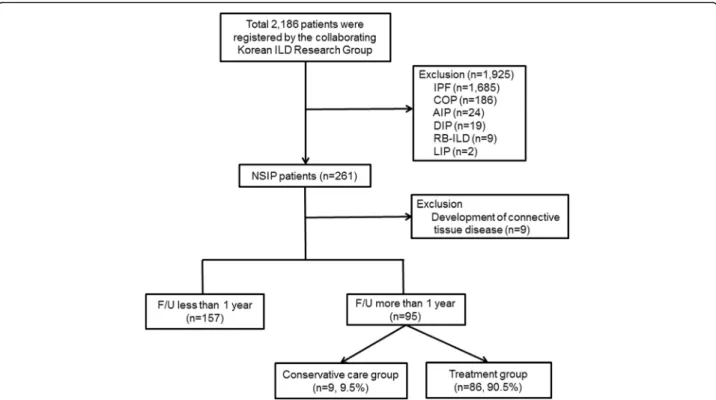
Figure 1 shows the patient-flow chart. In total, 2186 idiopathic interstitial pneumonias (IIP) patients were registered by the Korean ILD Research Group, which in-cludes pulmonologists from 54 University hospitals across the country with more than 500 beds starting 2006, from January 1, 2003, to December 31, 2007. Patients with a history of using medication that could provoke ILD (e.g., amiodarone or cytotoxic agent), and had a collagen-vascular disease were excluded from the study. In addition, patients with granulomatous diffuse parenchymal lung disease (e.g., sarcoidosis) or a rare
form of ILD (e.g., lymphangioleiomyomatosis or
pulmonary Langerhans cell histocytosis) were initially excluded from the study. NSIP was diagnosed based on the American Thoracic Society/European Respiratory
Society (ATS/ERS) 2002 guidelines via a multidisciplin-ary approach by a pulmonologist, a chest specific radi-ologist, and pathologists [12]. Patients diagnosed with IIP other than NSIP, including acute interstitial pneumo-nia, cryptogenic organizing pneumopneumo-nia, desquamative interstitial pneumonia, lymphocytic interstitial pneumo-nia, nonspecific interstitial pneumopneumo-nia, and respiratory bronchiolitis-associated interstitial lung disease, were ex-cluded. Patients diagnosed clinically without a surgical lung biopsy were excluded. Additionally, patients for whom definitive diagnoses could not be made at each hospital, were reviewed by the Scientific Committee of the Korean Academy of Tuberculosis and Respiratory Diseases. Among the 261 patients with NSIP, those who
developed a new CTD (n= 9) were excluded. Patients
with hypersensitivity pneumonitis (HP), as indicated by
the patient’s history, clinical symptoms, and serologic
test results, were also excluded from the study. Finally, 252 patients with iNSIP were analyzed in this study; 157 patients were followed-up within 1 year, and 95 patients were followed-up after >1 year. Clinical (age, gender, smoking status, smoking amount, respiratory symptom, comorbidity, and outcome), physiological (pulmonary function test [PFT]), and laboratory (arterial blood gas
analysis, C-reactive protein, anti-nuclear antibody
[ANA], and rheumatoid factor) findings were
retrospect-ively investigated. All patients’ data were recorded in a
web-based registry (www.ild.or.kr).
The 95 patients who were followed-up after >1 year were divided into two groups: a no treatment group
(n= 9) and a treatment group (n= 86). Patients who
were prescribed corticosteroid or immunosuppressive
agents were defined as “treatment group”. The mean
duration of treatment was 11.8 ± 8.3 months. The no treatment group patients were either only prescribed medication for symptom control or no medication at all.
To determine the effect of the treatment, the treat-ment group was further divided into two sub-groups. The response group was defined as patients with a
change of≥10% between the initial predicted forced vital
capacity (FVC) (%) and the 1-year follow-up predicted FVC (%). The non-response group was defined as patients with a change of <10% between the initial predicted FVC (%) and the 1-year follow-up predicted FVC (%).
Statistical analysis
Continuous variables were expressed as the mean ± standard deviation (SD) or median with interquartile
range and compared by Student’s t-test or a Mann
Whitney U-test according to the distribution of patients. Categorical variables were presented as frequency (n)
and percentage (%), and were analyzed by Fisher’s exact
rank test or Paired t-test were conducted to compare the effect of treatment (initial PFT results vs 1-year
follow-up results). A repeated measures ANOVA was
conducted to compare the change in lung function between the response group and non-response group at 1 year. A logistic regression model was used to investi-gate the factors affecting the treatment outcome. An adjustedpvalue <0.05 was considered to indicate
signifi-cance. SPSS™ Version 22.0 (SPSS, Chicago, Illinois,
USA), was used for all statistics analysis.
Results
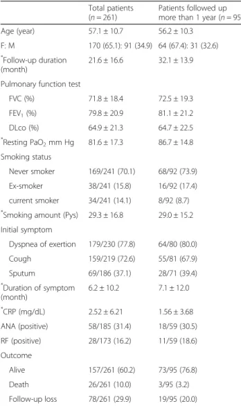
Among 252 patients with iNSIP, 95 patients who were followed-up for over 1 year were analyzed to evaluate treatment response in this study. Table 1 shows the characteristics of the patients with iNSIP. The mean age was 57.1 ± 10.7 years, and females (65.1%) were
predom-inant. The mean follow-up duration was 21.6 ±
16.6 months among all patients, and 32.1 ± 13.9 months in patients followed-up after >1 year. Overall lung func-tion was slightly decreased compared to normal; FVC
(%) was 71.8 ± 18.4, forced expiratory volume (FEV1) (%)
was 79.8 ± 20.9, and diffusing capacity of the lung for
carbon monoxide (DLCO) (%) was 64.9 ± 21.3. Most of
the patients were not smokers. Dyspnea (77.8%) and cough (72.6%) were the most common respiratory symptoms.
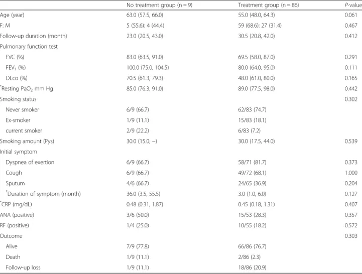
Clinical, physiologic, and laboratory data for the no treatment group and treatment group are shown in Table 2. More than 95% of patients were treated with steroid (Additional file 1: Table S1). In the treatment group, the median age was lower than that in the treat-ment group, although the difference was not significant
(p= 0.061). Gender, follow-up duration, PFT results,
smoking, initial respiratory symptoms, laboratory results, and comorbidities did not differ significantly between the groups (Table 2 and Additional file 1: Table S2). The change in lung function between the initial visit and the 1-year follow-up was investigated (Additional file 1: Table S3). In the no treatment group, although the FVC
(%), FEV1(%), and DLCO(%) were increased 1 year after
diagnosis, compared to the initial assessment, these
changes in PFT were not significant (p= 0.276,p= 0.400,
and p= 0.489, respectively). However, in the treatment
group, these values were all significantly improved after 1 year (p< 0.001, all).
Table 3 also shows the difference in lung function be-tween the initial PFT and 1-year follow-up PFT per the sero-positivity of ANA. The ANA results were available in 59 patients (62.1%). Forty-one patients (69.5%) with an initial negative ANA showed a significant improve-ment in lung function after 1 year; FVC (%) increased
by 11.1%, FEV1 (%) by 11.3%, and DLCO (%) by 12.1%
(p= 0.008, p= 0.005, and p < 0.001, respectively). How-ever, sero-positive patients did not show a significant improvement in lung function after 1-year.
We compared the baseline characteristics between the response group and non-response groups (Table 4). Age and composition proportion of gender were similar
between the groups (p= 0.895 and p= 0.705). In the
re-sponse group, the follow-up duration was significantly
longer than in the non-response group (p= 0.006), but
the duration of respiratory symptoms was shorter (3.4 ±
4.4 months vs 7.1 ± 9.3 months, respectively; p= 0.038).
With regard to pulmonary function, initial FVC (%),
FEV1 (%), and DLCO (%) were significant higher in
the non-response group than in the response group
(p< 0.001, p< 0.001, and p= 0.008, respectively).
Current smokers were only found in the
non-response group (p= 0.022). There were no significant
differences in the initial respiratory symptoms, labora-tory results, and comorbidities between the two groups (Table 4 and Additional file 1: Table S2). However, the proportion of ANA sero-positivity was higher in the non-response group than in the response
group (p= 0.013). Furthermore, in the non-response
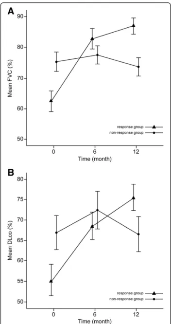
group, two patients (4.5%) died during the follow-up period. Figure 2 shows the change in pulmonary lung function over time (initial, 6 month, and 12 month) be-tween the two groups. The FVC improved by 24.6%, and
DLCO improved by 20.2% after 1 year in the response
group. However, in the non-response group, lung function after 1 year did not differ greatly from baseline. Therefore,
there were significant differences in FVC and DLCO
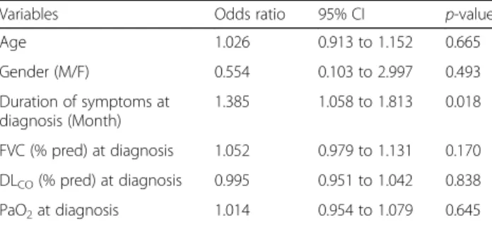
be-tween the two groups over time (p< 0.001, andp= 0.002). Multivariate analysis with logistic regression was conducted to investigate the risk factors for the non-response group (Table 5). Age, gender, duration of
respiratory symptoms, FVC (%), DLCO (%), and arterial
oxygen tension (PaO2) were examined. Although ANA
showed a significant difference between the response group and non-response group, only 15 patients showed positivity (Table 4). Thus, ANA was excluded from the multivariate analysis. The duration of symptoms at diag-nosis was significantly associated with the response to
treatment (hazard ratio (HR), 1.385; 95% CI, 1.058–
1.813; p= 0.018). In addition, to identify the factors
related to treatment failure, we defined patients with at
least a 5% reduction in lung function reduction as“
treat-ment failure” and performed logistic analysis. Thus, we
found that age was significantly related to treatment failure. Older patients showed a tendency to experience treatment failure (Additional file 1: Table S4).
Discussion
Although the prognosis of iNSIP is better than that of IPF, the 5-year mortality rate is estimated to be 17.7% [2]. To this date, there is no generally accepted guideline for the treatment of iNSIP; however, a previous study showed that corticosteroid and/or immunosuppressant therapy helped in maintaining or improving lung func-tion in 81% patients with iNSIP [9]. Our study showed that the serologic negativity of ANA was related with an
Table 1Characteristics of the study population
Total patients (n= 261)
Patients followed up more than 1 year (n= 95)
Age (year) 57.1 ± 10.7 56.2 ± 10.3
F: M 170 (65.1): 91 (34.9) 64 (67.4): 31 (32.6)
*Follow-up duration
(month)
21.6 ± 16.6 32.1 ± 13.9
Pulmonary function test
FVC (%) 71.8 ± 18.4 72.5 ± 19.3
FEV1(%) 79.8 ± 20.9 81.1 ± 21.2
DLco (%) 64.9 ± 21.3 64.7 ± 22.5
*
Resting PaO2mm Hg 81.6 ± 17.3 86.7 ± 14.8
Smoking status
Never smoker 169/241 (70.1) 68/92 (73.9)
Ex-smoker 38/241 (15.8) 16/92 (17.4)
current smoker 34/241 (14.1) 8/92 (8.7) *
Smoking amount (Pys) 29.3 ± 16.8 29.0 ± 15.2
Initial symptom
Dyspnea of exertion 179/230 (77.8) 64/80 (80.0)
Cough 159/219 (72.6) 55/81 (67.9)
Sputum 69/186 (37.1) 28/71 (39.4)
*
Duration of symptom (month)
6.2 ± 10.2 7.1 ± 12.0
*CRP (mg/dL) 2.52 ± 6.21 1.56 ± 3.68
ANA (positive) 58/185 (31.4) 18/59 (30.5)
RF (positive) 28/173 (16.2) 11/59 (18.6)
Outcome
Alive 157/261 (60.2) 73/95 (76.8)
Death 26/261 (10.0) 3/95 (3.2)
Follow-up loss 78/261 (29.9) 19/95 (20.0)
Note: Values in parentheses are percentages
F:Mfemale:male,FVC, forced vital capacity,% predpercentage of the predicted value,FEV1forced expiratory volume,DLCOdiffusing capacity of the lung for carbon monoxide,PaO2arterial oxygen tension,CRPC-reactive protein, ANAantinuclear antibody,RFrheumatoid factor
*
Follow-up duration, smoking amount, duration of symptoms, and CRP showed a non-normal distribution in all patients
*
Resting PaO2mm Hg, duration of symptom, and CRP showed a non-normal
improvement in pulmonary function, and patients who had a relatively shorter duration of initial respiratory symptoms responded to corticosteroids better than iNSIP patients with a longer duration of initial respira-tory symptoms.
Since Bjoraker et al. [13] reported the importance of the differentiation of NSIP from IPF, there has been
much progression in the diagnosis of NSIP as a formally approved disease entity, but there are still no clear guidelines for diagnosis and treatment [2, 5, 12, 14]. Furthermore, some medical conditions (CTD-related ILD, hypersensitivity pneumonitis, cryptogenic organiz-ing pneumonia, infection, or drug-induced lung disease) are related with NSIP, and therefore, the histologic
Table 2Characteristics according to treatment in patients followed-up for more than one year
No treatment group (n = 9) Treatment group (n = 86) P-value
Age (year) 63.0 (57.5, 66.0) 55.0 (48.0, 64.3) 0.061
F: M 5 (55.6): 4 (44.4) 59 (68.6): 27 (31.4) 0.467
Follow-up duration (month) 23.0 (20.5, 43.0) 30.5 (20.8, 42.0) 0.412
Pulmonary function test
FVC (%) 83.0 (63.5, 91.0) 69.5 (58.0, 87.0) 0.291
FEV1(%) 100.0 (75.0, 104.5) 80.0 (64.0, 95.0) 0.111
DLco (%) 70.5 (61.3, 79.3) 48.0 (61.0, 80.0) 0.165
*
Resting PaO2mm Hg 85.0 (76.3, 91.0) 89.0 (77.5, 98.0) 0.442
Smoking status 0.302
Never smoker 6/9 (66.7) 62/83 (74.7)
Ex-smoker 1/9 (11.1) 15/83 (18.1)
current smoker 2/9 (22.2) 6/83 (7.2)
Smoking amount (Pys) 30.0 (15.0,−) 30.0 (17.5, 44.0) 0.539
Initial symptom
Dyspnea of exertion 6/9 (66.7) 58/71 (81.7) 0.373
Cough 6/9 (66.7) 49/72 (68.1) 1.000
Sputum 4/6 (66.7) 24/65 (36.9) 0.204
*
Duration of symptom (month) 36.0 (3.5, 55.5) 3.0 (1.0, 6.0) 0.127
*
CRP (mg/dL) 0.48 (0.31, 1.87) 0.45 (0.18, 1.31) 0.407
ANA (positive) 3/6 (50.0) 15/53 (28.3) 0.357
RF (positive) 1/4 (25.0) 10/55 (18.2) 0.572
Outcome 0.303
Alive 7/9 (77.8) 66/86 (76.7)
Death 1/9 (11.1) 2/86 (2.3)
Follow-up loss 1/9 (11.1) 18/86 (20.9)
Note: Values in parentheses are percentages
F:Mfemale:male,FVCforced vital capacity,% predpercentage of the predicted value,FEV1forced expiratory volume,DLCOdiffusing capacity of the lung for carbon monoxide,PaO2arterial oxygen tension,CRPC-reactive protein,ANAantinuclear antibody,RFrheumatoid factor
Note: data are expressed as the median with interquartile range or number with proportion (%) *
CRP showed non-normal distribution in the no treatment group *
Resting PaO2mm Hg, duration of symptoms, and CRP showed a non-normal distribution in the treatment group
Table 3Comparison between initial and 1-year follow-up lung function according to antinuclear antibody (ANA) positivity
ANA negative (n= 41) ANA positive (n= 18)
Initial Follow-up p-value Initial Follow-up p-value
FVC (%) 72.1 ± 20.6 83.2 ± 15.5 0.008 68.6 ± 19.5 76.4 ± 18.1 0.102
FEV1(%) 80.1 ± 23.7 91.4 ± 19.7 0.005 75.4 ± 19.0 83.7 ± 19.1 0.091
DLco (%) 68.7 ± 24.9 80.8 ± 26.0 <0.001 62.9 ± 21.1 64.9 ± 23.4 0.568
characteristics of NSIP can be found in these diseases
[15–18]. Due to the complexity of the diagnosis and low
prevalence of NSIP, the factors predicting the response to treatment or the therapeutic effect are not well known [1, 6].
In the treatment group, lung function significantly im-proved after 1 year compared to the initial assessment;
FVC (%) increased by 10.0%, FEV1 by 9.8%, and DLCO
by 8.4%. Park et al. [9] showed that the change in FVC (%) occurred according to histopathological type and the survival outcome; treatment response was better in cellular-type, and there was a 25% increase in FVC (%)
in the cellular-type/survivor group after 1 year. However, there was no improvement in FVC (%) in the fibrotic-type/non-survivor group. Additionally, in their study, the initial mean FVC (%) was 63.6 ± 14.6, which was lower than what we observed. Xu et al. [8] also investigated the change in PFT results, but there was no significant improvement between the initial and follow-up lung function, possibly due to the non-fixed follow-up dur-ation in their study. From our results, in the early stages of iNSIP, a clinician could anticipate that treatment
Table 4Comparison of clinical characteristics between the response group and non-response group
Response group (n= 42)
Non-response group (n= 44)
p-value
*
Age (year) 55.5 ± 11.3 55.8 ± 9.7 0.895
F: M 28 (66.7): 14
(33.3)
31 (70.5): 13 (29.5)
0.705
Follow-up duration (month) 36.6 ± 14.1 28.6 ± 12.2 0.006
Pulmonary function test
FVC (%) 63.6 ± 17.6 79.9 ± 18.4 <0.001
FEV1(%) 71.7 ± 20.8 88.6 ± 19.9 <0.001
DLco (%) 56.9 ± 19.0 70.5 ± 25.0 0.008
Resting PaO2mm Hg 81.4 ± 17.0 92.5 ± 11.0 0.004
Smoking status 0.022
Never smoker 30/40 (75.0) 32/43 (74.4)
Ex-smoker 10/40 (25.0) 5/43 (11.6)
current smoker . 6/43 (14.0)
Smoking amount (Pys) 26.5 ± 17.2 32.4 ± 15.1 0.415
Initial symptom
Dyspnea of exertion 28/35 (80.0) 30/36 (83.3) 0.717
Cough 26/36 (72.2) 23/36 (63.9) 0.448
Sputum 13/33 (39.4) 11/32 (34.4) 0.675
*Duration of Symptom
(month)
3.4 ± 4.4 7.1 ± 9.3 0.038
*
CRP (mg/dL) 1.97 ± 5.01 1.19 ± 2.19 0.416
ANA (positive) 3/25 (12.0) 12/28 (42.9) 0.013
RF (positive) 2/25 (8.0) 8/30 (26.7) 0.092
Outcome 0.105
Alive 30/42 (71.4) 36/44 (81.8)
Death . 2/44 (4.5)
Follow-up loss 12/42 (28.6) 6/44 (13.6)
Note: Values in parentheses are percentages
F:Mfemale:male,FVCforced vital capacity,% predpercentage of the predicted value,FEV1forced expiratory volume,DLCOdiffusing capacity of the lung for carbon monoxide,PaO2arterial oxygen tension,CRPC-reactive protein, ANAantinuclear antibody,RFrheumatoid factor
*
Duration of symptoms, and CRP showed a non-normal distribution in the response group
*
Age, duration of symptoms, and CRP showed a non-normal distribution in the non-response group
Fig. 2Changes in lung function over time between the response group and non-response group.aChange in functional vital capacity (FVC) (%) over time between the two groups (p< 0.001, Mean ± standard error (SE),bChange in the diffusing capacity of the lung for carbon monoxide (DLCO) (%) over time between the two groups
would result in a 10% improvement in FVC (%) after 1 year. This information could help physicians predict the clinical course of patients with iNSIP, and plan adequate treatment modality.
Lee et al. [10] reported similar results to our study, showing that the presence of ANA was significantly re-lated with disease progression and a poor response to corticosteroids. They suggested that sero-positivity of ANA could be an early manifestation of systemic dis-eases associated with a poor outcome of NSIP. Xu et al. [19] also showed that systemic autoimmune disease was significantly associated with increased mortality in NSIP patients (p= 0.023). Felicio et al. [20] reported a higher production of collagen and elastic fibers in NSIP with collagen vascular disease than in iNSIP; in a cohort of 41 NSIP patients, an increase in elastic fibers >1.5% was
a significant risk factor for poor outcome (p= 0.01). In
this study, the response group showed a lower pro-portion of ANA positivity than the non-response
group (p= 0.013). Furthermore, negative ANA was
associated with a significant improvement in lung function after 1 year.
Sawata et al. [21] studied the influence of smoking in 31 NSIP patients over 2 years. They showed that the smoking group had a significantly worse outcome than the non-smoking group; non-smoking was significantly related with a
lower %DLCO/alveolar ventilation (DLCO/VA) in both
iNSIP (p= 0.009) and CTD-NSIP (p= 0.044), and
progres-sion free survival was worse in the smoker group (p=
0.0489). Furthermore, they showed that exacerbation was common in a heavy smoker. Similarly, in our study, the non-response group had a relatively higher total smoking patient number than the response group, and current smokers were only observed in the non-response group
(Table 4,p= 0.022). Marten et al. [22] suggested that
em-physema is higher in smokers with NSIP; therefore, cigarette smoking could be a pathogenic factor in a subset of NSIP patients. Thus, we presumed that smoking is re-lated with NSIP pathogenesis, and could provoke a poor response to corticosteroids, causing a worse outcome.
To assess the severity in the study population, we cal-culated the ILD-GAP score, which is a clinical prognosis prediction model using age, gender, and two lung
func-tion parameters (FVC (%), DLCO(%)) [23, 24]. Although
the initial PFT results were lower in the response group, the majority of patients in this study had ILD-GAP stage I (96.6%, data not shown). Moreover, a relatively longer duration of respiratory symptoms was a risk factor for poor response to corticosteroids (Table 5). This could mean that early treatment with corticosteroids and/or immunosuppressants might be more beneficial in the early stage of iNSIP, especially in ILD-GAP stage I patients. The physician should consider treatment of idiopathic NSIP in patients with respiratory symptoms, even if the severity of iNSIP is low.
There are some limitations to this study. First, it had a
patient selection bias. This study was performed
retrospectively and patients with ILD were enrolled in each hospital without a specific visit protocol. Therefore, 1-year
follow-up PFT results exist in only 95 patients.
Additionally, there have been major advances in the conceptualization of NSIP in recent years. In particular, it is currently speculated that iNSIP could be a type of auto-immune disease that is limited to the lungs or the respira-tory manifestation of undifferentiated CTD [5, 18, 25]. Initially, we only enrolled patients without autoimmune disease and nine patients who developed CTD were excluded from this study. Nevertheless, there could be
differences between this study population’s patients and
currently diagnosed NSIP patients. Second, the NSIP sub-type (cellular sub-type, fibrotic sub-type, or mixed) was not exam-ined in this study. Previous studies showed that fibrotic NSIP was related with a poor prognosis and more frequent hospitalization [9, 11, 26]. If the subtype was in-vestigated, it would be more informative. Third, the initial dose of corticosteroids or immunosuppressive agents was not examined. Lee et al. [10] reported that a low cortico-steroid dose was significantly related with relapse, which could mean that the dose of corticosteroids administered could affect the response to treatment.
Conclusion
Our study showed that corticosteroid and/or immunosup-pressant therapy was effective in iNSIP, resulting in an im-provement in lung function after 1 year. Corticosteroid-based treatment was especially effective in iNSIP patients who showed sero-negativity for ANA and those who had a shorter duration of respiratory symptoms. These findings suggest that early treatment with corticoste-roids and/or immunosuppressants could be therapeut-ically beneficial in iNSIP patients, even if the disease is at an early stage. Further prospective, large, and well-designed studies are needed to confirm the factors af-fecting treatment effect.
Table 5Analysis of risk factors that affect treatment response by logistic regression
Variables Odds ratio 95% CI p-value
Age 1.026 0.913 to 1.152 0.665
Gender (M/F) 0.554 0.103 to 2.997 0.493
Duration of symptoms at diagnosis (Month)
1.385 1.058 to 1.813 0.018
FVC (% pred) at diagnosis 1.052 0.979 to 1.131 0.170
DLCO(% pred) at diagnosis 0.995 0.951 to 1.042 0.838
PaO2at diagnosis 1.014 0.954 to 1.079 0.645
Additional file.
Additional file 1: Table S1.Treatment modality in treatment group (n= 86).Table S2.Comorbidities of study population.Table S3. Comparison between initial and 1-year follow-up lung function according to treatment.Table S4.Analysis of risk factors that associated with treat-ment failure (by logistic regression). (DOCX 26 kb)
Abbreviations
% pred:percentage of the predicted value; ANA: anti-nuclear antibody; CI: confidence interval; CTD: connective tissue disease; DLCO: diffusing
capacity of the lung for carbon monoxide; FEV1: forced expiratory volume;
FVC: forced vital capacity; GAP: gender, age, and 2 lung physiology variables (FVC and DLCO); HR: hazard ratio; ILD: interstitial lung disease; IPF: idiopathic
pulmonary fibrosis; NSIP: nonspecific interstitial pneumonia; PaCO2: arterial
carbon dioxide tension; PaO2: arterial oxygen tension; PFT: pulmonary
function test; SD: standard deviation; SEM: standard error; TLC: total lung capacity
Acknowledgements
We are grateful to all the members of The Korean Interstitial Lung Disease Research Group, comprising 54 hospitals, who helped with data collection, and the Medical Research Collaborating Center at Seoul National University Bundang Hospital for statistical analyses.
Funding None.
Availability of data and materials
All data were available in the ILD web-based registry (www.ild.or.kr).
Authors’contributions
JSP and SHL conceived and designed the study. All authors contributed to participant recruitment, and data collection/acquisition. SYK and DSK analyzed the data and performed the statistical analysis. JSP and SHL wrote the first draft of the manuscript. All authors critically evaluated the data, reviewed the manuscript, and approved the final version.
Ethics approval and consent to participate
The Institutional Review Board (IRB) of Yonsei University Health Service, Severance Hospital, reviewed and approved the study protocol (Reference number for ethics approval: 4–2009-0372).
Consent for publication Not applicable.
Competing interests
The authors declare that they have no competing interests.
Publisher’s Note
Springer Nature remains neutral with regard to jurisdictional claims in published maps and institutional affiliations.
Author details
1Department of Internal Medicine, Division of Pulmonary and Critical Care
Medicine, Seoul National University Bundang Hospital, 82 Gumi-ro, 173 Beon-gil, Bundang-gu, Seongnam-si, Gyeonggi-do 463-707, Republic of Korea.2Yonsei University College of Medicine, 50-1 Yonsei-ro, Seodaemun-gu, Seoul 120-752, South Korea.3Department of Internal Medicine, Division of Pulmonology, Severance Hospital, Institute of Chest Diseases, Yonsei University College of Medicine, 50-1 Yonsei-ro, Seodaemun-gu, Seoul 120-752, South Korea.4Division of Pulmonary and Critical Care Medicine, University of Ulsan College of Medicine, Asan Medical Center, Seoul, South Korea.5Department of Internal Medicine and Lung Institute, Division of Pulmonary and Critical Care Medicine, Seoul National University College of Medicine, Seoul, South Korea.6Division of Pulmonary and Critical Care Medicine, Samsung Medical Center, Sungkyunkwan University School of Medicine, Seoul, South Korea.7Department of Internal Medicine, Division of Allergy and Respiratory Medicine, Soonchunhyang
University Seoul Hospital, Seoul, South Korea.8Department of Internal Medicine, Division of Allergy and Respiratory Medicine, Soonchunhyang University Bucheon Hospital, Bucheon, South Korea.9Department of Internal Medicine, Division of Pulmonology, Gachon University Gil Medical Center, Incheon, South Korea.10Department of Internal Medicine, Division of Pulmonary, Allergy & Critical Care Medicine, Kangdong Sacred Heart Hospital, Hallym University, Seoul, South Korea.11Department of Internal Medicine, Pulmonary Division, Inha University Hospital, Incheon, South Korea. 12Department of Internal medicine, Division of Pulmonary Medicine, Chung
Ang University College of Medicine, Seoul, South Korea.13Department of Internal Medicine, Division of Respiratory and Critical Care Medicine, Korea University Anam Hospital, Korea University College of Medicine, Seoul, South Korea.14Department of Internal Medicine, Ewha Medical Research Institute, Ewha Womans University School of Medicine, Seoul, South Korea. 15Department of Internal Medicine, Division of Pulmonary Medicine, Ulsan
University Hospital, University of Ulsan College of Medicine, Ulsan, South Korea.16Department of Internal Medicine, Division of Critical Care and Pulmonary Medicine, Inje University Busan Paik Hospital, Busan, South Korea. 17Department of Internal Medicine, Division of Allergy and Pulmonology,
Bucheon St. Mary’s Hospital, The Catholic University of Korea School of Medicine, Bucheon, South Korea.
Received: 29 June 2017 Accepted: 20 November 2017
References
1. Flaherty KR, Martinez FJ. Nonspecific interstitial pneumonia. Semin Respir Crit Care Med. 2006;27:652–8.
2. Travis WD, Hunninghake G, King TE Jr, Lynch DA, Colby TV, Galvin JR, Brown KK, Chung MP, Cordier JF, du Bois RM, et al. Idiopathic nonspecific interstitial pneumonia: report of an American Thoracic Society project. Am J Respir Crit Care Med. 2008;177:1338–47.
3. Belloli EA, Beckford R, Hadley R, Flaherty KR. Idiopathic non-specific interstitial pneumonia. Respirology. 2016;21:259–68.
4. Kligerman SJ, Groshong S, Brown KK, Lynch DA. Nonspecific interstitial pneumonia: radiologic, clinical, and pathologic considerations. Radiographics. 2009;29:73–87.
5. Fischer A, Antoniou KM, Brown KK, Cadranel J, Corte TJ, du Bois RM, Lee JS, Leslie KO, Lynch DA, Matteson EL, et al. An official European Respiratory Society/American Thoracic Society research statement: interstitial pneumonia with autoimmune features. Eur Respir J. 2015;46:976–87.
6. Flaherty KR, Thwaite EL, Kazerooni EA, Gross BH, Toews GB, Colby TV, Travis WD, Mumford JA, Murray S, Flint A, et al. Radiological versus histological diagnosis in UIP and NSIP: survival implications. Thorax. 2003;58:143–8.
7. Riha RL, Duhig EE, Clarke BE, Steele RH, Slaughter RE, Zimmerman PV. Survival of patients with biopsy-proven usual interstitial pneumonia and nonspecific interstitial pneumonia. Eur Respir J. 2002;19:1114–8.
8. Glaspole I, Goh NS. Differentiating between IPF and NSIP. Chron Respir Dis. 2010;7:187–95.
9. Park IN, Jegal Y, Kim DS, Do KH, Yoo B, Shim TS, Lim CM, Lee SD, Koh Y, Kim WS, et al. Clinical course and lung function change of idiopathic nonspecific interstitial pneumonia. Eur Respir J. 2009;33:68–76.
10. Lee JY, Jin SM, Lee BJ, Chung DH, Jang BG, Park HS, Lee SM, Yim JJ, Yang SC, Yoo CG, et al. Treatment response and long term follow-up results of nonspecific interstitial pneumonia. J Korean Med Sci. 2012;27:661–7. 11. Travis WD, Matsui K, Moss J, Ferrans VJ. Idiopathic nonspecific interstitial
pneumonia: prognostic significance of cellular and fibrosing patterns: survival comparison with usual interstitial pneumonia and desquamative interstitial pneumonia. Am J Surg Pathol. 2000;24:19–33.
12. American Thoracic Society. European Respiratory Society international multidisciplinary consensus classification of the idiopathic interstitial pneumonias. This joint statement of the American Thoracic Society (ATS), and the European Respiratory Society (ERS) was adopted by the ATS board of directors, June 2001 and by the ERS executive committee, June 2001. Am J Respir Crit Care Med. 2002;165:277–304.
14. Travis WD, Costabel U, Hansell DM, King TE, Jr., Lynch DA, Nicholson AG, Ryerson CJ, Ryu JH, Selman M, Wells AU, et al. An official American Thoracic Society/European Respiratory Society statement: update of the international multidisciplinary classification of the idiopathic interstitial pneumonias. Am J Respir Crit Care Med 2013, 188:733–748.
15. Bouros D, Wells AU, Nicholson AG, Colby TV, Polychronopoulos V, Pantelidis P, Haslam PL, Vassilakis DA, Black CM, du Bois RM. Histopathologic subsets of fibrosing alveolitis in patients with systemic sclerosis and their relationship to outcome. Am J Respir Crit Care Med. 2002;165:1581–6. 16. Douglas WW, Tazelaar HD, Hartman TE, Hartman RP, Decker PA, Schroeder
DR, Ryu JH. Polymyositis-dermatomyositis-associated interstitial lung disease. Am J Respir Crit Care Med. 2001;164:1182–5.
17. Kim S, Tannock I, Sridhar S, Seki J, Bordeleau L. Chemotherapy-induced infiltrative pneumonitis cases in breast cancer patients. J Oncol Pharm Pract. 2012;18:311–5.
18. Nunes H, Schubel K, Piver D, Magois E, Feuillet S, Uzunhan Y, Carton Z, Tazi A, Levy P, Brillet PY, et al. Nonspecific interstitial pneumonia: survival is influenced by the underlying cause. Eur Respir J. 2015;45:746–55. 19. Xu W, Xiao Y, Liu H, Qin M, Zheng W, Shi J. Nonspecific interstitial
pneumonia: clinical associations and outcomes. BMC Pulm Med. 2014;14:175.
20. Felicio CH, Parra ER, Capelozzi VL. Idiopathic and collagen vascular disease nonspecific interstitial pneumonia: clinical significance of remodeling process. Lung. 2007;185:39–46.
21. Sawata T, Bando M, Nakayama M, Mato N, Yamasawa H, Sugiyama Y. Influence of smoking in interstitial pneumonia presenting with a non-specific interstitial pneumonia pattern. Intern Med. 2016;55:2939–44. 22. Marten K, Milne D, Antoniou KM, Nicholson AG, Tennant RC, Hansel TT,
Wells AU, Hansell DM. Non-specific interstitial pneumonia in cigarette smokers: a CT study. Eur Radiol. 2009;19:1679–85.
23. Ryerson CJ, Vittinghoff E, Ley B, Lee JS, Mooney JJ, Jones KD, Elicker BM, Wolters PJ, Koth LL, King TE Jr, Collard HR. Predicting survival across chronic interstitial lung disease: the ILD-GAP model. Chest. 2014;145:723–8. 24. Ley B, Ryerson CJ, Vittinghoff E, Ryu JH, Tomassetti S, Lee JS, Poletti V,
Buccioli M, Elicker BM, Jones KD, et al. A multidimensional index and staging system for idiopathic pulmonary fibrosis. Ann Intern Med. 2012;156:684–U658.
25. Kinder BW, Collard HR, Koth L, Daikh DI, Wolters PJ, Elicker B, Jones KD, King TE Jr. Idiopathic nonspecific interstitial pneumonia: lung manifestation of undifferentiated connective tissue disease? Am J Respir Crit Care Med. 2007;176:691–7.
26. Wang P, Jones KD, Urisman A, Elicker BM, Urbania T, Johannson KA, Assayag D, Lee J, Wolters PJ, Collard HR, Koth LL. Pathological findings and prognosis in a large prospective cohort of chronic hypersensitivity pneumonitis. Chest. 2017;
• We accept pre-submission inquiries
• Our selector tool helps you to find the most relevant journal • We provide round the clock customer support
• Convenient online submission • Thorough peer review
• Inclusion in PubMed and all major indexing services • Maximum visibility for your research
Submit your manuscript at www.biomedcentral.com/submit