Original Research Article
Microbiology and antibiotic sensitivity pattern of peritonsillar abscess
Anwar Sadath Choolakkaparambu Aboobakker, Sandeep Sreedhar*, Amith Jacob,
Abdulla Anchukandan
INTRODUCTION
Peritonsillar abscess (quinsy) refers to collection of pus between the fibrous capsule of the pharyngeal tonsil and the superior constrictor muscles of the pharynx.1 It is one
of the most frequently encountered emergencies in the Head and Neck region.2 The incidence of these infections
has reduced drastically due to administration of modern antibiotics and enhancement of oral hygiene and dental care. The incidence of deep space infections of the head and neck is estimated at around 10 per 100, 000 individuals per year currently.3
Peritonsillar abscess is a life threatening infection that often arises as a complication acute tonsillitis. Though
peritonsillar abscess is commonly prevalent among young adults and it is rare in children.4 The change in
microbiological flora of the tonsil with increase in age has been attributed as a cause of increased incidence in younger age.5
Intensive therapy will be required in few patients because it may lead to serious complications, such as deep neck abscess and if untreated, may rupture spontaneously with a risk of aspiration or sometimes it may progress to parapharyngeal space and also along the neck vessels to the mediastinum causing descending necrotizing mediastinitis.6 Proper management is therefore of
foremost importance and surgical drainage as well as proper antimicrobial therapy is warranted.7
ABSTRACT
Background: Peritonsillar abscess is a life threatening infection which requires immediate attention and care. If not treated immediately it may lead to several complications. But the culture and sensitivity results can be obtained after a minimum of 72 hours and hence empirical antibiotic therapy is needed. The objective of the study was to study the microbiological profile of peritonsillar abscess and the antibiotic sensitivity pattern of peritonsillar abscess.
Methods: This cross sectional study was carried out on 47 quinsy patients who attended the ENT OPD of a tertiary care hospital. Abscess was drained and the pus was sent for culture and sensitivity.
Results: 65.95% of the study population were males. prevalence of peritonsillar abscess was high in the age group of 21-30 (42.55%). Only one patient had bilateral abscess. Group-A beta hemolytic streptococcus was the common organism isolated followed by Staphyloccus aureus. Fluroquinolones and cefotaxime had a better sensitivity to nearly all the organisms.
Conclusions: Staphylococcus and Streptococcus were the highly prevalent organisms. Immediate empirical antibiotic therapy must be advocated after draining the collection.
Keywords: Peritonsillar abscess, Culture and sensitivity, Antibiotic, Antimicrobial sensitivity, Antimicrobial resistance
Department of Otorhinolaryngology, MES Medical College, Malappuram, Kerala, India
Received: 27 April 2020 Accepted: 14 May 2020
*Correspondence: Dr. Sandeep Sreedhar,
E-mail: sreedhar.sandeep@gmail.com
Copyright: © the author(s), publisher and licensee Medip Academy. This is an open-access article distributed under the terms of the Creative Commons Attribution Non-Commercial License, which permits unrestricted non-commercial use, distribution, and reproduction in any medium, provided the original work is properly cited.
The drug of choice for antimicrobial therapy should ideally be based on the culture and sensitivity reports of the pus drained from the abscess. But peritonsillar abscess needs to be treated immediately and cannot await the sensitivity reports since it takes more than 72 hours for the culture sensitivity report. So, it is desirable to know the organisms most commonly isolated from cases of peritonsillar abscess in region, so that the antibiotic to which most of these are sensitive can be used as standard drug in the treating peritonsillar abscess.
Hence this study was undertaken with the objective to study the microbiological profile of peritonsillar abscess and the antibiotic sensitivity pattern of peritonsillar abscess.
METHODS
This cross-sectional study was conducted on 47 peritonsillar abscess patients attending the ENT OPD of a tertiary care medical college in Kerala. Prior ethical committee clearance was obtained before the start of the study. All the patients with peritonsillar abscess who attended the ENT OPD between November 2013 and March 2015 who were not critically ill were included in the study, if they were willing to participate in the study. Written informed consent was obtained from all the participants before the start of the study. The patients were briefed about the need and purpose of the study, confidentiality of information and participant’s rights before getting informed consent. A pretested semi-structured questionnaire was used. A detailed history along with socio demographic details was taken in patients presenting with peritonsillar abscess and thorough examination done.
Patients underwent incision and drainage or fine needle aspiration according to the clinical condition. The pus was then sent to the laboratory for direct smear examination by Gram stain, culture and sensitivity studies.
One to two ml of the pus was inoculated in brain heart infusion broth (BHI) to nullify the effect of drugs and antibodies in the sample. The rest of the pus was inoculated in blood agar, MacConkey’s agar and chocolate agar plates and was incubated at 370 C for 24
hours. The organisms isolated were identified on the basis of their colonial, morphological and cultural characteristics and biochemical reactions. Antibiotic sensitivity test for the isolates were performed in Muller Hinton agar by disc diffusion method of Kirby-Bauer. The zone of inhibition was measured and recorded as sensitive, intermediate sensitive and resistant as indicated in the Kirby-Bauer method.
Patients who were able to take foods orally were discharged on the same day with oral antibiotics and analgesics.
The data was entered in the proforma, entered and analysed in Microsoft office excel and the results were expressed in percentages.
RESULTS
65.95% of the study population were males and the remaining 34.05% were females. The prevalence of peritonsillar abscess was high in the age group of 21 to 30 (42.55%), followed by 41 to 50 years (23.4%). The mean age of the study population was 29.4±7.04. 53.19% of the study population had abscess only in the right tonsil, while another 44.68% in their left tonsil. Around 2% of the study population had both tonsils affected.
Figure 1: Distribution of study population according to age.
Figure 2: Distribution of study populations according to clinical findings.
Everyone who presented with peritonsillar abscess invariably had throat pain, 98% of them had fever and around 96% of the study participants had difficulty in swallowing. Trismus was found in 80.85% of the study population and around 66% had referred otalgia. Around 38.29% of study population reported history of acute tonsillitis previously.
4.26 8.51
42.55
17.02 23.4
4.26 0
5 10 15 20 25 30 35 40 45
1-10 yrs 11-20 yrs
21-30 yrs
31-40 yrs
41-50 yrs
51-60 yrs
100 95.74 97.87
80.85
65.95
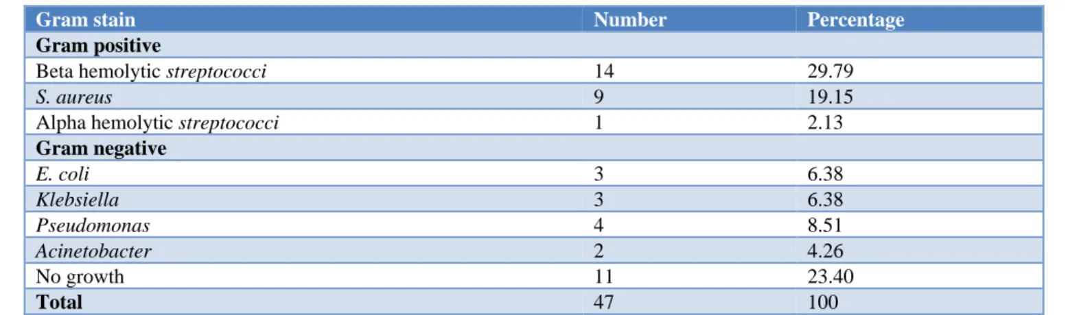
Table 1: Distribution of study population according to bacteria isolated.
Gram stain Number Percentage
Gram positive
Beta hemolytic streptococci 14 29.79
S. aureus 9 19.15
Alpha hemolytic streptococci 1 2.13
Gram negative
E. coli 3 6.38
Klebsiella 3 6.38
Pseudomonas 4 8.51
Acinetobacter 2 4.26
No growth 11 23.40
Total 47 100
Table 2: Antibiotic sensitivity pattern of bacteria isolated.
Antibiotics
Beta hemolytic
streptococci
(n=14)
S. aureus
(n=9)
Alpha hemolytic
streptococci
(n=1)
E. coli
(n=3)
Klebsiella
(n=3)
Pseudomonas
(n=4)
Acineto- bacter
(n=2)
Amikacin 64.28 100 100 100 33.33 25 0
Ampicillin 35.71 33.33 0 0 33.33 75 50
Amoxicillin 64.28 55.55 0 33.33 33.33 75 50
Gentamycin 35.71 33.33 0 0 66.66 25 0
Cotrimoxazole 28.57 44.44 100 0 66.66 50 100
Cefotaxime 58.71 88.88 100 66.66 100 100 100
Erythromycin 50 44.44 0 0 0 0 0
Ciprofloxacin 100 100 100 33.33 100 100 0
Ofloxacin 71.42 88.89 100 0 66.66 50 50
Levofloxacin 78.57 66.66 100 66.66 100 100 0
*Sensitivity to the antibiotic is expressed in percentages.
51% of the study population had gram positive infection, while 25.53% of the study population had gram negative infection. 23.4% of the pus samples had no growth. Around 30% of the total cultures had group-A beta hemolytic streptococcus. The next commonly isolated bacterium was Staphylococcus aureus (20%) followed by
Pseudomonas (8.5%), Klebsiella (6.38%), and E. coli
(6.38%). Acinetobacter was isolated in two cases (4.26%) and alpha hemolytic streptococci in one case (2.13%).
Anaerobes may play a causative role in the etiology of peritonsillar infections. This may be due to the fact that anaerobes form a part of the normal flora of the oral cavity. Also, the fact that the lesion is located in a closed space that does not communicate with the outside and is contiguous with the oral mucosa may also contribute to the anaerobic etiology in peritonsillar infections.
In our study, isolation of anaerobes could not be done due to non-availability of facilities like special anaerobic transport medium, anaerobic culture jars etc.
Group-A beta hemolytic streptococcus was found to be 100% sensitive to ciprofloxacin. Sensitivity to
S. aureus showed 100% sensitivity to amikacin and ciprofloxacin sensitivity to ofloxacin was 89%. The sensitivity of E. coli was 100% for amikacin. Resistance was seen for gentamicin and ampicillin. Klebsiella
showed 100% sensitivity to ciprofloxacin, levofloxacin and cefotxime. Sensitivity to gentamycin, cotrimoxazole and ofloxacin was 66%. Pseudomonas aeruginosa
showed 100% sensitivity to ciprofloxacin, levofloxacin and cefotaxime.
Alpha hemolytic streptococcus was sensitive to co-trimoxazole, ciprofloxacin, cefotaxime, levofloxacin, ofloxacin and amikacin. Acinetobacter was sensitive to co- trimoxazole but resistant to amikacin, gentamicin, erythromycin, levofloxacin and ciprofloxacin.
Fluroquinolones and cefotaxime had a better sensitivity to nearly all the organisms. Co trimoxazole, amoxycillin and ampicillin had a moderate sensitivity to all the organisms.
DISCUSSION
antibiotic sensitivity pattern. Male predominance was found in the study and the male to female ratio was nearly 2:1. Wolf et al in their study in Israel and Santhosh et al in their study in India observed a male to female ratio of 2:1.8,9 In a study done by Aparna et al in Kerala,
total males in the study were 65%.10 Singh et al in their
study in central India during 2013 found that there is a male predominance (78.57%, approximately 4:1) in the prevalence of quinsy.11 Har et al in his study reported
58% males.12 In a study done by Beeden et al, Rega et al
and Brito et al, there was a slight male predominance. In a study done by Acharya et al in Nepal the males were 29% (male: female ratio - 1:2) and by Thimmappa et al the females were 54%.1,3,13-15
The prevalence of peritonsillar abscess was high in young adults (21-30 years with a mean age of 29.4 years) and very less in children and geriatric population. Similar results were obtained in the studies done by Ophir et al, Sreekanth et al, Shilpa and Acharya et al.1,2,16,17
Bilateral quinsy is rare but reported by Brook et al in 1981.18 Mosby mentions that most peritonsillar abscesses
are unilateral; however, there is a 3% to 7% incidence of bilaterality.19 In our study 53.19% abscess were in the
right, 44.68% were in the left and 2% were bilateral. Similar results were observed in a study done by Ophir et al and Gupta et al.16,20
Brown mentions that peritonsillar abscess may occur as a complication of acute tonsillitis or it may apparently arise de novo with no preceding tonsillitis.4 Stringer et al in
their study found that 36% of patients had a prior history of tonsillitis, including 18% of patients with a prior history of peritonsillar abscess.21 Savolainen et al in a
study of 98 patients with peritonsillar abscess found that 21.4% of the patients had had three or more tonsillitis episodes in the past.22 In our study 38% had history of
previous tonsillar infection.
The culture positivity rate was 76.6% in our study. Similar results were obtained by studies done by Savolainen et al (86.7%), Wolf et al (75.58%), Guru et al (91.5%), Brito et al (84.5%) and Aparna et al.3,8,10,22,23
The wide use of antibiotics prior to admission negative growth in bacteriologic culture of nearly 25% of the samples.8
The most commonest organism found in the culture was group-A beta hemolytic Streptococcus and S. aureus. Similar results were obtained by studies done by Aparna et al, Flavio et al, Shilpa and Har et al.10,12,17,24
Beta hemolytic streptococci and S. aureus had a moderate to good sensitivity to most of the antibiotics. Klebsiella
and Pseudomonas had a moderate sensitivity. E. coli and
Acinetobacter had a poor sensitivity or resistance to most of the drugs. Similar results were obtained in other studies.1,9,11
CONCLUSION
Staphylococcus and Streptococcus were the most commonly associated organisms. If not adequately treated at the right time it can lead to serious complications. Immediate empirical antibiotic therapy is warranted after drainage without waiting for the culture and sensitivity report.
ACKNOWLEDGEMENTS
The authors acknowledge the guidance and support of Dr. E. Balakrishnan, Professor and HOD of Otorhino-laryngology, Government Medical College, Kannur, Kerala.
Funding: No funding sources Conflict of interest: None declared
Ethical approval: The study was approved by the Institutional Ethics Committee
REFERENCES
1. Acharya A, Gurung R, Khanal B, Ghimire A. Bacteriology and Antibiotic Susceptibility pattern of Peritonsillar Abscess. J Nepal Med Assoc. 2010;49(178):139-42.
2. Cherkuri S, Benninger MS. Use of Bacteriological studies in the outpatient management of peritonsillar abscess. Laryngoscope 2002;112:18-20.
3. Brito TP, Hazboun IM, Fernandes FL, Bento LR, Zappelini CEM, Chone CT, et al. Deep neck abscesses: study of 101 cases. Braz J Otorhinolaryngol. 2017;83(3):341-8.
4. Cowan DL, John H. Acute and chronic infection of the pharynx and tonsils. In : Hibbert John (ed) Scott- Brown’s Laryngology and Head and Neck surgery. Vol.5, 6th edition. Butterworth and Co ltd: Oxford; 1997: 543-547.
5. Akhtar MJ, Shinefield HR. Staphylococcus aureus
peritonsillar abscess in a 11-week-old infant. J Laryngol Otol. 1996;110(1):78-80.
6. Takenaka Y, Takeda K, Yoshii T, Hashimoto M. Gram Staining for the Treatment of Peritonsillar Abscess. Int J Otolaryngol. 2012;2012:464973. 7. Klug TE, Henriksen JJ, Fuursted K, Ovesen T.
Significant pathogens in peritonsillar abscesses. Eur J Clin Microbiol Infect Dis. 2011;30:619-27. 8. Wolf M, Even CI, Kronenberg J. Peritonsillar
abscess: Repeated Needle Aspiration Versus Incision and Drainage. Annals Otol Rhinol Laryngol. 1994;103(7):554-7.
9. Santosh AN, Viresh AN, Sharmada BK. Microbiology and antibiotic sensitivity of odontogenic space infection. Int J Med Dent Sci. 2014;3(1):303-13.
Tertiary Care Hospital in Kerala. J Med Sci Clin Res. 2018;6(9):429-35.
11. Walia IS, Borle RM, Mehendiratta D, Yadav AO. Microbiology and Antibiotic Sensitivity of Head and Neck Space Infections of Odontogenic Origin. J Maxillofac Oral Surg. 2014;13(1):16-21.
12. Gady HE, Aroesty JH, Shaha A, Lucente FE, Brooklyn, N.Y. Changing trends in deep neck abscess A retrospective study of 110 patients. Oral Surg Oral Med Oral Pathol. 1994:77:446-50. 13. Beeden AG, Evans JNG. Qunisy Tonsillectomy-A
further report. J Laryngol Otol. 1970;84:443-8. 14. Rega AJ, Aziz SR, Ziccardi VB. Microbiology and
Antibiotic Sensitivites Of Head and Neck Space Infections of Odontogenic Origin. J Oral Maxillofac Surg. 2006;64:1377-80.
15. Thimmappa TD, Ramesh S, Nagraj M, Gangadhara KS.. A study of deep space infections of neck. Int J Otorhinolaryngol Head Neck Surg. 2017;3(1):116-21.
16. Ophir D, Bawnik J, Poria Y, Porat M, Marshak G. Peritonsillar abscess-A Prospective evaluation of outpatient management by Needle aspiration. Arch Otolaryngol Head Neck Surg. 1998;114:661-3. 17. Shilpa C. Microbiology of Peritonsillar Abscess: A
Prospective Study. Int J Sci Stud. 2016;4(4):17-9. 18. Laxmipathi G, Mahendra N, Kumar V. Uncommon
complication of Quinsy. Indian J Otoryngyol Head Neck Surg. 2003;55(4):289-91.
19. Otto AR, Noorily DA, Otto MP. Deep Neck Infections. In: Shockley WW, Pillsbury CH (eds)
The Neck Diagnosis and Surgery. Mosby - Year Book: Missouri; 1994: 163-165.
20. Gupta G, McDowell RH. Peritonsillar Abscess. In: StatPearls. Treasure Island (FL): Available at: https://www.ncbi.nlm.nih.gov/books/NBK519520/. Accessed on 16 December 2019.
21. Loftus S, Ahuja K, Van-Hasselt Y. Diagnosis of peritonsillar infections: a prospective study of ultrasound, computerized tomography and clinical diagnosis. J Laryngol-Otol. 1999;113(3):229-32. 22. Savolainen S, Jousimies-Somer HR, Makite AA.
Peritonsillar abscess- clinical and Microbiological Aspects and Treatment Regimens. Arch Otolaryngol Head Neck Surg. 1993;119(5):521-4.
23. Guru K, Moghe S, Pillai A, Gupta MK, Pathak A. Incidence of Anaerobic bacteria in 118 patients with Deep space Head and Neck infections from the People’s University Hospital of Maxillofacial Surgery, Bhopal, India. J Orofacial Res. 2012;2:121-6.
24. Sakae FA, Imamura R, Sennes LU, Filho BCA, Tsuji DH. Microbiology of Peritonsillar Abscesses. Rev Bras Otorrinolaringol. 2006;72(2):247-51.