Dr Pradeep Vel. M et al JMSCR Volume 06 Issue 10 October 2018 Page 864
Role of VR 3D Imaging in temporal bone pathologies
Authors
Dr Pradeep Vel. M
1*, Dr Adaikappan.M
2, Dr Ruta Shanmugam
3*1
Resident, Department of Radio Diagnosis, Rajah Muthiah Medical College & Hospital, Annamalainagar, Chidambaram
Email: [email protected]
2
Professor & HOD, Department of Radio Diagnosis, Rajah Muthiah Medical College & Hospital, Annamalainagar, Chidambaram
3
Professor & HOD, Department of Otorhinolaryngology, Rajah Muthiah Medical College & Hospital, Annamalainagar, Chidambaram, India
Abstract
Background: Temporal bone contains the most complex anatomic intricacies and so, becomes arduous for abnormality interpretation. In the era of MDCT with post processing tools, a question how much these tools are valuable in detecting the complex temporal bone pathologies interested us to conduct this study.
Method: Prospective correlation study, by Department of Radio diagnosis & Otorhinolaryngology from January 2017 to June 2018 in RMMCH, Chidambaram. 73 Cases with symptoms pertaining to temporal bone underwent HRCT in a 4 slice MDCT. Post-processed VR 3D images correlated with axial and coronal image findings.
Results: Common age group of the study was 21- 30 years with more or less similar gender distribution. Most common indication was ear discharge followed by ear bleeding. Otitis media, cholesteatoma & fracture had highest percentage of HRCT diagnosis. VR 3D images demonstrated pathology in only 21.9% which were fractures, bone erosions of glomus jugulare and other bone tumours.
Conclusion: VR 3D images of 4 Slice MDCT could demonstrate confidently gross pathologies involving temporal bone margins.
Keywords: VR-Volume rendered, 3D-Three dimensional, MDCT- Multi Detector Computed Tomography, RMMCH-Rajah Muthiah Medical College & Hospital, HRCT-High Resolution Computed Tomography.
Introduction
MDCT is in use a for prolonged period but more recently Cone beam CT is being used for temporal bone imaging along with MRI.[1] Software advancements has helped in quick image reconstruction from axial images and better depiction of structures. 3D images can be generated by either volume rendering or surface rendering techniques. Radiologists utilize
reformation techniques less commonly for interpretation.[2]
Radiologists interpretation depends mostly through axial imaging whereas Otorhinolaryn-gologist get their image orientation based on coronal sections which more or less provides a surgical type of view. 3D images could provide a overview of the pathology and orientation when looking at 2D images for the surgeons.[9]
www.jmscr.igmpublication.org Impact Factor (SJIF): 6.379
Index Copernicus Value: 79.54 ISSN (e)-2347-176x ISSN (p) 2455-0450
Dr Pradeep Vel. M et al JMSCR Volume 06 Issue 10 October 2018 Page 865 3D images could help in better understanding of
anatomy, surgical planning and educational purposes[3]. It helps surgeon for planning excision of temporal bone tumours, aural deformities & implantation of cochlear devices[4].
Aim
To correlate temporal bone HRCT interpretation with those of post processed VR 3D images.
Materials & Methods
Institutional ethical committee approval was taken for the conduct of this prospective correlation study. After excluding patients with skull base electrical devices, contrast allergy, renal failure and pregnancy, 73 cases with symptoms pertaining to the temporal bone referred for HRCT from department of Otorhinolaryngology between January 2017 & June 2018 were included in the study.
All cases were subjected to axial and coronal HRCT in a 4 slice MDCT Toshiba Alexion. Axial scans performed in supine position with slight flexion of neck so that the scan plane doesn't need gantry tilt and avoided radiation to lens. Scan plane was along the OMBL from the base of EAC cranially extended to the arcuate eminence of temporal bone visualised on lateral tomogram. Coronal images acquired in prone position perpendicular to the axial plane from cochlea to the posterior SCC.
Scan was performed using FOV of 15 to 20 cms, 1mm thin collimation, 1mm slice thickness, 512 x 512 large reconstruction matrix,120kVp, 100 mA & high spatial frequency(bony) algorithm. Images were reconstructed to form slice interval of 0.5 mm.
VR 3D images were formed using Osrix software in workstation from acquired HRCT data sets. VR 3D images were analysed in all planes and angles to identify the pathology interpreted through axial and coronal images. Parameter alteration for VR done using provided options viz., basic, bone & soft tissue. The best possible option which could demonstrate the pathology
were used to correlate with HRCT images. Based on correlation, pathologies that VR 3D demonstrate were identified.
Results
Data obtained were utilised in SPSS software for statistical analysis. Most results expressed in percentages.
Common age group of the study was 21-30 years with more or less similar gender distribution. [Fig.1 & 2]
Most common indication was ear discharge (52.1%) followed by ear bleeding (20.5%). [Fig 3] Otitis media, cholesteatoma & fracture had highest percentage (each 17.8%) of HRCT diagnosis. [Fig.4]
VR 3D images demonstrated pathology in only 21.9% which were fractures, glomus jugulare, and other bone tumours. [Fig.4 & 5]
VR 3D had statistically significant correlation (p value-0.0018) with HRCT findings in traumatic fractures, tumours, erosions of temporal bone and adjacent bones.
Fig 1 Showing sex distribution
MALE 51% FEMALE
49%
Dr Pradeep Vel. M et al JMSCR Volume 06 Issue 10 October 2018 Page 866
Fig.2 Showing distribution of study patients
Fig.3 Showing distribution of clinical indication
Fig.4 Showing HRCT diagnosis & VR3D demonstrated cases distribution
0 5 10 15 20 25 30
< 10 yrs 10 - 20 yrs 20 - 30 yrs 31 - 40 yrs 41 - 50 yrs > 50 yrs 2.6
21.6
27.3
19.2
13.2
15.1
%
AGE (in years)
0 10 20 30 40 50 60
20.5 52.1
20.5 19.2
1.4 2.7 1.4 1.4 1.4 1.4 1.4 %
CLINICAL INDICATION
0 5 10 15 20
%
Diagnosis
HRCT
Dr Pradeep Vel. M et al JMSCR Volume 06 Issue 10 October 2018 Page 867
Fig.5 Showing percentage of VR 3D demonstrated cases
Fig.6 VR3D of zygomatic process fracture
Fig.7 VR 3D of anterior wall of external auditory canal(EAC).
Fig.8 VR 3D of vertical mastoid fracture
Fig.9 VR 3D of anterior wall of EAC, zygomatic arch & squamous part fracture
0 20 40 60 80
No Demonstration Demonstration
78.1
21.9
%
Dr Pradeep Vel. M et al JMSCR Volume 06 Issue 10 October 2018 Page 868
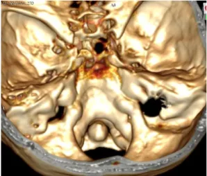
Fig. 10 VR3D of erosion of right petrous bone by glomus jugulare as viewed from vertex.
Fig.11 VR3D of right condyle osteochondroma seen from posterior aspect.
Discussion
This study of 73 cases had most patients of the age group 21-30yrs without any major difference for gender predilection for temporal bone involve-ment. Temporal bone pathologies evaluated by Vivek. R et al[6] by studying 50 patients had similar age group 20-40 years as the most affected with slight male preponderance for temporal pathologies.
Ear discharge (52.1%) followed by traumatic ear bleeding (20.5%) was the common clinical indication for HRCT in the present study. Bagul.M et al[7] studied 120 cases had deafness (65%) as the most common clinical presentation followed by ear discharge (58%). The variation could be due to more appropriate imaging protocol followed for hearing loss patients in our institution.
Frequent pathology in this study was inflammatory (36.9%), followed by traumatic fracture (17.8%). This in accordance with pathology distribution as reported by Bagul.M et al[7].
Among the 13 fracture cases, 1 case could not be demonstrated with VR 3D. The reason was a small fracture line of medial bony EAC near the tympanic membrane in axial image which VR 3D failed to demonstrate.
In this Middle, inner ear abnormalities and external ear soft tissue pathologies were not clearly demonstrated in VR 3D as the tools like autosegmentation, which help in removal of seperate bone parts were not available for the study. Moreover most of the studies showing excellent depiction of middle and inner ear structures used higher slice MDCT machines which could provide sub millimetre collimation, less than 0.5mm slice interval and still more increased spatial resolution. Further these studies had used different software applications, developed specifically for 3D imaging.[8,9] One suspected case of eagle's syndrome was studied but styloid elongation did not meet the criteria for the diagnosis and so was VR 3D images. Styloid being a part of the temporal bone and pathology of it could be clearly depicted using VR 3D.[10]
Fractures, tumours and erosions of temporal bone margins demonstrated by VR 3D in this study is in accordance with the study of Howard JD.et al [11]
describing VR 3D perspectives of demonstrating complex destructive lesions.
Conclusion
VR 3D images of 4 Slice MDCT could demonstrate confidently gross pathologies involving temporal bone margins.
References
Dr Pradeep Vel. M et al JMSCR Volume 06 Issue 10 October 2018 Page 869 2. Cody DD (2002) A APM/RSNA physics
tutorial for residents: topics in CT. Image processing in CT Radiographics. Sep-Oct;22(5):1255-68.
3. JunBC, Song SW, Cho JE, et al. Three-dimensional reconstruction based on images from spiral high-resolution computed tomography of the temporal bone: anatomy and clinical application. J Laryngol Otol2005; 119(9): 693–698 4. Schubert O, Sartor K, Forsting M, Reisser
C (1996 )Three-dimensional computed display of otosurgical operation sites by spiral CT. Neuroradiology 38:663–668 5. Klingebiel.R , Bauknecht. H.C , Rogalla.P.
et. al.(2001) High-resolution petrous bone imaging using multi-slice computerized tomography. Acta Otolaryngol. Jul; 121(5): 632–636.
6. R. Vivek, P. Gunasekaran, S. Sethurajan, M. Adaikappan. “Evaluation of HRCT Temporal Bone and Pathologies”.Journal of Evolution of Medical and Dental Sciences 2014; Vol. 3, Issue 52, October 13; Page: 12118-12126,DOI: 10.14260/jemds/2014/3601
7. Bagul M. High-resolution Computed Tomography Study of Temporal Bone Pathologies. Int J Sci Stud 2016;4(4):60-65.
8. Fatterpekar GM, Doshi AH, Dugar M, Delman BN, Naidich TP, Som PM. Role of 3DCT in the evaluation of the temporal bone. Radiographics. 2006 Oct; 26 Suppl1:S117-32. Review.
9. Rodt T, Ratiu P, Becker H, Bartling S, Kacher DF, Anderson M, Jolesz FA, Kikinis R. 3D visualisation of the middle ear and adjacent structures using reconstructed multi-slice CT datasets, correlating 3D images and virtual endoscopy to the 2D cross-sectional images. Neuroradiology. 2002;44(9):783– 790.
10.Kalmath.S et al .Role of 3D Multidetector Computed Tomography in diagnosis of Elongated styloid Process. DOI:
10.1594/ecr2016/C-0171