Dr Arun Kumar Pandey et al JMSCR Volume 07 Issue 11 November 2019 Page 562
Funcutional Outcome of Intra-Articular Joint Depressed Fractures of
Calcaneum Treated by Lateral Plating- A Case Series of 35 Patients
Followed For an Average Duration of 24 Months
Authors
Dr Arun Kumar Pandey
1, Dr Ranjeet Kumar Patel
2*, Prof. Rahmat Ali
3,
Dr Saurabh Agarwal
41
MS (Ortho), 2S.R. (Dept. of Orthopaedics)
Maharshi Vashishtha Autonomous State Medical College, Basti
3
Associate Professor and HOD (Dept. of Orthopaedic), Maharshi Vashishtha Autonomous State Medical College, Basti
4
Associate Professor, M.L.B. Medical College, Jhansi *Corresponding Author
Dr Ranjeet Kumar Patel
S.R. (Dept. of Orthopaedics), Maharshi Vashishtha Autonomous State Medical College, Basti
Abstract
Background: There is no consensus as to which is the best treatment for intraarticular fractures of the
calcaneus. Many people treat it conservatively whereas many go for operative procedures. Even for surgical treatment many do it by percutaneous technique many go for open reduction and internal fixation with plate or cancellous screws.
Purpose: To present a case series of 35 patients with intra-articular joint depressed fractures of
calcaneum treated by open reduction and internal fixation with plate through a lateral approach.
Material and Methods: A retrospective study was done in a level 1 trauma centre in which 35 patients (29
males and 6 females)of joint depressed type of calcaneum fractures were treated by open reduction and fixation by plate. All had acute trauma. Based on 2-dimensional CT scans, the fractures were categorized using Sanders classification. Radiographs and Maryland foot scores were used for evaluation of the results. Average follow-up was 24 months.
Results: 30 patients had full sub-talar range of movement and 5 patients had restricted range of
movement. 11 of 12 patients returned to manual labour jobs, the others were not employed at the time of injury. 26 patients had an excellent Maryland foot score and 9 patients had agood score .
Interpretation: Intra-articular calcaneum fractures are associated with high chances of sub-talar arthritis
with loss of sub-talar movement if not treated properly. Reduction of posterior facet is more important in joint depressed type of fracture. Lateral plating for these complicated fractures resulted in good to excellent sub-talar joint function with restoration of heel height, width and normal heel valgus.
Keyword: Intra-articular calcaneum fractures, trauma, plate, treatment outcome.
Introduction
Injury mechanisms and fracture patterns largely determine treatment results of calcaneal fractures.1 Controversy has existed over closed7,13,14,21 versus
open1-4,155,22 treatments. A number of treatment classifications have been proposed based on plain radiography.7,17,18,20
http://jmscr.igmpublication.org/home/ ISSN (e)-2347-176x ISSN (p) 2455-0450
Dr Arun Kumar Pandey et al JMSCR Volume 07 Issue 11 November 2019 Page 563 Improvement in imaging technology has allowed
a better understanding of fracture pathology and provided the basis for newer classifications.19,5 Intra-articular fractures of the calcaneum are amongst the most challenging fractures for orthopaedic surgeon because of complicated anatomy and difficulty in evaluating the fractures properly. Those who sustain them face a slow recovery, with possible permanent deformity and disability. When the fracture is joint depressed type of fracture, formal open reduction of fracture through a lateral approach, elevation of depressed posterior facet and fixation with a plate is required. This is a case series using a lateral approach for intra-articular fractures of calcaneum as previously described.
Patient and Methods
(a) Patient Selection: This is a retrospective
review of case series 30 intra-articular joint depressed fractures of calcaneum that came to our institute from April,2013 to August,2016. All the patients had joint depressed type of fracture according to Essex-Lopresti classification and had Sander’s type ranging from Type 1 to Type 4.There were 27 males and 3 females with age range from 20-60 years. All the injuries were due to road traffic accident {RTA}.32 fractures were closed and 3 were compound Grade 1 (Gustilo-Anderson type). 21 patients had right sided calcaneal fracture whereas 11 patients had left side and 3 patient had bilateral calcaneal fracture. The mean time from injury to surgery was 5 days after confirming the “wrinkle test”. All the patients were operated by open reduction and internal fixation with a plate. Patients with more than 3 week old or general conditions precluding surgery were excluded. The mean follow up duration was 24 months(range11 to 36 months). The procedure was performed in lateral position. A pre-operative planning after studying the x-rays {antero-posterior view, lateral view, Harris axial view(Fig 1(b)), Brodens view (Fig 1(a))}, CT scan and the 3-D reconstruction was done. The major fragments were labeled and the set of instruments
and implants were kept ready accordingly. The depressed posterior facet in depressed type of fractures was first elevated and then the plate was fixed. The void created after elevation of depressed posterior facet was filled by bone graft in 2 patients. Congruity of anterior facet, middle facet and calcaneo-cuboid joint was also confirmed. Newer plates like calcaneum locking compression plates {LCP} were used in 2 cases.
(b) Operative Procedure: After induction of
Dr Arun Kumar Pandey et al JMSCR Volume 07 Issue 11 November 2019 Page 564 The wound was irrigated and closed over a
suction drain. Post-operative patient was given a below knee plaster splint which was converted to a below knee plaster cast after a week once the wound condition was good. The plaster cast was cut after 4 weeks. The patient was kept non weight bearing 6 weeks post-operatively. Routine radiographs were obtained 4 weeks after surgery and every 4 weeks after until fracture healing.
The Bohler and Gissane angles as well as calcaneal height and width before and after surgery were compared. Subtalar movement was compared with that of the normal foot and expressed as a percentage. Functional assessment was carried out at the one-year follow-up, using the Maryland Foot Score.19 (Table 1).
Fig 1(a) Brodens View Fig 1(b) Harris Axial View
Dr Arun Kumar Pandey et al JMSCR Volume 07 Issue 11 November 2019 Page 565
Table 1
* Based on the Maryland Foot Score Excellent = 90-100 points, good = 75-89 points, fair = 50-74 points, and failure less than 50 points.
Results
26 patients had full sub-talar range of movement and 4 patients had restricted range of movement. All the patients returned back to their routine activities. 2 patients with compound fractures had superficial infection at the incision site which was treated with debridement11 and antibiotic impregnated beads insertion after which the infection subsided. 23 cases had post operative
Bohlers angle in normal limits, that is 25-400.7 cases had Bohlers angle <250. 23 cases had post operative crucial angle of Gissane in normal limits. 23 cases had normal heel height and 7 patients had minimal decrease in heel height. At the latest follow-up, there were 3 patients who had early sub-talar arthritis (figure 4) with restricted range of motion.27 patients had normal heel valgus, 2 had neutral heel alignment,1 had varus heel alignment in the last follow-up. 2 patients had partial wound dehiscence8,16 which healed by daily dressing by secondary intention. All the fractures went on to full healing without signs of collapse of posterior facet. None of the patients required any fusion. None of the patients had any peroneal tendon entrapment especially in the patients who had post-operative varus heel (figure 2) alignment. 23 patients had Maryland foot score which was excellent and 7 patients had score which was good(Table 2).
Table 2 S r. N o . A g e / S ex San de r’ s C T cl a ss if ic at io
n Pla
te t y p e S u b ta lar R o m B o h le rs a n gl e p re –
op Bo
h le rs a n gl e p o st
-op Gis
san es a n gl e p re
-op Gis
san es a n gl e p o st -op C o n g ru it y o f su b ta lar jo in t H ee l a li gn m en t H ee l H ei gh t {M M } [P OS T O P E R A T I V E ] H ee l a li gn m en t[ P O S T – O P E R A T IV E M a ry la n d F o ot S co re
1 24/F 3A Mod.sanders Full 2 25 140 105 Congruent N.valg 34 50 valgus 92
2 28/M 3A Mod.sanders Full 18 29 124 110 Congruent N.valg 32 60 valgus 96
3 34/M 3B Mod.sanders Full 5 26 125 106 Congruent N.valg 32 50 valgus 91
4 22/M 1 LCP Full 3 25 150 108 Congruent N.valg 33 40 valgus 93
5 53/M 2C 3.5mmDCP Full 40 40 123 110 Congruent N.valg 34.5 60 valgus 90
6 52/M 3B 1/3rd tubular Full 6 25 122 110 Congruent N.valg 33.5 70 valgus 85 7 37/M 4 3.5mmDCP Full 7 27 129 104 Congruent N.valg 32.5 40 valgus 93
8 29/M 2B Mod.sanders Full 2 14 119 84 Congruent N.valg 28 Neutral 96 9 22/M 2A 1/3rd tubular Full 20 34 141 106 Congruent N.valg 32 50 valgus 91
10 26/M 1 Mod.sanders Full 30 30 136 101 Congruent N.valg 33 70 valgus 91
11 33/F 3C 3.5mmDCP Full 5 28 138 106 Congruent N.valg 33 70 valgus 94
12 47/M 3A 3.5mmDCP Full 3 17 160 90 Incongruent N.valg 29 60 valgus 92
13 27/M 4 Mod.sanders Full 5 32 137 98 Congruent N.valg 31.5 50 valgus 79
14 31/F 1 1/3rd tubular Full 12 28 144 110 Congruent N.valg 34 50 valgus 90
15 25/M 2A 1/3rd tubular Restricted 15 15 146 96 Incongruent N.valg 28 60 valgus 88
16 55/M 3B Mod.sanders Full 17 32 138 104 Congruent N.valg 32.5 60 valgus 93
17 24/M 2C Mod.sanders Restricted 14 17 135 88 Congruent Neutral 28 Neutral 95 18 44/M 3A 1/3rd tubular Full 8 38 152 104 Congruent N.valg 32 70 valgus 92
19 22/M 4 1/3rd tubular Full 2 27 141 101 Congruent N.valg 32 50 valgus 86
20 21/M 2A LCP Full 7 30 128 101 Congruent N.valg 31.5 50 valgus 94
21 27/M 1 1/3rd tubular Full 12 19 122 93 Congruent N.valg 30 70 valgus 77
22 28/M 3C 1/3rd tubular Full 19 37 131 102 Congruent Neutral 34.5 40 valgus 96
23 29/M 3C Mod.sanders Full 4 33 127 106 Congruent N.valg 32.5 40 valgus 91
24 23/M 3A Mod.sanders Full 16 31 120 100 Congruent N.valg 31 50 valgus 91
25 38/M 4 Mod.sanders Full 11 29 118 103 Congruent N.valg 32.5 40 valgus 94
26 26/M 3C 1/3rd tubular Restricted 7 12 143 90 Incongruent Varus 27 70 valgus 97
27 21/M 3B Mod.sanders Full 14 33 126 107 Congruent N.valg 31 50 valgus 93
28 21/M 3C Mod.sanders Restricted 11 12 133 88 Incongruent N.valg 27 50 varus 93 29 27/M 1 3.5mmDCP Full 20 34 132 103 Congruent N.valg 32 40 valgus 79
30 28/M 4 3.5mmDCP Full 13 30 121 107 Congruent N.valg 31 70 valgus 84
Dr Arun Kumar Pandey et al JMSCR Volume 07 Issue 11 November 2019 Page 566 Thus the difference between pre-operative
Bohlers angle and Gissanes angle and
post-operative Bohlers and Gissanes angle is significant after T-TEST with probability<0.01.
MEAN DEVIATION STANDARD T – TEST PROBABILITY PRE –OP BOHLERS ANGLE 11.6 8.518 7.40 P<0.01 POST – OP BOHLERS ANGLE 26.966 7.53
PRE-OP GISSANES ANGLE 133.366 10.6 13.67 P < 0.01 POST – OP GISSANES ANGLE 101.366 7.29
Discussion
Intra-articular fractures of calcaneum can be treated either conservatively in the form of closed reduction and cast or it can be treated operatively with open reduction and fixation with screws and plate. Buckley et. al8 have done a comparative study of non-operative treatment in the form of closed reduction and cast with operative treatment in the form of open reduction and cast.
They found that the short term results of both the modalities of treatment is the same but medium
term and long term results of the operative treatment were much better as compared to cast treatment. The patients treated with cast had residual pain and over a course of time gradually developed sub-talar arthritis and were performing functionally less than the patients in operative group. The patient with operative approach were able to perform better and functionally had no residual pain and had minimum chances for developing sub-talar arthritis.
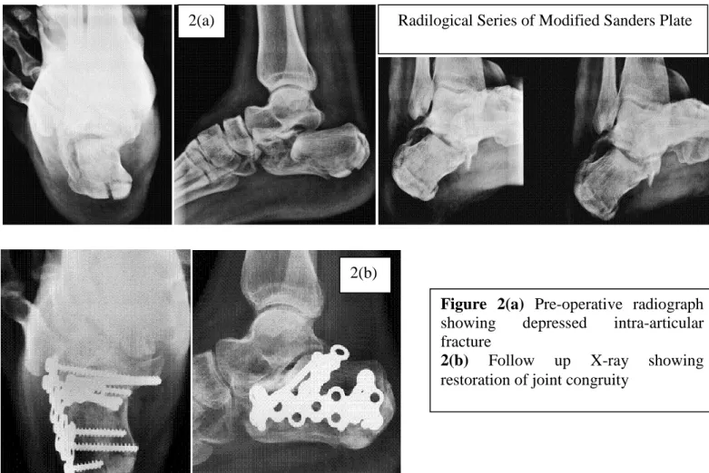
Radilogical Series of Modified Sanders Plate 2(a)
2(b)
Figure 2(a) Pre-operative radiograph
showing depressed intra-articular fracture
Dr Arun Kumar Pandey et al JMSCR Volume 07 Issue 11 November 2019 Page 567
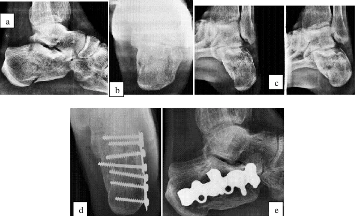
Figure 3:- (a)Lateral (b)Harris axial (c)Brodens view radiographs of intra-articular calcaneum fracture. Post
operative follow up (d) Harris axial (e)lateral views showing restoration of anatomy after open reduction. The guideline principles for treating these
fractures were
1) Reduction of depressed posterior facet, anterior and middle facet.
2) Stabilization of the fragment by 4.0mm cancellous screws
3) Proper sized plate fixation laterally with screws directed from lateral to medial 4) Restoration of heel width, height, valgus
alignment.
5) Intra-operative fluoroscopy to judge the reduction of the fracture and calcaneo-cuoid joint.
6) Decompression of subfibular space available for the peroneal tendons.
Controversy exists over non-operative versus operative treatment. 17 of 27 fractures treated by conservative means attained fair to poor results.14 Results are worse with increasing degrees of comminution of the posterior facet.5,6 The results of operative treatment are variable, mostly related to the quality of the posterior facet reduction; 80% of patients with successful reductions had satisfactory results.7 In another series, 76% of patients attained satisfactory results based on CT assessment of the fracture reduction.12
Unsatisfactory results were associated with failure to obtain or maintain a satisfactory reduction.
a
b
d
c
Dr Arun Kumar Pandey et al JMSCR Volume 07 Issue 11 November 2019 Page 568
Figure 4:- (a) Harris axial, (b)Broden view radiograph of depressed fracture of calcaneum. Immediate post
operative(c) lateral and (d) harris axial view. Follow up (e) lateral, (f) Harris axial radiographs showing sub-talar arthritis with fracture united.
Lateral, axial, anteroposterior and Broden view radiographs are used to examine calcaneal fractures. Extension of the fracture into the posterior facet is clearly visualized using the Broden view, but overlap of tarsal bones and articular surfaces makes assessment of the exact fracture anatomy difficult. 2-Dimensional CT scans helps in pre-operative planning and give additional information on
Calculation of Bohlers and Gissanes angle.
Calculation of heel height and width.
Size and number of fracture fragments.
Proper labeling the major fragments and methods to stabilize them.
Size and displacement of sustantacular tali relative to superomedial fragments.
Presence of step or diastasis of the posterior facet.
Impingement of the fibular malleolus on the tuberosity of calcaneum10.
Such scans also provide information regarding fractures involving the sinus tarsi, calcaocuboid joint, and anterior calcaneal process, all of which could be relevant while planning lateral surgical approach.
Buckley R, Meek R compared closed and open reduction of intra-articular fractures of calcaneum and found good results in open reduction methods for this fractures. Freeman BJC, Duff S, Allen PE, et al advocated extended lateral approach for treating intra-articular fractures of calcaneum. With direct visualization of fracture through a lateral approach, anatomic reduction of the fracture was possible, also the elevation of depressed posterior facet fragment was possible, easier decompression of the lateral wall and the plate was applied on the lateral surface of calcaneum which acted as a buttress.
e d
b a
c
Dr Arun Kumar Pandey et al JMSCR Volume 07 Issue 11 November 2019 Page 569
Conclusion
Intra-articular fractures of calcaneum are challenging fractures with a significant potential for complications. However ORIF (open reduction and internal function) utilizing lateral incision can result in excellent sub-talar and hindfoot function for these patients.
In conclusion, this study confirms that the intra-articular fractures of calcaneum can be best treated by open reduction and internal fixation with a plate and function of the calcaneus and subtalar joint be restored.
References
1. Benirschke SK, Sangeorzan BJ. Extensive intraarticular fractures of the foot. Surgical management of calcaneal fractures. Clin Orthop Relat Res 1993;292:128–34. 2. Burdeaux BD. Reduction of calcaneal
fractures by the McReynolds medial approach technique and its experimental basis. Clin Orthop Relat Res 1983;177:87–103.
3. Carr JB. Surgical treatment of the intra-articular calcaneus fracture. Orthop Clin North Am 1994;25:665–75.
4. Chan S, Ip FK. Open reduction and internal fixation for displaced intra-articular fractures of the os calcis. Injury 1995;26:111–5.
5. Crosby LA, Fitzgibbons T. Computed tomography scanning of acute intra-articular fractures of the calcaneus. A new classification system. J Bone Joint Surg Am 1990;72:852–9.
6. Crosby LA, Fitzgibbons T. Intraarticular calcaneal fractures. Results of closed treatment. Clin Orthop Relat Res 1993;290:47–54.
7. Essex-Lopresti P. The mechanism, reduction technique, and results in fractures of the os calcis, 1951-52. Clin Orthop Relat Res 1993;290:3–16.
8. Folk JW, Starr AJ, Early JS. Early wound complications of operative treatment of
calcaneus fracture:Analysis of 190 fractures. J Orthop Trauma 1999;13:369-370.
9. Gould, N. Lateral approach to the Os calcis. Foot Ankle.1984;4:218-220
10. Heger L, Wulff K, Seddiqi MS. Computed tomography of calcaneal fractures. AJR Am J Roentgenol 1985;145:131–7.
11. Heir Ka, Infante AF, Walling AK,et al. Open fractures of calcaneus:Soft tissue injury determined outcome.J Bone Joint Surg Am 2003;85:2276-2282.
12. Hutchinson F 3rd, Huebner MK. Treatment of os calcis fractures by open reduction and internal fixation. Foot Ankle Int 1994;15:225–32.
13. Jarvholm, U, Korner L, Thoren O, Wiklund L.M. Fractures of the calcaneus. A comparison of open and closed treatment. Acta Orthop. Scand 1984; 55: 652-6.
14. Kitaoka HB, Schaap EJ, Chao EY, An KN. Displaced intra-articular fractures of the calcaneus treated non-operatively. Clinical results and analysis of motion and ground-reaction and temporal forces. J Bone Joint Surg Am 1994;76:1531–40.
15. Letournel E. Open treatment of acute calcaneal factures. Clin Orthop Relat Res 1993;290:60–7.
16. Levin LS, Nunley JA. The management of soft tissue problem associated with calcaneus fractures. Clin Orthop 1993;290:151-156.
17. Nade S, Monahan PR. Fractures of the calcaneum: a study of the long-term prognosis. Injury 1973;4:200–7.
18. Paley D, Hall H. Intra-articular fractures of calcaneus. A critical analysis of results and prognostic factors. J Bone Joint Surg Am 1993;75:342–54.
Dr Arun Kumar Pandey et al JMSCR Volume 07 Issue 11 November 2019 Page 570 tomography scan classification. Clin
Orthop Relat Res 1993;290:87–95.
20. Soeur R, Remy R. Fractures of the calcaneus with displacement of the thalamic portion. J Bone Joint Surg Br 1975;57:413–21.
21. Thordarson DB, Krieger LE. Operative vs. nonoperative treatment of intra-articular fractures of the calcaneus: a prospective randomized trial. Foot Ankle Int 1996;17:2–9. 22. Zwipp H, Tscherne H, Thermann H,