ORIGINAL PAPERS
Dorota B. Namiot
1, A, B, D, F, Katarzyna Leszczyńska
2, B, Andrzej Namiot
3, C–E,
Urszula M. Leszczyńska
4, B, Robert Bucki
5, D, E, Robert Milewski
6, C,
Zbigniew Namiot
7, 8, B, D, EThe Influence of Oral Health Status
and Dental Plaque Removal Practices on the Occurrence
of
Helicobacter Pylori
Antigens in Saliva
Wpływ stanu zdrowia jamy ustnej i zabiegów usuwania płytki nazębnej
na występowanie antygenów
Helicobacter pylori
w ślinie
1 Department of Prosthetic Dentistry, Medical University of Białystok, Białystok, Poland 2 Department of Diagnostic Microbiology, Medical University of Białystok, Białystok, Poland 3 Department of Human Anatomy, Medical University of Białystok, Białystok, Poland 4 Student of Medical University of Białystok, Białystok, Poland
5 Institute for Medicine and Engineering, University of Pennsylvania, Philadelphia, USA
6 Department of Statistics and Medical Informatics, Medical University of Białystok, Białystok, Poland 7 Department of Physiology, Medical University of Białystok, Białystok, Poland
8 Institute for Medicine, State College of Computer Science and Business Administration, Łomża, Poland
A – concept, B – data collection, C – statistics, D – data interpretation, E – writing/editing the text,
F – compiling the bibliography
Abstract
Background. Helicobacter pylori can colonise both the dental plaque and saliva.
Objectives. The aim of this study was to evaluate the influence of oral health status and dental plaque removal practices on the occurrence of H. pylori antigens in saliva of subjects with H. pylori infected and non-infected dental plaque.
Material and Methods. Two hundred and twenty-five patients were included in the study. The presence of H. pylo-ri antigens in saliva and dental plaque was determined with a kit designed to detect H. pylori antigens in feces.
Results. H. pylori antigens were found in the dental plaque and saliva of 58.7% and 36.4% of subjects, respectively. They were more frequently detected in saliva of subjects with H. pylori infected than non-infected dental plaque. The oral health status and dental plaque removal practices from natural teeth did not influence the presence of
H. pylori antigens in saliva. The use of dentures decreased the incidence of H. pylori antigens in saliva, and this was related to the method of dentures storage after their removal for the night.
Conclusions. The lower occurrence of H. pylori antigens in saliva than in dental plaque was associated with neither oral health status nor dental plaque removal procedures from natural teeth, but was related to dentures usage and the means of their storage after removal (Dent. Med. Probl. 2013, 50, 3, 275–281).
Keywords: Helicobacter pylori, oral hygiene, dental plaque, saliva.
Streszczenie
Wprowadzenie. Helicobacter pylori może kolonizować zarówno płytkę nazębna, jak i ślinę.
Cel pracy. Celem pracy była ocena wpływu stanu zdrowia jamy ustnej i zabiegów usuwania płytki nazębnej na wystę-powanie antygenów bakterii H. pylori w ślinie osób z zakażoną i niezakażoną bakterią H. pylori płytką nazębną.
Materiał i metody. 225 pacjentów obu płci w wieku 25–70 lat uczestniczyło w badaniu. Stan zdrowia jamy ustnej był oceniany przez stomatologa. Informacje dotyczące usuwania płytki nazębnej pochodziły z ankiety. Obecność antygenów H. pylori w ślinie i płytce nazębnej określano za pomocą zestawu służącego do wykrywania antygenów
H. pylori w kale.
Dent. Med. Probl. 2013, 50, 3, 275–281
H. pylori can occur both in the stomach and in the oral cavity. H. pylori enters the oral cavi-ty mainly with consumed food, with regurgitat-ed stomach contents, or through direct contact with the saliva of an infected subject [1]. Since on-ly some cases of oral infection lead to a stomach infection [2, 3], this suggests a lack of a direct re-lationship between an infection in the oral cavi-ty and the stomach. Within the oral cavicavi-ty, H. py-lori is most often isolated from the dental plaque; however, it can be also found in the saliva, peri-odontal pocket, palatine tonsils and mucous mem-brane [4–7]. A significant number of H. pylori bac-teria present in the dental plaque are either dead or in a non-culturable form [8]; therefore, the ability of the swallowed dental plaque bacteria to infect the stomach is difficult to predict. Theoretically, infected saliva should be much more contagious than dental plaque, as an adult swallows about 1500 ml of saliva each day, but similar to dental plaque, the contagiousness of saliva for the stom-ach has yet to be established. It has been shown previously that the oral health status and dental plaque removal practices from natural teeth and dentures are not associated with the presence of
H. pylori antigens in the dental plaque [9].
The aim of this study was to determine the in-fluence of oral health status and dental plaque re-moval practices from the natural teeth and remov-able dentures on the incidence of H. pylori anti-gens in saliva of subjects with H. pylori infected and non-infected dental plaque.
Material and Methods
Subjects
Two hundred and twenty-five patients from the Gastroenterology and Medical Department of District Hospital in Białystok, aged 25 to 70 years, both men and women with natural teeth or with natural teeth and dentures, were included in the study. The study was conducted in the years 2009– –2010. The inclusion criteria were good general condition (anamnesis, physical examination), no
chronic or devastating diseases, no history of an-tibiotic therapy in the past month and no histo-ry of H. pylori eradication. Each patient completed a questionnaire concerning dental plaque removal procedures from the natural teeth (brushing) and removable dentures (rinsing after meal, brush-ing, removing for the night, storage after remov-ing, and disinfection) and underwent a dental ex-amination, including the evaluation of the condi-tion of natural teeth (number of preserved natural teeth, caries index, plaque index, and calculus in-dex), prosthetic restorations and periodontium. The plaque and calculus indices were calculated according to 4-point scale proposed by Green and Vermillion [10], while the periodontal status was evaluated using Russell’s periodontal index [11].
The study was approved by the Ethics Com-mittee for Research on Humans and Animals of Medical University of Białystok. Written consent was obtained from all subjects before study entry.
Samples Collection
and Processing
At least 2 mg of dental plaque from the natural teeth was collected by scraping. The samples were taken in the morning, before eating and perform-ing daily oral hygienic procedures, and transferred to Eppendorf tubes filled with 50 µl of 0.15 mol/l saline. To determine the mass of the collected den-tal plaque, the Eppendorf tube containing saline was weighed before and after plaque sampling.
The microbiological laboratory received each Eppendorf tube directly after plaque collection. Then, 50 µl of culture medium (Brucella broth) was added to the tube and the whole volume was incubated at 37°C for 72 hours under microaero-philic conditions. This preliminary incubation aimed to increase the number of H. pylori bacte-ria in the sample, and consequently the sensitiv-ity of the method used for detecting the presence of H. pylori antigens. After the incubation, 100 µl of diluent, included in the manufacturer’s kit, was added to the culture, mixed and centrifuged for 5 minutes at 5000 rpm. Fifty µl of obtained super-natant was used for further analysis.
Wyniki. AntygenyH. pylori w płytce nazębnej i ślinie stwierdzono odpowiednio u 58,7% i 36,4% osób badanych. Były one częściej wykrywane w ślinie osób z zakażoną niż niezakażoną bakterią H. pylori płytką nazębną. Stan zdrowia jamy ustnej i zabiegi usuwania płytki nazębnej z zębów naturalnych pozostawały bez wpływu na obecność antygenów w ślinie. Użytkowanie protez zębowych zmniejszało częstość występowania antygenów H. pylori w śli-nie, co pozostawało w związku ze sposobem ich przechowywania po wyjęciu na noc.
Wnioski. Rzadsze występowanie antygenów H. pylori w ślinie niż w płytce nazębnej nie było związane ani ze sta-nem zdrowia jamy ustnej, ani zabiegami usuwania płytki nazębnej z zębów naturalnych, ale z użytkowaniem protez zębowych i sposobem ich przechowywania po wyjęciu (Dent. Med. Probl. 2013, 50, 3, 275–281).
Directly before the collection of plaque, each subject provided 5 ml of unstimulated saliva. The saliva was initially centrifuged for 5 minutes at 5000 rpm. Four hundred µl of obtained sedi-ment was dissolved in 400 µl of the diluent pro-vided with each kit, thoroughly mixed, and recen-trifuged for 5 minutes at 5000 rpm. Fifty µl of ob-tained supernatant was used for further analysis.
Helicobacter Pylori
Antigen
Test Performance
The detection of H. pylori antigens in den-tal plaque and saliva was performed according to the manufacturer’s instruction provided with kit, which was originally designed for detecting H. py-lori antigens in feces using monoclonal antibodies (Amplified IDEIATM Hp StAR TM, Oxoid, Ely, UK).
In brief, the prepared supernatant was transferred from studied samples to a plate with microwells coated by the manufacturer with monoclonal an-tibodies against H. pylori. A solution containing monoclonal antibodies against H. pylori conjugat-ed with horseradish peroxidase was also addconjugat-ed to the microwells. The whole volume was incubated in the room temperature for 60 minutes. Unbound antibodies were washed away and tetramethylben-zidine, a substrate for horseradish peroxidase, was added. After a 10-minute incubation the reaction was stopped with sulphuric acid, which resulted in a solution colour change from blue to yellow. The colour intensity was measured spectrophotometri-cally at 450 nm.
Statistics
The results (mean value ± standard deviation, median and range) were analysed statistically us-ing the Mann-Whitney U test, c2 test or
Kruskal-Wallis test. Statistical significance was accepted at p < 0.05.
Results
H. pylori antigens were detected in the den-tal plaque and saliva of 58.7% (132/225) and 36.4% (82/225) of subjects, respectively (p < 0.001). In to-tal, 67.1% (151/225) of subjects were found to have an H. pylori infection in the oral cavity (H. pylo-ri antigens in either the saliva, dental plaque or in both) (Table 1). Almost half of the subjects (47.7%) with a confirmed presence of H. pylori antigens in the dental plaque also had H. pylori antigens in their saliva, while only 20.4% of subjects without
H. pylori antigens in the plaque had detected H. py-lori antigens in saliva (p < 0.001). In 76.8% of sub-jects with a confirmed presence of H. pylori anti-gens in saliva, H. pylori antigens were present also in the dental plaque, while only 39.1% of subjects without H. pylori antigens in saliva demonstrat-ed their presence in the dental plaque (p < 0.001). Comparing the four groups in which H. pylori an-tigens in saliva and dental plaque were analysed together, it was found that the number of natural teeth, carious teeth, filled teeth, plaque index, cal-culus index and periodontal status were the same (Table 2). No differences regarding the type of toothbrush used, its yearly frequency of chang-ing or performed hygienic procedures concernchang-ing natural teeth (number of daily brushings, dura-tion of a single brushing event, total time of daily brushing) or removable dentures (removal for the night, storage after removing, rinsing after meals, brushing and disinfecting) were observed (Table 3 and 4). However, it was shown that H. pylori anti-gens were less frequently present in saliva of sub-jects using dentures (p=0.007) (Table 2). The com-parison of four groups, in which H. pylori antigens in saliva and dental plaque were analysed together, demonstrated that only the storage of dentures af-ter their removal for the night was associated with a presence of H. pylori antigens in saliva (p=0.039) (Tables 2–4).
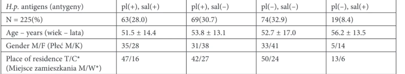
Table 1. The patients’ characteristics Tabela 1. Charakterystyka pacjentów
H.p. antigens (antygeny) pl(+), sal(+) pl(+), sal(–) pl(–), sal(–) pl(–), sal(+)
N = 225(%) 63(28.0) 69(30.7) 74(32.9) 19(8.4)
Age – years (wiek – lata) 51.5 ± 14.4 53.8 ± 13.1 52.7 ± 17.0 56.2 ± 13.5
Gender M/F (Płeć M/K) 35/28 31/38 33/41 5/14
Place of residence T/C*
(Miejsce zamieszkania M/W*) 47/16 42/27 50/24 13/6
H.p. – Helicobacter pylori; pl – dental plaque (płytka nazębna); sal – saliva (ślina). * Place of residence includes the town (T) and countryside (C).
Table 2. Data of patients with regard to oral health status
Tabela 2. Dane pacjentów uwzgledniające stan zdrowia jamy ustnej
H.p. antigens (antygeny) pl(+), sal(+) pl(+), sal(–) pl(–), sal(–) pl(–), sal(+)
Dentist visits – regular/non-regular*
(Wizyty u stomatologa – regularne/nieregularne*) 20/43 15/54 25/49 8/11
Number of teeth (Liczba zębów) 18.6 ± 8.1 15.0 ± 9.1 17.3 ± 9.0 15.9 ± 8.3
Number of decayed teeth
(Liczba zębów z próchnicą) 2.9 ± 3.4 1.9 ± 2.4 1.8 ± 2.6 2.8 ± 4.7
Number of filled teeth
(Liczba zębów wypełnionych) 5.1 ± 4.3 5.4 ± 4.9 5.3 ± 4.8 5.7 ± 5.1
Plaque index (Wskaźnik płytki) 1.4 (0–3) 1.4 (0–3) 1.2 (0–3) 1.3 (0.2–3)
Calculus index (Wskaźnik kamienia) 0.3 (0–1.6) 0.2 (0–2) 0.3 (0–3) 0.2 (0–1.4)
Periodontal index (Wskaźnik przyzębia) 0.6 (0–8) 0.7 (0–8) 0.5 (0–7.3) 0.5 (0–3.1) Denture wearers, n (%) (Użytkownicy protez) 31 (49.2) 52 (75.4) 48 (64.9) 11 (57.9)**
Fixed (Stałych), n (%) 6 (9.5) 12 (17.4) 14 (18.9) 2 (10.5)
Removable (Ruchomych), n (%) 11 (17.5) 23 (33.3) 16 (21.6) 4 (21.1)
Fixed and removable, n (%)
(Stałych i ruchomych) 14 (22.2) 17 (24.6) 18 (26.1) 5 (26.3)
* regular – at least one a year (*regularne – co najmniej raz w roku). ** p < 0.007.
Table 3. Hygienic practices concerning natural teeth of subjects with and without H. pylori antigens in the dental plaque (pl) and saliva (sal)
Tabela 3. Zabiegi higieniczne dotyczące zębów własnych osób z obecnymi i nieobecnymi antygenami H. pylori w płytce nazębnej (pl) i ślinie (sal)
H.p. antigens (antygeny) pl(+), sal(+) pl(+), sal(–) pl(–), sal(–) pl(–), sal(+)
Number of daily toothbrushing (Liczba codziennych szczotkowań zębów) 0 ×, n (%)
1 ×, n (%) 2 ×, n (%) 3 ×, n (%)
12 (19.0) 11 (17.5) 32 (50.8) 8 (12.7)
17 (24.6) 9 (13.0) 41 (59.4) 2 (2.9)
18 (24.3) 8 (10.8) 41 (55.4) 7 (9.5)
5 (26.3) 2 (10.5) 10 (52.6) 2 (10.5) Time of single toothbrushing (Czas pojedynczego szczotkowania zębów)
30 sec., n (%) 60 sec., n (%) 120 sec., n (%) 180 sec., n (%)
6 (11.8) 20 (39.2) 17 (33.3) 8 (15.7)
12 (23.1) 15 (28.8) 17 (32.7) 5 (15.4)
9 (16.1) 18 (32.1) 20 (35.7) 9 (16.1)
3 (21.4) 3 (21.4) 3 (21.4) 5 (35.7) Total time of daily toothbrushing (Całkowity czas codziennego szczotkowania zębów)
Up to 2 minutes (do 2 minut), n (%)
2 minutes or longer (2 minuty lub dłużej), n (%) 14 (27.4)37 (72.5) 15 (28.8)37 (71.2) 12 (21.4)44 (78.6) 2 (14.3)12 (85.7) Used toobrush (Używana szczoteczka do zębów)
Soft (Miękka), n (%) Medium (Średnia), n (%) Hard (Twarda), n (%)
9 (17.6) 34 (66.7) 8 (15.7)
3 (5.8) 40 (76.9) 9 (17.3)
7 (12.5) 41 (73.2) 8 (14.3)
1 (7.1) 11 (78.6) 2 (14.3) Frequency of yearly toothbrush changing (Częstość wymiany szczoteczki do zębów w ciągu roku)
1 ×, n (%) 2 ×, n (%)
3 × or more (lub częściej), n (%)
3 (5.9) 26 (51.0) 22 (43.1)
10 (19.2) 26 (50.0) 16 (30.8)
8 (14.3) 18 (32.1) 30 (53.6)
1 (7.1) 4 (28.6) 9 (64.3)
Discussion
The detection of H. pylori antigens using an immunoenzymatic assay is one of the methods documenting the presence of H. pylori in the den-tal plaque and saliva [8, 9, 12]. Its advantage lies in its simplicity and high level of reliability. This method has been used for the last decade to de-tect of H. pylori antigens in feces [13]. The results of currently performed studies reveal that subjects with the presence of H. pylori antigens both in the dental plaque and saliva, only in the dental plaque, only in saliva, and those demonstrating the pres-ence of antigens neither in the dental plaque nor saliva demonstrate the same oral health status and the same profile of hygienic procedures concern-ing dental plaque removal from the natural teeth.
The bacteria contained in the dental plaque are transferred to saliva in only a limited range, and such a phenomenon is observed in immature plaques only [14]. Assuming that dental plaque re-moval procedures disturb its structure and facili-tate the bacterial transfer into saliva, H. pylori an-tigens should be found more frequently in saliva of subjects who brush their teeth regularly than in those who do not do it at all. The results of the current study contradict this assumption; the number of subjects with H. pylori infected dental
plaque who either have or do not have the bacte-rial antigens in saliva, is the same. This phenom-enon could be attributed to the fact that subjects participating in this study present many losses in the natural dentition, especially molars and pre-molars, i.e. teeth H. pylori are most frequently iso-lated from [15]. On the other hand, the molars and premolars are not so easily available for brush-ing, therefore the structure of the dental plaque on their surfaces are disturbed probably only in part during brushing. The absence of H. pylori in sali-va and its presence in the dental plaque can be fa-cilitated by the fact that the dental plaque is not removed completely during routine procedures, and that the plaque removal is performed at least 12 hours before the collection of saliva, which en-ables the restoration of the plaque structure and makes the plaque a complex unable to release bac-teria to saliva. In 47.7% of subjects with positive test results in the dental plaque, bacterial antigens are present in the saliva as well, while in 76.8% of subjects with a positive test for the presence of
H. pylori antigens in saliva, bacterial antigens are also present in the dental plaque. Such a result in-dicates that H. pylori present in saliva colonizes dental plaque and not vice versa.
It is suggested that H. pylori can be present not only in the plaque on the natural teeth but also on
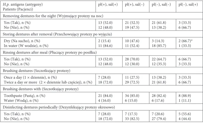
Table 4. Hygienic practices concerning removable dentures of subjects with and without
H. pylori antigens in the dental plaque (pl) and saliva (sal)
Tabela 4. Zabiegi higieniczne dotyczące ruchomych protez zębowych osób z obecnymi i nieobecnymi antygenami H. pylori
w płytce nazębnej (pl) i ślinie (sal)
H.p. antigens (antygeny)
Patients (Pacjenci) pl(+), sal(+) pl(+), sal(–) pl(–), sal(–) pl(–), sal(+)
Removing dentures for the night (Wyjmujący protezy na noc) Yes (Tak), n (%)
No (Nie), n (%) 13 (52.0)12 (48.0) 21 (52.5)19 (47.5) 21 (61.8)13 (38.2) 3 (33.3)6 (66.7) Storing dentures after removal (Przechowujący protezy po wyjęciu)
Dry (Na sucho), n (%)
In water (W wodzie), n (%) 2 (15.4)11 (84.6) 10 (47.6)11 (52.4) 3 (14.3)18 (85.7) 2 (66.7)*1 (33.3) Rinsing dentures after meal (Płuczący protezy po posiłku)
Yes (Tak), n (%)
No (Nie), n (%) 13 (52.0)12 (48.0) 28 (70.0)12 (30.0) 22 (64.7)12 (35.3) 6 (66.7)3 (33.3) Brushing dentures (Szczotkujący protezy)
Once a day (1 × dziennie), n (%)
Twice a day or more (2 × dziennie lub częściej), n (%) 7 (28.0)18 (72.0) 11 (27.5)29 (72.5) 13 (38.2)21 (61.8) 3 (33.3)6 (66.7) Brushing dentures with (Szczotkujący protezy)
Toothpaste (Pastą), n (%)
Water (Wodą), n (%) 21 (84.0)4 (16.0) 34 (85.0)6 (15.0) 28 (82.4)6 (17.6) 8 (88.9)1 (11.1) Disinfecting dentures periodically (Dezynfekujący protezy okresowo)
Yes (Tak), n (%)
No (Nie), n (%) 7 (28.0)18 (72.0) 7 (17.5)33 (82.5) 7 (20.6)27 (79.4) 5 (55.6)4 (44.4) * p < 0.039.
dentures [16, 17]. As the surface of removable den-tures is frequently greater than the surface of natu-ral teeth, and the plaque deposits are usually found both on the natural teeth and removable dentures, it can be speculated that subjects using removable dentures will demonstrate plaque and saliva infec-tion more frequently than subjects with natural dentition only. The result of the current study does not support this thesis, but shows that with the use of dental prostheses, the incidence of H. pylo-ri antigens in saliva is reduced and this is associ-ated with the storage of removable dentures after their removal for the night. One may think, there-fore, that the removal of dentures and their storage
dry eliminates the significant amount of the bac-teria colonising the prostheses, including H. pylo-ri. Moreover, it indicates that removable dentures, made in the majority of studied subjects with acryl, constitute a good bed for bacterial adhesion.
Based on the results of the current study it can be concluded that there was an association between the presence of H. pylori in the dental plaque and saliva, but this relationship does not result from the oral health status or dental plaque removal practic-es from the natural teeth. The lower incidence of
H. pylori antigens in saliva of subjects using den-tures may be associated with the storage condi-tions of the removable dentures for the night.
Acknowledgements
The study was supported by the Medical University of Białystok, grant No 3 –18800 L.
References
[1] Feldman R.A., Eccersley A.J., Hardie J.M.: Epidemiology of Helicobacter pylori: acquisition, transmission, pop-ulation prevalence disease-to-infection ratio. Br. Med. Bull. 1998, 54, 39–53.
[2] Czesnikiewicz-Guzik M., Loster B., Bielanski W., Guzik T.J., Konturek P.C., Zapala J., Konturek S.J.: Impli-cations of oral Helicobacter pylori for the outcome of its gastric eradication. J. Clin. Gastroenterol. 2007, 41, 145–151. [3] Miyabayashi H., Furihata K., Shimizu T., Ueno I., Akamatsu T.: Influence of oral Helicobacter pylori on the
success of eradication therapy against gastric Helicobacter pylori. Helicobacter 2000, 5, 30–37.
[4] Bago I., Bago J., Plećko V., Aurer A., Majstorovic K., Budimir A.: The effectiveness of systemic eradication therapy against oral Helicobacter pylori. J. Oral Pathol. Med. 2011, 40, 428–432.
[5] Umeda M., Kobayashi H., Takeuchi Y., Hayashi J., Morotome-Hayashi Y., Yano K., Aoki A., Ohkusa T., Ishikawa I.: High prevalence of Helicobacter pylori detected by PCR in the oral cavities of periodontitis patients. J. Periodontol. 2003, 74, 129–134.
[6] Gebara E.C.E., Faria C.M., Pannuti C., Chehter L., Mayer M.P.A., Lima L.A.P.A.: Persistence of Helicobacter pylori in the oral cavity after systemic eradication therapy. J. Clin. Periodontol. 2006, 33, 329–333.
[7] Kusano K., Inokuchi A., Fujimoto K., Miyamoto H., Tokunaga O., Kuratomi Y., Shimazu R., Mori D., Yamasaki F., Kidera K., Tsunetomi K., Miyazaki J.: Coccoid Helicobacter pylori exists in the palatine tonsils of patients with IgA nephropathy. J. Gastroenterol. 2010, 45, 406–412.
[8] Leszczyńska K., Namiot D.B., Namiot Z., Leszczyńska J.K., Jakoniuk P., Kemona A.: Application of immu-noassay for detection of Helicobacter pylori antigens in the dental plaque. Adv. Med. Sci. 2009, 54, 194–198. [9] Namiot D.B., Leszczyńska K., Namiot Z., Chilewicz M., Kemona A.: The occurrence of Helicobacter pylori
antigens in dental plaque; an association with oral health status and practices of oral hygiene. Adv. Med. Sci. 2010, 55, 167–171.
[10] Green J.C., Vermillion J.R.: The oral hygiene index: a method for classifying oral hygiene status. J. Am. Dent. 1960, 61, 173–179.
[11] Russell A.L.: A system of classification and scoring for prevalence of periodontal disease. J. Dent. Res. 1956, 35, 350–358.
[12] Leszczyńska K., Namiot D.B., Namiot A., Chilewicz M., Leszczyńska J.K., Bucki R., Kemona A., Namiot Z.: Application of immunoassay for detection of H. pylori antigens in saliva (abstract). Helicobacter 2010, 15, 363. [13] Gisbert J.P., Pajares M.: Stool antigen test for the diagnosis of Helicobacter pylori infection: a systematic review.
Helicobacter 2004, 9, 347–368.
[14] Rowshani B., Timmerman M.F., Van Der Velden U.: Plaque development in relation to periodontal condition and bacterial load of the saliva. J. Clin. Periodontol. 2004, 31, 214–218.
[15] Song Q., Lange T., Spahr A., Adler G., Bode G.: Characteristic distribution pattern of Helicobacter pylori in den-tal plaque and saliva detected with nested PCR. J. Med. Microbiol. 2000, 49, 349–353.
[16] Namiot D.B., Namiot Z., Kemona A., Bucki R., Gołębiewska M.: Oral health status and oral hygiene practices of patients with peptic ulcer and how these affect Helicobacter pylori eradication from the stomach. Helicobacter 2007, 12, 63–67.
[17] Namiot Z., Namiot D.B., Kemona A., Stasiewcz J.: Effect of antibacterial therapy and salivary secretion on the efficacy of Helicobacter pylori eradication in duodenal ulcer patients. Oral Surg. Oral Med. Oral Pathol. Oral Ra-diol. Endod. 2004, 97, 714–717.
Address for correspondence:
Dorota Beata Namiot
Medical University of Białystok 24A M. Skłodowskiej-Curie 15-276 Białystok, Poland Tel.: +48 85 748 57 69 E-mail: dorota.namiot@op.pl Received: 7.05.2013
Revised: 20.05.2013 Accepted: 12.07.2013
Praca wpłynęła do Redakcji: 7.05.2013 r. Po recenzji: 20.05.2013 r.