KEY WORDS
ultraviolet radiation
disinfectant
ABSTRACT
Constance M. Dumais; Phatareactivation of UV-1rradiated CDiifcrms in
Wastewater Effluent. (under the direction of Dr. J. Donald Johnson).
Ultraviolet (uv) radiation of wastewater is currently being
considered as an alternative to chI orination. Howeverj the problem
of the extent of photoreactivation (PR); end under what conditions it
would occur most severely has not yet been defined. The parameters
of optimal exposure timej sunlight intensity; nutrient content;
temperature; and uv dose were examined. These same parameters were
also studied under laboratory conditions; using an artificial PR
light source. During strong summer sunlight; hQ minutes of exposure
gave the maximum amount of repair at temperatures near 25"C. At
higher (3fe'^C) or lower (17°C) temperatures; the level of PR
declined. Nutrient attenuation; achieved by diluting the sample I'-ID
or 1=40 in receiving water did not decrease PR; however a very large
decrease in nutrients did negatively affect the level of survival
after PR. Under optimal conditions; the effective level of-TABLE OF CONTENTS
Page
Acknowledgements...v
List at Tab i es...v i List of Figures...yii I. LiteratureReview :' ^ Inact i vat i an by U\J \ i s^t...1
UV photDprDducts...2
Phatareactivatian...7
K i net i cs...12
,: _ Ex c i 5 i D n ( d a r k ) !" e ? a i r...14
Other repair schemes...17
H. Project Objectives...23
III. Materials and Methods Intensity measurements...24
Temperature control...26
Incubation conditions...29
Nutrient control...30
IV. ResuitsandDiscussicn Parameters exam i ned..._...33
~ " - ^-: Effect of exposure time on PR...34 '
' -' Effect of PR light intensity...41
-Effect of temperature on PR... 45
Effect of nutrient levels an PR...47
: Effect of UV dose on PR...53
-. Pyrex bottle vs. open beaker...58
V . . Conclusions...63
mmmm^m'
ACKNOWLEDGEMENTS
I would like to thank Dr, J. Donald Johnson far his guidance and support throughout the course of this project. A special note of appreciation extends to John Chang? far his input and help in
carrying out this research) and to Mark Darfmanj for ail his patience
and friendship. I would I ike tc thank the other members of the
research team: Jerry Quallsj David Lobej and Susan Ossoff. Finally? very special thanks to Aviva Jeruchim whose help was invaluable.
I would like to add a \jery special thanks to my famiiyj who have been supportive and loving throughout this time-" to Charles?
Lieseiotte? and Andrew Dumais? and Joanne Sullivan,
LIST OF TABLES
Table 1
Morsan Creak Water Quality...3D
Table 2 '^^
Final Effluent Quality...31
Tab Ie 3
Exposure T i me j 1 abar atory... .38
Table 4
Exposure Time? field...39
Tab i e 5a) b '
PR Light Intensity...,..".'...43
Table 6
Temperature... 45
Tab!e 7
Nutrients? sterile creek water...50
Table a
Nutrients? nnn-nutrient buffer...51
Tab!e 9
UV DD 5 e > i ab D r a t D r y...55
Table ID
UV Dose? field...S6
Table 11
Beaker vs. Battle...hi
'^^^KSI^^^'
LIST OF FIGURES
Figurel
U\J Photapraducts...5
Figure 2
AbBarbing cf PR Light...9
Figures
UV DDse Decrement...11
F i gure 4
Excision Repair... 16
Figi-'i'eS
Post-Replication Repair...22
Figure 6
Columinating Beam Apparatus...25
Figure?
Blacl< Light Spectrum...,...27
Figures
Response of SEE-4nG Detector...28
Figure?
UV Dose vs. Time? laboratory...37
Fi gure ID
UV Dose vs. Time? field...38
F i g u r e 11
UV Dose vs. Intensity...42
Figure 12 ' ; : ' ;-ͣ ,-^
UV Dose vs. Temperature...47
F i gurs 13
UV Dose v5. Lag Survival; labDratary...54
F i gure 14
U\J Dose v5. Lag Survival; field...,...55
F i gure 15
LITERATURE REVIEW
Recently? in public health literature? the safety of chlorine as
a disinfectant in water and wastewater treatment has came into
question? (31? 49). The formation of potential toxic nr carcinD3enic byproducts? such as trihalomethanes) or THM-'s ? has caused researchers to examine possible alternatives to chI orination . One such
possibility currently under consideration is ultraviolet (uv) light. UV light has long been known to be lethal to microoganisms. The first pubi ished evidence in 1878 by Downs and Blunt? demonstrated the bactericidal properties of short wave radiation (47). In 1903?
Bernard and Morgan determined the region of lethal action to be between 226.7 - 228.7nm (5). Two years later? Bang published
findings which placed the most effective germidical wavelengths at approximately 25nnm. In 191D? this property of uv I ight was first applied to disinfecting water by Cernovodeanu and Henri? and Henri? Helbronner? and Recklinghausen (47). They effectively disinfected
water? to which a variety of bacteria had been added.
There are several advantages to using uv as a disinfectant over chlorine. These include the high efficiency of uv disinfection at a cost which approaches that of conventional methods (35); that the chemical composition of the water is not altered; that no harmful byproducts are formed; that the taste of the water is not changed; and that the maintenance of the machinery involved is not difficult? needing oniy routine cleaning (5? 29? 47). However? because there is no disinfectant residual in the water? process control can be
" -:'ͣ' ͣ-ͣ,;..^ . ͣ-; . -2- - . ͣ
-contamination. Also the water quality prior to uv-irradiation must be relatively good; in order to achieve adequate disinfectionj thus
increasing the cost of operation. Finallyj there is the problem of regrowth of damaged? but not l< i I i ed microbes. There ars several
repair processes within the eel I > which can reverse uv caused lesions
in the ce!Is DNA. The most effective of these is a I ight induced enzymatic repair system? known as photoreactivation.
Initial !yj the mechanism which led to eel I death from irradiation
was unknown. It was speculated that the uv light might act directly
on cellular components) such as proteins? enzymes? or nucleic acids? or indicrectly? by forming toxins within the cell (3). Later
research showed the action spectrum of uv light on the cell
corresponded very we I I to the absorption spectrum of nucleic acids? suggesting that thsy might be the target ofthe uv photons. As the
importance of nucleic acids and DNA became understood? initiated by Watson and Crick''5 work in the 195D''5? it became apparent that damage
to this cellular component could be lethal? cause mutations? and
account for many of the other effects attributed to exposure to uv
light. By the early 197D''s? the uv photoproducts were well
characterized. , .
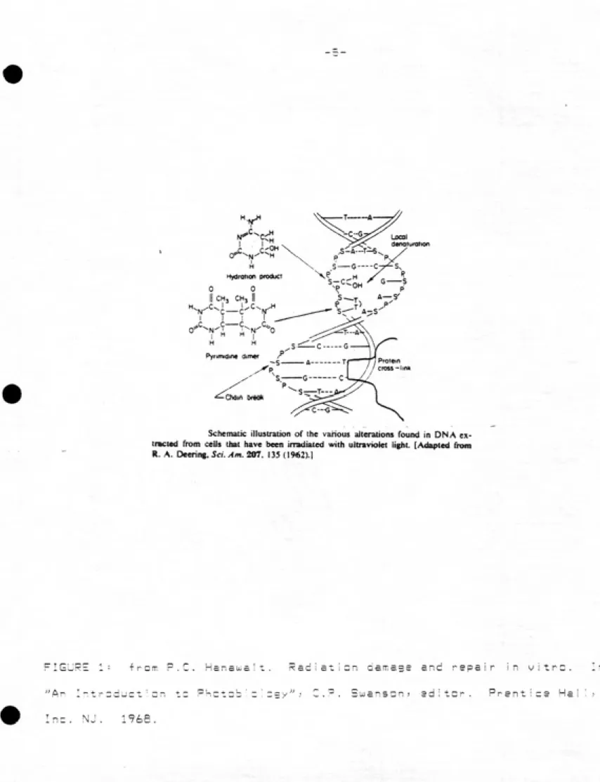
When photons of uv 1 ight are absorbed by the DNA? there is
usually some local denaturization of the double helix (17? 44). (See
Fig. 1) This is often caused by dimerization of adjacent pyrimidine
bases. These dimers can occur between any of the cyclobutanes? and all types have been isolated? including Thimidine—Thimidine (T—T)? Cytosine-Cytosine (C-C-)? and Thimidine-Cytosine (T-C). Thimidine dimers predominate? and are also the most easily reversed by the
-3-fairly comman eventsj and also causes denaturizatian at the DNA. The frequency of chain breakages increase? leaving the DNA susceptible to
forming cross linkages with other pieces of genetic material or
proteins. At least some of these cross I inkages may be attributed to
the error prone excision repair system; which is stimulated by
irradiating with germicidal wavelength light. These lesions can lead to the types of uw damage observed in microbes* including death?
mutations? and delay of DNA synthesis and cell replication cycles?
making uv light an effective germicidal agent.
Organismal survival is a function of both intensity and exposure
time? as per the equation-"
D (mWatt-sec/cm2) = I (MUl/cm^) x T (sec) (equ.l)
where D is the does? I is the light intensity? and T is the time. As early as 1729? Gates reported that the Bunson-Roscoe Law of Reciprocity did not apply to uv irradiation? over a large range of
intensities (11). That is to say -"Hhat when the product of intensity
and exposure time is constant? a constant photochemical reaction results''^. This was at least partially repudiated in subsequent
literature (24? 34). Survival can be represented by the following
equation:
lDg(N=/NG) = f(Da5e) (eqn.2)
where Nl|-, is the initial papulaticn? and Ng is the number of
survivors after irradiation. Equation 2 is derived from Luckiesh and
Holiday-'s (29? 47). Survival data is usually plotted as log survival
vs. uv dose. The resulting dose — survival curves for uv disinfection
-4-deviatian from one-hit or one-taraet kinetics? which may be due to
threshold dose levels or redundancy in the aenome (16). There has
also been speculation that the shoulder may also be caused from the
repslr of the photoproducts, This is somewhat substantiated by the
observation that LJlcrDcoccu5_radigdurans exhibits an exceptionally
large shouiderj and has one of the most efficient repair systems of
any organism tested (24). Dose-survival curves include a straight
I i ne portion which foi lows Chick-'s Law; where increases in uv dose
results in proportional iy lower survival and the Law of Reciprocity
applies. Often at high uv doses? there is a leveling off) after
which further increases in dose do not give the expected decrease in
survival. This has been largely attributed to particulate protection
in wastewater disinfection. Unsettled clumps of organic matter or
aggregates of eel Is can shield bacteria imbedded within them by
absorbing the uv light (33).
The efficient disinfection of water by uv light depends on a
variety of factors. Physical-chemical characteristics of the water
to be treated are important. Highly turbid or colored water may
absorb uv lightj making it unavailable for disinfection. The removal
of particles and aggregates will improve the efficiency of the
irradiating light (36). Some mixing of the water in the uv reactor
facilitates the exposure of microbes to the uv? effectively
increasing the dose seen by a given organism by increasing the
exposure time. The intensity can be enhanced by I ining the reactor
interior with reflective material. However? too much forewsrd
dispersion will a I I aw a significant portion of the microbes to short
circuit through the reactor > receiving a smaller uv dose?
Local aenaturation
Hydraiion praduc!
CH, CHj 11
H H
PyrMndine dimer
Protein cross-Iin*
Oxiin tmck
Schematic Illustration of the vanous aiterations found in ON A ex¬
tracted from cells that have been irradiated with ultraviolet light. [Adapted from R. A. Oeenng, Sci. Am. 207. 135 (1962).!
FIGURE 1: iron-.
ͣ
"Ar I-;tr::duct:D
Inc. NJ. 1768.
o r
rianauic Radiation damage arid repair in vitrc.
-6- - .
given volume of water passing through a reactor are those which short
circuit through the unit (35). There is a tradeoff between ai lowing
some degree of mixing and not causing too much foreward dispersion.
A recent study demonstrated that ideal plug flow is more effective
than completely mixed type flow patterns in uv disinfection (6) 35).
Although uv i ight absorbed by the DNA wi I! produce some type of
lesion; not all of these are fatal. There exists several
possibi I ities where uv induced damage could be biologically
undetected (15). For example; the lesion could be located in a
portion of the DNA molecule that does not contain vital information
or is dupI icated elsewhere in the genome or that is not being
transcribed or translated at the time of irradiation. Polyploid
organisms are known to require roughly twice the uv dose in order to
achieve a level of disinfection comparable to those containing only a
single copy of the genetic information. Kinetic studies show that
the inactivation curves; similar to those generated when there are
two-targets per eel!.
When measurable endpoints; such as mutation or death are observed; the level of survival can be greatly affected by
past-irradiation conditions. There are several processes by which
the damaged eel I can resume normal function. Some involve
spontaneous electrochemical reactions; such as the disintegration of
dimers; or dehydration of the bases. Some repair schemes involve
exchange of complementary genetic material to replace the damaged
portion of the chromosome. Some are enzymatic systems. Not ail the
repair is specific for uv induced lesions. The most affective of
these is photoreactivation; which is specific for reversing
Photoreactivation (PR) refers to the highly specialized enzymatic, removal of pyrimidine dimers? initiated by post-irradiation
iiiumination with i ie"t of wavelengths in the range of BDQ-BDDnm (23). The reversal of uv damage was first observed by Whitakerj in 1942. However) it was Kelnerj in 1949) who perceived the importance
of exposure to near-uv light after disinfection) and correlated it to
the abi I ity of bacteria to reverse the otherwise fatal effects of the
germicial light (25). Later that same year) Duibecco published
similar findings in bacteriophages (S). Since that times the ability
of organisms to use 3QD-5DDnm I ight to stimulate the enzymatic repair
of pyrimidine dimers has been studied extensively (See Fig. 2). It
has been found in almost al! bacteria) viruses) and yeast (23)) aswell as in protozoans and higher animal ceils (3; 12) 14) 27), PR is
able to reverse a variety of eel IuIar effects caused by uv I ight)including death) mutations) and delay of cell division) protein synthesis) and DNA replication. ''
-Early experimentation concentrated primarily on determining the
extent of photoreactivabiiity in cultured organisms) as well as the
influences of irradiation and illumination conditions on organismai
survival. Experiments often involved irradiation with commercially
available germicidal I ampsj having a peak output at 253.7nm.
Estimation of uv dose was made by plotting log survival vs. timS)
assuming the output of the lamps was the same? regardless of age)
manufacturer) etc. Disinfection was followed by iiiumination using
either fluorescent lights) black lightS) or Quartz-iodide lamps as
the pR source (1) 17). Both treatments were commonly performed on
-: ͣ,:•...
-8-researchers plated the uv-irradiated samples prior to exposure
to PR light. Phage studies were performed on irradiated viruses
-w h re h -were allo-wed to absorb onto host cells before being
photoreactivated. 'Definitions in early literature were inconsistent and confusing?
until Jaggerj in one of the first comprehensive reviews? suggested a
standardized nomenclature (23). Jagger defined PR as -'-'the
restoration of uv radiation lesions in biological systems with light
of wavelengths longer than that of the damaging radiation-'-'. He
proposed that PR be used in lieu of the ether terms sometimes used?
such as photorestoratianor photareversal. Since then? the term
photoreactivation has been further narrowed to include only that
enzymatic restoration of uv damage involving the photoreactivating
enzyme (PRE). Inactivation has been defined as when an organism is
unable to give rise to a colony under normal (i.e. standard
laboratory) growth condition. Varying growth conditions; in some
casesi may stimulate the formation of a colony which would not have
occurred had a standard nutrient media been used (3D) 45).
Irradiation refers to the germicidal light? and illumination to PR
I i ght , - \ "
ͣ
' ' -)['._: "' . :- _
PRE was first isolated by Rupert and Harm in 196Dj extracted from
yeast (4D). Prior to this? other evidence indicated the enzymatic
nature of PR. Factors known to influence the extent of reca^ery due
to PR included temperature? pH? exposure time? and to some degree?
light intensity and nutrient conditions of the post-irradiation
environment. Factors which inhibit ceil growth and division? i.e.
-9-2x10
5x10
2x10
334 355 366 385 405 435
WAVELENGTH(nm)
Absolute action spectra for photoenzymatic repair of various
biological systems. Stationary phase /;. loti Bs-i-160 cells (x); stationary
pha.se cells of Saccharoinyces cercvhiac uur\-2, which were mutated to in¬ crease the PRE content (•); llavmophihis transforming DNA in vitro with yeast PRE either at low purification ( ) or at high purification (<i). (From
W. Harm, in: Molecular Mechanisms lor Repair of DNA, Part A, P. C.
Hanawalt and R. B. Setlow, eds.. Plenum Publ. Co., New York, 1975 pp
8')-|OI.)
FIGURE 2 ͣͣ frcm Harmj Wa'tsr. Biaias'ra: Effects -* U^trav
.:" ͣ:- : . ^ .ͣ-.-:..-.,^^
-,-10-wIHj in general? promote survival (16). However? those which -•
interfere with enzyme mobility? or otherwise inhibit its ability to
function? will decrease the amount of repair pcssibie. FR is the
most effective of all the enzymatic repair schemes? because it is
-able to split the dimers directly. It has been estimated that
approximately 8D% of the uv lesions are T-T dimers (38). PR is also
the least error prone of the repair schemes,
Keiner initially considered the '-'photoreactivable sector-'-' of a
given microbe to be constant. That is to say? at a given uv dose?
the proportion of cells able to rsQa-\/er is constant? and is
calculated as the number of survivors after PR (Ni^) divided by the
number of survivors after irradiation (N^j). This led to the
proposal of the dose-reduction ratio? hypothesizing that the uv dose
could be reduced by a constant factor. PR curves? plotted as
iag-Burviva! vs. uv dose? yield para!lei curves with a higher
survival than that of the unphotoreactivated sample. This holds only
for pure cultures. The resulting recO'^ery was expressed as the dose
or fluence decrement? written as:
A D = D - D^ (eqn, 3)
(19? 2D)? where^D is the dose decrement? D is the uv dose? and D^
is the equivalent uv dose? after PR (see Fig. 3). Jagger reported PR
as a percentage of the initial population? represented by the
f o 1 I ow i ng equat i on
ͣͣ
ͣ
'
g
U
<
o z
>
IT 3
0)
\
UV FLUENCE
Representation of the extent of photoreactivation by the fluence
decrement AF (see text for details). The open circle, marked PR, represents the survival obtained after exposure to fluence F with subsequent photo-reactivation. (From W. Harm, in: Molecular Mechanisms for Repair of DNA, Part A, P. C. Hanawalt and R. B. Setlow, eds.. Plenum Publ. Co., New York,
1975, pp. 89-101.)
FIGURE 3: •'•-tiiT Harrii) Ws'ts'-) B;d;3s'c=: E-'fezts -
ͣ
., .- ͣ
-12-where Nq is the initial papulations N_j the number of survivors
after irradiationj and Ni_ the number of survivors after PR,
Dulbecco also thought the number of lesions which can be
photorepaired was constant (9)) and termed this fraction the
ͣ
'•'photoreactivable sector •'ͣ' which he defined as follows:
1 - a
slops of straight line portion of survival curve after PR
slope of straight portion of curve without PR
According to Kelner; low PR light intensity produced lower ratios or
recovery? given the same total dose of PR 1 ight. When the optimum PR
1 ight intensity was reduced by 1D%; recovery was reduced by 30%
(26). Dulbeccoj however? found no such decrease in efficiency with
decreasing intensity in bacteriophages. He did report a dec I ine in
infective units at high PR doses? when illumination was continued
after the maximum retzo'-jery level had been reached. This was due to
damage sustained by the host-phage complex by the near-uv I ight (7).
The kinetics of PR have been studied extensively in the l?6Q-'s
and 1970''s. The early reports show a nearly I i near response with
time under striciy controlled laboratory conditions (S). Kelner also
mentions a linear response with intensity and exposure time? but not
across as large a range as with phages (25). Temperature dependence
was also recognized early in the literature as an important factor in
the degree of photoreact i vat i on , In Ul. Harm's 1975 review of the
kinetics of PR? he included the spectrum and intensity of the PR
light) amount of PRE in the cell? temperature? pH? ionic strength?
and presence of inhibitors as mediating parameters in recovery (2D).
'R:
'T..
E3^ K^ (eqn. 5)
<^' ^v
where E is the PRE; S> the substrate; is the pyrimidine dimer; ES is
the enzyme-substrate complex; and Pj the product; is the repaired DNA
The MichaeI is-Menton equation differentitates between chemical
and enzyme kinetics; allowing for the expression of enzyme saturation
(28). At low substrate concentrations; the increase in velocity is
proportional to the increase in substrate. At higher concentrations;
velocity is no longer proportional to the substrate; and becomes
independent with respect to S; approaching zero order kinetics. The
velocity is then dependent on the enzyme concentration. A typical
Michae1 is-Menton reaction scheme is written as fallows
Ki
E + S ;£i± ES
S
E3 "-"^^ ^ -^
^K. (eqn. 6)
(notations are as previously described). PR follows Michael is-Menton
kinetics; with two exceptions: the final; photolytic step is not
reversible; therefore there is no k4; and this step is absolutely
dependent on i ight of wavelengths between 3DQ-5DDnm.
Each component of the reaction can be studied separately; by
exposing samples incubated in the dark for a known amount of time; to flashes of intense PR light. The dark incubation allows the EB to
form; and the flash of PR must be bright enough to convert all the ES
to P + S. The formation constant; k^ > can be determined in the
: ͣ - » ..; -14- : .;;.ͣ
given uv dose and organism) by converting E to the number of repaired
dimers per individual? which is converted to the number of ES
complexes present at the time of the light flash. Bysxperimentationj they were able to determine the optimum incubation
time and flash intensity to convert al! the ES to product. The
amount of PRE is control led by adding it as a ce! ' free extract?which allowed the calculation of the number of repaired dimers. From
the measured time? light intensity? and number of repaired dimers?
kji can be calculated. Similar manipulations of dark and light
phases permitted the determination of k2 and k3? as we I I as the
effect of the various parameters mentioned earlier. Both k^ and
k2 show a strong temperature dependence? however the photolytic
step is only affected by high or low temperature.
The final? dimer splitting step in the reaction is absolutely
dependent an I ight between the wavelengths 3DD-55nm. Uhen discrete
flashes of I ight were used? Rupert found that the disappearance of
the first half of the ES complex was temperature independent? and
proportional to the intensity of the I ight flash. The disappearance
of the second half of the complex was slower? temperature dependent?
and not proportional to the intensity. He attributed this to the
heterogeneous composition of the substrate? i.e. the dimers are no
longer primari !y T-T? the preferred substrate for PRE. As the repair
process continues? the propartian of C-C and T-C dimers increases?
and they are not as easi ly spI it by PRE? and so the process slows
(37) . "ͣ . ͣ / V
Photoreactivation is not the only enzymatic repair system
. •
-15-from PR in several ways. Excision repair is net specific far uv
induced damage; but rather functions more as a general repair system? operating continuously . It is not iight dependent; and it is much more error prone than is PR. Several enzymes systematica!ly remove and resynthesize the area around the lesion. The first step in the process is the recognition of the damage as a distortion in the double helix. An incision is made in several base
units before the damaged portion of the DNA; and the lesion is then excised. The DNA is resynthesized; and the new piece is reconnected to the strand by a polynucleotide ligase. (See Fig. 4) Unlike PR;
all organisms tested have this excision repair system; except for
some specially developed mutants (2D; 45).
Dark repair was first reported to reverse uv lesions by Roberts
and Aldous; in 1949 (36). They stipulated several factors for
controMing the considerable variability in organismal response to uv
irradiation; which include the phase of the growth cycle of the
culture ot be irradiated; growth media and temperatures used during
culturing as well as post-disinfection. Irradiated organisms; which are ai lowed to remain for a time in a nutrient poor I iquid suspension before plating wi I 1 have higher survival rates than those plated
immediately after treatment. This was later termed I iquid holding
recovery (LHR). They also noticed that synthetic media was morefavorable for survival than other types nf media commonly used in
enumeration techniques; eg. Difco Broth, For low to medium uv doses;
maxiurn survival rates were seen after 2-4 hours of pre-incubation in
non-nutrient buffers; while high uv doses require a longer period of
time to achieve maximum recovery. Although these trends correlate
-16-INCISION
{i0r4,S mdonuciwM)
I I I I I I I 1^ TTTTTTTTT
'''''''ͣ''...ͣ ͣ 1 ͣ ͣ
REPaW REPLICATION
(ONA polymarasa I)
-TTTT-miPP^i* ^TTTTTTTT
ͣ
'''ͣ'ͣ I '''''ͣ''''ͣͣ ͣ
', A general model for the major pathway of excision repair. An enzyme recognizes the lesion, shown here as a
cydobu-tMie-type pynmidine dimer, and makes an incision cut in the DNA
strand. Repair replication (heavy line) commences using the oppo¬ site strand of DNA as the template. Fmaily, the damaged section
of the DNA is excised, and the break in the DNA strand is sealed. The verticai arrows indicate the locations of nuclease cuts in the
damaged DNA strand and the horizontal arrow indicates the direc¬ tion of repair replication, beginning at the 3'OH end of the DNA
strand. [Modified fimn P^ C. Hanawalt. Endeavour 31, 83-87
(1»72).]
EXCISION
(S' exonucleose)rae)
I I I I I i-rWP^Pi I I I I I I I I
' ͣ '...I I I I I...Ill
REJOINING
(polynMCiwM* tigoM)
I I I I I I I -WiPPPrri I I I II I
I I I I I I I I I I I I 11 I I I I I I I
"IGURE 4: fr = m SrritK, K. C. The Science c- Fh = tab'2la9y
.;. .;..,.,...
-17-exclusion of light during the pre~incubatian by the authors? since
the effect of I ight was not recognized. However? in 1964? Caste!!ani
et al. reported that the degree of recovery after uv irradiation
between photoreactivated organisms and those held in LHR was
comparable) under conditions of strictly controlled light (4).
Maximum PR could be obtained within 5 - ID minutes of exposure to PR
I ight) whi le 4 - 6 hours was required to reach the same degree of recwjery using only the excision repair system. Over that 4 - ^ hour
period) approximately 1D%, of the organisms lost the ability to
photoreactivate. Since about the same level of recovery was obtained
by both systems) they concluded that excision repair functioned an
essentially all the photorepairabie lesions. However) of al! the
types of phatoproducts formed) PR only splits eyeiobutane dimerS)
while excision repair can reverse all types of damage. One would
therfore expect the dark repair process to be more efficient) except
that it is more prone to error than is PR) and so the recovery levels
are similar.
There are several other schemes by which organisms can reverse
the effects of uv. Viruses? in particular) have evolved a variety of ways to repair their damaged genome) using the host cei Is enzymatic repair systems. THese schemes have been reviewed by Hanawalt (lii)
and Harm (20)) and are briefly summarized below:
1) Multiplicity Reactivation: When a hast cell is infected by
both uv-irradiated and unirradiated phages of the same
specieS) there is a stimulation of recombinational events.
Through a random exchange of,genetic material between the
damaged and undamaged genomes) achieved by subverting the
.-.::--- - , : . ; -18"
senerated tor the irradiated phages? enhancing survival.
2) Cross Reactivation: As with multiciplicity reactivation) the
random exchange of genetic material can yield viable copies
of genomes of irradiated phages? ones 1 5 ! Cs the host cell.
However? in this case; the phages are of different types?
only one of which is irradiated. It is a ''ͣ'donor-recipient •'•' relationship? rather than a two-way exchange of DNA. Both
mechanisms rely an the stimulation of recombination of uv light? and the formation of viable viruses is a chance
event. The increased frequency of recombination simply improves the 1 ike! ihood that a viable genome can be formed.
. - It is thought that the excision 'repair system helps by
increasing the frequency of strand breakage? which in turn
P r o m o t e 5 r e c o m b i n a t i ͤ n .
3) Suppression of Prophage Induction: This tends to enhance the
survival of the host cell by the presence of uv inducible
prophages? i.e. conditions which inhibit induction of the prophage? such as a lesion in its DNA increases the uv
resistance in the host cell. This is also a chance
happening? and depends on whether or not the lesion is farmed
-intheprophagegenome.
4) Hast Cell Reactivation (HCR): Irradiated viruses may
actually subvert the host cells own repair enzymes in order
to repair their DNA. Although the en2yme5 do not function as . effectively on the foreign DNA? phages able to to use the
,.- , hastes excision repair system do have higher survivals than
irradiated! the phage DNA usual ly does not compete we I 1 for
the enz ymes.
5) UV Reactivation (UVR): Seemingly in contradiction to HCR;
phage survival is enhanced when the host is given a small
dose of uv after infection. It is frequently found in those instances where HCR is also possible. UVR can be reduced by
photoreactivationj and is found in excision repair proficient
bacteria? which may indicate that the stimulation of the dark
repair is at least partially responsible for the reversal of
the uv effects. ͣ '' ͣ „ '
6) Spontaneous Decay of Photoproducts= Within about 2-3 hours
of formation? the lesions may spontaneously degrade? i.e.
hydration products can dehydrate or dimers spI it due to chemical instability. After that time they may become
irreversible (4). Environmental conditions? including
temperature and pH influence the reversal to the
pre-irradiation state. When cells are incubated at 45°C
for up to 3 hours? beginning immediately after exposure to uv light? followed by normal growth conditions (37°)? the rate
of decay increases. Acid catalysis of T-T dimers may occur
spontaneously in the cell? under certain conditions. Catalase synthesis reportedly also enhances the decay of
photoproducts.
7) . Direct Reversal of Photoproducts: This occurs at very high
uv doses. Germicidal light energy is more efficient at "ͣ'
"forming dimers? howver itmaymanomerizsdimsrs? via
-2D- ͣ
being approximately BDD-IDDDX faster. At 'jsr-y high levels c +
uv irradiation? an equilibrium is established? where? as the
substrate of undimerized adjacent pyrimidines becomes exhausted? the rate between the two opposing processes
becomes roughly equal.
6) Photoprotection: Organisms illuminated with light of
wavelengths longer than those of the primary germicidal uv
light before irradiation are more resistant to the effects of
uv radiation. The increase in survival may be largely due to
the inhibition of DNA and protein synthesis by the near uv
I ight. This delay s!lows other repair systems to located and
reverse the damage caused by the germicidal light.
7) Indirect Phatoreactivation= The mechanism which promotes
survival survival is similar to photoprotection? in that
normal cell function is suspended temporarily? permitting the
detection amd repair of the lesions. It is different from PR
in that it is not temperature dependent? nor does it follow
the typical saturation kinetics? although it is the longer
wavelengths responsible for the increase in survival.
Pyrimidine dimers are not split directly? but are thought to
ID) Post-Rep! ication Repair or Recombinational Repair: This complements excision repair in the ceI!j using the parental DNA strand as a template to fill the gaps caused by excision 0+ photoproducts. Partial DNA rep! ication must have already
occurred before this mechanism will function. Dari< repair? without PR J may account far 95% recovery after uv
irradiation. However; there may be as many as 1DD+
potentially lethal lesions in the genome; possibly due to insufficient time or inability to complete repair at sM sites. Post-replication repair is able to further reverse the remaining damage. It is not possible to completely
eliminate all the effects of uv radiation; because the
genetic materia! is not the only target of the uv photons.
Other ce ! I u1ar components; such as proteins and so on; may be
severely damaged; and these contribute to the mortal ity of the microbes. However; any strategy which enhances repair of
_________ ________ _______^
(a)
(b) A model for postrepiication
lepair of UV radiation-damaged I»JA. (a) Dots indicate photochemical
lesions produced in the two strands of
DNA. (b) DNA synthesis proceeds
past the lesions in the parental strands ^i^Mi^^^ • mbm^hb ^a^^^Hi^ kavmg gaps in the daughter strands. /^ \
to) FQling of the gaps in the daughter strands with material from the paren¬ tal strands by a recombinational pro¬
cess (depends upon functional recA*
genes), (d) Repair of the gaps in the parental strands by repair replication.
The retder is cautioned that steps to) - "ͣ^ͣͣ^ \^A ^ m^^^^hb^w^^
and (d) are highly schematized, and (c)) i...n
wffl probaUy be modified as additional ,
dau become avaflaUe. (Modified from
nefierence 19.) \ ^^ • • 1<\^—
l^fe
IG'JRE 5: from Smith) K. C. The Science cf Photab i c i ngy . K
-23
PROJECT OBJECTIVES
Experiments were desisned to reach the fa! lowing objectives.
They were divided into two phases'- laboratory and field. The
laboratory phase was designed to determine the effects of the
following parameters on PR:
; 1> intensity of PR time
2) cptimai exposure time "'ͣ
3>. temperature 4 > nutrients
5) uv dose ." "
After the optimal conditions? as relevant to wastewater disinfection
were estabi ished in the laboratory; a simi Iar set of parameters was
tested in the field) i nc i ud i ng ͣͣ
1) exposure time
2> nutrients 3) uv dose
4) effect of incubation in Pyrex bottles
Field experiments were conducted between May 22> 1984 and July 3;
1784) when suni ight intensity was close to that of fulI summer sun.
•24-MATERIALS AND METHODS
• In all PR experiments? initial uv disinfection of samples of the
secondary effluent from the Mason Farm Wastewater Treatment faci I ity
in Chapel Hill; North Carol ina> was accomplishedby irradiating them
under the colluminating beam apparatus- (35). (See Fig. 6). This
allows the accurate repi ication of uv doses. Intensity of the uv
light at the surface of theeffluent was made with an IL-50D radiometer? equipped with an SEE-24n detector? cai ibrated to a
National Bureau of Standards lamp (See Fig. 7). Effluent samples of
l.Dcm depth were continuously stirred during irradiation. The average intensity was calculated as in Quails? et. al. (35).
Absorbance was measured at 253.7nm for each sample? and the uv dose corrected so as to be consistent over time. This was done by
adjusting the exposure time to compensate for the actual uv intensity
the organisms were exposed to? eg. by increasing the exposure time if
the uv absorbance of the sample was high or the lamp output low.
For the first phase of this study? an artificial PR light source
was chose? to permit the control of intensity. The I ight source was
required to emit light in the 30D-5D0nmrange? with as little as---'
possible below 3DDnm. Uave1engths above 5DDnm do not significantly
affect PR (25). To accomplish this? three 15 Watt GE black light
fluorescent lamps (F15T8-BL) were used. These lamps had the
advantage of emitting most of their energy at approximately 36Dnm?
with a significant portion of their spectrum between the optimumrange far PR. Although there was a small amount of light below
3DDnm? the glass tubing around the lamps did not transmit a
-25-SHIELD^
SUPPORT
STAND - 72 cm
ͣ
^ UV LAMP
.COLLI MATING
TUBE
PETRI DISH
KMAGNETIC STIRRER
FIGURE h- Cn ; urn i net i ng Beam Apparatus; frcm QuaMs) et. a'.
•a'e of suspended particles in uitrauioist
.9S3 .
^ ;
26 , ...
-radiometer at PR light intensities below D.5 mWatt/cm^. (See Fig.
7). .. ' \ ^ '- : ""
ͣͣ
';- \-S--:"}' ' '
ͣ
;
ͣ
. "
ͣ
PR light intensities were also measured wiht an IL-5DD radinmeter
and SEE-400 detector. The detector was fitted with two filtersj
SCB-32D and CF-47D) for wavelength measurement between 3DD-42Dnm) to
match the PR action spectrum of E._^_caXl> adapted from Jagger (23).
(See Fig. 8). Intensity inside the PR chamber could be increased by
lining it with reflective material; such as aluminum foil; and
decreased by removing one or two I amps.
Following uv irradiation in the iabaratory phase? samples were
fi Itered through D.45 micron Ge1 man fi Iters and placed on pads soaked
in m-Endo broth; as per Standard Methods procedure (46).
IlluminatiDn was performed in an enclosed chamber which effectively
excluded extraneous i ight. The temperature in the chamber was
approximately 25°C. Plates were kept moist by adding sterile?
non-nutrient phosphate buffer as necessary. Possible toxic effects
of the black I ight were determined on samples with either nutrient or
buffer soaked pads. No significant toxicity was seen in the first 60
minutes of i Ilumination; however after 2 hours; a decrease in
survival was seen in some samples placed on m-Endo broth soaked
pads. All handling of uv irradiated samples was performed excluding
light below 5DDnm> by using a GE Gold fluorescent lamp (25).
Dark controls were filtered and kept in the dark at room temperature
for the duration of the i I Ium i nat i on. ^
For experiments examining temperature effects; the PR chamber was
placed in either a 37°C or a 4'^C incubator; or kept at room
ͣ
27-t.s
\j^—FI^BIB (INTREGAL FILTER)
NANOMETERS
b/
\^
= 15*1 J 1 \ \ \ \ s iL
IS B« i
/ / f
11
1 11
i
\
\ 1 IV t
a
ͣ
F
\
l^^
"^^ r 1 1 1 1 11 n
1 1 1 1 1 1
300 350 400 450 500 550 «00 650 700 750 WAVELENGTH NANOMETERS
APPROXIMATE INITIAL SPECTRAL ENERGY DISTRIBUTION
F T
URE 7 ͣͣ Energy distribution a-*- FlbBLB black! 'sHt lamps? cDL,'rtesy
-7>fl-so
70
E. coll
PR act ion spectrum
/
/
calculated response
for
SE E 40(y SCS 329^CF 470
300 3SO 400
Wavelength (nm)
4S0
-29-of 6DQTiiUJatt~5ec/cin^> which was found to yield maximutn recovery; at
intensitites of 1.25mWatt/cm^ or lower. Optimum PR in the
laboratory was reached under the fallowing conditions: exposure at
25°C for 6D minutes at Q. 1 ImlJatt/cm^ j on samples f i I tered and
placed on pads soaked in nutrient broth.
Due to significant (i.e. greater than 1D%) differences in
intensity within a relatively small area? petri dishes the size of
the detector were used; so that the intensity could be measured at
exact intervals! and the dose control led for each repi ication. The
large volumes of irradiated effluent required for accurateenumeration made it impractical to illuminateunfilteredsamples.
In the field phase? disinfection was accomplished in the same
manner as previously described. However) irradiated samples were
incubated in Morgan Creeks exposed to sun! ight in Pyrex bottles
before filtering. Dark controls were also placed in Pyrex bottle;
and wrapped in aluminum foil to exclude light; and incubated in the
creek with the photoreactivated samples. Pyrex bottle do not absorb
.light above approximately 29Dnm. Absorbance increases rapidly below
that wavelengths while excluded some of the disinfecting effects of
sunlight. Therefore) we were able to examine the effects of PR;
without additional killing from shorter wave light emitted by the
sun .
Sunlight intensity was measured in the field) approximately 2D-'
from the creek. The detector was placed in an unshaded area?
-3D-alloui far both maximum intensity for exposure and accurate
measurement by the SEE4D0 detector.
-Since Chapel Hi i I wastewater effluent is generally of higher nutrient concentration than the receiving stream? the treated
effluent was diluted to simulate as closely as possible actual stream
conditions. In One series? sterile? non-nutrient phosphate buffer was used as the diluent? in 1 = 10 and 1'ͣ UU dilution ratios withirradiated effluent. In a second series? Morgan Creel< water was
filter sterilized and used to dilute the effluent in the same
ratios. During the time of experimentation? Morgan Creel< has the
following characteristics? as measured by the Orange Water and Sewer
Authority (OUIASA):TABLE 1: ͣ '
PARAMETER RANGE MEAN
POP. STD. DEV
BOD(mg/L) D D - 2 S COD<mg/L) L 1 _
23 7
AMONIA D Dl- 0 D6
DO 7 3 - S 3
TURBIDITY n
2 - 16 D
PH 7 1 - 7 5
D 94 + ͤ .81
12 5 + 4 55 0 D3 +..D D16 7 71 + a 33
2 76 + 4 99
1.37 + n.56 33.41 + 3.95
D.16 ± D.25 7.8 + D.41
D.21 + D . 02
6.95 4- n 1 ci
.
-31-Far the secondary effluent? during the same time period? the
characteristics are as faiiaws (also obtained from OUABA):
TABLE 2- :
PARAMEJER RANGE MEAN PQP^SID^DEV^
BOD(MG/L) n.5 - 2.7 COD(MG/L) 28.9 - 42.4
AMONIA O.Dl- D.85 .
DO 7.2 - 8.7 TURBIDITY 0.2 - 0.3
pH t.B - 7.2
The creek temperature ranged between 24 -2L°Z> nearly the optimum
far PR) as determined in the laboratory phase of this study. This
faci I itated good temperature control during the incubation of the
sample in the stream. Absorbance of I ight in the wavelength range of 300-55-nm was 0.012 - 0.112/cm) increasing below 3DDnm. At 254nm)
the absorbance of Morgan Creek water was approximately D.175/cm.
In determining optimum temperature? intensity? exposure time? and
nutrient canditians in both field and lab phases? uv doses cf 4 and 9or 16mU)att-5ec/cm- were used? yielding log survivals of -2.0 and
-3.0? respectively. For assessing the effect of uv dose? samples
were irradiated at levels of 4? 9? 16? and 26mUlatt-5ec/cm2, giving
-2.0? -3.D? -3.5? and -4.D logs survival? respectively. Actual log
survivals for each separate experiment varied somewhat with the
Results are presented as either log survivals? befare and after
PR) ar as %PR. Statistical evaIuatian was perfarmed usina a SAS
model for analysis? and p-va!ues reported refer to the '7'9% confidence
ͣ
AS*«w»f*«*5fV5*f"'
-33-RESULTS AND DISCUSSION
This study proposed to determine the optimum I ewe I of rstzawsry of
uv-irradiated coiifarmsj due to phatareactivation. Since PR is an
enzymatic process) !<nawn to be dependent on growth conditions; the
following parameters were examined: nutrient levels in wastewater
secondary effluent and receiving water? temperature? PR light
intensity and exposure time* and uv dose. One portion of the study
was performed in a laboratory setting? using an artificial PR light
source? in order to control the parameters of temperature and
intensity. UV irradiations were dons in such a way as to accurately
reproduce the disinfecting dose levels given on each samp! ing day?
for each effluent sample. Variations in log survivals after each
treatment are therefore presumably due to changes in the uvsensitivity of the coi iform population over time. This is not an
unreasonable assumption? since the types? relative proportions? and
physical state of organisms in the wastewater change with the organic
and inorganic composition of the water? time of day? etc. In order
to minimize this variation; samples were taken at approximately the
same time of day for each experiment. It was assumed that? although
the col iform group is a heterogenous population? their response wouid
be? at least qualitatively? similar to E^_cgli? which comprise the
largest portion of this group. Since the nineteenth century? E^. coll
has been routinely used as a model for disinfection processes.
However? there may be important physJa Iog;caI differences between
,:.:/. ..;...,
-34-relative resistance to disinfectants of same of the Gram negative
bacteria (7). We also assumed that the Law of Reciprocity applied
within the narrow ranges of intensity used in applying both uv and PR
doses. -.-:..
EFFECT OF EXPOSURE TIME TO PR LIGHT=
In both the laboratory and the fields samples of uv irradiated
secondary effluent were exposed to PR ' ight for 20; 6Dj and 12D
minutes. The level of photoreactivation increased with time at the
fixed intervals? unti i maximum r-eco'^er-y was obtained at 20 and hD
minutes? for the laboratory and field samples? respectively. Further
exposure had a negative effect on recovery? and survival dscl ined.
(See Figs. 9? ID and Tables 3? 4). There is precedence for this in
the literature. This may? in part? be due to the loss of the ability
to photoreactivate over time (4). Several authors report a time
limit of 2 - 4 hours at room temperature? beyond which damage becomes
permanent (8? 13? 20). Furthermore? light below 315nrn still causes
dimer formation? although not as efficiently as with the 254nmwavelength. As the organism loses its photorepair capabilities?
these new lesions became increasingly biologically important. The
other repair mechanisms? mentioned in the literature review section?
may not be able to keep pacs with the lesion formation. In addition?
longer wavelengths of PR light? (365nm) may also form lesions in the
DNA? again at a slower rate? and of a different type than the 254nm
light (22? 34). Thus? while PR is operating? the rate of lesion
formation due to damaging components in the near-uv light is much
-35- ;:i;.,
systems begin to slow? yet lesions are continual iy being formed) and
recD'^ery declines.
There may be another factor contributing to the declining
survival after prolonged exposure to PR I ight; which is saturation of
the enzyme system. There are a limited number of PRE molecules in
the cell? and no indication in the literature that the number
increases after irradiation (19) 38). Again? as the amount of damage
caused by elements of the PR light? the system becomes overloaded?
and unable to match the rate af lesion formation. Also? since PR
acts only on pyrimidine dimers? there is a certain amount of overlap
with other repair mechanisms? especially the excision repair. That
is to say? excision repair can mend ail types of photoproducts
(dimers? hydration products? strand breal<5? and protein
cross-l inloges) whereas PR is only capable of sp! itting T-T dimers
efficiently. The two mechanisms essential ly compete for the same
substrate (dimers)? which are gradual!y repaired? so that they are no
longer the major cause of lethality. The excision repair alone must
reverse the remaining lesions? which can comprise about 20%. of the
total damage from the original irradiation? and more from subsequent
illumination. This is not the most efficient use of the various
repair mechanisms by the cell? which can be seen as fighting against
time to repair as much of the damage as possible. As the organism
gets ready to replicate its DNA and divide? time for repair has
essentially run out? but for two rather inefficient repair systems
-34-In a study done in Northwest Bersen Countys New Jersey} on uv
treated secondary effluent? by O.K. ScheiblS) the Dptimum exposure time was also found to be 9D minutes (41) 42). In that report? samples of irradiated effluent were placed in borosi I icate battles?
either transparent to light or shielded. They were exposed to
suni ight for 1D> 3D? 9D; or ISD minutes. Ultraviolet doses were
larger than those used in our study? reported as 57 and 3BD
mU-s/cm'^ in NJ . The genera! trends were in good agreement?
increasing up to a point? beyond which continued i I I urnination had a
detrimental effect. The need for a longer exposure time may have been due to the higher uv doses? which may require more time to
-ͣ
'7-40
30
Q--20
8?
A.^UV Dose = 4mW-s/cm2
I
I
UVDose = 9mW-s/cm2
60
TIME (min.)
120
FISURE 9: Fercert =R vs. -;me cf exposure to sunlight ^or u^
and 9mWatt-5ec/c^2. Bars indicate range . :"
_-a«-30H
%-
20•
i\
/
^
UV Dose = 4mW-s/cm^
(sunlight)
/
g I
/
1/ \
UV Do5e= 9.mW- s/ cm'
(sunlight)
\ UV Dose -- A mW- s/ cm''
\ (blacklight)
\
"c---To---§0---T^---rto-ILME ^
-IGURE IG: °Brze-t ^-. ^5. "^ixs -f exposure to sunMaht for uv doses
4 and 9inuJatt-ss =/-t2 and hiackiicht +cr uv dose 4. Bars indicate
-37-Tab I e 3= Effect of Exposure Time? Laboratory Experiments. PR Light Intensity = D.22 mui/cm^
2/24/34
2/27/84
PR Ave.
UV exposure cfu/ml
Dose time (m i n) Lags %,PR
UV Dose
PR Ave exposure cfu/m
time (m i n) Less '/.PR
D ^ —
2683 — —
4 0 43 ͣ -i.ea 'ͣͣ
-20 29D -0.77 . 7.3
60 133 , -1.32 'ͣ : 3.3
120 45 ^ -1.66 : o.oa
0 4
1717 , D 80 20 ' 103 60 ID.D 120 7.0
-1.33
-1.22 \ 1.4 -2.23 -4.3 -2/37 -4.5 16 0 20 60 120
1.33.46 ' -6.0 -2.67 .: 0.2 14.0 -2.27 ' 0.5 17.0 -2.21 : 0.6
il6 D 1 .4 -3.D7 —
20 446 -1 .58 2.6
60 27.8 -1.77
1.5
-40-Tabie 4= Effect of Exposure Time. Incubated in Pyrex bottles in Morgan Creek
:^________^___;__________6/12/84_SiJniiahi_I_=_3^65_-_3^2S_____________
________6/i4/84_SuniiaHt__Jt_=_2^75_i_3^5PR Ave.
UV exposure cfu/ml
Dose time (m i n > Logs %PR Dose
PR Ave, exposure cfu/ml
t i me (m i n) Logs "APR
—
172D — ""'' ͣ —
0 5.7 -1.53 :'ͣ : ~
ID 313 -D.79 ^ 16
3D 787 -D.39 ^ . ^^
6D 8ia -D.37 :.: 42
12D ͣ 557 -D.54 ' '29
—
255D —
-D : 19 -2.3
-10 317 -D.91 11.0
3D \ 747 -D.53 23.6
60 9D7 ;. ' -0.45 30.4 120 ͣ 770 ͣ ͣͣ -D.52 24.6
D 1.63 -3.07 —
ID 117 -1.22 6.2
3D 219 :', -D.94 11.4
6D 89.0 -1.33 4.6
2D 31.6 -1.78 16.4
0 :; ^3.3 ;ͣ -2.28 —
ID . ,,, 51 ;, -1.7D 1.9 30 :
251 - :;; : ;. -1.01 9.8
60 383 -0.82 15.D
-41-:FFECT of PR LIGHT INTENSITY ' ''
During the first series of experiments far determining the
ͤ
ptimum exposure time; the PR light intensity was 0.225
mUlatt-sec/cm^! giving an optimum PR dose of 6QDmUi-s/cm^. If the
Law of Reciprocity holds for phatoreactivation of col iforms? then this dose could be reached at higher or lower intensities by altering the exposure time. If it dose not hoid? then light intensity must be
considered as an independent variable.
The effect of PR I ight intensity was tested at uv dose levels of
4 and 7 mW-s/cm'-. PR i i ght intensity was varied as fol lows: 1.27-1.12) D.S6 - D.eD; D.SD - D.43, D.22, and D.ll mW-s/cm^, as
measured by the IL-5DD radiometer with the SEE-40D detector. (See
Fig. 11 and Tables 5aj 5b). Increasing intensity approximately
ID-fold resulted in a statistically significant lower level of
recovery at both uv doses (p = D.DZ at uv dose 4? and O.ODOl at uv dose 9).
This was perhaps not unexpected; as germicidal wavelength energy
becomes increasingly efficient with intensity. According to Gates
(ll)j there is a 75%. loss in killing efficiency by germicidal lamps
with a 5D%> decrease in intensity of wavelengths between 239-3n2nm.
With blacklight) here is a near-uv component (i.e. 300 - 315nm)>
which may become mare important at high intensities. Effects from
other damaging portions af the PR spectrum may also became mare
biologically important. Peak (34) report increasing 365nm damage with increasing intensity. In a review of near-uv light effects of
bacteria? Jagger (22) reports a synergystic effect between near and
far-uv radiation at high fluencs rates (intensities); which can be
-kZ-'tU
-»
35
->
N
'T\ T
30
-UV Dose=4mW-s/cm2
25
-•r
•
a:
Q. 20
S5
-1
X. • 1
15 h
-^ \
10 -
ͣ
~N
5
-1 -1
UV Dose = 9mW-s/cm^
1 1 1 1 1
0 0.2 0.4 0.6 0.8 1.0 1.2
PR LIGHT INTENSITY (mW/cm^)
1.4
ͣ
:5URE 11: =er = er.t PR vs. Intensity far jv dcses 4 and 9mw-5/ =
•43-'able 5a: EFFECT OF PR INTENST
UV Dose Dark Contra 1
(rnW-s/cin^) ^
._Ceii5/ml. !^R Intens ,t^ii.
pn (mW/cm'-) bU ?6n son SBO
> 32- ͣ
;. .3D.. :
Los S -1.45 y, PR
182 266 250
162 . 174 244
18D 246 274
-D.7D -D.57 ͣD.54
17.6 27.7 25. a
15.4 19.2 25.1
17.5 25.3 28.6
-44-Table 5B: EFFECT OF =R INTENSITY
UV Dose
iW-s/cmi
i-S 1 13,
Dark Contro ___Pg.Intini: tii
.27 .8^ .43
0 7DG 4 19.0 ͣ
Los S -1.57
160 152 136
124 178 142
132 152 176
70
Los S -2.54 -1.51
-D.64
41
67
25 23 39
17 28 32
21 , . 31 32
31 ^The times of exposure were varied so that the PR Doses were the same
r -45- . .
the spectrum is thought to cause this increase in mortality. Jagger I
proposes the overstimulation of the excision repair system as a
passible mechanism to account for this. When the sections of excised DNA became too numerous or too large? the strand breaks may ovsriapj and so the integrity af the molecule is destroyed.
This does not appear to be the case for suni ight exposure in
Pyrex battles, (See Table lD)j where the sunlight intensity ranged from 1.7-4.BmW/cm^J with no signficant change in the amount of
photcreactivatian. Some of the light intensity may be lost due to reflection and refraction of light waves as it passes through the various interfaces of air; water;, glass? and effluent.
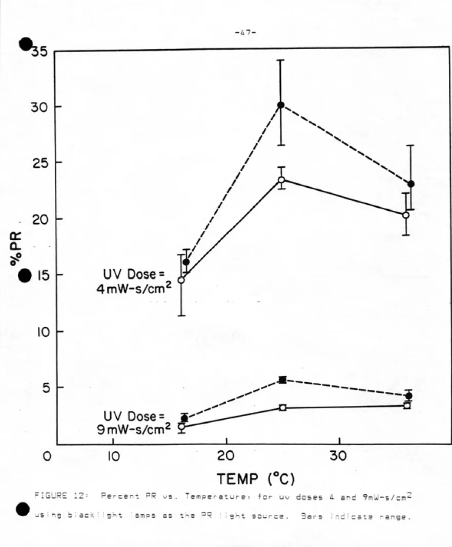
EFFECT OF TEMPERATURE ON PR ͣ .: :
Photareactivation is temperature dependent? largely due to the
ͣ
ormatian of the enzyme-substrate complex (eg, 18? 19? 39), The
second step? which is the photolytic reaction? is reported to be
stable over a wide range of temperatures? from 2 - 4Q°C (38). There was a significant temperature effect also found in this
study of uv-irradiated coliforms. (See Fig, 12 and Table 6).
Recovery increased almost 1ogrythmicaI Iy from 17-25°C at both uv doses tested. Raising the temperature further? to 36^0? did not
promote survival at either uv dose. The PR dose given in each case
was iDDmUsec/cm^. The difference in temperature had a
statistically significant effect at uv dose 9 (p=n.an25)? but not at
uv dose 4 (p=n.D748). This may be due to the high variability in log
survivals at the lowest uv dose? same of which showed lower levels of
J<illing than would be expected? rather than a lack of temperature
-46-ͣ
A3LE 6: EFFECT OF PR TEMPERATURE
''s/ml
______________________________________________________j_Sj___________________________
PR TemperatursW
UV Dose ,_ Dark_____________________________
control 16C 25C 3&C
.iiyzlZE!!!-!__________________________________________________________________
174D
0 1300
, ' ͣ 150D
16C 40,40 266 394 332 ͣ
. 25C 32;28 282 368 362
ͣ
.4 36C 34 J 34 202 378 306
Los S --.
ͣ
-/...'-".: ,.."\:. r'::y :i' -1.64 -0.78 -0.6D -D . 66
% PR J " 15.6 24.3 20.1
- : ͣͣ 16.7 22.5 22.1
11.3 , 23.2 18.4
16C 1.4,3.0 18.0 53.4 57.2
9 25C l.a;2.6 28.8 49.8 52.4 ' 36C 3.6,2.4 26.6 47.6 51.4 Log S -2.81 -1.79 -1.49 , : -1.45
%PR " -l.D 3.4 3.6
;,. ͣ .1 .S ͣ 3.1 3.3
.. \: ' ' 1.6 3.0 ' 3.2
^1
-LI-30
25
20
15
10
UV Dose =
4mW-s/cm^
UV Dose= i
9mW-s/cm2
10 30
TEMP rC)
---i
FIGURE 12: Percent PR vs. Temperature! for uv doses 4 and ^mUl-s/cm^
-4B-There is ampie precedent in_ the I iteraturej indicatins that a
variety of microbes reach maximum reccwery in the 24-26°C
temperature range (eg 1> 1^ 11). In a recent study compar i ng Ej^_cDil
and S^_aureus> Adl<ins and Ai i en found 23 - 25'^C to yield the
highest levels of photoreactivation in both organisms (1). Kelnerj
in the early literature showed PR to increase with temperature
between 2D - 40°C. for several bacteria tested {7.L) . In many early
experiments; however; temperature was not control led; nor was heat
from the lamps; which can be considerable. Increasing the
temperature from 25° to 3(i° decreased the intensity output of the lamps; as did decreasing the temperature from 25^ to 17°C. This
was controMed for in our study by adjusting exposure time to
compensate for the change in PR light intensity; giving a dose of
'^DDmUi-s/cm'^ for all treatments. AI though sever a I studies have used
fluorescent lamps; of both biacklight and day-glow types; the effect of changing temperatures on the lamps themselves were not reported. This has 1 ittle bearing on the field results per se? however should
be considered when laboratory setups are used to simulate •'-'real w o r ! d •'-' c o n d i t i o n s .
Harm (17) explained the decrease in PR at low temperatures by increasing viscosity within the eel I. The enzyme-'s movement from one
lesion to the next may be hampered; and so decrease the repair rate.
Only at ^jery low temperatures; i.e. below C^C; did the site
conformationai changes in the enzyiiie-substrats complex and decrease
in the metabolic rate as significantly affecting PR.
The Bergen County study included temperature as an independent
variable; and conducted a series of exposures between Feb. and Aug.
-49- ;.
eparted) with a correlation coefficient of D.75. Howeverj there was a high degree of variability in the photoreactivated samples? and the
increasing intensity of the sun was not considered as a variable.
Intensity does affect PR? and this increase in intensity is
concomitant with the rising water temperature in their experim.ents.
This report suggests that perhaps they should be considered
separately. These were ali conducted in either the chlorine contact
chamber; or in the spi ! I way leading to it. Battles were placed in
the channel in some cases? or samples were collected directly from
the contact chamber or downstream in others. The actual PR dose received by any sample would vary. Also? no attempt was made to .
measure the suni ight intensity? nor was there any mention of the
meteriological conditions (overcast? sunny? or variable)? and so
(general izations srs difficult to make from this data? even though the
authors attribute 5D% of the correlation coefficient to temperature
effects.
EFFECT OF NUTRIENT LEVELS ON PR
UV treated secondary effluent was di luted with either a sterile?
non-nutrient buffer? or fi Iter steri I ized Morgan Creek water
col lected 1/4 mi ie upstream from the effluent outfa! 1 . The fi Itered creak water contained less than one coliform per IDDml. Samples were
again irradiated at two uv dose levels? as previously described. No
significant difference was found between undiluted samples and those diluted with sterile creek water at either uv dose tested. The pvalues were Q.61 and Q.13 and 9mW-sec/cm'^ ? respectively. However?
.when diluted with phosphate buffer? sample (p=Q.u6 and D.DDn2 for uv
-50-•iBLE 7 . Effect of nutrient on photDr eac t i vat i an .
UV_________________________6/8___________________________________6/11_________
Dose Treatment Dilution* cfu/mi 7.PR Treatment Oil* cfu/mi %PR
3780
Darl< 78 Dark
Lial^t Undi 1 uted 1250 1170 1120 30.0 28.5 26.7 Li gl-it 1:10" 1570 1330 1550 38.2 32.1
37.7 ͣ; , -'
1:40 1456 1372 1168 35.3 33.7 27.7
Dark 0.4 Dark
Light Undi uted 117 164 185
2.7
4.1
4.6 :
L i ght
1:10 207 218 187 5.2 5.5: 4.7 1:40 147 206 210 3.7 5.2 5.3 un J i ght Intens i ty =
(mW/cm2)
3.7 - 4.5
Und
1 : 10
1:40
Und
1 = 10
1:40
387 120
530 32 4
530 32 4 440 25 3 680 44 2 570 35 5 560 34 7 568 35 4 488 27 n
560 34 7
-! C
67 4.7 82 5.8 71 5.0 108 7.7 87 6.2 87 6.3 150 10.7 77 7.1 SO 5.7
3.25 - 3.30
liable 8 Nutrient Effects Diluted an Sterile Phosphate Buffer
"ͣͣ;-. -,. . Sunlight I =
ͣ
: , , ͣ ; ^ .: ͣ- 3.2 ~ 3.35
UV Dilutian Ave. Ave. Lng Ave,
Dgse________EsiiS_____Eiy/siJ__________i^iliillidlJ___._______?»°S____________
D - 3653 ..-- . - ^ .
4 dar i<
cantrol 42.7 -1.93 ; , -^
.;.-.-_.-undiluted HID -D . 52 ͣͣ.29.6 'ͣ I"/ 1 = 10 ^ _ 12D7 : ;^ ,:-D.48 ,";:;^ ^ 32.2 /
1:4D .:;.626.7 . '-0.77 :ͣ 16.2
dark contra 1 und i 1uted 1 •• 10
1 :4n
2.D 14.6 113 85.3
-3.27
ͣ
1.40 -1 .51 -1.63