ACC/AHA/ACP-ASIM PRACTICE GUIDELINES
ACC/AHA/ACP-ASIM Guidelines
for the Management of
Patients With Chronic Stable Angina
A Report of the American College of Cardiology/
American Heart Association Task Force on Practice Guidelines
(Committee on Management of Patients With Chronic Stable Angina)
COMMITTEE MEMBERS
RAYMOND J. GIBBONS, MD, FACC,Chair
KANU CHATTERJEE, MB, FACC JENNIFER DALEY, MD, FACP JOHN S. DOUGLAS, MD, FACC STEPHAN D. FIHN, MD, MPH, FACP JULIUS M. GARDIN, MD, FACC MARK A. GRUNWALD, MD, FAAFP
DANIEL LEVY, MD, FACC BRUCE W. LYTLE, MD, FACC ROBERT A. O’ROURKE, MD, FACC WILLIAM P. SCHAFER, MD, FACC SANKEY V. WILLIAMS, MD, FACP
TASK FORCE MEMBERS JAMES L. RITCHIE, MD, FACC, Chair
RAYMOND J. GIBBONS, MD, FACC, Vice Chair
MELVIN D. CHEITLIN, MD, FACC KIM A. EAGLE, MD, FACC
TIMOTHY J. GARDNER, MD, FACC ARTHUR GARSON, JR, MD, MPH, FACC
RICHARD O. RUSSELL, MD, FACC THOMAS J. RYAN, MD, FACC SIDNEY C. SMITH, JR, MD, FACC
TABLE OF CONTENTS
Preamble ...2093
I. Introduction and Overview...2093
A. Organization of Committee and Evidence Review...2093
B. Scope of the Guidelines ...2094
C. Overlap With Other Guidelines ...2094
D. Magnitude of the Problem ...2095
E. Organization of the Guidelines ...2097
II. Diagnosis...2098
A. History and Physical ...2098
B. Associated Conditions ...2105
C. Noninvasive Testing ...2106
1. ECG/Chest X-Ray...2106
2. Exercise ECG for Diagnosis...2107
3. Echocardiography (Resting) ...2111
4. Stress Imaging Studies—Echo and Nuclear...2112
D. Invasive Testing: Value of Coronary Angiography ...2119
III. Risk Stratification...2121
A. Clinical Assessment ...2121
B. ECG/Chest X-Ray...2123
C. Noninvasive Testing ...2123 This document was approved by the American College of Cardiology Board of
Trustees in March 1999, the American Heart Association Science Advisory and Coordinating Committee in March 1999, and the American College of Physicians-American Society of Internal Medicine Board of Regents in February 1999.
When citing this document, please use the following citation format: Gibbons RJ, Chatterjee K, Daley J, Douglas JS, Fihn SD, Gardin JM, Grunwald MA, Levy D, Lytle BW, O’Rourke RA, Schafer WP, Williams SV. ACC/AHA/ACP-ASIM guidelines for the management of patients with chronic stable angina: a report of the American College of Cardiology/American Heart Association Task Force on Practice Guidelines (Committee on the Management of Patients With Chronic Stable Angina). J Am Coll Cardiol 1999;33:2092–197.
1. Resting LV Function (Echo/Radionuclide
Imaging) ...2123 2. Exercise Testing for Risk Stratification and
Prognosis ...2124 3. Stress Imaging Studies (Radionuclide and
Echocardiography) ...2127 D. Coronary Angiography and Left
Ventriculography ...2133
IV. Treatment ...2135 A. Pharmacologic Therapy ...2135 B. Definition of Successful Treatment and Initiation of
Treatment ...2145 C. Education of Patients with Chronic Stable Angina...2147 D. Coronary Disease Risk Factors and Evidence That
Treatment Can Reduce the Risk for Coronary
Disease Events ...2149 E. Revascularization for Chronic Stable Angina ...2161
V. Patient Follow-up: Monitoring of Symptoms and
Antianginal Therapy ...2167
References ...2170 Index...2191
PREAMBLE
It is important that the medical profession play a significant role in critically evaluating the use of diagnostic procedures and therapies in the management or prevention of disease states. Rigorous and expert analysis of the available data documenting relative benefits and risks of those procedures and therapies can produce helpful guidelines that improve the effectiveness of care, optimize patient outcomes, and have a favorable impact on the overall cost of care by focusing resources on the most effective strategies.
The American College of Cardiology (ACC) and the American Heart Association (AHA) have jointly engaged in the production of such guidelines in the area of cardiovascular disease since 1980. This effort is directed by the ACC/AHA Task Force on Practice Guidelines, whose charge is to develop and revise practice guidelines for important cardiovascular diseases and procedures. Experts in the subject under consideration are selected from both organi-zations to examine subject-specific data and write guidelines. The process includes additional representatives from other medical practitioner and specialty groups where appropriate. Writing groups are specifically charged to perform a formal literature review, weigh the strength of evidence for or against a particular treatment or procedure, and include estimates of expected health outcomes where data exist. Patient-specific modifiers, comorbidities and issues of patient preference that might influence the choice of particular tests or therapies are considered as well as frequency of follow-up and cost-effectiveness.
The ACC/AHA Task Force on Practice Guidelines makes every effort to avoid any actual or potential conflicts of interest that might arise as a result of an outside relationship or personal interest of a member of
the writing panel. Specifically, all members of the writing panel are asked to provide disclosure statements of all such relationships that might be perceived as real or potential conflicts of interest. These statements are re-viewed by the parent task force, reported orally to all members of the writing panel at the first meeting, and updated yearly and as changes occur.
These practice guidelines are intended to assist physicians in clinical decision making by describing a range of generally acceptable approaches for the diagnosis, management, and prevention of specific diseases or conditions. These guide-lines attempt to define practices that meet the needs of most patients in most circumstances. The ultimate judgment regarding care of a particular patient must be made by the physician and patient in light of all of the circumstances presented by that patient.
The executive summary and recommendations are pub-lished in the June 1, 1999 issue ofCirculation. The full text is published in the June 1999 issue of theJournal of the American
College of Cardiology.Reprints of the full text and the executive
summary are available from both organizations.
James L. Ritchie, MD, FACC Chair, ACC/AHA Task Force on Practice Guidelines
I. INTRODUCTION AND OVERVIEW
A. Organization of Committee and Evidence Review
The ACC/AHA Task Force on Practice Guidelines was formed to make recommendations regarding the diagnosis and treatment of patients with known or suspected cardio-vascular disease. Ischemic heart disease is the single leading cause of death in the U.S. The most common manifestation of this disease is chronic stable angina. Recognizing the importance of the management of this common entity and the absence of national clinical practice guidelines in this area, the task force formed the current committee to develop guidelines for the management of patients with stable angina. Because this problem is frequently encountered in the practice of internal medicine, the task force invited the American College of Physicians-American Society of Internal Medicine (ACP-ASIM) to serve as a partner in this effort by naming four general internists to serve on the committee.
trials with small numbers of patients, careful analyses of nonrandomized studies or observational registries. A low rank (C) was given when expert consensus was the primary basis for the recommendation.
The customary ACC/AHA classifications I, II and III are used in tables that summarize both the evidence and expert opinion and provide final recommendations for both patient evaluation and therapy:
Class I Conditions for which there is evidence or general agreement that a given procedure or treatment is useful and effective.
Class II Conditions for which there is conflicting ev-idence or a divergence of opinion about the usefulness/efficacy of a procedure or treat-ment.
Class IIa Weight of evidence/opinion is in favor of usefulness/efficacy. Class IIb Usefulness/efficacy is less well
es-tablished by evidence/opinion. Class III Conditions for which there is evidence and/or
general agreement that the procedure/ treatment is not useful/effective and in some cases may be harmful.
A complete list of many publications on various aspects of this subject is beyond the scope of these guidelines; only selected references are included. The committee consisted of acknowledged experts in general internal medicine from the ACP-ASIM, family medicine from the American Academy of Family Physicians (AAFP), and general cardiology as well as persons with recognized expertise in more special-ized areas, including noninvasive testing, preventive cardi-ology, coronary intervention, and cardiovascular surgery. Both the academic and private practice sectors were repre-sented. This document was reviewed by three outside reviewers nominated by the ACC, three outside reviewers nominated by the AHA, three outside reviewers nominated by the ACP-ASIM, and two outside reviewers nominated by the AAFP. This document was approved for publication by the governing bodies of the ACC, AHA, and ACP-ASIM. The task force will review these guidelines one year after publication and yearly thereafter to determine whether revisions are needed. These guidelines will be considered current unless the task force revises or withdraws them from distribution.
B. Scope of the Guidelines
These guidelines are intended to apply to adult patients with stable chest pain syndromes and known or suspected ischemic heart disease. Patients who have “ischemic equiv-alents,” such as dyspnea or arm pain with exertion, are included in these guidelines. Some patients with ischemic heart disease may become asymptomatic with appropriate therapy. As a result, the follow-up sections of the guidelines may apply to patients who were previously symptomatic. However, the diagnosis, risk stratification and treatment
sections of the guidelines are intended to apply to symp-tomatic patients. Asympsymp-tomatic patients with “silent isch-emia” or known coronary artery disease (CAD) that has been detected in the absence of symptoms are beyond the scope of these guidelines. Pediatric patients are also beyond the scope of these guidelines because ischemic heart disease is very unusual in such patients and is primarily related to the presence of coronary artery anomalies. Patients with chest pain syndromes following cardiac transplantation are also not included in these guidelines.
Patients with nonanginal chest pain are generally at lower risk for ischemic heart disease. Often their chest pain syndromes have identifiable noncardiac causes. Such pa-tients are included in these guidelines if there is sufficient suspicion of heart disease to warrant cardiac evaluation. If the evaluation demonstrates that ischemic heart disease is unlikely and noncardiac causes are the primary focus of evaluation, such patients are beyond the scope of these guidelines. If the initial cardiac evaluation demonstrates that ischemic heart disease is possible, subsequent management of such patients does fall within these guidelines.
Acute ischemic syndromes are not included in these guidelines. For patients with acute myocardial infarction (MI), the reader is referred to the “ACC/AHA Guidelines for the Management of Patients With Acute Myocardial Infarction” (1). For patients with unstable angina, the reader is referred to the Agency for Health Care Policy and Research (AHCPR) clinical practice guideline on unstable angina (2), which was endorsed by the ACC and the AHA. This guideline for unstable angina did describe some low-risk patients who should not be hospitalized but instead evaluated as outpatients. Such patients are indistinguishable from many patients with stable chest pain syndromes and are therefore within the scope of the present guidelines. Patients whose recent unstable angina was satisfactorily treated by medical therapy and who then present with a recurrence of symptoms with a stable pattern fall within the scope of the present guidelines. Similarly, patients with MI who subsequently present with stable chest pain symptoms
.30 days after the initial event are within the scope of the present guidelines.
The present guidelines do not apply to patients with chest pain symptoms early after revascularization by either percu-taneous techniques or coronary artery bypass grafting. Al-though the division between “early” and “late” symptoms is arbitrary, the committee believed that these guidelines should not be applied to patients who develop recurrent symptoms within six months of revascularization.
C. Overlap With Other Guidelines
This report includes text and recommendations from many of these guidelines, which are clearly indicated. Additions and revisions have been made where appropriate to reflect more recently available evidence. This report specifically indicates rare situations in which it deviates from previous guidelines and presents the rationale. In some cases, this report attempts to combine previous sets of similar and dissimilar recommendations into one set of final recommendations. Although this report includes a signifi-cant amount of material from the previous guidelines, by necessity the material was often condensed into a succinct summary. These guidelines are not intended to provide a comprehensive understanding of the imaging modalities, therapeutic modalities, and clinical problems detailed in other guidelines. For such an understanding, the reader is referred to the original guidelines listed in the references.
D. Magnitude of the Problem
There is no question that ischemic heart disease remains a major public health problem. Chronic stable angina is the initial manifestation of ischemic heart disease in approxi-mately one half of patients (3,4). It is difficult to estimate the number of patients with chronic chest pain syndromes in the U.S. who fall within these guidelines, but clearly it is measured in the millions. The reported annual incidence of angina is 213/100,000 population.30 years old (3). When the Framingham Heart Study (4) is considered, an addi-tional 350,000 Americans each year are covered by these guidelines. The AHA has estimated that 6,200,000
Amer-icans have chest pain (5); however, this may be a conserva-tive estimate.
The prevalence of angina can also be estimated by extrapolating from the number of MIs in the U.S. (1). About one half of patients presenting at the hospital with MI have preceding angina (6). The best current estimate is that there are 1,100,000 patients with MI each year in the U.S. (5); about one half of these (550,000) survive until hospitalization. Two population-based studies (from Olm-sted County, Minnesota, and Framingham, Massachusetts) examined the annual rates of MI in patients with symptoms of angina and reported similar rates of 3% to 3.5% per year (4,7). On this basis, it can be estimated that there are 30 patients with stable angina for every patient with infarction who is hospitalized. As a result, the number of patients with stable angina can be estimated as 30 3 550,000, or 16,500,000. This estimate does not include patients who do not seek medical attention for their chest pain or whose chest pain has a noncardiac cause. Thus, it is likely that the present guidelines cover at least six million Americans and conceivably more than twice that number.
Ischemic heart disease is important not only because of its prevalence but also because of its associated morbidity and mortality. Despite the well-documented recent decline in cardiovascular mortality (8), ischemic heart disease remains the leading single cause of death in the U.S. (Table 2) and is responsible for 1 of every 4.8 deaths (9). The morbidity associated with this disease is also considerable: each year
.1,000,000 patients have an MI. Many more are hospital-Table 1. Recent Clinical Practice Guidelines and Policy Statements Which Overlap With
This Guideline
Guideline Sponsor
Year of Publication
Radionuclide imaging (12) ACC/AHA 1995
Echocardiography (13) ACC/AHA 1997
Exercise testing (14) ACC/AHA 1997
Valvular heart disease (15) ACC/AHA 1998
Ambulatory monitoring (16) ACC/AHA 1999
Coronary angiography (17) ACC/AHA 1999
Percutaneous transluminal coronary angioplasty (18) ACC/AHA 1999 or 2000
Coronary artery bypass surgery (19) ACC/AHA 1999
National cholesterol education project (20) NHLBI 1996
National hypertension education (21) NHLBI 1997
Management of hypercholesterolemia (22) ACP-ASIM 1996
Bethesda Conference on risk factor reduction (23) ACC 1996
Clinical practice guideline: cardiac rehabilitation (24)
AHCPR 1995
Coronary artery calcification: pathophysiology, imaging methods, and clinical implications (25)
AHA 1996
Counseling postmenopausal women about preventive hormone therapy (26)
ACP-ASIM 1992
Bethesda Conference on insurability and
employability of the patient with ischemic heart disease (27)
ized for unstable angina and evaluation and treatment of stable chest pain syndromes. Beyond the need for hospital-ization, many patients with chronic chest pain syndromes are temporarily unable to perform normal activities for hours or days, thereby experiencing a reduced quality of life. According to the recently published data from the Bypass Angioplasty Revascularization Investigation (10), about 30% of patients never return to work following coronary revascularization, and 15% to 20% of patients rated their own health fair or poor despite revascularization. These data confirm the widespread clinical impression that ischemic heart disease continues to be associated with considerable patient morbidity despite the decline in cardiovascular mortality.
The economic costs of chronic ischemic heart disease are enormous. Some insight into the potential cost can be obtained by examining Medicare data for inpatient diagnosis-related groups (DRGs) and diagnostic tests. Ta-ble 3 shows the number of patients hospitalized under various DRGs during 1995 and associated direct payments by Medicare. These DRGs represent only hospitalization of patients covered by Medicare. The table includes estimates for the proportion of inpatient admissions for unstable angina, MI, and revascularization for patients with a history of stable angina. Direct costs associated
with non-Medicare patients hospitalized for the same diagnoses are probably about the same as the covered charges under Medicare. Thus, the direct costs of hospi-talization are .$15 billion.
Table 4 shows the Medicare fees and volumes of com-monly used diagnostic procedures in ischemic heart disease. Although some of these procedures may have been per-formed for other diagnoses and some of the cost of the technical procedure relative value units (RVUs) may have been for inpatients listed in Table 3, the magnitude of the direct costs is considerable. When the 1998 Medicare reimbursement of $36.6873 per RVU is used, the direct cost to Medicare of these 61.2 million RVUs can be estimated at $2.25 billion. Again, assuming that the non-Medicare patient costs are at least as great, the estimated cost of these diagnostic procedures alone would be about $4.5 billion.
These estimates of the direct costs associated with chronic stable angina obviously do not take into account the indirect costs of workdays lost, reduced productivity, long-term medication, and associated other effects. The indirect costs have been estimated to be almost as great as direct costs (4). The magnitude of the problem can be succinctly summarized: chronic stable angina affects many millions of Americans, with associated annual costs that are measured in tens of billions of dollars.
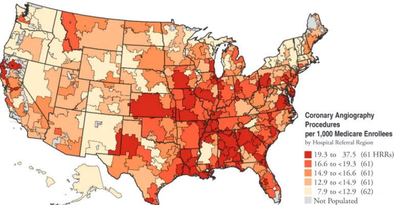
Given the magnitude of this problem, the need for practice guidelines is self-evident. This need is further reinforced by the available information, which suggests considerable regional differences in the management of ischemic heart disease. Figure 1 shows published informa-tion from the Medicare database for rates of coronary angiography in different counties of the country (11). Three- and four-fold differences in adjusted rates for this procedure in different counties within the same state are not uncommon, suggesting that the clinical management of such patients is highly variable. The reasons for such variation in management are unknown.
Table 2. Death Rates Due to Diseases of the Heart and Cancer, United States—1995
Death Rate per 100,000 Population
Group
Diseases of
the Heart Cancer
White males 297.9 228.1
Black males 244.2 209.1
White females 297.4 202.4
Black females 231.1 159.1
From Report of Final Mortality Statistics, 1995, Centers for Disease Control and Prevention (8). These rates are not adjusted for age.
Table 3. Medicare Experience With Commonly Used DRGs Involving Patients With Stable Angina
DRG # Description
1995 Discharges
Covered Charges (million)
Medicare Payments (million)
% of Pts with History of
Stable Angina
Medicare Payments for
Pts with Stable Angina
125 Coronary disease/cath 62,251 $ 519.8 $ 215.9 95* 205.1
143 Chest pain 139,145 641.8 268.1 100 268.1
124 Unstable angina 145,560 1,734.8 770.6 85† 655.0
121 MI with cath 167,202 2,333.5 1,020.8 55‡ 561.4
122 MI without cath 91,569 892.0 350.8 55‡ 192.9
112 PTCA 201,066 3,897.7 1,801.9 83§ 1,495.6
106 CABG with cath 101,057 5,144.0 3,626.9 83§ 3,010.3
107 CABG without cath 64,212 2,473.2 1,280.9 83§ 1,063.1
7,451.5
E. Organization of the Guidelines
These guidelines are arbitrarily divided into four sec-tions: diagnosis, risk stratification, treatment and patient follow-up. Experienced clinicians will quickly recognize
that the distinctions between these sections may be arbitrary and unrealistic in individual patients. However, for most clinical decision making, these divisions are helpful and facilitate presentation and analysis of the available evidence.
Figure 1. Map depicting coronary angiography rates in the U.S. HRR 5 hospital referral region. From Wennberg et al. (11) with permission.
Table 4. Medicare Fees and Volumes of Commonly Used Diagnostic Procedures for Chronic Stable Angina
Procedure
1998 CPT Code(s)
1998 Total (Professional and Technical) Medicare RVUs
Number Performed
(1996)
Estimated % for Stable
Angina
Estimated Total RVUs
Echocardiogram 93307 5.96 3,935,344 20% 4,690,930
Doppler echo 93320 2.61 3,423,899 20% 1,787,233
Treadmill exercise test 93015 or 93016–93018
3.25 689,851* 80% 1,793,612
Stress echocardiography 93350, 93015 6.81 303,047 80% 1,651,000
Stress SPECT myocardial perfusion imaging
78465, 93015 17.41 1,158,389 80% 16,134,041
Left heart catheterization with left ventriculogram and coronary angiography
93510, 93543, 93545, 93555,
93556
66.18 664,936† 80% 35,204,371
61,261,187
*Estimated by subtracting (93350178465) from (93015193018), since the total number of charges under 93015 and 93018 includes stress echo and stress SPECT. †Estimated from Medicare data. One source (David Wennberg, personal communication) has suggested this number could be as high as 771,925.
The three flow diagrams that follow summarize the management of stable angina in three algorithms: clinical assessment (Fig. 2), stress testing/angiography (Fig. 3), and treatment (Fig. 4). The treatment mnemonic (Fig. 5) is intended to highlight the 10 treatment elements that the committee considered most important.
Although the evaluation of many patients will require all three algorithms, this is not always true. Some patients may require only clinical assessment to determine that they do not belong within these guidelines. Others may require only clinical assessment and treatment if the probability of CAD is high and patient preferences and comorbidities preclude revascularization (and therefore the need for risk stratifica-tion). The stress testing/angiography algorithm may be required either for diagnosis (and risk stratification) in patients with a moderate probability of CAD or for risk
stratification only in patients with a high probability of CAD.
II. DIAGNOSIS
A. History and Physical
Recommendations
Class I: In patients presenting with chest pain, a detailed symptom history, focused physical examination, and directed risk-factor assess-ment should be performed. With this infor-mation, the clinician should estimate the probability of significant CAD (i.e., low, in-termediate, high).(Level of Evidence: B)
Definition of Angina
Angina is a clinical syndrome characterized by discomfort in the chest, jaw, shoulder, back or arm. It is typically aggravated by exertion or emotional stress and relieved by nitroglycerin. Angina usually occurs in patients with CAD involving$1 large epicardial artery. However, angina can also occur in persons with valvular heart disease, hypertro-phic cardiomyopathy and uncontrolled hypertension. It can be present in patients with normal coronary arteries and myocardial ischemia related to spasm or endothelial dys-function. Angina is also a symptom in patients with non-cardiac conditions of the esophagus, chest wall or lungs. Once cardiac causes have been excluded, the management of patients with these noncardiac conditions is outside the scope of these guidelines.
Clinical Evaluation of Patients With Chest Pain
History
The clinical examination is the most important step in the evaluation of the patient with chest pain, allowing the clinician to estimate the likelihood of clinically significant CAD with a high degree of accuracy (29). Significant CAD is defined angiographically as CAD with $70% diameter stenosis of $1 major epicardial artery segment or $50% diameter stenosis of the left main coronary artery. Although lesions of less stenosis can cause angina, they have much less prognostic significance (30).
The first step, a detailed description of the symptom complex, enables the clinician to characterize the chest pain (31). Five components are typically considered: quality,
location, duration of pain, factors that provoke the pain and factors that relieve the pain. Various adjectives have been used by patients to describe the quality of the anginal pain: “squeezing,” “griplike,” “pressurelike,” “suffocating” and
“heavy” are common. Not infrequently, patients insist that their symptom is a “discomfort” but not “pain.” Angina is almost never sharp or stabbing, and it usually does not change with position or respiration.
The anginal episode is typically minutes in duration. Fleeting discomfort or a dull ache lasting for hours is rarely angina. The location of angina is usually substernal, but radiation to the neck, jaw, epigastrium, or arms is not uncommon. Pain above the mandible, below the epigas-trium, or localized to a small area over the left lateral chest wall is rarely anginal. Angina is generally precipitated by exertion or emotional stress and commonly relieved by rest. Sublingual nitroglycerin also relieves angina, usually within 30 s to several minutes.
After the history of the pain is obtained, the physician makes a global assessment of the symptom complex. One classification scheme for chest pain in many studies uses three groups: typical angina, atypical angina or noncardiac chest pain (32) (Table 5).
Angina is further classified as stable or unstable (2). Unstable angina is important in that its presence predicts a much higher short-term risk of an acute coronary event. Unstable angina is operationally defined as angina that Figure 5. Treatment mnemonic: the 10 most important treatment elements of stable angina management.
Table 5. Clinical Classification of Chest Pain
Typicalangina (definite)
1) Substernal chest discomfort with a characteristic quality and duration that is 2) provoked by exertion or emotional stress and 3) relieved by rest or NTG.
Atypicalangina (probable)
- Meets 2 of the above characteristics. Noncardiacchest pain
- Meets one or none of the typical anginal characteristics.
presents in one of three principal ways: rest angina, severe new-onset angina, or increasing angina (Tables 6 and 7). Most important, unstable angina patients can be subdivided by their short-term risk (Table 8). Patients at high or moderate risk often have coronary artery plaques that have recently ruptured. Their risk of death is intermediate, between that of patients with acute MI and patients with stable angina. The initial evaluation of high- or moderate-risk patients with unstable angina is best carried out in the inpatient setting. However, low-risk patients with unstable angina have a short-term risk not substantially different from those with stable angina. Their evaluation can be accomplished safely and expeditiously in an outpatient setting. The recommendations made in these guidelines do not apply to high- and moderate-risk unstable angina but are applicable to the low-risk unstable angina group.
After a detailed chest pain history is taken, the presence of risk factors for CAD (23) should be determined. Ciga-rette smoking, hyperlipidemia, diabetes, hypertension and a family history of premature CAD are all important. Past history of cerebrovascular or peripheral vascular disease increases the likelihood that CAD will be present.
Physical
The physical examination is often normal in patients with stable angina (33). However, an exam made during an episode of pain can be beneficial. An S4 or S3 sound or
gallop, mitral regurgitant murmur, a paradoxically split S2or
bibasilar rales or chest wall heave that disappears when the pain subsides are all predictive of CAD (34). Even though the physical is generally not helpful for confirming CAD, a careful cardiovascular exam may reveal other conditions associated with angina, such as valvular heart disease or hypertrophic cardiomyopathy. Evidence of noncoronary atherosclerotic disease—a carotid bruit, diminished pedal pulse or abdominal aneurysm—increases the likelihood of CAD. Elevated blood pressure, xanthomas and retinal exudates point to the presence of CAD risk factors. Palpa-tion of the chest wall often reveals tender areas in patients whose chest pain is caused by musculoskeletal chest wall syndromes (35). However, pain produced by pressure on the
chest wall may be present even if the patient has angina due to ischemic heart disease. The presence of a rub will point to pericardial or pleural disease.
Developing the Probability Estimate
When the initial history and physical are complete, the physician and patient find themselves asking the same question: “Is it the heart?” In certain instances, the physician can confidently assure the patient that it is not. Patients with noncardiac chest pain are generally at lower risk for ischemic heart disease. As indicated on the flow diagram, the history and appropriate diagnostic tests will usually focus on noncardiac causes of chest pain. Appropriate treatment and follow-up for the noncardiac condition can be prescribed, and the patient can be educated about CAD and risk factors, especially if he or she rarely sees a physician. When there is sufficient suspicion of heart disease to warrant cardiac evaluation, the clinician should make a probability estimate of the likelihood of CAD. The impor-tance of doing so is obvious when considering how this estimate affects the utility of a commonly used diagnostic test: the standard exercise test. Consider how interpretation of the standard exercise test would be affected by varying the pretest probability of disease from 5% to 50% to 90% (36). In this example, the exercise test is considered positive if $1-mm ST-segment depression is observed. The test sensitivity is 50% and specificity 90% (14).
In patients with a low probability of CAD (5%), the positive predictive value of an abnormal test result is only 21%. If 1,000 low-probability patients are tested, 120 will test positive. Of these, 95 will not have significant CAD. Before testing such a Table 6. Three Principal Presentations of Unstable Angina (2)
Rest angina Angina occurring at rest and usually prolonged .20 minutes occurring within a week of presentation.
New onset angina Angina of at least CCSC III severity with onset within 2 months of initial presentation.
Increasing angina Previously diagnosed angina that is distinctly more frequent, longer in duration or lower in threshold (i.e., increased by at least one CCSC class within 2 months of initial presentation to at least CCSC III severity).
Note: CCSC5Canadian Cardiovascular Society Classification.
Table 7. Grading of Angina Pectoris by the Canadian Cardiovascular Society Classification System (46)
Class I
Ordinary physical activity does not cause angina, such as walking, climbing stairs. Angina (occurs) with strenuous, rapid or prolonged exertion at work or recreation. Class II
Slight limitation of ordinary activity. Angina occurs on walking or climbing stairs rapidly, walking uphill, walking or stair climbing after meals, or in cold, or in wind, or under emotional stress, or only during the few hours after
awakening. Angina occurs on walking more than 2 blocks on the level and climbing more than one flight of ordinary stairs at a normal pace and in normal condition.
Class III
Marked limitations of ordinary physical activity. Angina occurs on walking one to two blocks on the level and climbing one flight of stairs in normal conditions and at a normal pace.
Class IV
Inability to carry on any physical activity without discomfort– anginal symptoms may be present at rest.
group, the clinician must weigh the value of correctly diagnos-ing CAD in 25 patients against the cost of a stress test for all 1,000 patients plus the cost of misdiagnosis—undue anxiety, further invasive testing, unnecessary medications or higher insurance premiums—for the 95 patients with a false-positive test result. In patients with a high probability of CAD (90%), a positive test result raises the probability of disease to 98% and a negative test result lowers probability to 83%. Although exercise testing has prognostic value in these patients (see Section III, C-2) (37), a negative test result obviously does not allow the clinician to discard the diagnosis of CAD. In patients with a 50% probability of CAD, a positive test result increases the likelihood of disease to 83% and a negative test result decreases the likelihood to 36%. The test separates this group of patients into two distinct subgroups: one in whom CAD almost certainly exists and the other for whom the diagnosis, although far from being excluded, is doubtful. An accurate estimate of the likelihood of CAD is necessary for interpreta-tion of further test results and good clinical decision making about therapy.
Although it may seem premature to predict the probability of CAD after the history and physical, the clinicopathological study performed by Diamond and Forrester (38) demonstrated that it is possible. By combining data from a series of angiography studies performed in the 1960s and the 1970s, they showed that the simple clinical observations of pain type, age, and gender were powerful predictors of the likelihood of CAD. For instance, a 64-year-old man with typical angina has a 94% likelihood of having significant CAD. A 32-year-old woman with nonanginal chest pain has a 1% chance of CAD (14).
The value of the Diamond and Forrester approach was subsequently confirmed in prospective studies at Duke and Stanford. In these studies, both men and women were referred to cardiology specialty clinics for cardiac catheter-ization (39,40) or cardiac stress testing (41), and the initial clinical exam characteristics most helpful in predicting CAD were determined. With these characteristics, predic-tive models (logistic regression equations) were developed. When prospectively applied to another group of patients referred to the same specialty clinic, the models worked well. As in Diamond and Forrester’s original work, age, gender and pain type were the most powerful predictors. Other characteristics that strengthened the predictive abil-ities of the models were smoking (defined as a history of smoking half a pack or more of cigarettes per day within five years of the study or at least 25 pack-years), Q wave or ST-T-wave changes, hyperlipidemia (defined as a choles-terol level.250 mg/dL) and diabetes (glucose.140). Of these risk factors, diabetes had the greatest influence on increasing risk. Other significant risk factors, such as family history and hypertension, were not as strongly predictive and did not improve the power of equations.
Generalizability of the Predictive Models
Although these models worked well prospectively in the settings in which they were developed, clinicians must assess how reliable they will be when used in their own practices. The Diamond and Forrester probabilities were compared with those published in the Coronary Artery Surgery Study (CASS) (42), a large 15-center study that compared clinical and angiographic findings in .20,000 patients. In both studies, Table 8. Short-Term Risk of Death or Nonfatal Myocardial Infarction in Patients With Unstable Angina (2)
High Risk Intermediate Risk Low Risk
At least one of the following features must be present:
No high-risk features but must have any of the following:
No high- or intermediate-risk feature but may have any of the following features:
Prolonged ongoing (.20 min) rest pain
Prolonged (.20 min) rest angina, now resolved, with moderate or high likelihood of CAD
Increased angina frequency, severity, or duration
Pulmonary edema, most likely related to ischemia
Rest angina (.20 min or relieved with sublingual nitroglycerin)
Angina provoked at a lower threshold
Angina at rest with dynamic ST changes$1 mm
Nocturnal angina New onset angina with onset 2 weeks
to 2 months prior to presentation Angina with new or worsening MR
murmur
Angina with dynamic T-wave changes Normal or unchanged ECG
Angina with S3or new/worsening rales
New onset CCSC III or IV angina in the past 2 weeks with moderate or high likelihood of CAD
Angina with hypotension Pathologic Q waves or resting ST depression#1 mm in multiple lead groups (anterior, inferior, lateral) Age.65 years
CCSC5Canadian Cardiovascular Society Classification.
probability tables were presented in which patients were categorized by age, gender, and pain type. Tables with 24 patient groupings were published. With the exception of adults
,50 years old with atypical angina for whom the CASS data estimated a probability of disease 17% higher than the Diamond-Forrester data, the agreement between studies was very close: the difference averaged 5%. Because the results were so similar, the committee combined the probabilities from both studies in one evidence table (Table 9).
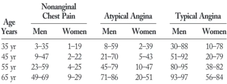
It is more difficult to compare the Duke data directly with the CASS and Diamond-Forrester tables because within each age, gender, and pain type grouping, the patient’s predicted probability of disease varies, depending on the presence or absence of electrocardiogram (ECG) findings (Q waves or ST-T changes) or risk factors (smoking, diabetes, hyperlipidemia). Table 10 presents the Duke data for mid-decade patients (35, 45, 55, and 65 years old). Two probabilities are given. The first is for a low-risk patient with no risk factors and a normal ECG. The second is for a high-risk patient who smokes and has diabetes and hyperlipidemia but has a normal ECG. The presence of ECG changes would increase the probability of coronary disease even more. When Tables 9 and 10 are compared, the correlation between studies is quite strong. Apparent in the Duke data is the importance of risk factors in modifying the
likelihood of disease. This becomes more important the younger the patient and the more atypical the pain. For example, the likelihood of disease for women,55 years old with atypical angina and no risk factors is ,10%, but if diabetes, smoking and hyperlipidemia are present, the likelihood jumps to 40%.
Applicability of Models to Primary-Care Practices
All the studies mentioned above were university-based. The patients used to develop the models were largely referred. The only study that directly looked at applica-bility of the university-derived model to primary-care practices was the Stanford study (40). The university-derived equation was used and the likelihood of CAD was predicted for patients presenting to two urban primary-care clinics. The equation worked well for typ-ical angina patients but substantially overpredicted CAD for patients at less risk.
Referral (or ascertainment) bias in these studies likely explains these differences (43,44) because the clinical decision-making process before the patient was referred is unknown. Primary-care providers do not unselectively refer all chest pain patients for cardiac evaluation. The disease probabilities for high-risk patients will vary little from the study because few primary-care physicians will fail to recommend cardiac evaluation for typical angina patients. However, younger patients with less classic pain stories will often be referred only after therapeutic trials, time or noncardiac diagnostic studies fail to eliminate CAD as a possibility. Correcting for referral bias is required before these models can be applied to primary-care practices. The Stanford study showed that it was possible to correct the model predictions by using the overall prevalence of CAD in the primary care population (40). Unfortunately, while Bayesian analysis might help a primary care provider im-prove the models, there are no studies examining how accurately providers calculate the prevalence of CAD among their chest pain patients, or how the prevalence of CAD varies among primary care settings. Primary-care physicians must therefore exercise caution when using these predictive equations, tables, or nomograms with patients presenting for the first time with chest pain. Whether the difference between the model estimates and actual likelihood of CAD is great enough to lead to a different diagnostic and therapeutic strategy is not known.
Ideally, the strategy a clinician uses to evaluate a patient with chest pain will also take into account the patient’s preferences. Two patients with the same pretest probability of CAD may prefer different approaches because of varia-tions in personal beliefs, economic situation or stage of life. Patient preference studies that inform physicians about what is an acceptable balance between the underdiagnosis and overdiagnosis of CAD have not been done.
Table 9. Pretest Likelihood of CAD in Symptomatic Patients According to Age and Sex* (Combined Diamond/Forrester and CASS Data) (38,42)
Age Years
Nonanginal
Chest Pain Atypical Angina Typical Angina
Men Women Men Women Men Women
30–39 4 2 34 12 76 26
40–49 13 3 51 22 87 55
50–59 20 7 65 31 93 73
60–69 27 14 72 51 94 86
*Each value represents the percent with significant CAD on catheterization.
Table 10. Comparing Pretest Likelihoods of CAD in Low-Risk Symptomatic Patients With High-Risk Symptomatic
Patients—Duke Database (41)
Age Years
Nonanginal
Chest Pain Atypical Angina Typical Angina
Men Women Men Women Men Women
35 yr 3–35 1–19 8–59 2–39 30–88 10–78
45 yr 9–47 2–22 21–70 5–43 51–92 20–79
55 yr 23–59 4–25 45–79 10–47 80–95 38–82
65 yr 49–69 9–29 71–86 20–51 93–97 56–84
B. Associated Conditions
Recommendations for Initial Laboratory Tests for Diagnosis
Class I
1. Hemoglobin.(Level of Evidence: C)
2. Fasting glucose.(Level of Evidence: C)
3. Fasting lipid panel, including total cholesterol, HDL cholesterol, triglycerides, and calculated LDL cholesterol.(Level of Evidence: C)
Using information gathered from the history and physical examination, the clinician should consider possibilities other than CAD in the differential diagnosis, because a number of other conditions can both cause and contribute to angina. In those patients with risk factors for CAD but an otherwise low probability history for angina, alternative diagnoses should be considered (Table 11).
In all patients, particularly those with typical angina, comorbid conditions that may precipitate “functional” an-gina (i.e., myocardial ischemia in the absence of significant anatomic coronary obstruction) should be considered. Gen-erally, these are pathological entities that cause myocardial ischemia either by placing increased myocardial oxygen demands on the heart or by decreasing the myocardial oxygen supply (Table 12).
Increased oxygen demand can be produced by such entities as hyperthermia, hyperthyroidism, and cocaine abuse. Hyperthermia, particularly if accompanied by volume contraction due to diaphoresis or other fluid losses, can precipitate angina in the absence of significant CAD (47).
Hyperthyroidism, with its associated tachycardia and increased metabolic rate, increases oxygen demand and, perhaps because of inceased platelet aggregation, may also decrease supply. These effects can readily lead to angina. In addition, elderly patients may not present with a typical clinical picture of thyrotoxicosis. Therefore, this possibility should be considered in the setting of minimal risk factors accompanied by a history of typical angina, particularly in older patients.
Sympathomimetic toxicity, of which cocaine is the prototype, not only increases myocardial oxygen demand but, through coronary vasospasm, simultaneously de-creases supply, sometimes leading to infarction in young patients. Long-term cocaine use may also lead to devel-opment of angina by causing premature develdevel-opment of CAD (48).
Angina may occur in patients with severe uncontrolled hypertension due to increased wall tension, which increases myocardial oxygen demand, and increased left ventricular (LV) end-diastolic pressure, which decreases subendocardial perfusion. These same mechanisms contribute to angina in hypertrophic cardiomyopathy and aortic stenosis; however, in these conditions, wall tension may be even greater due to an outflow tract gradient, and end-diastolic pressure may be even higher due to severe LV hypertrophy.
Table 11. Alternative Diagnoses to Angina for Patients With Chest Pain
Non-Ischemic Cardiovascular Pulmonary Gastrointestinal Chest Wall Psychiatric
Aortic dissection Pulmonary embolus Esophageal Costochondritis Anxiety disorders
Pericarditis Pneumothorax Esophagitis Fibrositis Hyperventilation
Pneumonia Spasm Rib fracture Panic disorder
Pleuritis Reflux Sternoclavicular arthritis Primary anxiety
Biliary Herpes zoster Affective disorders
Colic (before the rash) (e.g., depression)
Cholecystitis Somatiform disorders
Choledocholithiasis Thought disorders
Cholangitis (e.g., fixed delusions)
Peptic ulcer Pancreatitis
Table 12. Conditions Provoking or Exacerbating Ischemia
Increased Oxygen Demand Decreased Oxygen Supply
Non-Cardiac Hyperthermia Hyperthyroidism Sympathomimetic toxicity
(e.g., cocaine use) Hypertension Anxiety
Arteriovenous fistulae
Cardiac
Hypertrophic cardiomyopathy Aortic stenosis
Dilated cardiomyopathy Tachycardia
Ventricular Supraventricular
Non-Cardiac Anemia Hypoxemia
Pneumonia Asthma
Chronic obstructive pulmonary disease
Pulmonary hypertension Interstitial pulmonary fibrosis Obstructive sleep apnea Sickle cell disease Sympathomimetic toxicity
(e.g., cocaine use) Hyperviscosity
Polycythemia Leukemia Thrombocytosis
Hypergammaglobulinemia Cardiac
Aortic stenosis
Sustained tachycardia, either ventricular or supraventric-ular, may also increase myocardial oxygen demand. Parox-ysmal tachycardias are more frequent conditions that con-tribute to angina. Unfortunately, they are often more difficult to diagnose.
Conditions that reduce myocardial oxygen supply must also be considered in the differential diagnosis of patients with angina.
Anemia reduces the oxygen-carrying capacity of the blood and also increases the cardiac workload. An increased cardiac output is associated with ,9 g/dL of hemoglobin, and ST-T wave changes (depression or inversion) may be seen when hemoglobin drops below 7 g/dL.
Hypoxemia resulting from pulmonary disease (e.g., pneu-monia, asthma, chronic obstructive pulmonary disease, pul-monary hypertension, interstitial fibrosis, obstructive sleep apnea) may also precipitate angina. Obstructive sleep apnea should be seriously considered in patients with only noctur-nal symptoms.
Conditions that are associated with increased blood viscosity can increase coronary resistance and thereby de-crease coronary artery blood flow, precipitating angina in patients without severe coronary stenoses. Increased viscos-ity is seen with polycythemia, leukemia, thrombocytosis and hypergammaglobulinemia.
C. Noninvasive Testing
1. ECG/Chest X-Ray
Recommendations for Electrocardiography, Chest X-Ray, or Electron Beam Computed Tomography in the Diagnosis of Chronic Stable Angina
Class I
1. Rest ECG in patients without an obvious non-cardiac cause of chest pain.(Level of Evidence: B)
2. Rest ECG during an episode of chest pain.(Level of Evidence: B)
3. Chest X-ray in patients with signs or symptoms of congestive heart failure, valvular heart disease, pericardial disease, or aortic dissection/aneurysm.
(Level of Evidence: B)
Class IIa
Chest X-ray in patients with signs or symptoms of pulmonary disease.(Level of Evidence: B)
Class IIb
1. Chest X-ray in other patients.(Level of Evidence: C)
2. Electron beam computed tomography (EBCT).
(Level of Evidence: B)
A rest 12-lead ECG should be recorded in all patients with symptoms suggestive of angina pectoris; however, it will be normal in $50% of patients with chronic stable angina (49). A normal rest ECG does not exclude severe CAD. ECG evidence of LV hypertrophy or ST-T wave changes consistent with myocardial ischemia favor the
diagnosis of angina pectoris (50). Evidence of prior Q-wave MI on the ECG makes CAD very likely. However, certain Q-wave patterns are equivocal, such as an isolated Q in lead III or a QS pattern in leads V1and V2.
The presence of arrhythmias such as atrial fibrillation or ventricular tachyarrhythmia on the ECG in patients with chest pain also increases the probability of underlying CAD; however, these arrhythmias are frequently caused by other types of cardiac disease. Various degrees of AV block can be present in patients with chronic CAD but have many other causes and a very low specificity for the diagnosis. Left anterior fascicular block, right bundle-branch block and left bundle-branch block often occur in patients with CAD and frequently indicate the presence of multivessel CAD. How-ever, these findings also lack specificity in the diagnosis of chronic stable angina.
An ECG obtained during chest pain is abnormal in
'50% of patients with angina who have a normal rest ECG. Sinus tachycardia occurs commonly; bradyarrhyth-mia is less common. The ST-segment elevation or depression establishes a high likelihood of angina and indicates ischemia at a low workload, portending an unfavorable prognosis. Many high-risk patients need no further noninvasive testing. Coronary arteriography usu-ally defines the severity of coronary artery stenoses and the necessity and feasibility of myocardial revasculariza-tion. In patients with ST-T wave depression or inversion on the rest ECG, “pseudonormalization” of these abnor-malities during pain is another indicator that CAD is likely (51). The occurrence of tachyarrhythmias, atrio-ventricular block, left anterior fascicular block or bundle-branch block with chest pain also increases the probabil-ity of coronary heart disease (CHD) and often leads to coronary arteriography.
The chest roentgenogram is often normal in patients with stable angina pectoris. Its usefulness as a routine test is not well established. It is more likely to be abnormal in patients with previous or acute MI, those with a noncoronary artery cause of chest pain and those with noncardiac chest discom-fort. Cardiac enlargement may be attributable to previous MI, acute LV failure, pericardial effusion or chronic volume overload of the left ventricle such as occurs with aortic or mitral regurgitation. Abnormal physical findings, associated chest X-ray findings (e.g., pulmonary venous congestion), and abnormalities detected by noninvasive testing (echocar-diography) may indicate the correct etiology.
Enlargement of the upper mediastinum often results from an ascending aortic aneurysm with or without dissection. Pruning or cutoffs of the pulmonary arteries or areas of segmental oligemia may indicate pulmonary infarction/ embolism or other causes of pulmonary hypertension.
Ultrafast Computed Tomography
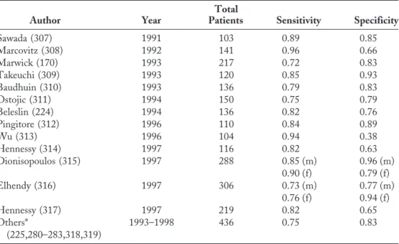
Ultrafast (electron beam) computed tomography is being used with increased frequency for the detection and quan-tification of coronary artery calcification (25). In seven studies including 50 to 710 patients, calcium of the coronary arteries detected by EBCT was an important indicator of angiographic coronary stenoses. In these studies of selected patients, the sensitivity of a positive EBCT detection of calcium for the presence of CAD varied from 85% to 100%; specificity ranged from only 41% to 76%; the positive predictive value varied considerably from 55% to 84% and negative predictive value from 84% to 100% (25). The presence and amount of calcium detected in coronary arteries by EBCT in two studies appeared to correlate with the presence and associated amount of atherosclerotic plaque (53,54). However, several studies (55–57) have shown a marked variability in repeated measures of coronary calcium by EBCT. Therefore, the use of serial EBCT scans in individual patients for identification and serial assessment of the progression or regression of calcium remains prob-lematic. The proper role of EBCT is controversial and will be the subject of future ACC/AHA statements.
2. Exercise ECG for Diagnosis
Recommendations for Diagnosis of Obstructive CAD With Exercise ECG Testing Without an Imaging Modality
Class I
Patients with an intermediate pretest probability of CAD based on age, gender and symptoms, including those with complete right bundle-branch block or <1 mm of ST depression at rest (exceptions are listed below in classes II and III).(Level of Evidence: B)
Class IIa
Patients with suspected vasospastic angina.(Level of Evidence: C)
Class IIb
1. Patients with a high pretest probability of CAD by age, gender and symptoms.(Level of Evidence: B)
2. Patients with a low pretest probability of CAD by age, gender and symptoms.(Level of Evidence: B)
3. Patients taking digoxin whose ECG has<1 mm of baseline ST-segment depression.(Level of Evidence: B)
4. Patients with ECG criteria for LV hypertrophy and <1 mm of baseline ST-segment depression.(Level of Evidence: B)
Class III
1. Patients with the following baseline ECG abnor-malities.
a. Pre-excitation (Wolff-Parkinson-White) syndrome.(Level of Evidence: B)
b. Electronically paced ventricular rhythm.
(Level of Evidence: B)
c. More than 1 mm of ST depression at rest.
(Level of Evidence: B)
d. Complete left bundle-branch block.(Level of Evidence: B)
2. Patients with an established diagnosis of CAD due to prior MI or coronary angiography; however, testing can assess functional capacity and progno-sis, as discussed in section III.(Level of Evidence: B)
Description of the Exercise Testing Procedure
Exercise testing is a well-established procedure that has been in widespread clinical use for many decades. Detailed descriptions of exercise testing are available in other publi-cations (58 – 60). This section provides a brief overview based on the “ACC/AHA Guidelines for Exercise Testing” (14).
Although exercise testing is generally a safe procedure, both MI and death occur at a rate of#1/2500 tests (61). The absolute contraindications to exercise testing include acute MI within two days, cardiac arrhythmias causing symptoms or hemodynamic compromise, symptomatic and severe aortic stenosis, symptomatic heart failure, acute pulmonary embolus or pulmonary infarction, acute myocar-ditis or pericarmyocar-ditis and acute aortic dissection (14,59). Additional factors are relative contraindications: left main coronary stenosis, moderate aortic stenosis, electrolyte ab-normalities, systolic hypertension .200 mm Hg, diastolic blood pressure.110 mm Hg, tachyarrhythmias or brady-arrhythmias, hypertrophic cardiomyopathy and other forms of outflow tract obstruction, mental or physical impairment leading to an inability to exercise adequately and high-degree atrioventricular block (14,59). In the past, unstable angina was a contraindication to exercise testing. However, new information suggests that exercise treadmill (62– 64) and pharmacologic (65– 68) testing are safe in low-risk outpatients with unstable angina and in low- or intermediate-risk patients hospitalized with unstable angina in whom an MI has been ruled out and who are free of angina and congestive heart failure.
Both treadmill and cycle ergometer devices are used for exercise testing. Although cycle ergometers have important advantages, fatigue in the quadricep muscles in patients who are not experienced cyclists usually makes them stop before reaching their maximum oxygen uptake. As a result, tread-mills are more commonly used in the U.S.
There are clear advantages in customizing the protocol to the individual patient to allow exercise lasting 6 to 12 min (69). Exercise capacity should be reported in estimated METs of exercise. (One MET is the standard basal oxygen uptake of 3.5 mL/kg per min.) If exercise capacity is also reported in minutes, the protocol should be described clearly.
defined by the Health Care Financing Administration [HCFA]) is not always required. The ECG, heart rate and blood pressure should be carefully monitored and recorded during each stage of exercise as well as during ST-segment abnormalities and chest pain. The patient should be mon-itored continuously for transient rhythm disturbances, ST-segment changes and other ECG manifestations of myo-cardial ischemia. Although exercise testing is commonly terminated when subjects reach a standard percentage (often 85%) of age-predicted maximum heart rate, there is great variability in maximum heart rates among individuals, so predicted values may be supramaximal for some patients and submaximal for others. Therefore, it is important to monitor the patient closely for other indications for stopping the test. Absolute indications for stopping include a drop in systolic blood pressure of.10 mm Hg from baseline blood pressure despite an increase in workload when accompanied by other evidence of ischemia; moderate to severe angina; increasing ataxia, dizziness or near syncope; signs of poor perfusion such as cyanosis or pallor; technical difficulties monitoring the ECG or systolic blood pressure; the subject’s desire to stop; sustained ventricular tachycardia; or ST elevation $1 mm in leads without diagnostic Q waves (other than V1
or aVR). Relative indications for stopping include a drop in systolic blood pressure of.10 mm Hg from baseline blood pressure despite an increase in workload in the absence of other evidence of ischemia; .2 mm of horizontal or downsloping ST-segment depression; marked axis devia-tion; arrhythmias such as multifocal premature ventricular complexes (PVCs), triplets of PVCs, supraventricular tachy-cardia, heart block or bradyarrhythmias; symptoms such as fatigue, shortness of breath, wheezing, leg cramps or clau-dication; bundle-branch block or IVCD that cannot be distinguished from ventricular tachycardia; increasing chest pain; systolic blood pressure .250 mm Hg; or diastolic blood pressure .115 mm Hg (59). Rating the level of perceived exertion with the Borg scale (71) helps measure patient fatigue, and fatigue-limited testing is especially important when assessing functional capacity.
Interpretation of the Exercise Test
Interpretation of the exercise test should include symp-tomatic response, exercise capacity, hemodynamic response, and ECG response. The occurrence of ischemic chest pain consistent with angina is important, particularly if it forces termination of the test. Abnormalities in exercise capacity, systolic blood pressure response to exercise and heart rate response to exercise are important findings. The most important ECG findings are ST depression and ST eleva-tion. The most commonly used definition for a positive exercise test is $1 mm of horizontal or downsloping ST-segment depression or elevation for$60 to 80 ms after the end of the QRS complex, either during or after exercise (14).
Cost and Availability
The exercise ECG is the least costly diagnostic test, with the cost of stress echocardiography $2-fold higher, stress SPECT myocardial imaging at least 5-fold higher, and coronary angiography 20-fold higher. A lower cost of the treadmill exercise test alone does not necessarily result in a lower overall cost of patient care, however, as the cost of additional testing and intervention may be higher because the exercise test is less accurate.
Treadmill exercise tests are performed frequently but somewhat less often than the most frequent imaging pro-cedure, which is stress SPECT myocardial perfusion imag-ing. An estimated two thirds of the treadmill exercise tests charged to Medicare in 1994 were performed as office procedures, and 33% of these charges were submitted by noncardiologists (14).
Rationale
Diagnostic Characteristics of Exercise Tests
The sensitivity of the exercise test measures the proba-bility that a patient with obstructive CAD will have a positive test result, whereas the specificity measures the probability that a patient without obstructive CAD will have a negative test result. Sensitivity and specificity are used to summarize the characteristics of diagnostic tests because they provide standard measures that can be used to compare different tests. Sensitivity and specificity alone, however, do not provide the information needed to interpret the results of exercise testing. That information can be calculated and expressed as predictive values. These calcu-lations require the sensitivity and specificity of the exercise test along with the pretest probability that the patient has obstructive CAD.
Positive Predictive Value5
~Pretest Probability!~Sensitivity! ~Pretest Probability!~Sensitivity!1 ~12Pretest Probability!~12Specificity! The numerator refers to positive test results that are true-positive, and the denominator refers to all positive test results, true-positive and false-positive. The positive predic-tive value is the probability that the patient has obstrucpredic-tive CAD when the exercise test result is positive.
Negative Predictive Value
negative. Therefore, knowing the sensitivity and specificity of the exercise test and the patient’s pretest probability of obstructive CAD is especially important when interpreting the results of exercise testing.
When interpreting estimates of the sensitivity and spec-ificity of exercise testing, it is important to recognize a type of bias called workup verification, or posttest referral bias. This type of bias occurs when the results of exercise testing are used to decide which patients have the diagnosis of CAD verified or ruled out with a gold-standard procedure. This bias also occurs when patients with positive results on exercise testing are referred for coronary angiography and patients with negative results are not. Such a selection process curtails the number of true-negative results. The result of this type of bias is to raise the measured sensitivity and lower the measured specificity in relation to their true values.
Sensitivity and Specificity of the Exercise Test
A meta-analysis of 147 published reports describing 24,074 patients who underwent both coronary angiography and exercise testing found wide variation in sensitivity and specificity (14). Mean sensitivity was 68% with a standard deviation of 16%; mean specificity was 77% with a standard deviation of 17%. When the analysis considered only results from the 58 studies that focused on diagnostic tests by excluding patients with a prior MI, mean sensitivity was 67% and mean specificity 72%. When the analysis was restricted to the few studies that avoided workup bias by including only patients who agreed before any testing to have both exercise testing and coronary angiography, sen-sitivity was 50% and specificity 90% (73,74). In a more recent study of 814 men that was carefully designed to minimize workup bias, sensitivity was 45% and specificity 85% (75). Therefore, the true diagnostic value of the exercise ECG lies in its relatively high specificity. The modest sensitivity of the exercise ECG is generally lower than the sensitivity of imaging procedures (12,13).
Although the sensitivity and specificity of a diagnostic test are usually thought to be characteristics of the tests themselves and not affected by patient differences, this is not always the case. For instance, the exercise test has a higher sensitivity in the elderly and persons with three-vessel disease than in younger persons and those with one-vessel disease. The test has a lower specificity in those with valvular heart disease, LV hypertrophy and rest ST depres-sion and those taking digoxin (14).
Physicians are often urged to consider more than just the ST segment when interpreting the exercise test, and some studies that use complex formulas to incorporate additional test information have found diagnoses made with this approach to be more accurate than those based only on the ST response (76,77). However, the diagnostic interpretation of the exercise test still centers around the ST response because different studies produce different formulas, and the
formulas provide similar results when compared with the judgment of experienced clinical cardiologists (75,78,79).
Pretest Probability
Diagnostic testing is most valuable when the pretest probability of obstructive CAD is intermediate: for exam-ple, when a 50-year-old man has atypical angina and the probability of CAD is '50% (see Table 9). In these conditions, the test result has the largest effect on the posttest probability of disease and thus on clinical decisions. The exact definition of the upper and lower boundaries of intermediate probability (e.g., 10% and 90%, 20% and 80%, 30% and 70%) is a matter of physician judgment in an individual situation. Among the factors relevant to the choice of these boundaries are the degree of uncertainty that is acceptable to physician and patient; the likelihood of an alternative diagnosis; the reliability, cost, and potential risks of further testing; and the benefits and risks of treatment in the absence of additional testing. Pauker and Kassirer (80) have described the application of decision analysis to this important issue. As indicated earlier, it should be recognized that the initial evaluation of patients with noncardiac pain will focus on noncardiac conditions. Clinical judgment in such patients may indicate that they are at low probability and do not require cardiac evaluation.
For the diagnosis of CAD, one possible arbitrary defini-tion of intermediate probability that appears in published research is between 10% and 90%. This definition was first advocated 20 years ago (81), and has been used in several studies (82,83) and the “ACC/AHA Guidelines for Exer-cise Testing” (14). Although this range may seem very broad, many sizable patient groups (e.g., older men with typical angina and younger women with nonanginal pain) fall outside the intermediate probability range. When the probability of obstructive CAD is high, a positive test result only confirms the high probability of disease, and a negative test result may not decrease the probability of disease enough to make a clinical difference. Although the exercise test is less useful for the diagnosis of CAD when pretest probability is high, it can provide information about the patient’s risk status and prognosis (see Section III). When the probability of obstructive CAD is very low, a negative test result only confirms the low probability of disease, and a positive test result may not increase the probability of disease enough to make a clinical difference.
Influence of Other Factors on Test Performance
Digoxin. Digoxin produces abnormal exercise-induced
ST depression in 25% to 40% of apparently healthy normal subjects (84,85). The prevalence of abnormal responses is directly related to age.
Beta-Adrenergic Blocking Agent Therapy. Whenever