Expanding the Proteome of an RNA Virus by Phosphorylation
of an Intrinsically Disordered Viral Protein
*
Received for publication, June 17, 2014, and in revised form, July 15, 2014Published, JBC Papers in Press, July 16, 2014, DOI 10.1074/jbc.M114.589911
Daniel G. Cordek‡, Tayler J. Croom-Perez‡, Jungwook Hwang§, Michele R. S. Hargittai¶,
Chennareddy V. Subba-Reddy储, Qingxia Han‡, Maria Fernanda Lodeiro‡, Gang Ning**, Thomas S. McCrory‡,
Jamie J. Arnold‡, Hasan Koc‡‡, Brett D. Lindenbach储, Scott A. Showalter§§, and Craig E. Cameron‡1
From the‡Department of Biochemistry and Molecular Biology, the**Huck Institutes of the Life Sciences, and the§§Department of Chemistry, Pennsylvania State University, University Park, Pennsylvania 16802, the§Graduate School of Biomedical Science and Engineering, Hanyang University, 222 Wangsimri-ro, Seongdong-gu, Seoul, 133-791, Korea, the¶Department of Chemistry, Saint Francis University, Loretto, Pennsylvania 15940, the储Department of Microbial Pathogenesis, Yale School of Medicine, New Haven, Connecticut 06536, and the‡‡Department of Pharmaceutical Science and Research, Marshall University School of Pharmacy, Huntington, West Virginia 25755
Background:How can HCV require only 10 proteins for decades-long evasion of the immune system?
Results:Phosphorylation of the intrinsically disordered domain (IDD) of NS5A changes its dynamics, inducing unique structure and function.
Conclusion:IDD phosphorylation expands the HCV proteome.
Significance:Post-translational modification of a viral IDD represents a strategy to expand a viral proteome when coding capacity is limited.
The human proteome contains myriad intrinsically disor-dered proteins. Within intrinsically disordisor-dered proteins, poly-proline-II motifs are often located near sites of phosphorylation. We have used an unconventional experimental paradigm to dis-cover that phosphorylation by protein kinase A (PKA) occurs in the intrinsically disordered domain of hepatitis C virus non-structural protein 5A (NS5A) on Thr-2332 near one of its poly-proline-II motifs. Phosphorylation shifts the conformational ensemble of the NS5A intrinsically disordered domain to a state that permits detection of the polyproline motif by using15N-, 13
C-based multidimensional NMR spectroscopy. PKA-depen-dent proline resonances were lost in the presence of the Src homology 3 domain of c-Src, consistent with formation of a complex. Changing Thr-2332 to alanine in hepatitis C virus gen-otype 1b reduced the steady-state level of RNA by 10-fold; this change was lethal for genotype 2a. The lethal phenotype could be rescued by changing Thr-2332 to glutamic acid, a phospho-mimetic substitution. Immunofluorescence and transmission electron microscopy showed that the inability to produce Thr(P)-2332-NS5A caused loss of integrity of the virus-induced membranous web/replication organelle. An even more extreme phenotype was observed in the presence of small molecule inhibitors of PKA. We conclude that the PKA-phosphorylated form of NS5A exhibits unique structure and function relative to the unphosphorylated protein. We suggest that post-transla-tional modification of viral proteins containing intrinsic disor-der may be a general mechanism to expand the viral proteome without a corresponding expansion of the genome.
Hepatitis C virus (HCV)2establishes chronic infections in
humans that can persist for decades before causing any clinical manifestations. The ability of a positive-strand RNA virus with such limited coding capacity to so effectively evade host defenses is extraordinary. A unique feature of HCV, relative to most acute RNA viruses of similar genetic size and encoded functions, is nonstructural protein 5A (NS5A). NS5A is a two-domain protein. The amino-terminal two-domain can form at least two structurally distinct homodimers (1, 2). One of these dimers binds to RNA, with a preference for GU-rich RNA (3, 4). The carboxyl-terminal domain is an intrinsically disordered domain (IDD) (5, 6) that contains numerous putative sites of phosphorylation and has been reported to bind dozens of cel-lular proteins (7). The ability of NS5A to antagonize and/or to hijack numerous cellular pathways may be a key determinant of HCV persistence.
Specific combinations of post-translational modifications, including phosphorylation, in the IDD of p53 confer the ability to interface with myriad cellular pathways (8). Conformational sampling of an IDD leads to multiple, unique structures capable of unique interactions (8). Although empirical examples are quite limited, the thought is that the addition of a post-transla-tional modification to a specific location will restrict conforma-tions sampled, thus channeling the IDD toward a single struc-ture or subset of strucstruc-tures. Therefore, the interaction of NS5A with so many proteins may rely on a similar mechanism, phos-phorylation-dependent acquisition of structure.
*This work was supported, in whole or in part, by National Institutes of Health, NIGMS, Grant R01GM0089001 (to Kevin D. Raney and C. E. C.) and National Institutes of Health, NIAID, Grant R21 AI100590 (to C. E. C.). 1To whom correspondence should be addressed: Dept. of Biochemistry and
Molecular Biology, Pennsylvania State University, 201 Althouse Labora-tory, University Park, PA 16802. Tel.: 814-863-8705; Fax: 814-865-7927; E-mail: [email protected].
Several observations suggest an important role for NS5A phosphorylation in HCV multiplication and/or pathogenesis. First, persistent replication of the prototypical genotype 1b sub-genomic replicon RNA in Huh-7 cells and sublines thereof requires an adaptive mutation that changes Ser-2204 of NS5A to Ile (9). The S2204I substitution prevents formation of the hyperphosphorylated/p58 form of NS5A (9). It is becoming increasingly clear that full-length genomes encoding adaptive mutations fail to cause disease in chimpanzees (10, 11), consis-tent with NS5A phosphorylation contributing to pathogenesis. Inhibitors of host cell kinases have been shown to impact HCV replication, presumably due to inhibition of NS5A phosphory-lation (12, 13). In addition, the most potent direct acting anti-HCV drug reported to date, daclatasvir, targets NS5A and alters the phosphorylation state of the protein (14, 15). Finally, a clear connection has been made between casein kinase II phosphor-ylation in the carboxyl-terminal region of the IDD and viral assembly (16, 17), although a molecular explanation for the gain of function caused by phosphorylation remains a mystery. Therefore, knowledge of the kinases responsible for NS5A phosphorylation and the corresponding sites of phosphoryla-tion (the NS5A phosphoproteome) is essential to understand the role of individual phosphorylation states during the HCV life cycle and to test the hypothesis that inhibitors of NS5A may function by perturbing one or more phosphorylated states of NS5A.
The inability to directly reveal the NS5A phosphoproteome (e.g.by using mass spectrometry to analyze NS5A isolated from HCV-infected hepatocytes) demands an alternative experi-mental approach. Here we define a new experiexperi-mental paradigm to study phosphorylation of NS5A. We show that protein kinase A (PKA) phosphorylation occurs near the poly-pro-line-II (PPII) motif of NS5A, leading to changes in the confor-mational sampling of this SH3 binding determinant. This phos-phorylation-dependent change in NS5A dynamics is important for replication because it contributes to the integrity of the rep-lication organelle. We suggest that phosphorylation and other post-translational modifications of viral IDPs represent an important strategy for expanding the functional proteome of viruses with limited coding capacity.
EXPERIMENTAL PROCEDURES
Construction of Recombinant NS5A Plasmids—NS5A 2005– 2419 was generated as described previously (18). Oligonucleo-tides 1 and 2 (Table 1) were used with pET26-Ub-⌬ 32-NS5A-C(His) (encoding residues 2005–2419 of the polyprotein) to amplify residues 2194 –2419. The NS5A deletion mutant 2005– 2306 was created using oligonucleotides 3 and 4 (Table 1). NS5A containing⌬P3, T2332A, or T2332E was amplified using oligonucleotides 2 and 3 (Table 1) on
pHCVbart.rep1/Ava-II-⌬P3 (3), pHCVbart.rep1/Ava-II-T2332A, or pHCVbart.rep1/ Ava-II-T2332E. The PCR fragments were digested and ligated into pET26Ub-CHIS vector. All plasmid constructs were veri-fied by restriction enzyme digestion and DNA sequencing.
Construction of NS5A Subgenomic Replicon Plasmids— Oligonucleotides 5– 8 (Table 1) were used to perform overlap extension PCR on pHCVbart.rep1/Ava-II to create the T2332A derivative. The overlap PCR fragment was digested and ligated
into vector to generate pHCVbart.rep1/Ava-II-T2332A (T2332A). The T2332E derivative, pHCVbart.rep1/Ava-II-T2332E, was constructed with oligonucleotides 7–10 (Table 1) in the same manner as the T2332A derivative. Both Thr-2332 derivatives were also cloned in the context of the NS5A cell culture adaptive mutation S2204I (SI), to generate SI/T2332A and SI/T2332E, using pHCVbart.rep1/Ava-II-SI (3), as described above.
JC1 Reporter Plasmid Constructs—The plasmids pJC1/ GLuc2A and pJC1/GLuc2A(GNN) were described previously (19). To construct the pJC1/GLuc2A(T2332A) and (T2332E) replicons, a 3,676-bp fragment from pYSGR-JFH(T2332A) and T2332E was subcloned into pJC1/GLuc2A using common SpeI and BsrGI restriction sites, generating pJC1/GLuc2A(T2332A) and (T2332E) replicons.
Expression and Purification of NS5A, NS3, and NS5B Pro-teins—All NS5A, NS3, and NS5B proteins (genotype 1b) used in this study were expressed and purified as described previously (4, 18, 20, 21).
In Vitro Phosphorylation Assays—PKA- or CK2-mediated phosphorylation of HCV non-structural proteins was per-formed in 50 mMHEPES, pH 7.5, 0.5 mM tris(2-carboxyethyl)-phosphine, 20 mM MgCl2, 100 mM NaCl, 125 M ATP, 0.5
Ci/l [␥-32P]ATP (MP Biomedicals), and 1
MNS3, NS5A, NS5A derivative, or NS5B. For reactions that did not require the use of radiolabeled ATP, the [␥-32P]ATP was omitted.
Reactions performed in the presence of PKA inhibitors or NS5A inhibitor BMS-790052 were performed by incubating 5 nMPKA with inhibitor at 37 °C for 30 min, after which point NS5A was added to 0.5M, and phosphorylation proceeded for the specified time. The phosphorylation reaction was quenched with an equal volume of 2⫻ SDS-PAGE sample buffer. The samples were resolved by 8% SDS-PAGE. For reactions con-taining radiolabeled ATP, the gels were analyzed with the Typhoon phosphorimager (GE Healthcare) and quantified with ImageQuant software. The amount of phosphorylation was normalized to the amount of radioactivity present in each lane. Gels of reactions not using radiolabeled ATP were analyzed by Western blot. PKA-phosphorylated NS5A IDD used for NMR was prepared as described above but with 100Misotopically labeled IDD, 250 M ATP, and 10MPKA. Reactions pro-ceeded for 60 min. Phosphorylation reactions were then desalted using a Zeba column pre-equilibrated with NMR buffer.
acquired spectra were processed using Xcalibur version 2.0. The raw tandem MS spectra were also converted to Mascot generic files (.mgf). Detection and mapping of the phosphory-lation site were achieved by database searching of tandem mass spectra of the proteolytic peptides against a current Swiss-Prot protein database using the Bioworks version 3.2 and Mascot version 2.2 (Matrix Science) search engines. The database searches were performed with cysteine carbamidomethylation as a fixed modification. The variable modifications were methi-onine oxidation (⫹16 Da) and phosphorylation (⫹80 Da) of Ser, Thr, and Tyr residues. Up to two missed cleavages were allowed for trypsin digestion. Peptide mass tolerance and frag-ment mass tolerance were set to 3 and 2 Da, respectively. Tan-dem MS spectra that are matched to phosphorylated peptides were manually evaluated at the raw data level with the consid-eration of overall data quality, signal/noise ratio of matched peaks, and the presence of dominant peaks that did not match to any theoreticalm/zvalue.
NS5A Alignment—HCV sequence alignments were per-formed using the Los Alamos HCV Sequence Database (22). Genotype references of the NS5A protein sequence were com-pared to determine the conservation of the PKA phosphoryla-tion site, residue Thr-2332.
Calculation of IC50Values of PKA Inhibitors—
Phosphoryla-tion of NS5A in the presence of specified PKA inhibitors was performed in 50 mMHEPES, pH 7.5, 0.5 mM tris(2-carboxyeth-yl)phosphine, 20 mMMgCl2, 100 mMNaCl, 50MATP, 0.5
Ci/l [␥-32P]ATP, and 0.005
MPKA. Reactions were incu-bated with 0 –50MPKA inhibitor H-7 dihydrochloride, H-89 dihydrochloride, KT-5720, or the myristoylated peptide frag-ment 14 –22 (Sigma), each in a final DMSO concentration of 2.5% in the reaction, at 37 °C for 30 min. After 30 min, NS5A was added to 0.5M, and phosphorylation proceeded for 30 min. The phosphorylation reaction was quenched with 2⫻ SDS-PAGE sample buffer. The samples were resolved by 8% SDS-PAGE. Data were fit to a hyperbolic equation to calculate the respective IC50for each inhibitor.
Antibodies—Rabbit polyclonal anti-NS5A, anti-NS3, and anti-NS5B were generated using purified protein immunogens by Covance, Inc. for our use. Rabbit polyclonal phospho-spe-cific anti-Thr(P)-2332-NS5A was generated using the synthetic peptide PPRRKRpTVVLSESC conjugated to keyhole limpet hemocyanin by Covance Research Products, Inc. for our use. Mouse monoclonal anti-NS5A was purchased from Advanced Immunochemical Services, Inc. Rabbit polyclonal anti-NS4B was a gift from Dr. Kouacou Konan (Albany Medical College). Rabbit polyclonal anti-giantin and mouse monoclonal anti- -actin were purchased from Abcam. Mouse monoclonal anti-PKAc was purchased from BD Transduction Laboratories. Goat monoclonal anti-calnexin was purchased from Origene. Alexa Fluor 488 goat mouse, Alexa Fluor 488 goat anti-rabbit, Alexa Fluor 594 goat anti-anti-rabbit, and Alexa Fluor 594 donkey anti-mouse IgG were purchased from Invitrogen. The goat anti-rabbit HRP, goat anti-rabbit AP, goat anti-mouse AP, bovine anti-goat HRP, and bovine anti-goat AP were purchased from Santa Cruz Biotechnology, Inc.
Cell Culture—Huh-7.5 cells were maintained in DMEM, as described previously (3). Stable cell lines were maintained in
supplemented DMEM containing 0.5 mg/ml G418. Where specified, cells were treated with DMSO; PKA inhibitor H-7, H-89, or KT-5720 or the myristoylated peptide fragment 14 –22 (all from Sigma); or NS5A inhibitor Daclatasvir (BMS-790052) for the designated times prior to harvesting.
Transient Transfection of HCV RNA—In vitro transcribed subgenomic RNA was transfected into Huh-7.5 cells using the TransMessenger transfection system (Qiagen). Briefly, 1.6⫻ 106cells were mixed with 2g of RNA per the manufacturer’s
protocol. For colony formation assays, 0.5 ⫻ 106 cells were
plated in 100-mm dishes. Cells were selected under DMEM containing 0.5 mg/ml G418 for 3 weeks, exchanging G418-con-taining media every 3 days.
Cell Culture and RNA Transfection for JC1/GLuc2A Replica-tion and Infectivity Experiments—Huh-7.5 cells were main-tained in DMEM supplemented with 10% fetal calf serum (HyClone, Logan, UT) and 1 mM nonessential amino acids (Invitrogen). Cells were seeded at 0.2⫻106cells/ml/well in
12-well plates and transfected with HCV RNA transcripts by using the TransIT-mRNA transfection kit (Mirus Bio, Madi-son, WI).
Replication and Infectivity Measurements Using JC1/GLuc2A Reporter Viruses—To measure the relative replication of JC1/ GLuc2A reporter viruses, cell culture medium was collected at various time points post-transfection, clarified by centrifuga-tion (16,000⫻gfor 5 min), and mixed with1⁄4volume ofRenilla
5⫻lysis buffer (Promega, Madison, WI) to kill infectious HCV. GLuc activity was measured, as described previously (19), on a Berthold Centro LB 960 luminescent plate reader with 20l of sample injected with 50l of Gaussia luciferase assay reagent (New England Biolabs), integrated over 10 s. To measure rela-tive infectivity, cell culture media containing infectious virus were collected at various times post-transfection, clarified by centrifugation, and stored at ⫺80 °C. The virus supernatants were used to infect naive Huh-7.5 cells seeded at 0.2 ⫻106
cells/ml/well in 12-well plates. After 6 h of virus adsorption, cells were washed three times with Dulbecco’s PBS and incu-bated with complete medium for an additional 72 h. Cell cul-ture media were collected, clarified, and assayed for GLuc activ-ity as described above.
RT-qPCR—Total RNA was extracted from subgenomic rep-licon transfected Huh-7.5 cells using the RNeasy Plus RNA extraction system (Qiagen). The RT-qPCR was performed at the Nucleic Acid Facility at Penn State. Oligonucleotides 11 and 12 were used for reverse transcription. The Taqman primer 5⬘-CGC CGC CAA GCT CTT CAG CAA-3⬘was used on an Applied Biosystems 7300 system.In vitrotranscribed RNA was used to quantify the copy number in cells.
Northern Blot Analysis—For Northern blotting, 0.5 and 2.0
g of total RNA from stable cell lines was separated on a 0.6% agarose gel of 0.8Mformaldehyde in 20 mMMOPS buffer con-taining 5 mMsodium acetate and 1 mMEDTA and transferred to a nylon membrane in 150 mM sodium chloride, 15 mM sodium citrate, pH 7.0. Additionally, 0.5, 1.0, 5.0, and 10.0 ng of in vitrotranscribed subgenomic replicon RNA was used as a positive control. The RNA was UV-cross-linked to the mem-brane and incubated with radiolabeled probes for 12 h. The probes were generated by PCR using oligonucleotides 13 and 14 (Table 1) with pHCVbart.rep1/Ava-II and labeled with [␣-32P]dATP (MP Biomedicals). RNA was visualized by the
Typhoon imager (GE Healthcare) and quantified using ImageQuant software.
Cycloheximide (CHX) Treatment—Huh-7.5 cells stably rep-licating the SI, SI/T2332A, and SI/T2332E subgenomic repli-cons were incubated in the presence of 100g/ml CHX for 48 h and harvested every 12 h for analysis via Northern and Western blotting and immunofluorescence.
Immunofluorescence—Cells were seeded on coverslips in 6-well plates. After the designated time and any specified treat-ment, the cells were washed with PBS and fixed for 15 min in 4% formaldehyde in PBS. Cells were washed with PBS, permeabi-lized for 5 min in 0.05% Triton X-100 in PBS, and washed with PBS. The cells were blocked with 3% BSA in PBS for 15 min and double-stained by incubation for 1 h each with primary anti-body A followed by primary antianti-body B. Cells were incubated for 1 h each with Alexa-488- or Alexa-594-conjugated second-ary antibodies (Invitrogen). After each antibody incubation, PBS was used to wash cells. The coverslips were mounted on glass slides in Vectashield with DAPI (Vectashield Laborato-ries, Inc., Burlingame, CA) and sealed with nail polish. Samples were analyzed by fluorescence microscopy (Zeiss Axiovert 200 M) with a⫻63 lens, and digital images were taken with an Axiocam MRm CCD camera. Optical sections were decon-volved using Axiovision software.
Confocal Microscopy—Slides prepared for immunofluores-cence were also used for confocal microscopy. Confocal analy-sis was performed on an Olympus Fluoview 1000 microscope with an Olympus IX70 inverted microscope with fluorescence burner and four single-line lasers with individual shutters that are software-controlled for sequential acquisition: violet (405 nm, 10 milliwatts), blue argon (488 nm, 10 milliwatts), green HeNe (543 nm, 10 milliwatts), and red HeNe (633 nm, 10
mil-liwatts). Images were taken using the PlanApo ⫻60/1.4 oil objective and the two-dimensionalX-Yscanning mode. Data were analyzed with the Olympus Fluoview version 3.0a soft-ware. Quantification was performed using Pearson’s coefficient of colocalization between the green (NS5A) and red (NS4B) channels, and the statistical significance was calculated using a two-tailed paired Student’sttest. Colocalization of NS5A and NS4B was calculated using 20 cells per cell line from three inde-pendent experiments.
Electron Microscopy—For ultrastructural analysis, cells were trypsinized, resuspended in complete DMEM, and processed as follows. Single cell suspensions were transferred to microcen-trifuge tubes and pelleted at 1,000⫻g for 10 min. The cell pellets were fixed with an ice-cold fixative containing 2% glutar-aldehyde in 0.1Mphosphate buffer, pH 7.4, at room tempera-ture for 1 h and then postfixed in 1% OsO4for 1 h. Samples were dehydrated with a serial treatment in graded ethanol and embedded in Eponite 12. Areas of interest were selected under a dissecting microscope, and 80-nm-thick sections were pro-duced with a Leica UC6 ultramicrotome. Sections were con-trasted with uranyl acetate/lead citrate and examined with an FEI Tecnai Spirit transmission electron microscope (FEI, Hills-borough, OR) at 120 kV.
Expression and Purification of 15
N-, 13C-Labeled NS5A IDD—NS5A IDD was expressed from the pSUMO-N(His)-NS5A IDD C2215S/C2314S plasmid (encoding residues 2212– 2417 of the polyprotein). Rosetta(DE3) cells containing the expression plasmid were grown in NZCYM supplemented with 25 g/ml kanamycin (K25) and 20 g/ml chloramphenicol (C20) at 37 °C toA600⫽0.8. The culture was used to inoculate
4 liters of NZCYM with K25 and C20 to A600⫽ 0.025. The
cultures were grown at 37 °C toA600⫽0.8 and then harvested and washed with M9 media without ammonium or dextrose. The cells were resuspended in 1 liter of M9 medium with
15NH 4Cl, [
13C]glucose, K25, and C20 and grown at 37 °C to
A600⫽5. The cultures were then cooled to 20 °C, and 0.8 mM
isopropyl 1-thio--D-galactopyranoside was added. The cul-tures were then grown at 20 °C for 12–15 h and then harvested. The cells were lysed in 100 mMpotassium phosphate, pH 8, buffer with 500 mMNaCl, 10 mM-mercaptoethanol, 10% glyc-erol, and 20 mMimidazole supplemented with protease inhibi-tors pepstatin A (10.0g/ml), leupeptin (10.0g/ml), and one protease inhibitor mixture tablet per 4 g of cell paste (Roche Applied Science, catalog no. 1-873-580) by passing through a
TABLE 1
Oligonucleotides used in this study
Number Name Sequence
1 HCV-NS5A-2194-NcoI-for 5⬘-CGC GCC ATG GAT CCT CTG GTT CTC CCC CCT CCT TGG CC-3⬘ 2 HCV-NS5A-HindIII-rev 5⬘-GGT ACC AAG CTT CTA TTA GCA GCA GAC GAC GTC CTC ACT-3⬘ 3 HCV-⌬32-NS5A-NcoI- for 5⬘-GCG GGT ACC CCA TGG ATC CTC TGG TGG AGT CCC CTT CTT-3⬘ 4 HCV-NS5A- 2307-HindIII-rev 5⬘-GGC AAG CTT CTA TTA GTA GTC CGG GTC CTT CCA-3⬘
5 HCV-NS5A-T2332A-for 5⬘-CCA CGG AGG AAG AGG GCG GTT GTC CTG TCA GAA-3⬘ 6 HCV-NS5A-T2332A-rev 5⬘-TTC TGA CAG GAC AAC CGC CCT CTT CCT CCG TGG-3⬘ 7 HCV-NS5A-XhoI-for 5⬘-GCG AAA TTC CCT CGA GCG ATG CCC ATA-3⬘
8 HCV-NS5B-MfeI-rev 5⬘-GCG GGT GGT GTC AAT TGG TGT CTC-3⬘
9 HCV-NS5A-T2332E-for 5⬘-CCA CGG AGG AAG AGG GAG GTT GTC CTG TCA GAA-3⬘ 10 HCV-NS5A-T2332E-rev 5⬘-TTC TGA CAG GAC AAC CTC CCT CTT CCT CCG TGG-3⬘ 11 HCV-rep-RT-PCR-for 5⬘-GGA AGC GGT CAG CCC AT-3⬘
French Press (SIM-AMINCO) twice at 16,000 p.s.i. Immedi-ately after lysis, PMSF was added to a final concentration of 1 mM, and Nonidet P-40 was added to 0.5%. The extract was clarified by ultracentrifugation for 30 min at 25,000 rpm (75,000 ⫻ g) at 4 °C. The clarified lysate was loaded onto a Ni-NTA-agarose column. The column was washed with 5 col-umn volumes of 5 and 50 mMimidazole. The protein was eluted with buffer containing 500 mMimidazole, and Ulp1 protease was used to cleave the SUMO tag. The cleaved NS5A IDD pro-tein was purified from the SUMO by running the sample over another Ni-NTA-agarose column. The sample was then passed over a Q-Sepharose, pH 6, column and an S-200 column. The purified protein was dialyzed into NMR analysis buffer (50 mM HEPES, pH 7.5, 100 mMNaCl, 10 mM-mercaptoethanol, 10% glycerol) and brought to a final concentration of 400M, and 10% (v/v) D2O was added right before analysis.
Expression and Purification of His-c-Src-SH3— His-c-Src-SH3 was expressed from the pET-Ub-based plasmids that are fused to yeast ubiquitin at the amino terminus (23). BL21(DE3)pCG1 cells containing the expression plasmid were grown in 100 ml of NZCYM with K25 and C20 at 37 °C to A600⫽1.0. The culture was used to inoculate 1 liter of autoin-ducing medium (24) with K25, C20 toA600⫽0.025. The cells
were grown at 37 °C toA600⫽0.8 and then cooled to 20 °C,
grown overnight (⬃12–14 h), and then harvested. Cells were lysed by passing through a French Press. PMSF was added to a final concentration of 1 mM, and Nonidet P-40 was added to 0.5%. The extract was clarified by ultracentrifugation for 30 min at 25,000 rpm (75,000⫻g) at 4 °C. The supernatant was loaded onto a Ni-NTA-agarose column and purified using the stan-dard manufacturer’s protocol. Protein was eluted with 500 mM imidazole and dialyzed into the NMR analysis buffer and brought to a final concentration of 1 mMand 10% (v/v) D2O.
NMR Spectroscopy—All spectra were acquired on uniformly
15
N and13C isotope-enriched samples at a concentration rang-ing from 0.4 to 0.7 mMin 50 mMHEPES buffer (pH 7.5), 100 mM NaCl, 10 mM-mercaptoethanol, 5% glycerol, and 10% D2O.
All of the NMR experiments were recorded at 11.7 T on a Bruker AVANCE-3 spectrometer operating at a1H frequency
of 500.13 MHz and equipped with a TCI cryoprobe. All spectra were collected at 298 K. Typical pulse times were 9.79 and 31s for hard1H and15N 90 pulses, respectively, with some
sample-based variation in the1H pulse time. All pulsed field gradients used in the experiments were applied for 1 ms with a sine shape. In all pulse sequences, unless otherwise noted by Sahu et al. (25), the 90 band-selective 13C pulses have the Q5 shape (or
time reversed, Q5tr), and the band-selective13C 180 pulses use
the Q3 shape with durations of 384 and 307s, respectively. The standard1H,15N HSQC, C_CON, HNCO, and HNCACO
experiments were used from the TopSpin pulse program library. Chemical shift assignments of residues found in the proline region of the15N,13C CON were generated as described
by Sahuet al. (25). Briefly, several of the proline resonances were uniquely assigned by their presence in the Ala-specific CON, Ser-specific CON, and TAVI-specific CON spectra. Additional resonance assignments were generated from (HN
-flip)N(CA)CON and (HN-flip)N(CA)NCO spectra (25). All NMR data were processed in TopSpin version 2.1 and
con-verted to Sparky (26) format for data analysis. For the c-Src SH3 titrations (0 –2 molar eq), purified c-Src SH3 was added to the same sample of15N-,13C-labeled NS5A IDD from a 1 mMstock, and the sample was concentrated back to its original volume, and spectra were collected. All15N,13C CON spectra for the
c-Src SH3 titrations were collected with 16 scans of signal aver-aging and 256 increments in the indirect dimension, yielding a total measurement time of 2 h/spectrum. Final data matrix dimensions were 512 ⫻ 2,048 points, with sweep widths of 36.00 and 20.00 ppm in the 15N and 13C dimensions,
respectively.
RESULTS
Phosphorylation of NS5A on Thr-2332 by PKA in Vitro— HCV NS5A is a phosphoprotein (27). Examples of a link between a specific kinase, a specific site of phosphorylation on NS5A, and a biological function/activity of NS5A remain the exception rather than the rule. This circumstance is further complicated by the fact that mass spectrometry of NS5A iso-lated from cells replicating HCV RNA has not yielded substan-tial biological insight (28 –32). Therefore, a new paradigm for studying NS5A phosphorylation and the function thereof is needed. We reasoned that as long as a protein is folded, specific phosphorylation might be observedin vitro, especially if our criteria also demanded a stoichiometry of phosphorylation that was greater than or equal to 1. We chose to study PKA because prior biochemical evidence suggested that NS5A is a substrate for this kinase (33). The NetPhosK server predicted one site of PKA phosphorylation at NS5A position Thr-360, correspond-ing to Thr-2332 of the HCV polyprotein. PKA was also a good test case because the NetPhosK server predicted six sites of phosphorylation in NS3 and three in NS5B. However, for the predicted phosphorylation sites in NS3 and NS5B to be acces-sible to the kinase active site, tertiary contacts would need to be lost, and, in some cases, secondary structure would need to be lost as well (Fig. 1A).
We observed stoichiometric phosphorylation of NS5A (Fig. 2A) but less than 0.1 eq of phosphate incorporated into NS3 or NS5B under the same conditions (Fig. 1B). That Thr-2332 was the site of phosphorylation was supported by biochemical anal-ysis of NS5A deletion constructs (Fig. 1C), mass spectrometry (Fig. 2B), and site-directed mutagenesis (Fig. 2C). Thr-2332 is located within a sequence that has been shown to function as a nuclear localization signal, although the biological relevance of the nuclear localization signal remains to be determined (34). Thr-2332 is also adjacent to one of the two PPII motifs known to be important for interactions of NS5A with SH3 domains of several cellular proteins (35, 36). Importantly, Thr-2332 is pres-ent in all genotypes that have been shown to replicate in cell culture and those that seem to be the most recalcitrant to the current standard of care in the clinic (Table 2).
2D). That Thr-2332 is the site of PKA phosphorylation was further supported by the inability of the antibody to recognize the T2332A derivative of NS5A that had been treated with PKA (lane 6ofpanel pT2332-NS5Ain Fig. 2D). The presence of a negative charge at position 2332 (T2332E derivative of NS5A) was not recognized by the antibody, consistent with the phos-phate moiety contributing to the epitope recognized by the antibody (lane 8ofpanel pT2332-NS5Ain Fig. 2D).
Phosphorylation of NS5A on Thr-2332 in Cells Replicating HCV RNA—It is now well established that sublines of the human hepatoma 7 cell line, in our case Huh-7.5 (37), can be used to efficiently establish a cell culture in which HCV RNA of genotype 1b (consensus 1 sequence) can be replicated persis-tently as long as an adaptive mutation(s) is present (9). Here we use a subgenomic replicon encoding the S2204I adaptive sub-stitution in NS5A (denoted SI throughout). The replicons also
contain coding sequence for neomycin phosphotransferase, thus permitting cells replicating HCV RNA to be selected in the presence of G418 (37).
We engineered the T2332A and T2332E substitutions into the SI replicon. We were able to transduce G418 resistance to Huh-7.5 cells as efficiently as observed for the SI replicon encoding Thr-2332 (Fig. 3A). It is worth noting that substitu-tions at position 2332 were not themselves adaptive (Fig. 3A) and had no significant impact on production of the p58 form of NS5A or the observed ratio of p58/p56 (Fig. 3B). Cell cultures persistently replicating SI/T2332A or SI/T2332E replicons expressed NS5A (lanes 5 and 6 of panel NS5A in Fig. 3C), although the steady-state level of NS5A was reduced 3-fold (Fig. 3D). Use of the Thr(P)-2332-NS5A antibody clearly showed reactivity in cells replicating the SI replicon (lane 4ofpanel pT2332in Fig. 3C) that was absent when Thr-2332 was changed
FIGURE 1.PKA specifically phosphorylates NS5Ain vitro.A, the conserved NetPhosK-predicted PKA phosphorylation sites on HCV NS3 and NS5B are inaccessible to the kinase without the loss of secondary and/or tertiary structure. The five putative PKA phosphorylation sites on NS3 protease (left), the one site on NS3 helicase (middle), and the three sites on NS5B polymerase (right) are shown inredon the crystal structure. Residue numbering corresponds to the HCV sequence of the respective crystal structures.B, PKA does not phosphorylate HCV NS3 or NS5Bin vitro.Left, purified recombinant HCV non-structural proteins, with 1g loaded per lane.Right top, stoichiometric phosphorylation of HCV non-structural proteins by PKA showed specificity of the kinase for NS5A.Right bottom, quantification of thetop panelwith numbers corresponding to the amount of phosphate incorporated/mol of viral protein after 60 min. Thenumbers
to alanine or glutamic acid (lanes 5and6ofpanel pT2332in Fig. 3C, respectively). Therefore, Thr-2332 of NS5A is phosphory-lated in cells replicating HCV RNA. When quantified, we found that only 20% of total NS5A was phosphorylated on Thr-2332 (Fig. 3D). Huh-7.5 cells express a protein that reacted with the Thr(P)-2332-NS5A antibody (lane 3ofpanel pT2332-NS5Ain Fig. 3C), consistent with the existence of one protein in the human proteome having the exact sequence as that found in NS5A that was used as the antigen (accession number Q9UBX2).
Given that only a subset of NS5A was phosphorylated on Thr-2332, we used immunofluorescence microscopy to deter-mine whether Thr(P)-2332-NS5A localized to a unique place in the cell relative to other/major form(s) of NS5A. Evaluation of Huh-7.5 cells showed that the Thr(P)-2332-NS5A antibody
stained the nucleus most intensely (panel mockin Fig. 3E). Eval-uation of cells replicating the SI replicon showed colocalization of Thr(P)-2332-NS5A with bulk NS5A (panel SIin Fig. 3E). As expected, substitution of Thr-2332 with alanine or glutamic acid failed to show any colocalization, thus confirming that the observed colocalization was not between NS5A and the cellular protein recognized by the Thr(P)-2332-NS5A antibody.
Thr(P)-2332-NS5A Influences Steady-state Levels of RNA for Genotype 1b HCV but Is Essential for Replication and Infectious Virus Production by Genotype 2a HCV—Because transduction of G418 resistance to cells provides no information on the levels of RNA produced in the cell, we were motivated to take a closer look at replication in cells in which the Thr(P)-2332/Thr-2332 ratio was perturbed. By using both RT-qPCR (Fig. 4A) and Northern blotting (Fig. 4B), it was clear that loss of the
2332/Thr-2332 ratio resulted in a nearly 10-fold reduction in the steady-state level of RNA. Interestingly, the impact of the perturbed Thr(P)-2332/Thr-2332 ratio was manifested only after multiple passages as if partitioning of replicated RNA dur-ing cell division was impacted (Fig. 4C). The lower steady-state level of RNA correlated directly with a lower steady-state level of the non-structural proteins (Fig. 4D) without a change in the stability of these proteins (Figs. 5,AandB) or the replicon RNA (Fig. 5,CandD). Collectively, these data reveal a unique
func-tion for a phosphorylated species of NS5A in maintenance of HCV RNA at maximal levels in cells persistently replicating genotype 1b HCV RNA.
Because of the known role of some NS5A phosphorylation events in infectious virus production, we turned to a model that would permit the role of Thr-2332 phosphorylation on virus assembly to be determined. For this, we used the Jc1/GLuc2A reporter virus, which is a genotype 2a HCV strain that expresses a secretable Gaussia luciferase (19). This reporter virus effi-ciently replicates and secretes luciferase in proportion to virus replication, facilitating rapid measurements of viral replication and infectivity. Furthermore, JC1/GLuc2A infectious virus pro-duction can be monitored by measuring the amount of secreted luciferase activity produced by reporter virus in infected cells, and the level of luciferase produced is relative to the amount of input virus (19).
In contrast to our observations with genotype 1b, the geno-type 2a replicon encoding the T2332A NS5A variant was inca-pable of replicating (T2332A in Fig. 4E). In fact, this change was as debilitating as a genome encoding an inactive polymerase (GNNin Fig. 4E). Unexpectedly, the T2332E NS5A variant rep-licated as well as wild type (T2332Ein Fig. 4E). The same phe-notypes were observed when infectious virus was monitored (Fig. 4F). Together, these results would suggest that Thr(P)-2332 is essential for replication of genotype 2A HCV and that T2332E phenocopies Thr(P)-2332 in this context.
Thr(P)-2332 Shifts the Conformational Ensemble of the SH3-binding PPII Motif in the IDD of NS5A—As shown in Fig. 6A, the NS5A IDD contains two PPII motifs, which have been termed PPII.1 and PPII.2 (35, 38). Thr(P)-2332 is located adja-cent to these motifs (Fig. 6A). Reverse-genetic, biochemical, and biophysical studies have implicated this region of NS5A in binding to a variety of cellular SH3 domains (35, 36, 38, 39). However, no study has monitored the PPII motifs directly. We reasoned that if we could accomplish this goal, then we would be able to determine whether and how Thr(P)-2332 impacts the PPII motifs and how these motifs interact with an SH3 domain. The recent realization that many of the phosphorylation sites in cellular IDPs are located adjacent to a PPII motif expanded the relevance of studying the relationship between PPII motifs and phosphorylation (40).
The absence of an amide proton for prolines located in a polypeptide chain precludes the use of an NMR strategy built around1H,15N HSQC experiments for direct visualization of
these prolines. We therefore built our NMR strategy around the
15N,13C CON experiment, using recently developed
multidi-mensional NMR techniques for the analysis and chemical shift assignments of IDPs that do contain resonances corresponding to proline residues (25). We expressed and purified isotopically
15N-,13C-labeled NS5A IDD and acquired double and triple
resonance spectra on both the unphosphorylated and PKA-phosphorylated proteins. The 15N,13C CON spectrum of
unphosphorylated HCV NS5A IDD shows nice chemical shift dispersion, with well resolved resonances in the proline region of the spectrum (Fig. 6B). Although NS5A IDD has 25 prolines, far fewer proline resonances were observed in the spectrum. The absence of resonances for many of the prolines is probably due to conformational exchange of the proline-rich region
TABLE 2
Conservation of PKA phosphorylation site in NS5A across HCV genotypes
Shown is an alignment of HCV genotypes using the Los Alamos HCV Sequence Database, with the PKA phosphorylation site shown in boldface type. The numbers are the genotype 1b reference.
1a._.H77.NC_004102 PP-RKKRTVVLTES 1a.US.99_38.DQ889300 PP-RRKRTVVLTES 1a.US.H77-H21.AF011753 PP-RKKRTVVLTES 1a.US.HC-TN.EF621489 PP-RKKRTVILTES 1b.DE.BID-V502.EU155381 2326
PP-RRKRTVVLSES2338
1b.JP.MD1–911.AF165046 PP-RRKKTVVLTES 1b.TR.HCV-TR1.AF483269 PP-RRKRTVVLTES 1c.ID.HC-G9.D14853 PP-RRKRTVVLDES 1c.IN.AY051292.AY051292 PP-RRKRTVVLDES 2a._.G2AK1.AF169003 PP-RSRRTVGLRES 2a.JP.AY746460.AY746460 PP-RRRRTVGLSES 2a.JP.JCH-6.AB047645 PP-RRRRTVGLSES 2b._.JPUT971017.AB030907 PP-RRRRARVLTQD 2b._.MD2B-1.AF238486 PP-RRRRAKVLTQD 2b.JP.MD2b1–2.AY232731 PP-RRRRAKVLTQD 2c._.BEBE1.D50409 PP-RRRRAVVLDQS 2i.VN.D54.DQ155561 PP-RRRRTVALDQS 2k.MD.VAT96.AB031663 PP-RRRRALVLSQS 3a.CH.452.DQ437509 PP-RRKRTVQLDGS 3a.DE.HCVCENS1.X76918 PP-RRKRTIQLDGS 3a.US.TN78–0.DQ430819 XP-RXKRTIQLDGS 3b.JP.HCV-Tr.D49374 PP-RRKRTIKLDGS 3k.ID.JK049.D63821 PP-RRKRTIVLSES 4a._.01–09.DQ418782 PP-RRKRTVQLTES 4a.EG.Eg7.DQ988076 PP-RRKRTVQLTES 4a.EG.Eg9.DQ988077 PP-RRKRTVQLTES 4d._.03–18.DQ418786 PP-RRKRAVVLSES 4d._.24.DQ516083 PP-RRKRTVALSES 4f.FR.IFBT84.EF589160 PPRRKKKTVVLSEA 4f.FR.IFBT88.EF589161 PPRRRKKTVVLSES 5a.GB.EUH1480.Y13184 LPRRKRKPMELSDS 5a.ZA.SA13.AF064490 PPRRKRKPVVLSDS 6a.HK.6a74.DQ480524 PP-RRKRLVHLDES 6a.HK.EUHK2.Y12083 PP-RRKRLVHLDES 6b._.Th580.NC_009827 PP-RRKRLIQLDES 6c.TH.Th846.EF424629 PP-RRKKVVKLDEA 6d.VN.VN235.D84263 PP-RKKRVVQLDEG 6e.CN.GX004.DQ314805 PP-RRKRVVKLDES 6f.TH.C-0044.DQ835760 PP-RRKRTVLLDSS 6f.TH.C-0046.DQ835764 PP-RRKRTVLLDSS 6g.HK.HK6554.DQ314806 PP-RRKKLVQLDDS 6g.ID.JK046.D63822 PP-RRKKLVQLDDS 6h.VN.VN004.D84265 PP-RRKKVVQLDSS 6i.TH.C-0159.DQ835762 PP-RRRKVVQLDAS 6i.TH.Th602.DQ835770 PP-RRRKVVQLDAS 6j.TH.C-0667.DQ835761 PP-RRKKMXQLDSS 6j.TH.Th553.DQ835769 PP-RRKKLXQXDSS 6k.CN.Km41.DQ278893 PP-RRKKVVRLDES
6k.CN.Km45.DQ278891 PP-RRKKVVHLDDS
6k.VN.VN405.D84264 PP-RRKSVVHLDDS 6l.US.537796.EF424628 PPX-RRKKVVHLDD 6m.TH.C-0185.DQ835765 PP-RRRKVVQLDQS 6m.TH.C-0192.DQ835766 PP-RRRKVVQLDRS 6m.TH.C-0208.DQ835763 PP-RKRKVVQLDQS 6n.CN.Km42.DQ278894 PP-RRKRTIHLDQS
within the IDD between multiple conformations on the milli-second time scale. Additional evidence supporting this conclu-sion is provided by the diverse line widths (and resulting signal intensities) of the proline resonances that are retained in the spectrum. We were able to assign three resonances in the
spec-trum of the unphosphorylated protein to Pro-2222, Pro-2369, and Pro-2389 (Fig. 6B); all of these are remote from the poly-proline motif (Fig. 6A).
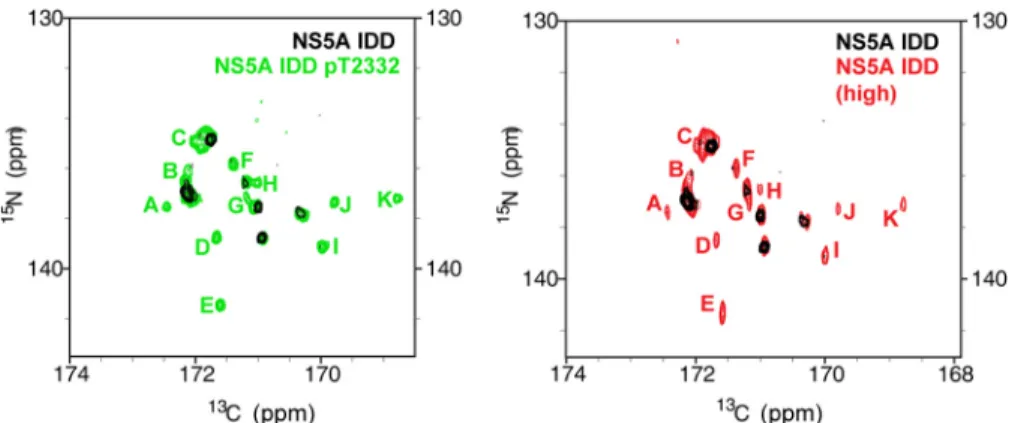
Upon phosphorylation by PKA, resonances for many more prolines were observed (Fig. 6C). With the assistance of amino
acid-specific15N,13C CON spectra (25), we were able to assign
three of these new resonances to 2315, 2322, and Pro-2325, suitable probes for both PPII motifs (Fig. 6A). It appears that the average conformations yielding the resonances observed in the Thr(P)-2332 spectrum are not created in response to phosphorylation, because we see them in the unphosphorylated IDD spectrum if the concentration is suffi-ciently high (Fig. 7). Therefore, it is likely that the ability of phosphorylation to bring resonances of the PPII motif into view originates from a reduction in the conformational sampling of this region of the IDD. Interestingly, an IDD with the T2332E substitution was indistinguishable from the IDD phosphory-lated on Thr-2332 (Fig. 6D), suggesting that T2332E-NS5A phenocopies the conformation of Thr(P)-2332-NS5A.
It is clear that NS5A interacts with several SH3-containing proteins (38). Recently, it has been shown that the NS5A PPII motif is essential for replication of genotype 2a (41). To deter-mine the impact of an SH3 domain on the PPII motifs of NS5A, we expressed and purified the SH3 domain from c-Src. We chose this SH3 domain because there is evidence that the NS5A-c-Src interaction is important for replication (42). Eval-uation of Thr(P)-2332䡠NS5A IDD䡠SH3 complexes produced at different molar ratios of the SH3 domain showed that the res-onances induced by phosphorylation disappear in the presence of the SH3 domain (red resonances are not present for any of the prolineslabeled greenin Fig. 6E). In contrast, none of the resonances remote from the PPII motifs are impacted by the presence of the SH3 domain (red resonances are present for all
FIGURE 4.Phosphorylation of NS5A Thr-2332 influences steady state levels of RNA for HCV genotype 1b but is essential for replication and infectious virus production for HCV genotype 2a.AandB, cells expressing the indicated genotype 1b replicons were used to measure the steady-state level of RNA by RT-qPCR inAor Northern blotting inB.C, reduced RNA levels lead to reduced protein levels. Extracts were prepared from the same cells used in the previous
of the prolineslabeled blackin Fig. 6E). Performing this exper-iment with T2332E-NS5A IDD yielded the same result as observed for Thr(P)-2332-NS5A IDD (Fig. 8A) without any per-turbations in the spectrum for the unphosphorylated protein (Fig. 8B). We conclude that both PPII motifs contribute to interactions with SH3 domains and that PKA phosphorylation near these motifs stabilizes a conformation competent for bind-ing to SH3 domains.
Because of the unique location of phosphothreonines in spectra produced by performing 1H,15N HSQC experiments,
we were able to identify and assign the resonance for Thr(P)-2332 (Fig. 6F). In the presence of the SH3 domain, there was a chemical shift perturbation followed by loss of the resonance. This observation suggests a possible interaction of Thr(P)-2332 with the SH3 domain as well. Given the substantial structural overlap that exists between SH3 domains, we were able to con-struct a model of the c-Src SH3 domain bound to the PPII.2 motif based on a structure of this motif bound to the Fyn SH3
domain (Fig. 6G) (43). This model places both Pro-2322 and Pro-2325 on c-Src SH3 domain and would suggest that Thr(P)-2332 should be close enough to interact with the SH3 domain as well.
Thr(P)-2332-NS5A Contributes to the Form and Function of the HCV Replication Organelle—At this point, we assumed that loss of Thr(P)-2332 negatively impacted interaction with one or more cellular SH3 domains. In order to further investigate the basis for the phenotype observed in cells producing T2332A- or T2332E-NS5A, we decided to evaluate the colocalization of NS5A with NS4B, a well accepted marker of the HCV membra-nous web/replication organelle (44). By using immunofluores-cence alone, it was obvious that the amount of NS5A-NS4B colocalization was reduced when Thr-2332 was changed to ala-nine (Fig. 9A). Mutation of position 2332 to glutamic acid yielded an intermediate phenotype (Fig. 9A). In order to per-form this experiment more quantitatively, we used confocal microscopy to obtain the Pearson’s coefficient for NS5A-NS4B
FIGURE 5.Thr-2332 phosphorylation does not affect HCV non-structural protein or RNA decay rates.AandB, T2332A and T2332E substitutions do not affect the decay of HCV non-structural proteins.A, Western blots of HCV non-structural proteins NS5A, NS3, and NS5B from SI, SI/T2332A, and SI/T2332E replicon-containing cells under 100g/ml CHX treatment.-Actin was detected as a loading control.B, plot of NS5A levels under CHX treatment at varying time points (tn) relative to 0 h (t0) showed an equal decay of the viral protein across SI, SI/T2332A, and SI/T2332E replicon-containing cell lines.CandD, T2332A
colocalization in single cells (45). In each case, 60 cells were evaluated, and a significant difference was observed in both the range and average colocalization observed when cells pro-ducing T2332A- or T2332E-NS5A were compared with those producing Thr(P)/Thr-2332-NS5A (Fig. 9B). We also analyzed the membranous web by using transmission elec-tron microscopy (Fig. 9C). Clustered, tubular-vesicular structures were very apparent in cells producing Thr(P)-2332/Thr-2332-NS5A (SI in Fig. 9C). In the absence of Thr(P)-2332, only vesicular structures were observed; these were not clustered (SI/T2332A in Fig. 9C). T2332E-NS5A led to the production of demonstrably larger vesicular struc-tures with very few tubular strucstruc-tures (SI/T2332E in Fig. 9C).
Some vesicles were clustered; others were not (SI/T2332E in Fig. 9C). Collectively, these data suggest that the Thr(P)-2332/Thr-2332-NS5A ratio is an important determinant of the assembly and/or stability of the replication organelle. The outcome associated with changing this ratio or this res-idue varies depending on the HCV genotype.
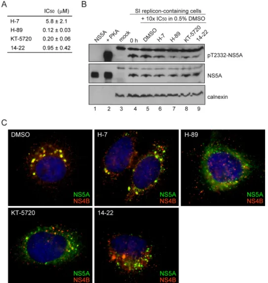
PKA Activity Is Required for HCV Replication beyond Phos-phorylation of NS5A on Thr-2332—In order to obtain evidence for PKA as the kinase responsible for phosphorylation of Thr-2332in vivo, we treated cells with the PKA inhibitor, KT-5720 (46), which caused a substantial reduction in the level of Thr(P)-2332-NS5A relative to untreated cells (comparelane 3 withlane 2ofpanel pT2332-NS5Ain Fig. 10A). In addition to
KT-5720, three other inhibitors of PKA have been used widely in the literature: H-7 (47), H-89 (48), and 14 –22 (49). All of these inhibitors prevent NS5A phosphorylation by PKAin vitro (Fig. 11A) and in cells replicating HCV RNA (Fig. 11B).
Because loss of NS5A phosphorylation on Thr-2332 caused perturbations in the integrity of the replication organelle, as determined by NS5A-NS4B colocalization (Fig. 9), we evalu-ated the impact of KT-5720 on integrity of the replication organelle (Fig. 10B). As expected, treatment of SI cells with KT-5720 caused disruption of the replication organelle (panel
⫹KT-5720in Fig. 10B). In order to determine whether loss of Thr(P)-2332-NS5A was reversible and would lead to reestab-lishment of the replication organelle (i.e.NS5A-NS4B colocal-ization), we removed the PKA inhibitor but added CHX to ensure that any Thr(P)-2332-NS5A observed would derive from existing pools of NS5A instead of newly translated NS5A. Under these conditions, Thr(P)-2332-NS5A was again pro-duced (lanes 4and5ofpanel pT2332in Fig. 10A). Interestingly, reestablishment of the replication organelle was not observed based on NS5A-NS4B colocalization (panels ⫹CHX in Fig.
10B). This suggests that once phosphorylation ceases and the replication organelle is disrupted, polyprotein synthesis may be required for reestablishment of the replication organelle.
If phosphorylation of NS5A on Thr-2332 is the only function of PKA for replication of HCV RNA, then treatment of SI cells with a PKA inhibitor should phenocopy the SI/T2332A mutant. When SI cells were treated for 24 h with 2 M(10 ⫻ IC50)
KT-5720, the steady-state level of HCV replicon RNA was reduced by an order of magnitude (Fig. 10C). Interestingly, the steady-state level of HCV replicon RNA was also reduced in SI/T2332A and SI/T2332E cells (Fig. 10C), suggesting that tar-gets of PKA phosphorylation other than NS5A contribute to HCV RNA replication. Consistent with the reduction in the steady-state level of RNA in SI/T2332A and SI/T2332E cells caused by the PKA inhibitor (Fig. 10C), KT-5720 treatment led to even further disruption of the replication organelles in these cells (panels SI/T2332AandSI/T2332Ein Fig. 10D). These data suggest that other viral and/or cellular substrates of PKA are required for assembly, stability, and/or function of the replica-tion organelle. All four inhibitors of PKA caused disrupreplica-tion of
FIGURE 7.PKA phosphorylation alters the observed resonances for NS5A IDD.Shown is a comparison of NMR spectra for unphosphorylated and PKA-phosphorylated NS5A IDD. In theleft panelis the15N,13C CON spectra of NS5A IDD (black) overlaid with the15N,13C CON spectra of NS5A IDD Thr(P)-2332 (green)
zoomedto the proline region of the spectrum. The proline residue resonances that appear upon phosphorylation are indicated withgreen letters. In theright panelis the15N,13C CON spectra of NS5A IDD (black) overlaid with15N,13C CON spectra of NS5A IDD at high concentration (red). Only at very high concentrations of unphosphorylated NS5A IDD do resonances begin to appear that are similar to resonances observed for PKA-phosphorylated NS5A IDD (resonances labeled with the sameletter codeas in theleft panelbut inred).
the replication organelle, although the fate of the NS5A and NS4B proteins differed for each inhibitor (Fig. 11C). We con-clude that PKA activity is essential for HCV genome replication.
Relocalization of PKA in Cells Replicating HCV RNA— Hav-ing established that PKA does indeed phosphorylate NS5A on Thr-2332, we asked whether this phosphorylation occurred by recruiting PKA into the replication organelle. In naive Huh-7.5 cells, PKAc showed a preferential localization to the Golgi (PKAcinpanel mockin Fig. 12A). In this experiment, the Golgi was stained with an antibody to the protein giantin (giantinin panel mockin Fig. 12A). In cells replicating HCV RNA, the Golgi-localized fraction of PKA was lost (PKAcinpanel SIin Fig. 12A). Importantly, the Golgi remained intact in cells repli-cating HCV RNA. (giantininpanel SIin Figs. 12Aand 13). The loss of Golgi-associated PKA in cells replicating HCV RNA did not require the ability of NS5A to be phosphorylated on Thr-2332 because the TThr-2332A and TThr-2332E mutants showed this phenotype (Fig. 12B). Although PKA did not accumulate in the replication organelle, the catalytic subunit was clearly present in the replication organelle, based on its colocalization with NS5A (Fig. 12B). Disruption of the replication organelle by
pre-cluding Thr-2332 phosphorylation was also sufficient to dis-rupt colocalization of PKA with NS5A (Fig. 12B). The ability of PKA and NS5A to colocalize in cells may facilitate NS5A phos-phorylation on Thr-2332. However, it is also possible that the relocalization of PKA from the Golgi to the cytoplasm may be sufficient for NS5A phosphorylation prior to its association with the replication organelle.
DISCUSSION
HCV NS5A contributes to genome replication, virion assem-bly, and myriad interactions with the host cell (7). How this protein interfaces with so many different viral and cellular pathways is unknown. It is well established that NS5A is a phos-phoprotein (27). These early studies showed that NS5A migrated as 56- and 58-kDa species in SDS-polyacrylamide gels. The fact that the calculated molecular mass of NS5A is 49 kDa led to the conclusion that all NS5A produced in cells is phosphorylated, with basal (56-kDa) and hyperphosphorylated (58-kDa) forms (27). Later, it became clear that even unphos-phorylated NS5A has a gel mobility of 56 kDa (18). The aberrant gel mobility is probably a reflection of the intrinsic disorder
FIGURE 9.Phosphorylation of Thr-2332 contributes to the integrity of the HCV membranous web/replication organelle.A, reduced colocalization of NS5A and NS4B in the absence of Thr(P)-2332-NS5A. Immunofluorescence was used to evaluate the replication organelle. NS4B is used as a marker for the replication organelle, with good colocalization implying integrity of this organelle (44). Cells replicating the indicated HCV 1b replicon were stained for NS5A (green) and NS4B (red), soyellowindicates colocalization. When there is afourth panelin arow, it shows the region in themerge panelthat is in thewhite box.
and/or proline-rich nature of the carboxyl-terminal two-thirds of NS5A (5, 6).
Phosphorylation and other post-translational modifications of an IDD of a protein can induce and/or stabilize structure, which, in turn, can confer unique function (8). Therefore, we
min-imum cut-off of 1 eq of phosphate/eq of protein is required for that protein to be considered a substrate of a kinase, PKA is only capable of phosphorylating NS5A, although NetPhosK predicts numerous sites in NS3 and NS5B (Fig. 1). Mass spectrometry, reverse genetics, and a phospho-specific antibody showed that Thr-2332 (polyprotein numbering; residue 360 of processed NS5A) was the site of phosphorylationin vitro(Fig. 2). The
latter two approaches permitted confirmation of this phosphor-ylation site in cells (Fig. 3).
Thr-2332 is located adjacent to two PPII motifs in NS5A (Fig. 6A). Our biophysical analysis (Fig. 6) is consistent with these motifs sampling multiple conformations on the millisecond time scale and Thr(P)-2332 restricting the sampling to confor-mations that are competent for binding to SH3 domains. It is
FIGURE 10.PKA targets other than NS5A are required for HCV replication.A, PKA phosphorylates NS5A at Thr-2332 in cells replicating HCV RNA. In order to demonstrate that PKA was indeed phosphorylating NS5A on Thr-2332 in cells, cells were treated with DMSO (lane 2) or the PKA inhibitor, KT-5720 (lane 3), for 24 h. The effect of treatment on Thr(P)-2332-NS5A and NS5A was determined by Western blotting. Huh-7.5 cells were used as a negative control (mock,lane 1); detection of calnexin served as a loading control. In order to determine whether dephosphorylated NS5A could be rephosphorylated, cycloheximide, a translation inhibitor, was added upon removal of the PKA inhibitor, KT-5720, and probed 12 (lane 4) or 24 h (lane 5) thereafter. Similar observations were made using other PKA inhibitors (Fig. 11).B, PKA inhibitors cause an irreversible loss in integrity of the replication organelle. The impact of the treatments performed inAon the replication organelle was determined by using immunofluorescence to evaluate NS5A-NS4B colocalization as described above.C, inhibition of PKA has a greater impact on HCV RNA levels than loss of pT2332. RT-qPCR was used to determine the copy number of HCV RNA in cells treated with DMSO or KT-5720. Errors represent S.E.n⫽3.D, inhibition of PKA further exacerbates loss of integrity of the replication organelle caused by substitutions at Thr-2332. Immunofluorescence was used to evaluate the integrity of the replication organelle for the wild-type replicon or the indicated Thr-2332 variant in the absence (DMSO) or presence (KT-5720) of the PKA inhibitor. Thewhite boxin themerge panelis enlarged in thebottom rightof each quadrant. This experiment was repeated with other PKA inhibitors (Fig. 11).
also possible that the phosphorylation-dependent changes were caused by acceleration of the conformational dynamics into the picosecond to microsecond time scale. Phosphoryla-tion-dependent changes in aggregation state were ruled out using dynamic light scattering (data not shown).
The ability of phosphorylation to regulate IDD conformation provides an explanation for cellular PPII motifs being located near phosphorylation sites (40). Phosphorylation need not always reduce conformational sampling; the context of the PPII motif would probably contribute to conformation. The rela-tionship between conformational sampling and binding affinity remains to be determined. In addition, it is easy to imagine that other post-translational modifications affect function in a sim-ilar manner by modulating conformational sampling to pro-mote or to interfere with a biomolecular interaction. Others have seen and/or proposed post-translational modification-de-pendent changes in motifs found in IDPs (50, 51). The novelty of this study is that we observe the prolines directly and provide an unambiguous link between a remote phosphorylation event and the conformation and dynamics of specific prolines. There-fore, these studies define the15N,13C CON NMR strategy as a
powerful approach for studying viral and cellular PPII motifs and interactions thereof.
The experimental paradigm used here to discover Thr(P)-2332 will be useful for identifying additional, specific sites of NS5A phosphorylation and the corresponding kinases. Two-dimensional gel analysis of NS5A present in Huh-7 cells persis-tently replicating HCV suggests many different phosphorylated species of the 56-kDa form, with pI values ranging from 4.0 to 5.5 (3). However, attempts to identify NS5A phosphorylation sites have yielded only a few sites (28 –32). None of these studies identified Thr-2332. The Thr(P)-2332-NS5A species repre-sents only 20% of total NS5A in cells (Fig. 3D). Therefore, this experimental paradigm clearly uncovers sites that have eluded direct approaches using NS5A isolated from HCV-replicating cells.
We conclude that Thr(P)-2332-NS5A, other PKA targets, and even other phosphorylated forms of NS5A contribute to form and function of the replication organelle. The NS5A IDD is among the most variable protein in the HCV proteome. It is possible that the NS5A phosphoproteome is equally variable. This variability may lead to differences in host factors used by
different genotypes and the ability to antagonize host defenses required to establish persistence. Clearly, the NS5A phospho-proteome encoded by Con1b/S2204I and Jc1–2a genotypes of HCV used in this study is different, because Jc1–2a produces the p58 form of NS5A, and Con1b/S2204I does not. The mag-nitude of the response of these genotypes to loss of Thr(P)-2332 is substantially different (Fig. 4).
Dozens of host factors have been implicated in HCV genome replication and many (re)localize to the replication organelle (52). Of these, several are thought to be recruited by interacting with NS5A. Interestingly, two NS5A-interacting and replica-tion organelle-associated host factors are regulated by PKA; these are annexin A2 (53, 54) and Raf-1 kinase (55). Annexin A2 interacts with phosphoinositides to form membrane microdo-mains of distinct protein composition, and these microdomicrodo-mains exhibit altered membrane dynamics (56). Phosphorylation of annexin A2 by PKA has been shown to promote its association with membranes. Raf-1 kinase interacts with Ras, and this interaction is disrupted by PKA phosphorylation of Raf-1 (57). Disruption of the Raf-1-Ras interaction may permit Raf-1 to localize to the replication organelle, thereby contributing to formation and/or stabilization of the replication organelle. Therefore, annexin A2 and Raf-1 are candidate host factors that may contribute to regulation of replication organelle structure by PKA.
It is possible that the ability to regulate assembly and disas-sembly of the replication organelle contributes to the ability of HCV to persist. This possibility is suggested by the observation that the difference in the steady-state level of RNA observed for the Thr-2332 mutants relative to wild type only manifests after multiple cell divisions (Fig. 4C). When the replication organelle
is assembled, HCV replicase components are perinuclear and in an asymmetric organization (Fig. 3E). Therefore, it is possible that in this state partitioning between daughter cells may be asymmetric. When disassembled, components of the replica-tion organelle are more homogeneous (Fig. 10D) and may be more equally distributed between daughter cells. Consistent with this possibility is the fact that cAMP concentration and PKA activity are highest at G1and lowest at mitosis (58).
The NS5A phosphoproteome may be a highly efficacious tar-get for development of anti-HCV agents. This idea is inspired by the recent discovery of daclatasvir, a drug thought to inhibit HCV genome replication by binding to NS5A, preventing hyperphosphorylation (14, 15) and disrupting the replication organelle (59). Evidence for direct binding of daclatasvir and related compounds to NS5A has been difficult to obtain (59). That NS5A is the target is supported primarily by drug resis-tance mutations (15). The effective concentration of daclatasvir is in the picomolar range; however, the concentration of NS5A in cells replicating HCV RNA is on the order of micromolar (14). Is it possible that the target is one of the many phosphor-ylated forms of NS5A? If this were the case, the picomolar effi-cacy could be related to binding of the drug to a NS5A species that is present at sub-picomolar levels. Analysis of the NS5A phosphoproteome after daclatasvir treatment may permit this possibility to be tested.
This study makes a very compelling case for the use of phos-phorylation in the intrinsically disordered domain of NS5A to expand the functional proteome of HCV. Interestingly, evalua-tion of phosphorylated proteins of other RNA viruses reveals that these proteins have intrinsically disordered domains, and known phosphorylation sites map to these domains (Table 3). We speculate that phosphorylation in IDDs of viral proteins may be a general mechanism employed by RNA viruses to expand their functional proteomes.
REFERENCES
1. Love, R. A., Brodsky, O., Hickey, M. J., Wells, P. A., and Cronin, C. N. (2009) Crystal structure of a novel dimeric form of NS5A domain I protein from hepatitis C virus.J. Virol.83,4395– 4403
2. Tellinghuisen, T. L., Marcotrigiano, J., and Rice, C. M. (2005) Structure of the zinc-binding domain of an essential component of the hepatitis C
FIGURE 13.Replication of HCV RNA does not alter Golgi structure.Shown is immunofluorescence analysis of NS5A (green) and the Golgi marker giantin (red) in naive Huh-7.5 cells (mock) and SI, SI/T2332A, and SI/T2332E HCV rep-licon-containing cells. The stable replication of HCV RNA had no effect on Golgi structure relative to mock cells.
TABLE 3
Intrinsic disorder and phosphorylation of (ⴙ)-strand RNA viral proteins
Virus Protein Disordera
Longest IDRb
PO4in IDRc
Bovine viral diarrhea virus NS5A 31 82 TBD Cucumber mosaic virus 2a 23 61 Yes Cucumber necrosis virus p33 27 70 Yes Dengue virus type 2 NS5 37 45 TBD Hepatitis C virus NS5A 55 68 Yes
Potato virus A VPg 32 20 Yes
Semliki Forest virus nsP3 32 81 Yes
Sindbis virus nsP3 49 58 Yes
Tick-borne encephalitis virus NS5 29 30 TBD Turnip crinkle virus p28 38 40 Yes Turnip yellow mosaic virus 66K 53 115 Yes Yellow fever virus NS5 25 47 TBD
aPercentage disorder was determined by PONDR (Predictor of Natrually
Disor-dered Regions).
bLongest intrinsically disordered region in length of amino acids.
cIf specific sites have been mapped as phosphorylated residues within the viral