A STRUCTURAL STUDY OF CONSERVED CENTRIOLE DUPLICATION MACHINERY
Lauren K. Slevin
A dissertation submitted to the faculty of the University of North Carolina at Chapel Hill in partial fulfillment of the requirements for the degree of Doctor of Philosophy in the
Department of Biology in the College of Arts and Sciences.
Chapel Hill 2014
ABSTRACT
Lauren K. Slevin: A structural study of conserved centriole duplication machinery (Under the direction of Kevin C. Slep)
ACKNOWLEDGEMENTS
I would like to acknowledge my advisor, Kevin Slep, and my committee, Kerry Bloom, Bob Duronio, Matt Redinbo, and Marcey Waters, for their creative input and encouragement throughout my dissertation progress, as well as our collaborator Gregory Rogers (U. of Arizona). Additionally, many thanks to Ashutosh Tripathy at the UNC MacInFac and Mike Miley at the UNC Crystallography Core, without whom I would have found instrumentation at UNC much more daunting! I especially thank Bob Goldstein and Mark Peifer, who have provided years of constructive feedback and incredible mentorship. I owe much of my passion for science and fortitude in graduate school to both of them.
I would also like to thank the members of the Slep lab, past and present, especially my undergraduate assistants, Derek Pinkerton and Mary Dandulakis. I could not ask for better colleagues. A special thanks goes to Erin Romes, Jaime Fox, Alka Das, and Tom Lane for their collegiality and countless sushi nights. Huge thanks to my friends from all the places I’ve called home, including Casey Stephens, Justin Hanks, Lily Wong, Megan Auvil, Mary Beth Campbell, Jill Christiansen, Danielle Slomberg, Chelsea Lane, Kathryn Trogden, Ahyeon Koh, Rebecca Hunter, Alessa Gambardella, Amy Entwistle, and Rabbi Jen Feldman as well as the community at the Chapel Hill Kehillah Synagogue. Thanks for the laughter, support, and memories.
PREFACE
Chapter 2 is a published manuscript describing work done in collaboration with the lab of Gregory Rogers (University of Arizona). This work was published in Structure in November 2012. Jonathan Nye carried out centriole count assays while Dan Buster completed all pull-down experiments and Western Blots (both from the Rogers lab). Derek Pinkerton (an undergraduate in the Slep lab) assisted with centriole localization experiments. I purified Plk4 PB1-PB2, obtained crystals, and assisted Kevin Slep with structure refinement, as well as designed, cloned, and performed the centriole localization assay (as well as the basic patch mutant assay, unpublished; described in detail in Chapter 6). Kevin Slep, Gregory Rogers and I designed experiments and drafted the manuscript.
Slevin, L.K., Nye, J., Pinkerton, D.C., Buster, D.W., Rogers, G.C., and Slep, K.C. (2012). The structure of the Plk4 cryptic polo box reveals two tandem polo boxes required for centriole duplication. Structure. 20, 1905-1917.
Chapter 5 is a published manuscript co-authored by Erin Romes, a former Slep lab graduate student, and me. This work was published in The Journal of Biological Chemistry in August 2014. Erin Romes cloned LC8, designed Ana2 peptides, purified proteins for
crystallization trials, crystallized LC8-Ana2 pep1, and assisted with ITC. I performed ITC, solved both structures as presented, analyzed comparative LC8 binding targets, and designed and carried out the SEC-MALS assays. Mary Dandulakis (a Slep lab undergraduate student) crystallized the LC8-Ana2 pep2 complex. I drafted all figures and wrote the manuscript. Kevin Slep designated the proposed LC8 binding sites within Ana2 and oversaw
experimental design.
Slevin, L.K.,* Romes, E.M.,* Dandulakis, M.G., and Slep, K.C. (2014). The mechanism of dynein light chain LC8-mediated oligomerization of the Ana2 centriole duplication factor. Journal of Biological Chemistry. 289, 20727-20739. *indicates equal
TABLE OF CONTENTS
LIST OF TABLES ... xiii
LIST OF FIGURES ... xiv
LIST OF ABBREVIATIONS AND SYMBOLS ... xvi
CHAPTER 1 : INTRODUCTION ... 1
Centrioles are microtubule-based cylindrical structures essential in nucleating polarized microtubule networks ... 1
Centrioles undergo duplication in a cell-cycle- and Plk4-dependent manner ... 4
Centriole duplication is driven by a conserved set of proteins ... 5
The Plk4 phosphorylation targets include itself, cell cycle regulator proteins, and unknown centriole components ... 9
Plk4 is a divergent member of the Polo-like family of kinases ... 10
The mechanism of Plk4’s licensing activity followed by swift degradation is conserved among opisthokonts ... 12
The Polo Box (PB) domain is used to regulate Plk activity and subcellular targeting by employing a structurally defined fold ... 15
PB3: the odd one out ... 23
References. ... 24
CHAPTER 2 : THE STRUCTURE OF THE PLK4 CRYPTIC POLO BOX REVEALS TWO TANDEM POLO BOXES REQUIRED FOR CENTRIOLE DUPLICATION ... 31
Summary ... 31
Introduction ... 31
Cloning and protein purification ... 35
Crystallization, data collection, and structure determination ... 37
Size exclusion chromatography and multi-angle light scattering (SEC-MALS) ... 38
Dynamic light scattering ... 38
Centriole localization assay ... 39
Centriole count assay ... 40
Immunoblotting ... 41
Immunoprecipitation ... 42
In vitro kinase assay... 42
Results ... 43
Crystallization and structure determination of the Plk4 Cryptic Polo Box ... 43
The Cryptic Polo Box comprises two structurally unique Polo Box domains ... 44
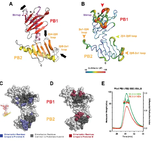
Plk4 PB1-PB2 forms a pseudo-symmetric homodimer ... 47
Unique features of Plk4 PB1 and PB2 and implications for target binding ... 50
Plk4 PB1-PB2 has a novel inter-domain and homodimeric arrangement ... 52
The conserved Plk4 PB1-PB2 inter-domain groove corresponds to the Plk1 PB1 target binding site ... 52
The full Plk4 PB1-PB2 cassette is required for Asterless binding ... 53
The PB1-PB2 cassette is necessary and sufficient for robust centriole targeting ... 56
PB1-PB2 scaffolds Plk4 trans-autophosphorylation to limit centriole duplication ... 57
Discussion ... 62
References ... 66
Tables ... 71
CHAPTER 3 : PLK4 PB3 IS A DIVERGENT PB DOMAIN WITH
SPECIES-DEPENDENT ROLES AND A VARIABLE STRUCTURE ... 77
Summary ... 77
Experimental Procedures ... 78
Cloning and protein purification ... 78
Crystallization, data collection, and structure determination ... 79
Size exclusion chromatography and multi-angle light scattering (SEC-MALS) ... 80
Centriole localization assay ... 80
Results ... 82
Crystallization and structure determination of Plk4 PB3 ... 82
The structure of D.m. PB3 contains two chains in the ASU but lacks a dimerization interface ... 83
Comparison of D.m. and M.m. structures reveals both conserved PB3 features and unique PB3 oligomer configurations ... 85
D.m. PB3 is a Monomer in Solution, while Mammalian PB3s Exist as Dimers ... 88
H.s. PB3 is Not Sufficient for Centriole Subcellular Localization ... 90
Discussion ... 91
References ... 94
Tables ... 96
CHAPTER 4 : A MOTOR-INDEPENDENT FUNCTION OF DYNEIN LIGHT CHAIN IN MITOTIC SPINDLE ORIENTATION ... 97
Asymmetric divisions play a critical role in tissue specification in the developing embryo ... 97
ACDs require cooperation between the cortex, molecular motors, dynamic cytoskeletal polymers, and the centrosome ... 100
An interaction among Ana2, LC8, and Mud regulates divisions within the developing Drosophila neuroblast ... 101
References. ... 104
CHAPTER 5 : THE MECHANISM OF DYNEIN LIGHT CHAIN LC8-MEDIATED OLIGOMERIZATION OF THE ANA2 CENTRIOLE DUPLICATION FACTOR ... 106
Summary ... 106
Introduction ... 107
Experimental Procedures ... 112
Cloning and expression of full-length LC8 ... 112
Protein purification for crystallization ... 113
Synthesis of Ana2 peptides ... 113
Crystallization of the LC8/Ana2 peptide complexes ... 113
Data collection, structure determination, and refinement ... 114
Isothermal microtitration calorimetry ... 115
Cloning and expression of LC8 and Ana2M constructs for SEC-MALS ... 115
Ana2M-LC8 complex purification for SEC-MALS ... 116
Mutagenesis of Ana2M ... 116
SEC-MALS ... 117
Results ... 117
Ana2 contains two high-affinity LC8 binding sites ... 117
Crystallization of the LC8 Ana2 Pep1 and Pep2 complexes ... 120
The structure of LC8 bound to Ana2 Pep1 ... 120
The structure of LC8 bound to Ana2 Pep2 ... 121
The LC8 binding pocket undergoes structural shifts to accommodate Ana2 peptides ... 125
Ana2 employs a unique tandem set of LC8 binding motifs ... 125
LC8 mediates Ana2M’s solubility and oligomerization state ... 127
References ... 136
Tables ... 141
CHAPTER 6 : DISCUSSION AND FUTURE WORK ... 142
Plk4 employs an assemblage of Polo Box domains to license centriole duplication ... 142
LC8 potentiates Ana2 oligomerization, with potential implications for the role of Ana2 in asymmetric cell division and centriole duplication ... 149
Concluding remarks ... 152
LIST OF TABLES
Table 2.1 Plk4 PB1-PB2 crystallographic data, phasing, and refinement ... 71
Table 2.2 Plk4 PB1-PB2 dynamic light scattering ... 72
Table 3.1 Plk4 PB3 crystallographic data, phasing, and refinement ... 96
LIST OF FIGURES
Figure 1.1 Centrioles form the basis of centrosomes and cilia ... 3 Figure 1.2 Canonical centriole duplication requires cell-cycle-dependent cues and
mother centriole templating ... 6 Figure 1.3 Centriole duplication requires a set of proteins functionally and spatially
conserved across species ... 7 Figure 1.4 The architectural map of the Plk family of kinases highlights the
divergence of Plk4.. ... 11 Figure 1.5 Plk4 is functionally conserved and maintains specific structural modules ... 13 Figure 1.6 Plk4 depends on the PB1-PB2 homodimerization interface to
autophosphorylate in trans and bind F-box proteins for its degradation ... 14 Figure 1.7 The structure of each solved PB domain takes on a conserved fold ... 17 Figure 1.8 Plks employ PB domains to take on differential dimeric configurations
for cellular function ... 20 Figure 1.9 PB1-PB2 pairs diverge in both sequence and structure. ... 22 Figure 2.1 The Plk4 Cryptic Polo Box is composed of tandem PB domains, PB1
and PB2. ... 46 Figure 2.2 Plk4 PB1-PB2 is an asymmetric homodimer with plastic stirrups and
loops ... 49 Figure 2.3 Plk4 PB1 and PB2 diverge from Plk1 PB domain structures and form a
unique inter-domain interaction. ... 51 Figure 2.4 Plk4 PB1 and PB2 form a composite inter-domain groove delineated by
conserved and basic residues ... 55 Figure 2.5 The Plk4 PB1-PB2 cassette is required for robust centriole localization. ... 58 Figure 2.6 Plk4 PB1-PB2 promotes centriole amplification and protects full-length
Plk4 in trans. ... 61 Figure 2.S1 Plk4 PB1-PB2 forms numerous crystallographic interfaces but is a
homodimer in solution.. ... 73 Figure 2.S2 The presence of either the PB1-PB2 or PB3 domain of Plk4 does not
Figure 2.S3 Expression of Plk4 PB1-PB2 is sufficient to promote centriole
amplification. ... 75
Figure 2.S4 Titrated expression of Plk4 PB1-PB2 causes differential centriole amplification ... 76
Figure 3.1 The structure of D.m. PB3 reveals a single PB domain with two protomers in the asymmetric unit.. ... 84
Figure 3.2 D.m. and M.m. Plk4 PB3 form similar folds but maintain different spatial crystallographic arrangements. ... 87
Figure 3.3 PB3 takes on a species-dependent oligomerization state in solution... 89
Figure 3.4 H.s. PB3 is not a robust centriole localization domain in cultured RPE1 cells ... 91
Figure 4.1 The Drosophila larval neuroblast marshals cell fate determinant complexes and the mitotic spindle structure to achieve asymmetric cell division along an apicobasal polarity axis. ... 99
Figure 4.2 LC8 acts to homodimerize its targets in parallel. ... 103
Figure 5.1 Ana2 contains two conserved LC8 binding sites. ... 111
Figure 5.2 LC8 binds two Ana2 sites with different affinities ... 119
Figure 5.3 Structures of LC8-Ana2 complexes reveal LC8 homodimers bound to two parallel Ana2 peptides. ... 122
Figure 5.4 Ana2's binding sites 1 and 2 employ both shared and unique LC8-binding determinants ... 124
Figure 5.5 Ana2's two LC8 binding sites differentially bind LC8. ... 128
Figure 5.6 SEC-MALS of Ana2M co-purified with LC8 shows a stable complex corresponding to LC88-Ana2M4. ... 131
Figure 5.7 A proposed model of LC8-mediated Ana2 oligomerization. ... 134
LIST OF ABBREVIATIONS AND SYMBOLS
Å Ångstrom
Asl Asterless
ACD asymmetric cell division ASU asymmetric unit
β-ME β-mercaptoethanol BSA bovine serum albumin
C.e. Caenorhabditis elegans (nematode) CMV cytomegalovirus
CuSO4 copper sulfate
Da Dalton
DIC Dynein intermediate chain
D.m. Drosophila melanogaster (fruit fly) DMEM Dulbecco’s modified eagle medium DRE downstream regulatory element eGFP enhanced green fluorescent protein FBS fetal bovine serum
FL full-length
IPTG isopropyl β-D-1-thiogalactopyranoside
kDa kiloDalton
L liter (or leucine) LC8 Dynein light chain 8
M molar (or mitosis, or methionine; context-dependent)
mg milligram
min minute
mL milliliter
M.m. Mus musculus (mouse)
mM millimolar
NaCl sodium chloride
nm nanometer
PAGE polyacrylamide gel electrophoresis
PB Polo Box
PBS phosphate-buffered saline
D-PLP Pericentrin-Like Protein (Drosophila melanogaster) PCM pericentriolar material
Pctn Pericentrin
PDB Protein Data Bank (pdb.org) PEG polyethylene glycol
Plk Polo-like Kinase
RMSD root-mean-square deviation
RNAi RNA interference
RPE1 Human retinal pigment epithelial cells S cell cycle synthesis phase
SAS Spindle Assembly Abnormal Protein SDS sodium dodecyl sulfate
SEC-MALS Size Exclusion Chromatography coupled with Multi-Angle Light Scattering SeMet selenomethionine
µg microgram
µl microliter
µM micromolar
WT wild type
CHAPTER 1: INTRODUCTION
Centrioles are microtubule-based cylindrical structures essential in nucleating polarized microtubule networks
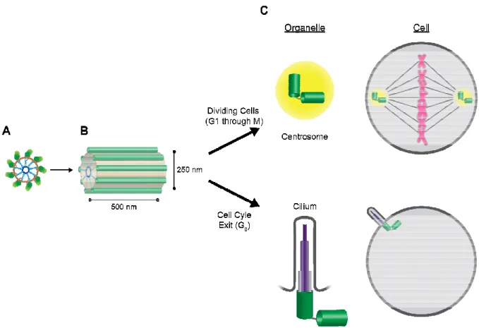
Centrioles are cylindrical subcellular structures observed in nearly all metazoans.
Centrioles take species- and cell type-specific dimensions; human somatic cells undergoing
mitotic divisions typically have centrioles 250 nm in diameter and 500 nm long (Winey and
O’Toole, 2014). Centriolar structure consists of a cartwheel-like formation with 9 spokes that emanate radially (Figure 1.1). This cartwheel forms the basis of the 9-fold radial symmetry of the microtubule triplet blades (van Breugel et al., 2011; Kitagawa et al., 2011), which decorate the length of the centriolar barrel and are evident in electron microscopic images of centrioles (Guichard et al., 2013; Li et al., 2012). Centrioles play critical roles in centrosomes and cilia, organelles that nucleate and organize dynamic polarized microtubule networks
(Figure 1.1).
In cells that have exited the cell cycle (in G0), centrioles are recruited to the cell
cortex, where they become modified centrioles called basal bodies. Basal bodies then
nucleate either a single cilium (called a primary cilium) or many cilia (motile cilia),
depending on the tissue type and centriole number (Scholey and Anderson, 2006). The primary cilium emanates from the cell in a finger-like projection, and employs its extended surface area to sense environmental cues and facilitate cell-to-cell communication. The
formation of the primary cilium requires exactly two centrioles, which form the physical base
formation of multiple motile cilia, which aid in various tissues (including the fallopian tubes,
airway, and brain of human cells (Bylander et al., 2013)) to create movement and propagate
signals.
In actively dividing cells, however, centrioles play an important role in forming the
centrosome, an organelle that nucleates and organizes the bipolar mitotic spindle (Figure
1.1). Centrosomal centrioles play a key role in regulating these activities in most vertebrate
somatic cells, though centrioles are interestingly absent in higher fungi, higher plants, and
many oocyte types (Carvalho-Santos et al., 2010), in which acentriolar structures govern mitotic spindle formation. While centrioles themselves do not nucleate microtubule growth,
data from several labs in recent years suggest that centrioles are central in recruiting,
stabilizing, and spatially organizing the pericentriolar material complex (PCM), which
contains such microtubule nucleators as the γ-TuRC complex (Mennella et al., 2012). A centrosome consists of a pair of centrioles and their associated shells of PCM components.
The centriole pair is connected orthogonally, forming an “L” shape between an older
(“mother”) centriole and a nascent (“daughter”) centriole (Rodrigues-Martins et al., 2007). The centrioles maintain a physical connection, with proteinaceous tethers including C-Nap1,
LRRC45 and Rootletin, within the mature centrosome through mitosis (Bornens et al., 1987;
He et al., 2013). The centrosome seeds growth of the microtubule polymers, and maintains secure connections with the minus ends of microtubules (both astral and spindle
microtubules), while the plus ends extend towards the kinetochores. During mitosis, exactly
two centrosomes (and therefore a total of exactly four centrioles) form the bipolar mitotic
spindle. Supernumerary centrosomes have been implicated in forming multi-polar spindles, a
Misregulated centriole duplication is also associated with diseases including dwarfism, male
sterility, and primary microcephaly
Dias et al., 2011), underlining the importance of learning how cells and regulate their biogenesis.
Figure 1.1. Centrioles form the basis of centrosomes and cilia.
the proximal end of a centriole (see Figure 1.3 for greater molecular detail). Microtubule triplet blades are depicted in green, surrounding the inner barrel. (B) Side view of a vertebrate centriole. Microtubules are depicted as single green tubes for simplicity. (C) Mother-daughter centriole pairs (shown as connected green barrels) form the basis of centrosomes (cycling cells) or cilia (G
and an associated ordered cloud of PCM components (yellow), and nucleate the mitotic spindle to accurately segregate sister chromatids (magenta) during
employ centrioles to nucleate inner microtubules of various lengths and multiplicity (purple) to form a finger-like protrusion from the cell.
Misregulated centriole duplication is also associated with diseases including dwarfism, male
microcephaly (Chavali et al., 2014; Nigg and Raff, 2009; Bettencourt underlining the importance of learning how cells “count” their centrioles
and regulate their biogenesis.
Centrioles form the basis of centrosomes and cilia. (A) Cross
the proximal end of a centriole (see Figure 1.3 for greater molecular detail). Microtubule t blades are depicted in green, surrounding the inner barrel. (B) Side view of a vertebrate centriole. Microtubules are depicted as single green tubes for simplicity. (C)
daughter centriole pairs (shown as connected green barrels) form the basis of centrosomes (cycling cells) or cilia (G0). Top, centrosomes consist of two centrioles (green) and an associated ordered cloud of PCM components (yellow), and nucleate the mitotic spindle to accurately segregate sister chromatids (magenta) during cell division. Bottom, cilia employ centrioles to nucleate inner microtubules of various lengths and multiplicity (purple)
like protrusion from the cell.
Misregulated centriole duplication is also associated with diseases including dwarfism, male
Chavali et al., 2014; Nigg and Raff, 2009; Bettencourt-“count” their centrioles
Centrioles undergo duplication in a cell-cycle- and Plk4-dependent manner
Despite the fact that cell biologists have studied centrioles for more than a century (Boveri 1909), the manner by which cells create their centrioles has only recently begun to come to light. Using microscopic and molecular techniques, researchers have discovered a couple of mechanisms by which cells undertake centriole biogenesis. In the less understood de novo mechanism, nascent, unconnected centrioles are made in the cytoplasm due to the presence of excessive duplication components. The de novo mechanism has largely been described in oocyte systems (Rodrigues-Martins et al., 2007; Peel et al., 2007; Eckerdt et al., 2011), in which ectopic overexpression of key components leads to the formation of nascent centrioles incapable of PCM recruitment. De novo centriole biogenesis is thought to play a biological role in cells that have exited the cell cycle, differentiated, and require motile cilia (Zhao et al., 2013). To meet this requirement, the cell must make hundreds of modified centrioles to undergo multiciliogenesis; thus, the de novo pathway is an important mechanism through which differentiated cells create centrioles. However, it is not the dominant pathway through which most centrioles are made in dividing cells; therefore, I will focus herein on the axiomatic “templating” duplication mechanism.
enters G1, the cell-cycle-dependent enzyme Separase and signaling kinase Plk1 function to sever the connection between mother and daughter centrioles. The loss of physical connection “resets” the identities of the centrioles, making both new mothers to begin centriole duplication anew.
By the end of G1, the kinase Plk4 (Polo-like Kinase 4) is recruited to the site of centriole duplication to act as the “master licenser” of centriole duplication. While little is known about its mechanism of action, Plk4 is known to be required for centriole duplication; additionally, its licensing activity requires its catalytic activity (Bettencourt-Dias et al., 2005). After Plk4 licenses centriole duplication by its unknown mechanism, daughter centrioles begin to grow orthogonally to their respective mother centrioles. The formation of the initial structures is commensurate with DNA replication during S phase (Figure 1.2). As the cell progresses through G2 and prepares for mitotic entry, the daughter centriole completes elongation and the two mature mother-daughter pairs each finish recruiting the PCM components required to build and organize the mitotic spindle. The cell undergoes mitosis, and each new cell undergoes the process again.
Centriole duplication is driven by a conserved set of proteins
Figure 1.2. Canonical centriole duplicatio mother centriole templating.
and four centrioles; when it completes cytokinesis, each resulting (daughter) cell receives one centrosome and two centrioles
Separase (red) and Plk1 (not shown)
centrioles, resetting the identity of both to “mother”
Plk4 (blue) licenses centriole duplication. During Synthesis phase, nascent daughter centrioles (chartreuse) begin to grow orthogonally to their respective mothers (forest green). As the cell completes G2 and prepares for mitosis, the daughter centrioles elongate, ar capped to pre-determined lengths, and the mother
PCM proteins (yellow). As the cell builds the mitotic spindle, the two mature centrosomes are separated on opposite sides, and the cyclical process begins again.
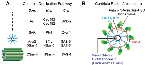
within centriolar eukaryotes. Shown in Figure 1.3 (A) is a comparative list of the components required for centriole duplication
melanogaster (D.m.), humans (
elegans (C.e.). While many more proteins contribute to procentriole assembly, elongation, and capping (length determination), shown in Figure 1.3 (A) are the five basic players necessary for centriole duplication.
Canonical centriole duplication requires cell-cycle-dependent cues and mother centriole templating. Clockwise from top, a mitotic cell contains two centrosomes and four centrioles; when it completes cytokinesis, each resulting (daughter) cell receives one centrosome and two centrioles (forest green cylinders). As the cell enters the first Gap phase, and Plk1 (not shown) cleaves the linkage between mother and daughter centrioles, resetting the identity of both to “mother” (Tsou et al., 2009). By the end of G
licenses centriole duplication. During Synthesis phase, nascent daughter centrioles (chartreuse) begin to grow orthogonally to their respective mothers (forest green).
and prepares for mitosis, the daughter centrioles elongate, ar determined lengths, and the mother-daughter centriole pair recruits a cloud of PCM proteins (yellow). As the cell builds the mitotic spindle, the two mature centrosomes are separated on opposite sides, and the cyclical process begins again.
within centriolar eukaryotes. Shown in Figure 1.3 (A) is a comparative list of the components required for centriole duplication (Goshima et al., 2007) in the fruit fly
humans (Homo sapiens, H.s.), and the nematode
). While many more proteins contribute to procentriole assembly, elongation, and capping (length determination), shown in Figure 1.3 (A) are the five basic players necessary for centriole duplication.
dependent cues and Clockwise from top, a mitotic cell contains two centrosomes and four centrioles; when it completes cytokinesis, each resulting (daughter) cell receives one (forest green cylinders). As the cell enters the first Gap phase, cleaves the linkage between mother and daughter . By the end of G1, licenses centriole duplication. During Synthesis phase, nascent daughter centrioles (chartreuse) begin to grow orthogonally to their respective mothers (forest green). and prepares for mitosis, the daughter centrioles elongate, are daughter centriole pair recruits a cloud of PCM proteins (yellow). As the cell builds the mitotic spindle, the two mature centrosomes
While Plk4 (also known as SAK/ZY the “master licenser” of centriole duplication (
2009), it requires centriole scaffolding components to localize to the site of nascent centrioles. In D.m., Asterless is the scaffolding protein responsible for binding and recruiting Plk4 to nascent centrioles (Dzhindzhev et al., 2010
redundant system to recruit Plk4 to centrioles, presumably due to its essential function in centriole duplication; in humans, either Cep152 (the Asterless homolog) or Cep192 (the SPD-2 homolog) is sufficient for Plk4 localization
2010). Interestingly, C.e. have lost the Asterless homolog, and make use of recruit Plk4, indicative that the
specific interactions (Shimanovskaya et al., 2014
Figure 1.3. Centriole duplication requires a set of proteins functionally and s
conserved across species. (A) Schematic pathway of conserved components. Three species are compared: Drosophila melanogaster
elegans (C.e.), with the resulting structures shown to the left. See text
components as they are known to localize to the cartwheel structure (omitting Asl for clarity and Plk4, which is transient). Sas
cartwheel (shown in dark
(N-is unknown, but its N-terminus binds Sas 2013) and its C-terminus binds Sas
While Plk4 (also known as SAK/ZYG-1 in D.m. and C.e., respectively) is considered nser” of centriole duplication (Cunha-Ferreira et al., 2009; Rogers et al., , it requires centriole scaffolding components to localize to the site of nascent , Asterless is the scaffolding protein responsible for binding and recruiting Dzhindzhev et al., 2010). However, humans have developed a redundant system to recruit Plk4 to centrioles, presumably due to its essential function in centriole duplication; in humans, either Cep152 (the Asterless homolog) or Cep192 (the
2 homolog) is sufficient for Plk4 localization (Hatch et al., 2010; Cizmecioglu et al., have lost the Asterless homolog, and make use of
recruit Plk4, indicative that the C.e. Plk4 and SPD-2 proteins have coevolved to form their Shimanovskaya et al., 2014).
Figure 1.3. Centriole duplication requires a set of proteins functionally and s
(A) Schematic pathway of conserved components. Three species Drosophila melanogaster (D.m.), Homo sapiens (H.s.), and
), with the resulting structures shown to the left. See text for details. (B) Map of components as they are known to localize to the cartwheel structure (omitting Asl for clarity and Plk4, which is transient). Sas-6 oligomerizes into an 18-mer and builds the central -terminus) and light (C-terminus) blue. The localization of Ana2 terminus binds Sas-4 (highlighted in top orange box, Cottee et al., terminus binds Sas-6 (highlighted in bottom orange box
, respectively) is considered Ferreira et al., 2009; Rogers et al., , it requires centriole scaffolding components to localize to the site of nascent , Asterless is the scaffolding protein responsible for binding and recruiting . However, humans have developed a redundant system to recruit Plk4 to centrioles, presumably due to its essential function in centriole duplication; in humans, either Cep152 (the Asterless homolog) or Cep192 (the Hatch et al., 2010; Cizmecioglu et al., have lost the Asterless homolog, and make use of SPD-2 alone to 2 proteins have coevolved to form their
2010a). Sas-4 (red) forms a scaffold along the outer wall of the centriole to recruit and stabilize the triplet microtubule blades (Cottee et al., 2013).
It remains unknown exactly what Plk4’s function is at the centriole after its recruitment; it is only known that centriole duplication requires catalytically active Plk4, though recent evidence suggests that a direct interaction between Plk4 and Sas-6 is sufficient to recruit Sas-6 to the site of the nascent centriole (Lettman et al., 2013).
The first structure observed through cryotomography in the emerging daughter centriole is the cartwheel with 9-fold symmetry. Structural breakthroughs occurred in 2011, when several labs demonstrated that a higher-order oligomer of the conserved Sas-6 (spindle-assembly abnormal-6 (Leidel et al., 2005)) creates cartwheels with an inherent 9-fold radial symmetry in vitro (van Breugel et al., 2011; Kitagawa et al., 2011). It has since been shown (van Breugel et al., 2014) that 18 Sas-6 protomers associate in two ways to create the 9-spoked cartwheel: first, the helical region at the Sas-6 C-terminus interacts with other protomers to form a high-affinity coiled-coil dimer. Second, the globular Sas-6 N-terminus interacts weakly and laterally with the same domain in other protomers, forming the hub of the cartwheel; the coiled-coil regions emanate out from the hub, forming the spokes of the wheel (Figure 1.3 B, shown in blue).
µM in C.e. (Kitagawa et al., 2011)). Thus, while Ana2 likely promotes Sas-6-based cartwheel structures, the mechanism by which Ana2 does so remains unknown. Ana2 does contain several known binding motifs (presented in greater detail in Chapter 5), including a Sas-6- and a Sas-4-binding domain, each of which has been mapped using biochemical techniques and yeast two-hybrid screens (Stevens et al., 2010a; Cottee et al., 2013). The presumptive binding sites between Ana2 and its partners Sas-6 and Sas-4 are mapped in Figure 1.3 B, as well as all currently known protein positions relative to the daughter cartwheel (Guichard et al., 2013; Mennella et al., 2012).
Finally, Sas-4 aids in elongating the nascent centriole, mostly through recruiting additional PCM components required for building the surrounding microtubule triplet blades (Pelletier et al., 2006; Dammermann et al., 2008; Gopalakrishnan et al., 2011; Cottee et al., 2013). At the distal end of the centriole, further components including Poc5, CP110 and Klp10A cooperate to regulate centriole length and prevent centrosomal centrioles from converting to basal bodies (Azimzadeh et al., 2009; Delgehyr et al., 2012).
The Plk4 phosphorylation targets include itself, cell cycle regulator proteins, and unknown centriole components
Pagan and Pagano, 2011) and Chk2 (Petrinac et al., 2009), though loss of either of these targets has no direct effect on centriole number in D.m. (Rogers et al., 2009). In C.e., earlier experiments indicated that SAS-6 is a phosphorylation target of the Plk4 ortholog ZYG-1 (Kitagawa et al., 2009), though further work demonstrated that ZYG-1 directly binds SAS-6 for its recruitment to nascent centrioles in a phosphorylation-independent manner (Lettman et al., 2013). Thus, the identity of the direct Plk4 phosphorylation target(s) remains unknown and contested. Of specific interest to the field is whether Plk4 even directly targets a centriole duplication player, or whether Plk4 indirectly licenses centriole duplication by preventing degradation of daughter centriole components (Puklowski et al., 2011; Pagan and Pagano, 2011).
Plk4 is a divergent member of the Polo-like family of kinases
(Figure 1.4); furthermore, it is only
central nervous system, underlining its evolutionary departure from the other Plk’s roles in proliferative tissues (de Carcer et al., 2011
though in different measures: its kinase domain more closely resembles that of the Aurora family of kinases than the Plk family (
Oegema), and unlike all other Plk members, Plk4 contains three structurally defined Polo Box (PB) domains rather than two
throughout embryonic development and in adult proliferative tissues in humans, i bone marrow and the male testis
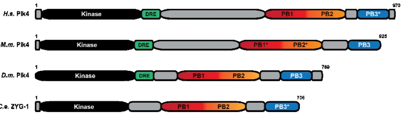
Figure 1.4. The architectural map of the Plk family of kinases highlights the divergence of Plk4. Each Homo sapiens
All Plks share an N-terminal kinase domain (black), though Plk4’s kinase domain more closely resembles Aurora kinases. Plks 1, 2, 3, and 5 share a C
domains (PB1-PB2, light and dark purple) that generally regulate the activity and targe the full-length molecule (see the following sections for in
function). Plk1 contains an inter cis to inhibit kinase activity in a PB1
a downstream regulatory element (DRE, green) immediately C
used to regulate protein levels preceding centriole duplication (see the following section for in-depth discussion of the DRE). Plk4 al
terminal array of three distinct PB domains
(Figure 1.4); furthermore, it is only expressed in fully differentiated tissue types including the central nervous system, underlining its evolutionary departure from the other Plk’s roles in de Carcer et al., 2011). Plk4 is likewise divergent from the Plk4 family, gh in different measures: its kinase domain more closely resembles that of the Aurora han the Plk family (personal communication, Yao Liang Wong and Karen , and unlike all other Plk members, Plk4 contains three structurally defined Polo Box (PB) domains rather than two (Slevin et al., 2012). Plk4, like Plk1, is largely expressed throughout embryonic development and in adult proliferative tissues in humans, i
bone marrow and the male testis (de Carcer et al., 2011).
Figure 1.4. The architectural map of the Plk family of kinases highlights the divergence Homo sapiens major isoform is shown here, with each cartoon drawn to scale. terminal kinase domain (black), though Plk4’s kinase domain more closely resembles Aurora kinases. Plks 1, 2, 3, and 5 share a C-terminal pair of Polo Box
PB2, light and dark purple) that generally regulate the activity and targe
length molecule (see the following sections for in-depth discussion regarding PB function). Plk1 contains an inter-domain linker (IDL, tan) that binds Plk1’s kinase domain in
to inhibit kinase activity in a PB1-PB2-dependent fashion (Xu et al., 2013
a downstream regulatory element (DRE, green) immediately C-terminal to its kinase domain, used to regulate protein levels preceding centriole duplication (see the following section for depth discussion of the DRE). Plk4 also differs from the Plk family by containing a C terminal array of three distinct PB domains (Slevin et al., 2012; Park et al., 2014;
expressed in fully differentiated tissue types including the central nervous system, underlining its evolutionary departure from the other Plk’s roles in . Plk4 is likewise divergent from the Plk4 family, gh in different measures: its kinase domain more closely resembles that of the Aurora Yao Liang Wong and Karen , and unlike all other Plk members, Plk4 contains three structurally defined Polo . Plk4, like Plk1, is largely expressed throughout embryonic development and in adult proliferative tissues in humans, including
Figure 1.4. The architectural map of the Plk family of kinases highlights the divergence major isoform is shown here, with each cartoon drawn to scale. terminal kinase domain (black), though Plk4’s kinase domain more terminal pair of Polo Box PB2, light and dark purple) that generally regulate the activity and targeting of depth discussion regarding PB domain linker (IDL, tan) that binds Plk1’s kinase domain in , 2013). Plk4 contains terminal to its kinase domain, used to regulate protein levels preceding centriole duplication (see the following section for so differs from the Plk family by containing a
Shimanovskaya et al., 2014). PB1-PB2 (red and orange) collectively bind scaffolding proteins, localizing Plk4 to its centriolar targets, and homodimerize in trans to afford DRE-dependent polyubiquitination (Dzhindzhev et al., 2010; Slevin et al., 2012). *Plk5’s kinase domain is truncated and non-functional, followed by another start site that initiates the remainder of the Plk5 polypeptide.
The mechanism of Plk4’s licensing activity followed by swift degradation is conserved among opisthokonts
represents a new class of bona fide
Biochemical and structural techniques as well as functional studies in cell culture have revealed the architecture and modules within both human and
et al., 2002; Habedanck et al., 2005; Dzhindzhev et a al., 2013; Shimanovskaya et al., 2014
domain is the downstream regulatory element (DRE) (Figure 1.5), a phospho region with important roles in down
al., 2009; Rogers et al., 2009
region for F-box binding (β-TrCP in
degradation. This degradation event is essential to limit Plk4 levels in cells, as ectopic Plk4 constructs that are phosphorylation
leading to centriole amplification al., 2010).
Figure 1.5. Plk4 is functionally conserved and maintains specific structural modules. architectural comparison of the functional Plk4 orthologs reveals conservation within the domain architecture. H.s., Homo sapiens
Drosophila melanogaster (fruit fly);
homologs share a similar N-terminal kinase domain (black), while most orthologs (excluding C.e. ZYG-1) share the DRE (green), a region u
central pair of PB domains (PB1
degradation of the FL molecule. The function of the C bona fide Plk4 orthologs.
Biochemical and structural techniques as well as functional studies in cell culture have revealed the architecture and modules within both human and Drosophila
et al., 2002; Habedanck et al., 2005; Dzhindzhev et al., 2010; Slevin et al., 2012; Sonnen et al., 2013; Shimanovskaya et al., 2014). Immediately C-terminal to the conserved kinase domain is the downstream regulatory element (DRE) (Figure 1.5), a phospho
region with important roles in down-regulating the full-length molecule (Cunha al., 2009; Rogers et al., 2009). Active Plk4 autophosphorylates the DRE in trans,
TrCP in H.s.; Slimb in D.m.), polyubiquitination, and proteolytic gradation event is essential to limit Plk4 levels in cells, as ectopic Plk4 constructs that are phosphorylation-resistant in both H.s. and D.m. cells become stabilized, leading to centriole amplification (Cunha-Ferreira et al., 2009; Rogers et al., 2009; H
Figure 1.5. Plk4 is functionally conserved and maintains specific structural modules. architectural comparison of the functional Plk4 orthologs reveals conservation within the
H.s., Homo sapiens (human); M.m., Mus musculus
(fruit fly); C.e., Caenorhabditis elegans (nematode). All Plk4 terminal kinase domain (black), while most orthologs (excluding 1) share the DRE (green), a region used to regulate FL protein stabilization. A central pair of PB domains (PB1-PB2, red and orange) collectively regulate localization and degradation of the FL molecule. The function of the C-terminal PB3 (blue) remains debated Biochemical and structural techniques as well as functional studies in cell culture Drosophila Plk4 (Leung l., 2010; Slevin et al., 2012; Sonnen et terminal to the conserved kinase domain is the downstream regulatory element (DRE) (Figure 1.5), a phospho-regulated Cunha-Ferreira et trans, priming the ), polyubiquitination, and proteolytic gradation event is essential to limit Plk4 levels in cells, as ectopic Plk4 cells become stabilized, Ferreira et al., 2009; Rogers et al., 2009; Holland et
and undefined. *Asterisks indi
similarity, but not structurally verified as PB domains. The other PB structures and their references are as follows: (H.s.
D.m. PB1-PB2: Slevin et al., 2012 and Shimanovskaya et al., 2014; data, unpublished; C.e. PB1-PB2: Shimanovskaya et al., 2014
The C-terminus of Plk4 comprises an array of three PBs. PB1
orange, respectively, in Figure 1.5) together bind Asterless (Asl), affording localization of the full-length molecule to the site of daughter centriole assembly
Shimanovskaya et al., 2014). Additional work from our lab has shown that PB homodimer in trans, effectively dimerizing the full
binding and, ultimately, polyubiquitination and degradation (Figure 1.6) The PB1-PB2 cassette is a conserved feature of Plk4s ac
(shown in Figure 1.5). Unique to Plk4 is the presence of the C
unknown function in the context of the FL molecule. Early work determined that the PB3 also forms a dimer; however, work from
PB3 is not sufficient for robust centriole localization The role of PB3 in Plk4 function remains unclear.
and undefined. *Asterisks indicate PB modules that have been identified via sequence similarity, but not structurally verified as PB domains. The other PB structures and their H.s. PB1-PB2: Park et al., 2014; M.m. PB3, Leung et al., 2002; PB2: Slevin et al., 2012 and Shimanovskaya et al., 2014; D.m. PB3: author’s own
PB2: Shimanovskaya et al., 2014).
terminus of Plk4 comprises an array of three PBs. PB1-PB2 (shown in red and nge, respectively, in Figure 1.5) together bind Asterless (Asl), affording localization of the length molecule to the site of daughter centriole assembly (Slevin et al., 2012;
. Additional work from our lab has shown that PB
effectively dimerizing the full-length molecule and allowing for F binding and, ultimately, polyubiquitination and degradation (Figure 1.6) (Slevin et al., 2012
PB2 cassette is a conserved feature of Plk4s across the species commonly studied (shown in Figure 1.5). Unique to Plk4 is the presence of the C-terminal PB3, a domain with unknown function in the context of the FL molecule. Early work determined that the PB3 also forms a dimer; however, work from our lab as well as other labs demonstrated that PB3 is not sufficient for robust centriole localization (Leung et al., 2002; Slevin et al., 2012 The role of PB3 in Plk4 function remains unclear.
cate PB modules that have been identified via sequence similarity, but not structurally verified as PB domains. The other PB structures and their PB3, Leung et al., 2002; PB3: author’s own
Figure 1.6. Plk4 depends on the PB1-PB2 homodimerization interface to autophosphorylate in trans and bind F-box proteins for its degradation. (A) Active Plk4 (activity notated by a yellow star in the kinase domain) dimerizes via a strong homodimerization interface between PB1-PB2 cassettes in two protomers (Slevin et al., 2012), allowing for phosphorylation of the DRE in trans. (B) Schematic of the FL molecules following dual phosphorylation events in trans. The phosphorylated DRE region is able to then bind an F-box protein (Slimb in D.m., purple; β-TrCP in H.s.) for its degradation mechanism. Note that 1) both diagrams refer to information gleaned in D.m. Plk4, 2) the molecules are translated horizontally relative to each other in (B), and 3) the oligomerization state of PB3 remains contested in D.m. The author’s unpublished data suggest that PB3 is a monomer in D.m., thus the schematic presents a monomeric form.
The Polo Box (PB) domain is used to regulate Plk activity and subcellular targeting by employing a structurally defined fold
The PB domain takes a conserved, simple structural fold: a 6-stranded anti-parallel β -sheet packed perpendicularly against a single α-helix (Figure 1.7 A and B). Though the sequences and binding partners vary greatly among Plks (Yun et al., 2009; Slevin et al., 2012; Xu et al., 2013; Park et al., 2014; Shimanovskaya et al., 2014), serial structural alignments of single PB domains reveal little structural divergence among the individual domains, with RMSD values ranging from 1.5 to 4 Å (Figure 1.7 C). However, three trends emerge when comparing both structural alignments as well as their sequence conservation (Figure 1.7 C,D): 1) PB structures share greater primary and tertiary structure elements within the same spatial orthologs (i.e., PB1s are more similar in both structure and sequence to other PB1s, PB2s are more similar to other PB2s, etc.); 2) As predicted, C.e. ZYG-1 is the least similar in primary structure for each respective PB than any other species; 3) PB2 architecture deviates from the prototypical PB structure in that its β6 strand is parallel to its neighbor and occurs C-terminal to the single α-1 helix (Figure 1.7 E, blue arrow pointing to
Figure 1.7. The structure of each solved PB domain takes on a conserved fold. (A) The structure of a single PB domain (D.m. Plk4 PB1, PDB accession code 4G7N, Slevin et al., 2012) reveals a 6-stranded antiparallel β-sheet packed (cyan, labeled β1-β6) orthogonally against a single α-helix (olive, labeled α1). Loops are shown in dark gray and have been smoothened for simplicity. (B) A schematic depicting the architecture of a typical PB domain, mimicking the geometry of the PB domain shown in A. Note that the N-terminus of the protein begins at β1, the middle forms the serpentine β-sheet, and then the C-terminus consists of the final α1. The dotted lines indicate that the loop connecting β6 and α1 goes back into the plane of the field of view. (C) A series of structural alignments (using the DALI pairwise alignment server, Holm and Rosenström, 2010) reveals little differences among the solved PB domain structures, regardless of species or Plk origin. The species, Plk, and PB number are noted for each PB tested; each was aligned to every other PB within the grid, and the RMSD and percent identity is reported within the corresponding box. Each box within the grid is colored according to a green/red scale, with green indicating the lowest RMSD (best alignment) and red indicating the highest RMSD (poor alignment). (D) A grid showing the sequence conservation between the same individual pairs as in C, with green indicating high conservation and red indicating low conservation. (E) Alignment between D.m. Plk4 PB1 (shown in eggplant) and either H.s. Plk4 PB1 (PDB accession code 4N9J (Park et al., 2014), shown in bronze) or C.e. ZYG-1 PB2 (PDB accession code 4NKB (Shimanovskaya et al., 2014), shown in blush) reveals conservation among Plk4 PB1 homologs and structural differences between Plk4 PB1 and PB2 homologs.
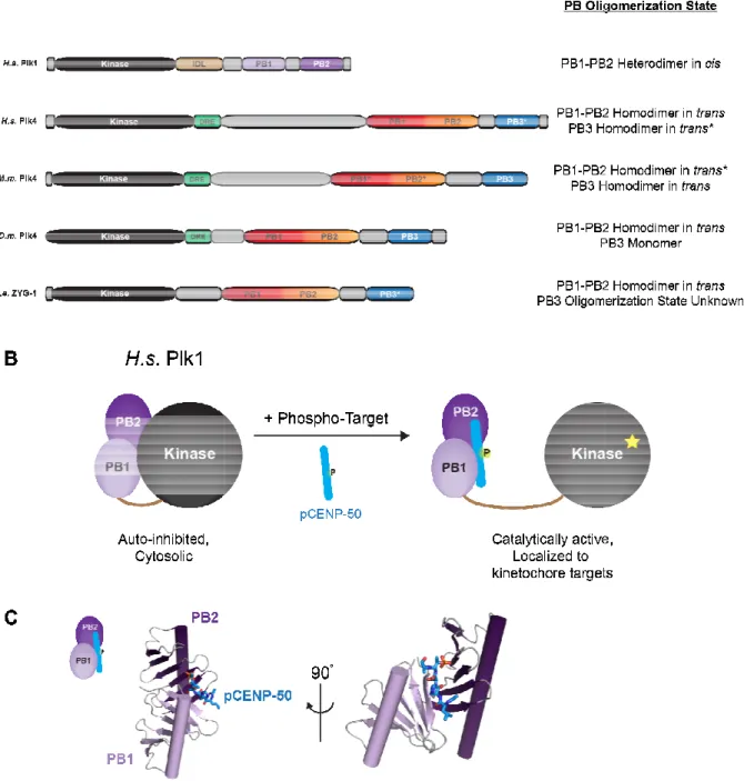
Figure 1.8. Plks employ PB domains to take on differential dimeric configurations for cellular function. (A) A list of the compared Plks and their known PB oligomeric states. Those states labeled with asterisks (*
conservation between mouse and human Plk4. (B) Plk1 PB1
architecture to bind the kinase and allosterically inhibit its catalytic activity
Following phosphorylation events of centrosomal and/or centromeric targets, the PB1 structural unit binds the phospho
domain. (C) The crystal structure of PB1 CENP-50 (PDB accession code 3FVH shown in two orientations.
Figure 1.8. Plks employ PB domains to take on differential dimeric configurations for (A) A list of the compared Plks and their known PB oligomeric states. Those states labeled with asterisks (*) indicate predicted oligomeric states based on the conservation between mouse and human Plk4. (B) Plk1 PB1-PB2 take on a
to bind the kinase and allosterically inhibit its catalytic activity (
tion events of centrosomal and/or centromeric targets, the PB1 binds the phospho-primed targets, releasing the interaction with the kinase domain. (C) The crystal structure of PB1-PB2 bound to a phospho-peptide representative of (PDB accession code 3FVH, Yun et al., 2009) with the same color scheme as B,
for a thorough discussion).
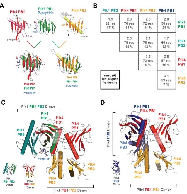
Figure 1.9. PB1-PB2 pairs diverge in both sequence and structure. alignment (Holm and Rosenström, 2010
reveals large differences between Plk1 PB pairs and Plk4 PB pairs (top row, ranging from 12-18% sequence identity). All known Plk4 homolog PB1
closely, with sequence identities ranging from 18 color-code sliding scale, with white
representing complete identity. accession code 4G7N, in oranges;
PB2 pairs diverge in both sequence and structure. Holm and Rosenström, 2010) of all structurally characterized PB1
reveals large differences between Plk1 PB pairs and Plk4 PB pairs (top row, ranging from 18% sequence identity). All known Plk4 homolog PB1-PB2 pairs align much more closely, with sequence identities ranging from 18-40%. Sequence similarity is shown on a
code sliding scale, with white squares representing low similarity and cobalt squar identity. The structures of 3 specific PB1-PB2 pairs (
greens) are shown in the sidebar. (B) Structural alignments between PB1-PB2 pairs demonstrate large differences between Plk1 and Plk4 PB arrays. Plk4 PB1-PB2 from both D.m. and H.s. align well with similar folds and primary sequences, while Plk1 PB1-PB2 collectively form a “pincer,” interacting side-to-side rather than head-to-tail.
PB3: The odd one out
REFERENCES
1. Azimzadeh, J., Hergert, P., Delouvée, A., Euteneuer, U., Formstecher, E., Khodjakov, A., Bornens, M. (2009) hPOC5 is a centrin-binding protein required for assembly of full-length centrioles. The Journal of Cell Biology. 185(1), 101–114.
2. Bettencourt-Dias, M., Rodrigues-Martins, A., Carpenter, L., Riparbelli, M., Lehmann, L., Gatt, M.K., Carmo, N., Balloux, F., Callaini, G., Glover, D.M. (2005) SAK/PLK4 is required for centriole duplication and flagella development. Current Biology. 15(24), 2199–2207.
3. Bettencourt-Dias, M., Hildebrandt, F., Pellman, D., Woods, G., Godinho, S.A. (2011) Centrosomes and cilia in human disease. Trends in Genetics. 27(8), 307–315.
4. Bornens, M., Paintrand, M., Berges, J., Marty, M.C., Karsenti, E. (1987) Structural and chemical characterization of isolated centrosomes. Cell Motility and the Cytoskeleton. 8(3), 238–249.
5. Boveri, T. (1909) Die Blastomerenkerne von Ascaris megalocephala und die Theorie der Chromosomenindividualität. Archiv für Zellforschung. 3, 181-268.
6. Bylander, A., Lind, K., Goksör, M., Billig, H., Larsson, D.J. (2013) The classical progesterone receptor mediates the rapid reduction of fallopian tube ciliary beat frequency by progesterone. Reproductive Biology and Endocrinology. 11(1), 33–9. 7. Carvalho-Santos, Z., Machado, P., Branco, P., Tavares-Cadete, F.,
Rodrigues-Martins, A., Pereira-Leal, J.B., Bettencourt-Dias, M. (2010) Stepwise evolution of the centriole-assembly pathway. Journal of Cell Science. 123(Pt 9), 1414–1426.
8. Chavali, P.L., Pütz, M., Gergely, F. (2014) Small organelle, big responsibility: the role of centrosomes in development and disease. Philosophical transactions of the Royal Society of London. Series B, Biological sciences. 369(1650), 20130468– 20130468.
9. Cizmecioglu, O., Arnold, M., Bahtz, R., Settele, F., Ehret, L., Haselmann-Weiss, U., Antony, C., Hoffmann, I. (2010) Cep152 acts as a scaffold for recruitment of Plk4 and CPAP to the centrosome. The Journal of Cell Biology. 191(4), 731–739.
10.Cottee, M.A., Muschalik, N., Wong, Y.L., Johnson, C.M., Johnson, S., Andreeva, A., Oegema, K., Lea, S.M., Raff, J.W., van Breugel, M. (2013) Crystal structures of the CPAP/STIL complex reveal its role in centriole assembly and human microcephaly. eLife. 2(0), e01071.
ligase limits centrosome amplification through degradation of SAK/PLK4. Current Biology. 19(1), 43–49.
12.Dammermann, A., Müller-Reichert, T., Pelletier, L., Habermann, B., Desai, A., Oegema, K. (2004) Centriole assembly requires both centriolar and pericentriolar material proteins. Developmental Cell. 7(6), 815–829.
13.Dammermann, A., Maddox, P.S., Desai, A., Oegema, K. (2008) SAS-4 is recruited to a dynamic structure in newly forming centrioles that is stabilized by the gamma-tubulin-mediated addition of centriolar microtubules. The Journal of Cell Biology. 180(4), 771–785.
14.de Cárcer, G., Manning, G., Malumbres, M. (2011) From Plk1 to Plk5: Functional evolution of polo-like kinases. Cell Cycle. 10(14), 2255–2262.
15.Delattre, M., Canard, C., Gönczy, P. (2006) Sequential protein recruitment in C. elegans centriole formation. Current Biology. 16(18), 1844–1849.
16.Delgehyr, N., Rangone, H., Fu, J., Mao, G., Tom, B., Riparbelli, M.G., Callaini, G., Glover, D.M. (2012) Klp10A, a microtubule-depolymerizing kinesin-13, cooperates with CP110 to control Drosophila centriole length. Current Biology. 22(6), 502–509. 17.Dzhindzhev, N.S., Yu, Q.D., Weiskopf, K., Tzolovsky, G., Cunha-Ferreira, I.,
Riparbelli, M., Rodrigues-Martins, A., Bettencourt-Dias, M., Callaini, G., Glover, D.M. (2010) Asterless is a scaffold for the onset of centriole assembly. Nature. 467(7316), 714–718.
18.Eckerdt, F., Yamamoto, T.M., Lewellyn, A.L., Maller, J.L. (2011) Identification of a polo-like kinase 4-dependent pathway for de novo centriole formation. Current Biology. 21(5), 428–432.
19.Elia, A.E.H., Rellos, P., Haire, L.F., Chao, J.W., Ivins, F.J., Hoepker, K.,
Mohammad, D., Cantley, L.C., Smerdon, S.J., Yaffe, M.B. (2003) The molecular basis for phosphodependent substrate targeting and regulation of Plks by the Polo-box domain. Cell. 115(1), 83–95.
20.Fırat-Karalar, E.N., Stearns, T. (2014) The centriole duplication cycle. Philosophical transactions of the Royal Society of London. Series B, Biological sciences.
369(1650), 20130460–20130460.
21.Godinho, S.A., Pellman, D. (2014) Causes and consequences of centrosome
abnormalities in cancer. Philosophical transactions of the Royal Society of London. Series B, Biological sciences. 369(1650), 20130467–20130467.
scaffold for cytoplasmic complexes and tethers them in a centrosome. Nature Communications. 2, 359–11.
23.Goshima, G., Wollman, R., Goodwin, S.S., Zhang, N., Scholey, J.M., Vale, R.D., Stuurman, N. (2007) Genes required for mitotic spindle assembly in Drosophila S2 cells. Science. 316(5823), 417–421.
24.Guderian, G., Westendorf, J., Uldschmid, A., Nigg, E.A. (2010) Plk4 trans-autophosphorylation regulates centriole number by controlling βTrCP-mediated degradation. Journal of Cell Science. 123(Pt 13), 2163–2169.
25.Guichard, P., Hachet, V., Majubu, N., Neves, A., Demurtas, D., Olieric, N., Flückiger, I., Yamada, A., Kihara, K., Nishida, Y., Moriya, S., Steinmetz, M.O., Hongoh, Y., Gönczy, P. (2013) Native architecture of the centriole proximal region reveals features underlying its 9-fold radial symmetry. Current Biology. 23(17), 1620–1628.
26.Habedanck, R., Stierhof, Y.-D., Wilkinson, C.J., Nigg, E.A. (2005) The Polo kinase Plk4 functions in centriole duplication. Nature Cell Biology. 7(11), 1140–1146. 27.Hatch, E.M., Kulukian, A., Holland, A.J., Cleveland, D.W., Stearns, T. (2010)
Cep152 interacts with Plk4 and is required for centriole duplication. The Journal of Cell Biology. 191(4), 721–729.
28.He, R., Huang, N., Bao, Y., Zhou, H., Teng, J., Chen, J. (2013) LRRC45 is a centrosome linker component required for centrosome cohesion. Cell Reports. 4(6), 1100–1107.
29.Holland, A.J., Lan, W., Niessen, S., Hoover, H., Cleveland, D.W. (2010) Polo-like kinase 4 kinase activity limits centrosome overduplication by autoregulating its own stability. The Journal of Cell Biology. 188(2), 191–198.
30.Holm, L., Rosenström, P. (2010) Dali server: conservation mapping in 3D. Nucleic Acids Research. 38, W545–9.
31.Kang, Y.H., Park, J.-E., Yu, L.-R., Soung, N.-K., Yun, S.-M., Bang, J.K., Seong, Y.-S., Yu, H., Garfield, Y.-S., Veenstra, T.D., Lee, K.S. (2006) Self-regulated Plk1
recruitment to kinetochores by the Plk1-PBIP1 interaction is critical for proper chromosome segregation. Molecular Cell. 24(3), 409–422.
33.Kitagawa, D., Vakonakis, I., Olieric, N., Hilbert, M., Keller, D., Olieric, V., Bortfeld, M., Erat, M.C., Flückiger, I., Gönczy, P., Steinmetz, M.O. (2011) Structural basis of the 9-fold symmetry of centrioles. Cell. 144(3), 364–375.
34.Kleylein-Sohn, J., Westendorf, J., Le Clech, M., Habedanck, R., Stierhof, Y.-D., Nigg, E.A. (2007) Plk4-induced centriole biogenesis in human cells. Developmental Cell. 13(2), 190–202.
35.Lee, K.S., Park, J.-E., Kang, Y.H., Kim, T.-S., Bang, J.K. (2014) Mechanisms Underlying Plk1 Polo-Box domain-mediated biological processes and their physiological significance. Molecules and Cells. 37(4), 286–294.
36.Leidel, S., Delattre, M., Cerutti, L., Baumer, K., Gönczy, P. (2005) SAS-6 defines a protein family required for centrosome duplication in C. elegans and in human cells. Nature Cell Biology. 7(2), 115–125.
37.Lettman, M.M., Wong, Y.L., Viscardi, V., Niessen, S., Chen, S.-H., Shiau, A.K., Zhou, H., Desai, A., Oegema, K. (2013) Direct binding of SAS-6 to ZYG-1 recruits SAS-6 to the mother centriole for cartwheel assembly. Developmental Cell. 25(3), 284–298.
38.Leung, G.C., Hudson, J.W., Kozarova, A., Davidson, A., Dennis, J.W., Sicheri, F. (2002) The Sak polo-box comprises a structural domain sufficient for mitotic subcellular localization. Nature Structural Biology. 9(10), 719–724.
39.Li, S., Fernandez, J.-J., Marshall, W.F., Agard, D.A. (2011) Three-dimensional structure of basal body triplet revealed by electron cryo-tomography. The EMBO Journal. 31(3), 552–562.
40.Mennella, V., Keszthelyi, B., McDonald, K.L., Chhun, B., Kan, F., Rogers, G.C., Huang, B., Agard, D.A. (2012) Subdiffraction-resolution fluorescence microscopy reveals a domain of the centrosome critical for pericentriolar material organization. Nature Cell Biology. 14(11), 1159–1168.
41.Nigg, E.A., Raff, J.W. (2009) Centrioles, centrosomes, and cilia in health and disease. Cell. 139(4), 663–678.
42.Ohno, S. (1970). Evolution by gene duplication. London: George Alien & Unwin Ltd. Berlin, Heidelberg and New York: Springer-Verlag.
43.Pagan, J., Pagano, M. (2011) FBXW5 controls centrosome number. Nature Cell Biology. 13(8), 888–890.
45.Park, S.-Y., Park, J.-E., Kim, T.-S., Kim, J.H., Kwak, M.-J., Ku, B., Tian, L., Murugan, R.N., Ahn, M., Komiya, S., Hojo, H., Kim, N.-H., Kim, B.Y., Bang, J.K., Erikson, R.L., Lee, K.-W., Kim, S.J., Oh, B.-H., Yang, W., Lee, K.S. (2014)
Molecular basis for unidirectional scaffold switching of human Plk4 in centriole biogenesis. Nature Structural & Molecular Biology. 21(8), 696–703.
46.Peel, N., Stevens, N.R., Basto, R., Raff, J.W. (2007) Overexpressing centriole-replication proteins in vivo induces centriole overduplication and de novo formation. Current Biology. 17(10), 834–843.
47.Pelletier, L., O'Toole, E., Schwager, A., Hyman, A.A., Müller-Reichert, T. (2006) Centriole assembly in Caenorhabditis elegans. Nature. 444(7119), 619–623. 48.Petrinac, S., Ganuelas, M.L., Bonni, S., Nantais, J., Hudson, J.W. (2009) Polo-like
kinase 4 phosphorylates Chk2. Cell Cycle. 8(2), 327–329.
49.Puklowski, A., Homsi, Y., Keller, D., May, M., Chauhan, S., Kossatz, U., Grünwald, V., Kubicka, S., Pich, A., Manns, M.P., Hoffmann, I., Gönczy, P., Malek, N.P. (2011) The SCF–FBXW5 E3-ubiquitin ligase is regulated by PLK4 and targets HsSAS-6 to control centrosome duplication. Nature Cell Biology. 13(8), 1004–1009.
50.Rodrigues-Martins, A., Riparbelli, M., Callaini, G., Glover, D.M., Bettencourt-Dias, M. (2007) Revisiting the role of the mother centriole in centriole biogenesis. Science. 316(5827), 1046–1050.
51.Rogers, G.C., Rusan, N.M., Roberts, D.M., Peifer, M., Rogers, S.L. (2009) The SCF Slimb ubiquitin ligase regulates Plk4/Sak levels to block centriole reduplication. The Journal of Cell Biology. 184(2), 225–239.
52.Scholey, J.M., Anderson, K.V. (2006) Intraflagellar transport and cilium-based signaling. Cell. 125(3), 439–442.
53.Shimanovskaya, E., Viscardi, V., Lesigang, J., Lettman, M.M., Qiao, R., Svergun, D.I., Round, A., Oegema, K., Dong, G. (2014) Structure of the C. elegans ZYG-1 cryptic polo box suggests a conserved mechanism for centriolar docking of Plk4 kinases. Structure. 22(8), 1090-1104.
54.Sillibourne, J.E., Tack, F., Vloemans, N., Boeckx, A., Thambirajah, S., Bonnet, P., Ramaekers, F.C.S., Bornens, M., Grand-Perret, T. (2010) Autophosphorylation of polo-like kinase 4 and its role in centriole duplication. Molecular Biology of the Cell. 21(4), 547–561.
56.Sonnen, K.F., Gabryjonczyk, A.-M., Anselm, E., Stierhof, Y.-D., Nigg, E.A. (2013) Human Cep192 and Cep152 cooperate in Plk4 recruitment and centriole duplication. Journal of Cell Science. 126(Pt 14), 3223–3233.
57.Soung, N.-K., Park, J.-E., Yu, L.-R., Lee, K.H., Lee, J.-M., Bang, J.K., Veenstra, T.D., Rhee, K., Lee, K.S. (2009) Plk1-dependent and -independent roles of an ODF2 splice variant, hCenexin1, at the centrosome of somatic cells. Developmental Cell. 16(4), 539–550.
58.Stevens, N.R., Dobbelaere, J., Brunk, K., Franz, A., Raff, J.W. (2010a) Drosophila Ana2 is a conserved centriole duplication factor. The Journal of Cell Biology. 188(3), 313–323.
59.Stevens, N.R., Roque, H., Raff, J.W. (2010b) DSas-6 and Ana2 coassemble into tubules to promote centriole duplication and engagement. Developmental Cell. 19(6), 913–919.
60.Tsou, M.-F.B., Stearns, T. (2006) Mechanism limiting centrosome duplication to once per cell cycle. Nature. 442(7105), 947–951.
61.Tsou, M.-F.B., Wang, W.-J., George, K.A., Uryu, K., Stearns, T., Jallepalli, P.V. (2009) Polo kinase and Separase regulate the mitotic licensing of centriole duplication in human cells. Developmental Cell. 17(3), 344–354.
62.van Breugel, M., Hirono, M., Andreeva, A., Yanagisawa, H.-A., Yamaguchi, S., Nakazawa, Y., Morgner, N., Petrovich, M., Ebong, I.-O., Robinson, C.V., Johnson, C.M., Veprintsev, D., Zuber, B. (2011) Structures of SAS-6 suggest its organization in centrioles. Science. 331(6021), 1196–1199.
63.van Breugel, M., Wilcken, R., McLaughlin, S.H., Rutherford, T.J., Johnson, C.M. (2014) Structure of the SAS-6 cartwheel hub from Leishmania major. eLife. 3(0), e01812.
64.van de Weerdt, B.C.M., Littler, D.R., Klompmaker, R., Huseinovic, A., Fish, A., Perrakis, A., Medema, R.H. (2008) Polo-box domains confer target specificity to the Polo-like kinase family. Biochimica et Biophysica Acta- Molecular Cell Research. 1783(6), 1015–1022.
65.Winey, M., O'Toole, E. (2014) Centriole structure. Philosophical transactions of the Royal Society of London. Series B, Biological sciences. 369(1650), 20130457– 20130457.
67.Yun, S.-M., Moulaei, T., Lim, D., Bang, J.K., Park, J.-E., Shenoy, S.R., Liu, F., Kang, Y.H., Liao, C., Soung, N.-K., Lee, S., Yoon, D.-Y., Lim, Y., Lee, D.-H., Otaka, A., Appella, E., McMahon, J.B., Nicklaus, M.C., Burke, T.R., Jr, Yaffe, M.B., Wlodawer, A., Lee, K.S. (2009) Structural and functional analyses of minimal
phosphopeptides targeting the polo-box domain of polo-like kinase 1. Nature Structural & Molecular Biology. 16(8), 876–882.
68.Zhao, H., Zhu, L., Zhu, Y., Cao, J., Li, S., Huang, Q., Xu, T., Huang, X., Yan, X., Zhu, X. (2013) The Cep63 paralogue Deup1 enables massive de novo centriole biogenesis for vertebrate multiciliogenesis. Nature Cell Biology. 15(12), 1434–1444. 69.Zimmerman, W.C., Erikson, R.L. (2007) Polo-like kinase 3 is required for entry into
CHAPTER 2: THE STRUCTURE OF THE PLK4 CRYPTIC POLO BOX REVEALS TWO TANDEM POLO BOXES REQUIRED FOR CENTRIOLE DUPLICATION 1
Summary
Centrioles are key microtubule polarity determinants. Centriole duplication is tightly controlled to prevent cells from developing multipolar spindles, a situation that promotes chromosomal instability. A conserved component in the duplication pathway is Plk4, a polo kinase family member that localizes to centrioles in M/G1. To limit centriole duplication, Plk4 levels are controlled through trans-autophosphorylation that primes ubiquitination. In contrast to Plks 1-3, Plk4 possesses a unique central region called the “Cryptic Polo Box”. Here, we present the crystal structure of this region at 2.3-Å resolution. Surprisingly, the structure reveals two tandem, homodimerized polo boxes, PB1-PB2, that form a unique, winged architecture. The full PB1-PB2 cassette is required for binding the centriolar protein Asterless as well as robust centriole targeting. Thus, with its C-terminal polo box (PB3), Plk4 has a novel, triple polo box architecture that facilitates oligomerization, targeting, and
promotes trans-autophosphorylation, limiting centriole duplication to once per cell cycle.
Introduction
Centrioles are cylindrical, microtubule-based structures that form the core
_____________________________
1 Slevin, L.K., Nye, J., Pinkerton, D.C., Buster, D.W., Rogers, G.C., and Slep, K.C. (2012). The structure of the
Plk4 Cryptic Polo Box reveals two tandem Polo Boxes required for centriole duplication. Structure. 20(11), 1905-1917.