Simvastatin and Atorvastatin inhibit DNA replication
licensing factor MCM7 and effectively suppress
RB-deficient tumors growth
Juan Li1,2,9, Jie Liu1,2,9, Zheyong Liang1,2, Fang He1,2, Lu Yang1,2, Pingping Li1,2, Yina Jiang3, Bo Wang1,2, Can Zhou4, Yaochun Wang1,2, Yu Ren4, Jin Yang5, Jianmin Zhang6, Zhijun Luo7, Cyrus Vaziri8and Peijun Liu*,1,2
Loss or dysfunction of tumor suppressor retinoblastoma (RB) is a common feature in various tumors, and contributes to cancer cell stemness and drug resistance to cancer therapy. However, the strategy to suppress or eliminate Rb-deficient tumor cells remains unclear. In the present study, we accidentally found that reduction of DNA replication licensing factor MCM7 induced more apoptosis in RB-deficient tumor cells than in control tumor cells. Moreover, after a drug screening and further studies, we demonstrated that statin drug Simvastatin and Atorvastatin were able to inhibit MCM7 and RB expressions. Further study showed that Simvastatin and Atorvastatin induced more chromosome breaks and gaps of Rb-deficient tumor cells than control tumor cells.
In vivoresults showed that Simvastatin and Atorvastatin significantly suppressed Rb-deficient tumor growth than control in
xenograft mouse models. The present work demonstrates that ‘old’ lipid-lowering drugs statins are novel weapons against
RB-deficient tumors due to their effects on suppressing MCM7 protein levels.
Cell Death and Disease(2017)8,e2673; doi:10.1038/cddis.2017.46; published online 16 March 2017
Retinoblastoma (RB) gene, a well-studied tumor suppressor, plays important roles in cell-cycle regulation and other cellular processes.1–3Loss or dysfunction of RB is a common feature in various tumors, and contributes to tumor cell stemness and drug resistance.4,5Therefore, it is urgent to explore a way to suppress RB-deficient tumor cells.
We accidentally found that acute depletion of mini-chromosome maintenance protein 7 (MCM7), a DNA replica-tion licensing factor, could induce more apoptosis in RB-deficient tumor cells than in control cells. Therefore, MCM7 might be an ideal target for suppressing RB-deficient tumor cell growth. MCM7 is one component of MCM2-7 hexamer (MCMs). The MCM2-7 complex forms the core of the DNA helicase and is responsible for melting and unwinding the double helix during DNA synthesis.6–8Recent studies have demonstrated that the chromatin-bound excess MCM com-plex plays an important role in maintaining genomic integrity under conditions of replicative stress in human cells, and that acute ablation of MCMs induces chromosome fragility in cells.9–11 DNA replication licensing factor MCM2-7 proteins are highly expressed in various clinical tumor tissues.12–16 Reduction of MCMs causes tumor cells to become sensitive to chemotherapy drugs;11,17thus, excess MCMs in tumor cells might serve as a shield to resist antitumor chemotherapy. Remarkably, depletion or mutation of a single MCM in mammalian cells by siRNA-mediated approaches results in
suppression of all functional MCMs due to the hexameric dependency of the MCM complex for helicase activity,9,11,18,19 and cells might own a sensing mechanism that maintains equal MCM subunit stoichiometry.20,21
In the present study, we demonstrated that reduction of MCM7 induces much moreγ-H2AX expression and apoptosis in RB deficient or inactive tumor cells than in RB-proficient tumor cells. Small molecular drug screening and further experiments demonstrated that Simvastatin (SVA) and Atorvastatin (ARO) could suppress MCM7 protein expression effectively. SVA and ARO are members of statins, well known as small-molecule inhibitors of 3-hydroxy-3-methylglutaryl coenzyme A (HMG-CoA) reductase that can lower cholesterol. Numerous studies, employing a variety of tumor cells, have demonstrated their antitumor effects.22–24Nevertheless, the mechanisms for these anticancer effects are still not well characterized.
The aim of this study is to verify whether MCMs is a target for anti-RB-deficient tumor therapy, and to further clarify the antitumor effect and potential mechanism of the statin drugs.
Results
Acute depletion of MCM7 induces γ-H2AX expression
and apoptosis in RB-deficient or inactive tumor cells.
Numerous studies have shown that DNA replication licensing factors are overexpressed in various tumor cells and clinical
1Center for Translational Medicine, The First Affiliated Hospital of Xian Jiaotong University, Xi’an 710061, Shaanxi, China;2Key Laboratory for Tumor Precision Medicine of
Shaanxi Province, The First Affiliated Hospital of Xian Jiaotong University, Xi’an 710061, Shaanxi, China;3Department of Pathology, The First Affiliated Hospital of Xian Jiaotong University, Xi’an 710061, Shaanxi, China;4Department of Breast Surgery, The First Affiliated Hospital of Xian Jiaotong University, Xi’an 710061, Shaanxi, China;
5
Department of Oncology, The First Affiliated Hospital of Xi’an Jiaotong University, Xi’an 710061, Shaanxi, China;6Department of Cancer Genetics, Roswell Park Cancer Institute, Buffalo 14263, NY 14263, USA;7Department of Biochemistry, Boston University School of Medicine, Boston 02118, MA, USA and8Department of Pathology and
Laboratory Medicine, University of North Carolina at Chapel Hill, Chapel Hill, NC, USA
*Corresponding author: P Liu, Center for Translational Medicine or Key Laboratory for Tumor Precision Medicine of Shaanxi Province, The First Affiliated Hospital of Xian Jiaotong University, 277 Yanta West Road, Xi’an, Shaanxi 710061, PR China. Tel: +86 18991232306; Fax: +86 029 85324628; E-mail: [email protected]
9
These two authors contributed equally to this work and should be considered co-first authors.
Received 17.10.16; revised 17.1.17; accepted 18.1.17; Edited by A Stephanou
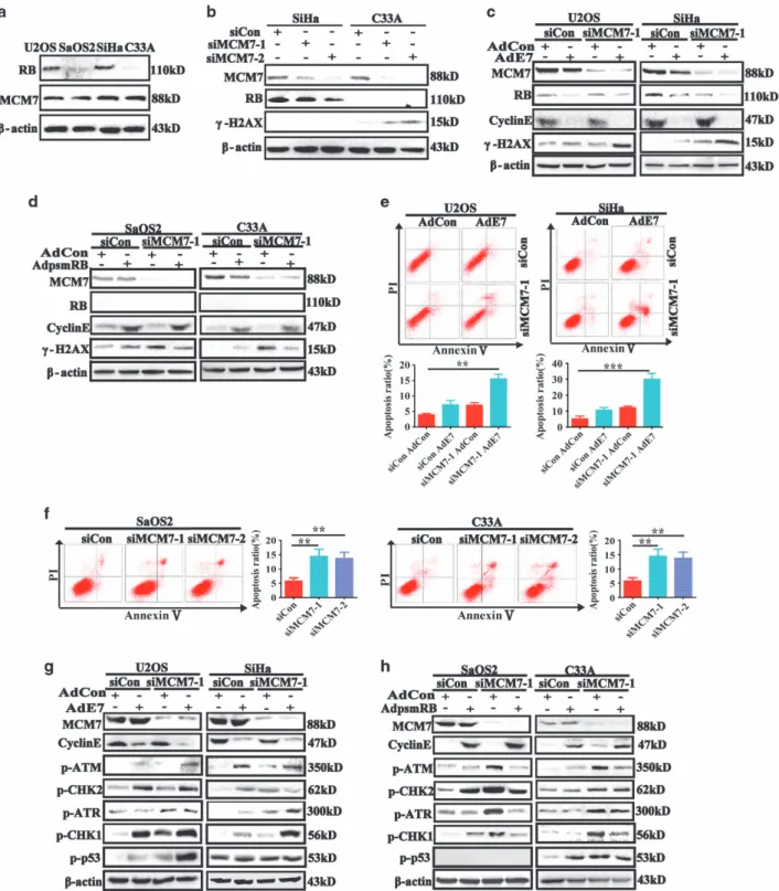
tissues;12–14 therefore, excess MCMs in tumor cells might serve as a shield to protect tumor cells against antitumor chemotherapy. Studies have also shown that depletion of MCM7 results in disruption of whole MCMs hexamer complex.9,11,18,19 To test the role of MCMs in maintaining cell chromosomal integrity, the RB expression levels of U2OS/SaOS2/SiHa/C33A cancer cells were detected (Figure 1a). Then, MCM7 was silenced in SiHa and C33A cells, and DNA double-strand break (DSB) marker protein
γ-H2AX was detected. As shown in Figures 1b, significant reduction of MCM7 expression was achieved, and depletion of MCM7 led to a reduction in RB protein levels. This result is fully expected based on our published work: in MCM7-depleted cells, there is reduced replication licensing leading to attenuation of several cell-cycle-regulated genes, including RB whose expression is transcriptionally induced in G1.26 Interestingly, γ-H2AX was also detectable in RB-deficient C33A cells after MCM7 knockdown. These results indicated that RB status may be important for the DNA DSBs after MCM7 depletion. To further investigate the effect of MCM7 depletion in treatment of RB-proficient and RB-deficient tumors, AdE7 adenoviruses, which express human papillomavirus (HPV) E7 oncoprotein and degrade RB protein,27–29was used to infect RB-proficient U2OS and SiHa cells; and AdPSM-RB adenoviruses, which express an active allele of RB protein,30was used to restore active RB in RB-deficient SaOS2 and C33A cells. In this study, the active RB allele PSM is a truncation mutant that contains the pocket domain but is smaller than full-length RB protein. PSM-RB lacks epitopes that are recognized by most commercial RB antibodies, including the antibodies used in our study. Therefore, as a surrogate for measuring RB activity in AdPSM-RB-infected cells, we examined expression of Cyclin E (a repressed transcriptional target of RB–see Figures 1c and d). We also confirmed expression of PSM-RB mRNA (using RT-PCR) in AdPSM-RB-infected cells (Supplementary Figure S1b). As shown in Figures 1c and d, MCM7 depletion resulted in stronger expression of γ-H2AX after HPV E7 adenovirus infection in SiHa and U2OS cells, and MCM7 depletion induced a weaker expression of γ-H2AX in PSM-RB adenovirus-infected C33A and SaOS2 cells. Then, MCM7 and RB were silenced using siRNA in SiHa and U2OS cells and the effects of MCM7/RB-ablation onγ-H2AX were determined. As shown in Supplementary Figure S1a, co-depletion of MCM7 and RB led to increased γ-H2AX, thereby fully recapitulating the effects of HPV E7 onγ-H2AX when MCM7 is depleted. These results strengthen our conclusion that RB allows cells to tolerate reduced MCM7 expression.
γ-H2AX is known as a sensor for DNA DSBs, and apoptosis is one of the results of DNA DSB.31,32Thus, we asked whether depletion of MCM7 gave rise to more apoptosis in RB-deficient tumor cells. The FACS results showed that MCM7 depletion resulted in more apoptosis in HPV E7 adenovirus-infected U2OS and SiHa cells (Figure 1e), and that MCM7 depletion led to a larger apoptotic population in SaOS2 and C33A RB-deficient tumor cells (Figure 1f).
DNA DSBs trigger DNA damage response (DDR) check-point to repair the damaged DNA, and cell apoptosis is one of the outcomes of DDR response.33–35Here, we asked whether
RB status is crucial for activation of DDR checkpoints. RB inactive (AdE7 infected) and deficient tumor cells showed elevated p-ATM, p-ATR, p-Chk1, p-Chk2 and p-p53 expres-sions, and that PSM-RB restored C33A and SaOS2 cells showed weaker expression of those proteins, and that total DNA damage checkpoint proteins were not changed (Figures 1g, h, Supplementary Figures S1c and d). Taken together, our results showed that RB inactive or deficient tumor cells were more sensitive to acute MCM7 depletion, and that acute MCM depletion induced stronger DDR response in RB-deficient tumor cells than in control tumor cells.
SVA or ARO inhibits MCM7 and RB protein expressions
and induces apoptosis in Hep3B cells. Based on the
above observation, to further explore which small molecular drug(s) can inhibit MCM7 protein expression, a small molecular drug screening was carried out and SVA showed the strongest inhibition effect on MCM7 protein (Figure 2a). To identify which statins were more effective to inhibit MCM7 expression, we screened five clinically used statins and identified their capacities for inhibiting MCM7 expression. We found that SVA and ARO exhibited more inhibitory effect on MCM7 protein expression than the other statins. Moreover, we found that SVA and ARO could effectively reduce both the MCM7 and RB protein expressions (Figure 2b).
To determine the effects of SVA or ARO on cell-cycle progression, Hep3B cells were treated with different concen-trations of SVA or ARO. The percentages of the G2/M phase cells were reduced, and that the percentages of apoptotic cells in the sub-G1 phase were significantly increased at 72 h following the treatment (Figure 2c). Our experiments showed that expressions of MCM7, RB, p-RB and S-phase proteins CDC45 and PCNA were reduced while expressions of G1 phase inhibitors p21 and p27 proteins were increased after SVA or ARO treatment (Figure 2d).
Antiapoptotic protein Bcl2 was reduced, apoptosis protein cleaved-caspase-3 increased and DSB sensor proteinγ-H2AX, which is an apoptosis-related protein,31increased 72 h after SVA or ARO treatment (Figure 2e). Immunofluorescence assay showed the presence ofγ-H2AX after SVA or ARO treatment (Figure 2f). These data suggest that SVA or ARO decreased G2/M phase population and induced apoptosis.
SVA suppresses MCM7 and RB protein expressions via activating endoplasmic reticulum stress and autophagy
signaling pathway. To investigate the mechanism(s) by
which SVA led to reduced expression of MCM7 and RB, the mRNA expression levels of MCM2, MCM4, MCM7 and RB were analyzed. Interestingly, the mRNA expression levels of all licensing factors and RB increased after SVA treatment (Figure 3a), while the protein expression levels of MCM7 and RB decreased after SVA treatment (Figure 3b). Our results showed that reduction of MCM7 and RB protein after SVA treatment was not due to the reduced transcription activity.
We next investigated whether MCM7 and RB were degraded by ubiquitin proteasome system (UPS). Our experi-ments showed that MG132 did not recover the SVA-reduced MCM7 and RB protein expression levels (Figure 3c). These data indicated that reduction of MCM7 and RB proteins after SVA treatment were not due to UPS activation.
Reportedly, statins can activate AMPK,35–37endoplasmic reticulum (ER) stress and autophagy signaling pathway,38,39 and inhibit the mTOR pathway.40 We tested whether serum starvation-induced autophagy affected MCM7 and RB expression. After serum starvation, MCM7 and RB mRNA expressions were significantly increased, while their protein expressions showed no change (Figures 3d and e). Then, we
determined whether SVA activated ER stress and autophagy signaling pathway. We found that p-AMPK, p-PERK, p-eIF2α and LC3A/B were increased and p70S6 was decreased after SVA treatment (Figures 3f, g and i). We also found that compound C (AMPK inhibitor) and GSK2606414 (PERK inhibitor) effectively reversed the MCM7 and RB protein expression (Figures 3h and j). Taken together, our results Figure 3 Simvastatin reduced MCM7 and RB expressions via activating cell autophagy and ER stress signaling pathway. (a) Hep3B cells were treated with 5 or 10μM Simvastatin for 24 or 72 h. The mRNA expression levels of MCM2, MCM4, MCM7 and RB in the resulting cells were analyzed by RT-PCR. (b) MCM7 and RB expressions were decreased after Simvastatin treatment. Hep3B cells were treated with 5 or 10μM Simvastatin for 24 or 72 h, and then analyzed by immunoblotting with MCM7 or RB antibodies. (c) Hep3B cells were treated with 2μM MG132 and 10μM Simvastatin for 48 h, then harvested and analyzed by immunoblotting with MCM7 and RB antibodies. (dande) Hep3B cells were cultured with 10% FBS or 0.01% FBS for 24 h and subjected to DNA content analysis using a FACS flow cytometer; or analyzed the expression of MCM7 and RB by RT-PCR and immunoblotting. (f) Hep3B cells were treated with 5 or 10μM Simvastatin for 24 or 72 h, and then cell extracts were analyzed by immunoblotting with AMPK cell signaling or cell autophagy-related antibodies. (g) Hep3B cells were treated with serum starvation or 5μM Simvastatin for 72 h. Cell monolayers were fixed and stained with LC3A/B, and immunofluorescence images were presented (Scale bars, 10μm). (h) Hep3B cells were treated for 48 h with 5μM compound C (AMPK inhibitor; Sigma-Aldrich, St. Louis, MO, USA) or 10μM Simvastatin, then harvested and analyzed by immunoblotting with p-AMPK, MCM7 or RB antibodies. (i) Hep3B cells were treated with 5 or 10μM Simvastatin for 24 h or 72 h, and then cell extracts were analyzed by immunoblotting with PERK, p-PERK and p-eIF2αantibodies. (j) Hep3B cells were treated for 48 h with 2μM GSK2606414 (PERK inhibitor; Millipore, Bedford, MA, USA) or 10μM Simvastatin, then harvested and analyzed by immunoblotting with p-PERK, MCM7 or RB antibodies
indicated that SVA inhibited MCM7 and RB protein expres-sions by activating ER stress and the autophagy signaling pathway.
SVA inhibits tumor proliferation and sensitizes
RB-deficient or inactive tumor cells. To further
demon-strate the broad inhibitory effect of SVA on different tumor cells, the RB and p-RB protein expression of paired U2OS and SaOS2 human osteosarcoma cells, SiHa and C33A human cervical cancer cells, and LNCaP and Du145 human prostate cancer cells were examined. RB proteins were not detectable or weakly expressed in SaOS2, C33A and Du145 cells, in contrast with their paired U2OS, SiHa and LNCaP cells (Figure 4a). Then the paired cancer cells were treated with different dosages of SVA. Cells that survived were harvested and counted at different time points (Figure 4b). We found that deficient cells were more sensitive to SVA than RB-proficient cells (Figure 4c). Apoptosis proteins cleaved-PARP and cleaved-caspase-3 were detectable after 72 h SVA treatment, and apoptosis inhibitory protein Bcl2 were decreased after SVA treatment for 72 h (Figure 4d). Flow cytometry experiments also showed that the pre-G1 apoptotic cell populations were significantly increased in RB-deficient C33A and Du145 cells (Supplementary Figures S2a–c).
Taken together, our results demonstrated that SVA could inhibit proliferation of various tumor cells, and that RB-deficient cells were more sensitive to the SVA treatment than RB-proficient cells.
SVA increases chromosome instability in RB-deficient
cells. We investigated the possible mechanism(s) by which
SVA induced apoptosis in RB-proficient and RB-deficient cells. Chemical cytotoxicity is usually associated with endogenously generated reactive oxygen species (ROS), but we found that ROS was not detected after SVA treatment (Supplementary Figure S3). It suggested that SVA exerted apoptotic effects were not induced by the increase in oxidative stress.
Then we detectedγ-H2AX expression in RB-proficient and -deficient cells. The result showed detectable γ-H2AX in SaOS2 and C33A cells, but noγ-H2AX in U2OS and SiHa cells after SVA treatment (Figure 5a). After SVA treatment, RB-depleted U2OS cells showed a significantly increased Pre-G1 apoptotic cell population, andγ-H2AX was detectable in RB-depleted U2OS cells, but barely detected in control cells (Figures 5b–e).
Since SVA induced the expression of γ-H2AX in RB-depleted tumor cells, we asked whether SVA could activate DDR response. After SVA treatment, DDR checkpoint proteins (p-ATM, p-CHK2 and p-p53) were increased at an earlier time points in SaOS2 and C33A cells than in U2OS and SiHa cells. Seventy-two hours after SVA treatment, those active DDR checkpoint proteins were significantly impaired in SaOS2 and C33A cells, compared with those in U2OS and SiHa cells (Figure 5f).
Ibarra and others11 have reported that cells with reduced backup MCMs are sensitive to replicative stress and display chromosome instability after treatment with DNA replication inhibitor aphidicolin. Therefore, we asked whether SVA could induce chromosome instability and whether RB status was
related to chromosome instability. We found that RB-deficient SaOS2 cells displayed more breaks and gaps than U2OS after SVA treatment (Figure 5g).
Taken together, compared with proficient cells, RB-deficient cells showed highly increased chromosome instabil-ity and significantly impaired DDR checkpoint proteins 72 h after SVA treatment, which suggests that irreparable chromo-some damage might be a cause of cell apoptosis.
RB-deficient tumors were more sensitive to SVA than
RB-proficient tumors in vivo. To investigate whether SVA
can inhibit tumor growthin vivo, mouse melanoma B16 cells were subcutaneously injected into C57 mice, and mouse breast carcinoma 4T1 cells were injected into the fat pad of BalB/C mice. When tumors grew to the size of 4 mm × 4 mm, mice were treated with water or SVA (60 mg/kg/day) via taking gavage. B16 and 4T1 tumors were significantly redu-ced after SVA treatment (Figures 6a–c and Supplementary Figures S4a–c). Immunohistochemistry showed that MCM7-positive and RB expression were reduced after SVA treat-ment (Figures 6d, e and Suppletreat-mentary Figure S4d).
Next, we determined whether RB-deficient tumor was more sensitive to SVA than RB-proficient tumorin vivo. SiHa human cervical cancer cells were infected with control adenovirus and HPV E7 adenovirus. Forty-eight hours after the infection, adenovirus-infected cells were subcutaneously injected into nude mice. The growth of AdE7-infected SiHa tumor was significantly decreased after SVA treatment, and the inhibitory effects were dosage-dependent (Figures 6f–h). Immunohisto-chemistry showed that MCM7 and RB proteins were reduced in AdE7-infected tumor cells after SVA treatment. These experi-ments showed that MCM7 and RB were reduced after SVA treatment and that RB-deficient tumor was more sensitive to SVA than RB-proficient tumorin vivo(Figures 6i and j).
Taken together, the present study demonstrated that statin drugs such as SVA could effectively inhibit MCM7 and RB via activation of ER stress and autophagy signaling cascade, and that reduction of MCM7 and RB induced more chromosome breaks or gaps and further gave rise to apoptosis in RB-deficient tumor cells (Figure 6k).
Discussion
Pre-RC.15,16 Our finding will provide a potential strategy for RB-deficient tumor therapy since loss or inactivation of RB is a common feature of more than half of human tumors.
Interestingly, we found that MCM7 depletion resulted in stronger expression ofγ-H2AX in HPV E7 adenovirus-infected SiHa and U2OS cells than in control adenovirus-infected cells. As is well known, high-risk HPVs (HPV16, 18, etc.) are important risk factors in various tumors; most cases of cervical cancer are associated with HPV infections, such as HPV16 and HPV 18 infections.29,41,42Therefore, the results of our study also identify MCM7 as a potential therapeutic target not
only in RB-deficient tumors but also in HPV-expressing malignancies, thereby broadening the significance of our findings.
After a small molecular drug screening, we found that SVA could inhibit MCM7 protein levels more effectively. Statins, inhibitors of HMG-CoA reductase and well-known cholesterol-lowering drugs, have been used in clinic for almost 40 years and have been proved to be safe, effective drugs with minor side effects.43–45 Randomized controlled clinical trials have indicated that statins have provocative and unexpected benefits for reducing the incidence of colorectal cancer and Figure 4 RB-deficient or inactive tumor cells were more sensitive to Simvastatin than RB-proficient cells. (a) The expression status of RB in different cells. Extracts of U2OS/ SaOS2/SiHa/C33A/LNCaP/Du145 cells were analyzed by immunoblotting with MCM7 or RB antibodies. (b) The cell survival rates of U2OS, SaOS2, SiHa, C33A, LNCaP and Du145 cells after treatment with Simvastatin for 24, 48 or 72 h. The viable cells were harvested and counted three times for each treatment. (c) U2OS and SaOS2 cells were treated with 5μM Simvastatin for 72 h, incubated with annexin V-FITC and PI, and then examined by FACS. (d) U2OS/SaOS2 and SiHa/C33A were treated with Simvastatin (2.5 or 5μM) for 24 or 72 h, and then cell extracts were analyzed by immunoblotting with apoptosis-related antibodies
melanoma.46–48 Statins can also inhibit tumor growth via different mechanisms, such as inhibition of angiogensis or inflammation.49,50 Our results showed that SVA or ARO displayed a stronger effect on inhibiting MCM7 expression in various tumor cells. Interestingly, SVA or ARO not only effectively reduced the MCM7 protein expression in various
Several reports have shown that various RB-deficient tumors are drug resistant.4,5,51 For instance, tamoxifen (TAM)-resistant breast cancer cells display stem cell-like traits and loss of RB function.4,5Statins can re-sensitize those TAM-resistant breast cancer cells to chemo-drugs.52,53 Our work present here may provide a clue for exploring the mechanisms by which statins inhibit proliferation of TAM-resistant breast cancer cells or other chemo-drug-resistant tumor cells in the process of tumor therapy.
Ibarra and co-workers have reported that depletion of licensing factor MCMs induces chromosome break and gap.11 As DNA damage or chromosome instability is a common cause of cell-cycle arrest and apoptosis, one of the earliest events in DNA damage is the initiation of histone H2AX phosphorylation on Ser139 to generateγ-H2AX, which forms nuclear foci and recruits DNA repair factors.54,55Our results showed that SVA and ARO treatment resulted higher γ-H2AX expression in RB-deficient tumor cells than in RB-proficient tumor cells. We also found that RB-deficient SaOS2 cells displayed more breaks and gaps than RB-proficient U2OS cells after SVA treatment. Since DNA damage initiates DDR response in cells and DDR inhibition triggers apoptosis signaling cascade, we examined the expression of DDR response checkpoint proteins at different time points after SVA treatment. Our results demonstrated that SVA or ARO inhibited DDR checkpoint proteins activation at later time points in RB-deficient tumor cells than in RB-proficient tumor cells. SVA or ARO also induced more chromosome breaks or gaps in RB-deficient tumor cells than in RB-proficient tumor cells. Evidence indicates that tumor development is associated with perturbed DDR repair pathways. Defects in one DDR repair pathway can be compensated by other pathways, which may contribute to resistance to DNA damage chemotherapy and radiotherapy. Therefore, DDR pathways make an ideal target for therapeutic intervention.33,35,55,56 Our results demonstrated that SVA or ARO effectively inhibited activities of RB and other DDR checkpoint proteins in various tumor cells 72 h after treatments. These results support the point of view that DDR pathways are ideal targets for tumor therapy.
In vivoresults showed that SVA effectively reduced the size and weight of xenograft tumors and inhibited MCM7 and RB protein expressions in mice. What is more, although the mice remained healthy after treatment with high-dosage SVA (60 mg/ day/kg in mice amount to about 5.4 mg/kg/day in human), a dosage much higher than what is used for patients, whether high-dosage SVA can effectively inhibit tumor development in clinic should be investigated. This finding also provided evidence for the potential of statins in tumor treatment.
Although previous reports have shown that MCMs serve as potential targets for tumor therapy, an important problem should be pointed out: partial suppression of MCMs function can give rise to increased genomic instability and DNA damage.11,17MCM4 knockout mice display genomic instability and mice with sustained, partially defective MCMs function display increased cancer risk.57–60We provide here that SVA or ARO reduces MCM7 and RB protein expressions, induces chromosome instability and gives rise to apoptosis in various tumor cells. Statins have been used almost four decades and have been proved safe. No evidence shows that statins result in tumor; instead randomized controlled clinical trials indicate that statins have unexpected benefits of reducing tumors.43-45 To our knowledge, this is the first report showing that: (1) RB-deficient or inactive tumor cells are more sensitive to MCMs reduction; (2) statin drug SVA or ARO inhibits the protein expression of licensing factor MCM7 and RB via activating ER and autophagy signaling pathway; (3) SVA or ARO induces a higher apoptosis rate in RB-deficient cells than in RB-proficient cells; (4) SVA or ARO triggers DDR checkpoint cascade at earlier time point and induces more chromosome breaks or gaps in RB-deficient tumor cells than in RB-proficient tumor cells. These findings suggest that statin drugs, such as SVA and ARO, may be potential anti-RB-deficient tumor drugs, and may provide new insights for application of‘old’lipid-lowering statin drugs to tumor therapy.
Materials and Methods
Cell culture and treatments. SiHa and C33A human cervical cancer cells, Hep3B human liver cancer cells and 4T1 mouse mammary gland cells were cultured in high-glucose DMEM (HyClone, Logan, UT, USA) and 10% fetal bovine serum (Gibco, Grand Island, NY, USA). LNCaP and Du145 human prostate cancer cells and B16 mouse melanoma cells were cultured in RPMI-1640 medium (Hyclon) and 10% fetal bovine serum (Gibco). U2OS human osteosarcoma cells were cultured in McCoy's 5a medium (Hyclon) and 5% fetal bovine serum (Gibco) and 10% horse serum (Gibco). SaOS2 human osteosarcoma cells were cultured in McCoy's 5a medium and 15% fetal bovine serum. All cell lines purchased from Shanghai Institute of Biochemistry and Cell Biology, Chinese Academy of Sciences (Shanghai, China).
Cell lines were authenticated by short-tandem repeat analysis by the cell bank. For siRNA (Invitrogen, Carlsbad, CA, USA), cells were transfected using Lipofectamine 2000 (Invitrogen) according to the manufacturer’s instructions. Target sequences of oligonucleotides used were as follows: Control (Non-Targeting), 5′ -UUC UCCGAA CGU GUC ACG U-3′and 5′-ACG UGA CAC GUU CGG AGA A-3′; MCM7-1, 5′-AUC GGA UUG UGA AGA UGA A-3′ and 5′-UUC AUC UUC ACA AUC CGA U-3′; MCM7-2, 5′-GCU CCA GAU UCA UCA AAU U-3′and 5′-AAU UUGAUGAAU CUG GAG C-3′; RB1, 5′-AAU GGU UCA CCU CGA ACA C-3′ and 5′-GGGUGU UCG AGG UGA ACC A-3′; RB-2, 5′-GAA ACA GAA GAA CCU GAU U-3′and 5′-AAU CAG GUU CUU CUG UUU C-3′.
The Simvastatin prodrug (Sigma-Aldrich) was subjected to activation as described by Sadeghi et al.25 Atorvastatin, Lovastatin, Fluvastatin and Pravastatin were purchased from Sigma-Aldrich. Atorvastatin was dissolved in methanol. Lovastatin
Figure 5 Simvastatin was more likely to induce chromosome instability andγ-H2AX expression in RB deficient or inactive tumor cells than in RB-proficient cells. (a) Simvastatin inducedγ-H2AX expression in RB deficient or inactive tumor cells. U2OS, SaOS2, SiHa and C33A cells were treated with 5μM Simvastatin for 72 h, and then analyzed by immunofluorescence staining withγ-H2AX antibody. (bandc) U2OS cells were transfected with siRB-1 and siRB-2 for 48 h to deplete RB. Then cells were treated with 5μM Simvastatin for 72 h, incubated with PI and analyzed with FACS. The result was compared with that of control cells, statistical analysis of the difference in sub-diploid peak between drug-treated cells and control cells. (dande) U2OS cells were depleted of RB by siRB and treated with Simvastatin (2.5, 5 or 10μM) for 72 h. The resulting cells were analyzed by immunofluorescence staining or immunoblotting withγ-H2AX antibody. (f) U2OS, SaOS2, SiHa and C33A cells were treated with 2.5 or 5μM Simvastatin for 24 or 72 h; then cells extracts were analyzed by immunoblotting with p-ATM, p-ATR, p-CHK1 or p-CHK2 antibodies. (g) Simvastatin induced more DNA damage in RB deficient or inactive tumor cells. U2OS and SaOS2 cells were treated with 5μM Simvastatin for 72 h. Chromosomal instability was examined as described and chromosome gaps (white boxes) and breaks (black boxes) were scored
was dissolved in ethanol. Fluvastatin and Pravastatin were dissolved in double distilled H2O.
Immunoblot analysis. Protein extracts were prepared from cell cultures. For immunoblotting analysis, protein extracts were separated on 6–12% SDS-PAGE gels and transferred onto PVDF membrane (Millipore, Bedford, MA, USA). The membrane reacted with primary anti-MCM7, anti-p27, anti-Bcl2 (Santa Cruz, CA, USA),
anti-γ-H2AX (Millipore) and the other antibodies (Cell Signaling Technology, Beverly, MA, USA). Proteins were visualized by chemiluminescence reagent (Cell Signaling).
reactions were visualized according to the TaKaRa product manual. Each experiment was performed in triplicate and standardized to GAPDH levels. The sequences of primers (TaKaRa) are as follows:
PSM-RB1-F 5′-CAACTGCACAGTGAATCCAAAAGA-3′ PSM-RB1-R 5′-ATATGTGGCCATTACAACCTCAAG-3′ RB1-F 5′-GACCAACTGATCACCTTGAATC-3′ RB1-R 5′-ATTTCAATGGCTTCTGGGTCTG-3′ MCM2-F 5′-CTCAACCAGATGGACCAGGA-3′ MCM2-R 5′-TCGATCACATAGTCCCgCAGA-3′ MCM4-F 5′-AGCATGGCACTCATCCACAAC-3′ MCM4-R 5′-TCAACGAGATCATTGTGCAAA-3′ MCM7-F 5′-GCTGATGCCGTACAAGAG-3′ MCM7-R 5′-AGCAGGGTACTGGTTCTG-3′
Flow cytometry.For cell-cycle analysis, cells were fixed in 70% ethanol for at least 1 h at 4 °C. Then cells were washed and resuspended in 1 ml PBS containing 8μg RNAse A and 50μg propidium iodide. The cell suspensions were incubated in the dark at room temperature for 30 min prior to FACS analysis. Flow cytometry was performed on a Becton Dickinson Canto instrument (BD Biosciences, San Jose, CA, USA) and red fluorescence PI-labeled cells were measured through a 600 nm wave-length filter. Analysis was performed according to the manufacturer’s instructions.
Immunofluorescence.Cells were fixed with 4% paraformaldehyde and permeabilized with 0.1% Triton-X100. Anti-mouse and anti-rabbit Alexa Fluor 488 secondary antibodies were from Invitrogen. Images were taken by a Leica TCS SP5 II Microscope (Leica, Wetzlar, Germany).
Immunohistochemistry staining.Tissues were fixed in 10% neutralized formaldehyde for 24 h and embedded in paraffin. Primary antibodies anti-MCM7 and anti-RB (1:50 dilution) were applied overnight at 4 °C, followed by a second antibody for 30 min at room temperature. Images were taken by a Leica SCN400 slide scanner (Leica).
Chromosomal instability assays. Cells were treated with 0.5%μg/ml colchicine for 4 h at 37 °C before collection. To prepare metaphase chromosome spreads, cells were resuspended in 0.56 M KCl, incubated for 25 min at 37 °C, centrifuged and resuspended in fixation solution (3:1 vol/vol methanol/acetic acid) for 5 min at 37 °C. Cells were dropped onto microscope slides and dried for 2 h at 65 °C. Images were recorded with a chromosome karyotype analyzer (ZEISS, Oberkochen, Germany).
ELISA buffers and antigen preparation. The ELISA buffers used regularly include (a) coating buffer, 50 mM carbonate buffer (pH 9.6):1.59 g Na2CO3, 2.93 g NaHCO3 in 1 l distilled water; (b) dilution buffer, 10 mM PBS (pH=7.4): 2.9 g Na2HPO4·12H2O, 8 g NaCl, 0.2 g KCl and 0.2 g KH2PO4in 1 l distilled water; (c) washing buffer (PBST), 10 mM PBS (pH 7.4) containing 0.05% (v/v) Tween 20; (d) double blocking buffer, 10 mM PBS (pH 7.4) containing 0.05% (m/v) BSA and 0.05% (m/v) skim milk powder solution; (e) TMB solution, 1 ml sodium citrate buffer (pH 3.6) containing10μl 10 mg/ml TMB and 1μl 30% (v/v) H2O2; and (f) stop solution: 2 M H2SO4.
Hep3B was cultured in high-glucose DMEM medium supplemented with 10% FSB and 88 compounds of Selleck Customized Library as complete medium for 72 h. Cells were washed in PBS and scraped and whole-cell lysates were prepared and centrifuged at 12 000 r.p.m. for 10 min at 4 °C.
Detection of MCM7 by double antibody sandwich ELISA. The
microwells of the ELISA plate were coated with MCM7 capture antibody (SC-9966) in coating buffer at 4 °C overnight. Three washes with washing buffer were performed to remove unbound antibody and each well was blocked with 200μl of 0.05% skim milk powder and incubated at 37 °C for 60 min. Add 100μl sample per well. Cover with the bottom of microwells. Incubate for 2 h at room temperature. Washes were repeated and 100μl of the Detection antibody (CST-4018) at a 1:1000 dilution in PBS was added to each well and incubated at 37 °C for 30 min. Washing was repeated and 100μl goat anti-Rabbit IgG horseradish peroxidase conjugate (at a 1:5000 dilution in PBS) was then added to each well and was incubated at 37 °C for 30 min. The plate was then washed three times and 100μl of TMB solution was added to each well and incubated at 37 °C for 15 min. The reaction was then terminated by the addition of 50μl of 2 M H2SO4 and the absorbance values at 450 nm were measured.
Animals. The mice were from Laboratory Animal Center of Xi’an Jiaotong University (Xi’an, China). The experimental protocol was approved by the Ethical Committee and the Institutional Animal Care and Use Committee of Xi’an Jiaotong University. B16 cells (1 × 10⁶cells) were injected subcutaneously into female C57/B16 mice (6–8 weeks, body weight 20 g). 4TI cells (5 × 10⁵cells) were injected into the fat pad of female BalB/C mice (6–8 weeks, body weight 20 g). SiHa cells or SiHa knockdown RB cells (1 × 10⁶cells) were injected subcutaneously into female nude mice (6–8 weeks, body weight 20 g). The mice received 60 mg/kg/day SVA by gavage.
Statistical analysis. Each experiment was repeated at least three times. Results are expressed as mean±S.D. or S.E.M. as indicated. Statistical analysis was performed using GraphPad Prism 5 and presented in the following manners: *Po0.05, **Po0.01 and ***Po0.001.
Conflict of Interest
The authors declare no conflict of interest.
Acknowledgements. National Natural Science Fund of China (NSFC, No. 81071876 and No. 81272342) and New Century Excellent Talents (No. NCET-10-0677).
Author contributions
All authors meet the authorship requirements. JL and JL designed and performed most of experiments and analyzed the data. YJ, ZL, PL, BW, FH, LY, JJ and CZ conducted experiments. YW and YR analyzed data, with input and scientific advice from JY, JZ, ZL and CV. PL supervised all experiments and wrote the manuscript.
1. Massague J. G1 cell-cycle control and cancer.Nature2004;432: 298–306.
2. Knudsen ES, Knudsen KE. Tailoring to RB: tumour suppressor status and therapeutic response.Nat Rev Cancer2008;8: 714–724.
3. Burkhart DL, Sage J. Cellular mechanisms of tumour suppression by the retinobla-stoma gene.Nat Rev Cancer2008;8: 671–682.
4. Lehn S, Ferno M, Jirstrom K, Ryden L, Landberg G. A non-functional retinoblastoma tumor suppressor (RB) pathway in premenopausal breast cancer is associated with resistance to tamoxifen.Cell Cycle2011;10: 956–962.
5. Lin X, Li J, Yin G, Zhao Q, Elias D, Lykkesfeldt AEet al.Integrative analyses of gene expression and DNA methylation profiles in breast cancer cell line models of tamoxifen-resistance indicate a potential role of cells with stem-like properties.Breast Cancer Res2013;15: R119. Figure 6 Simvastatin more significantly inhibited tumor growth in RB-depleted tumors than in RB-proficient tumorsin vivo. (a) Simvastatin inhibited tumor growth in primary tumor. The B16 cancer cell xenograft model was established by subcutaneously injecting 1 × 10⁶B16 cells into C57 mice. Then the mice were given ddH2O or 60 mg/kg/day Simvastatin by gavage until the tumor sizes were more than 4 mm × 4 mm. Tumors were taken after 10 days and images were presented. Simvastatin inhibited tumor size and weight. Tumor weight (b) was evaluated 10 days after treatment and tumor volumes (c) were recorded every 2 days. (d) Simvastatin inhibited MCM7 and RB expressionsin vivo. Tumor specimens were analyzed by immunohistochemical staining with MCM7 or RB antibodies. (e) Statistical difference of the number of MCM7-positive cells and RB-positive cells between control and drug-treated group. (f) RB-depleted tumors were more sensitive to Simvastatin. Nude mice bearing SiHa (cells infected with Adcon or AdE7 for 48 h) human cervical tumor xenografts were treated with ddH2O or Simvastatin (30 and 60 mg/kg/day of body weight) through taking gavage. Taken the tumors after 12 days and imaged in figures are shown. Simvastatin more notably inhibited the tumor size and weight in RB-negative tumor than in control tumor. Tumor weight (g) was evaluated 12 days after treatment and tumor volume (h) were recorded every 2 days. (i) Simvastatin inhibited MCM7 and RB expressions in RB-negative tumor. Tumor specimens were analyzed by immunohistochemical staining with MCM7 or RB antibodies. (j) The statistics of MCM7-positive cells and RB-positive cells between control and drug-treated group. (k) Schematic model. Stains suppress MCM7 and RB protein expressions via activating ER stress and autophagy signaling pathway. The deficiency of MCM7 and RB protein induces chromosome instability, and gives rise to apoptosis in various tumor cells
6. Fragkos M, Ganier O, Coulombe P, Mechali M. DNA replication origin activation in space and time.Nat Rev Mol Cell Biol2015;16: 360–374.
7. Bell SP, Dutta A. DNA replication in eukaryotic cells.Annu Rev Biochem2002;71: 333–374. 8. Takahashi TS, Wigley DB, Walter JC. Pumps, paradoxes and ploughshares: mechanism of
the MCM 2-7 DNA helicase.Trends Biochem Sci2005;30: 437–444.
9. Woodward AM, Gohler T, Luciani MG, Oehlmann M, Ge X, Gartner Aet al.Excess Mcm2-7 license dormant origins of replication that can be used under conditions of replicative stress. J Cell Biol2006;173: 673–683.
10. Ge XQ, Jackson DA, Blow JJ. Dormant origins licensed by excess Mcm2-7 are required for human cells to survive replicative stress.Genes Dev2007;21: 3331–3341.
11. Ibarra A, Schwob E, Mendez J. Excess MCM proteins protect human cells from replicative stress by licensing backup origins of replication.Proc Natl Acad Sci USA2008;105: 8956–8961. 12. Gonzalez MA, Tachibana KE, Laskey RA, Coleman N. Control of DNA replication and its
potential clinical exploitation.Nat Rev Cancer2005;5: 135–141.
13. Jackson AP, Laskey RA, Coleman N. Replication proteins and human disease.Cold Spring Harb Perspect Biol2014;6: a013060.
14. Dudderidge TJ, Stoeber K, Loddo M, Atkinson G, Fanshawe T, Griffiths DFet al.Mcm2, Geminin, and KI67 define proliferative state and are prognostic markers in renal cell carcinoma.Clin Cancer Res2005;11: 2510–2517.
15. Di Fiore R, D'Anneo A, Tesoriere G, Vento R. RB1 in cancer: different mechanisms of RB1 inactivation and alterations of pRb pathway in tumorigenesis.J Cell Physiol2013;228: 1676–1687.
16. Knudsen ES, Knudsen KE. Retinoblastoma tumor suppressor: where cancer meets the cell cycle.Exp Biol Med2006;231: 1271–1281.
17. Bryant VL, Elias RM, McCarthy SM, Yeatman TJ, Alexandrow MG. Suppression of reserve MCM complexes chemosensitizes to gemcitabine and 5-Fluorouracil.Mol Cancer Res2015;
13: 1296–1305.
18. Kang YH, Galal WC, Farina A, Tappin I, Hurwitz J. Properties of the human Cdc45/Mcm2-7/ GINS helicase complex and its action with DNA polymerase epsilon in rolling circle DNA synthesis.Proc Natl Acad Sci USA2012;109: 6042–6047.
19. Labib K, Tercero JA, Diffley JE. Uninterrupted MCM2-7 function required for DNA replication fork progression.Science2000;288: 1643–1647.
20. Chuang CH, Wallace MD, Abratte C, Southard T, Schimenti JC. Incremental genetic perturbations to MCM2-7 expression and subcellular distribution reveal exquisite sensitivity of mice to DNA replication stress.PLoS Genet2010;6: e1001110.
21. Chuang CH, Yang D, Bai G, Freeland A, Pruitt SC, Schimenti JC. Post-transcriptional homeostasis and regulation of MCM2-7 in mammalian cells.Nucleic Acids Res2012;40: 4914–4924. 22. Holstein SA, Hohl RJ. Synergistic interaction of lovastatin and paclitaxel in human
cancer cells.Mol Cancer Ther2001;1: 141–149.
23. Feleszko W, M"ynarczuk I, Olszewska D, Jalili A, Grzela T, Lasek Wet al.Lovastatin potentiates antitumor activity of doxorubicin in murine melanoma via an apoptosis dependent mechanism.Int J Cancer2002;100: 111–118.
24. Khanzada UK, Pardo OE, Meier C, Downward J, Seckl MJ, Arcaro A. Potent inhibition of small-cell lung cancer cell growth by simvastatin reveals selective functions of Ras isoforms in growth factor signaling.Oncogene2006;25: 877–887.
25. Sadeghi MM, Collinge M, Pardi R, Bender JR. Simvastatin modulates cytokine-mediated endothelial cell adhesion molecule induction: involvement of an inhibitory G protein. J Immunol2000;165: 2712–2718.
26. Liu P, Slater DM, Lenburg M, Nevis K, Cook JG, Vaziri C. Replication licensing promotes cyclin D1 expression and G1 progression in untransformed human cells.Cell Cycle2009;8: 125–136. 27. Darnel GA, Schroder WA, Antalis TM, Lambley E, Major Let al.Human papillomavirus E7 requires the protease Calpain to degrade the retinoblastoma protein.J Biol Chem2007;282: 37492–37500.
28. Oh K-J, Kalinina A, Bagchi S. Destabilization of Rb by human papillomavirus E7 is cell cycle dependent: E2-25K is involved in the proteolysis.Virology2010;396: 118–124. 29. Wang J, Sampath A, Raychaudhuri P, Bagchi S. Both Rb and E7 are regulated by the ubiquitin
proteasome pathway in HPV-containing cervical tumor cells.Oncogene2001;20: 4740–4749. 30. Hamel PA, Gill RM, Phillips RA, Gallie BL. Regions controlling hyperphosphorylation and conformation of the retinoblastoma gene product are independent of domains required for transcriptional repression.Oncogene1992;7: 693–701.
31. Cook PJ, Ju BG, Telese F, Wang X, Glass CK, Rosenfeld MG. Tyrosine depho-sphorylation of H2AX modulates apoptosis and survival decisions.Nature2009;e458: 591–596. 32. Banath JP, Klokov D, MacPhail SH, Banuelos CA, Olive PL. Residual gammaH2AX foci as
an indication of lethal DNA lesions.BMC Cancer2010;10: 4.
33. Roos WP, Thomas AD, Kaina B. DNA damage and the balance between survival and death in cancer biology.Nat Rev Cancer2016;16: 20–33.
34. Al-Ejeh F, Kumar R, Wiegmans A, Lakhani SR, Brown MP, Khanna KK. Harnessing the complexity of DNA-damage response pathways to improve cancer treatment outcomes. Oncogene2010;29: 6085–6098.
35. Helleday T, Petermann E, Lundin C, Hodgson B, Sharma RA. DNA repair pathways as targets for cancer therapy.Nat Rev Cancer2008;8: 193–204.
36. Sun W, Lee TS, Zhu M, Gu C, Wang Y, Zhu Yet al.Statins activate AMP-activated protein kinasein vitroandin vivo.Circulation2006;114: 2655–2662.
37. Ma L, Niknejad N, Gorn-Hondermann I, Dayekh K, Dimitroulakos J. Lovastatin induces multiple stress pathways including LKB1/AMPK activation that regulate its cytotoxic effects in squamous cell carcinoma cells.PLoS ONE2012;7: e46055.
38. Chen JC, Wu ML, Huang KC, Lin WW. HMG-CoA reductase inhibitors activate the unfolded protein response and induce cytoprotective GRP78 expression.Cardiovasc Res2008;80: 138–150.
39. Yang PM, Liu YL, Lin YC, Shun CT, Wu MS, Chen CC. Inhibition of autophagy enhances anticancer effects of atorvastatin in digestive malignancies.Cancer Res2010;70: 7699–7709. 40. Roudier E, Mistafa O, Stenius U. Statins induce mammalian target of rapamycin (mTOR)-mediated inhibition of Akt signaling and sensitize p53-deficient cells to cytostatic drugs.Mol Cancer Ther2006;5: 2706–2715.
41. Muñoz N, Bosch FX. The causal link between HPV and cervical cancer and its implications for prevention of cervical cancer.Bull Pan Am Health Organ1996;30: 362–377.
42. Zhai K, Ding J, Shi HZ. HPV and lung cancer risk: a meta-analysis.J Clin Virol2015;63: 84–90.
43. Betteridge DJ, Carmena R. The diabetogenic action of statins-mechanisms and clinical implications.Nat Rev Endocrinol2016;12: 99–110.
44. Armitage J. The safety of statins in clinical practice.Lancet2007;70: 1781–1790.
45. Law M, Rudnicka AR. Statin safety: a systematic review.Am J Cardiol2006;97: 52–60. 46. Demierre MF, Higgins PD, Gruber SB, Hawk E, Lippman SM. Statins and cancer prevention.
Nat Rev Cancer2005;5: 930–942.
47. Poynter JN, Gruber SB, Higgins PD, Almog R, Bonner JD, Rennert HSet al.Statins and the risk of colorectal cancer.N Engl J Med2005;352: 2184–2192.
48. Collisson EA, Kleer C, Wu M, De A, Gambhir SS, Merajver SDet al.Atorvastatin prevents RhoC isoprenylation, invasion, and metastasis in human melanoma cells.Mol Cancer Ther 2003;2: 941–948.
49. Lee SJ, Lee I, Lee J, Park C, Kang WK. Statins, 3-hydroxy-3-methylglutaryl coenzyme A reductase inhibitors, potentiate the anti-angiogenic effects of bevacizumab by suppressing angiopoietin2, BiP, and Hsp90alpha in human colorectal cancer.Br J Cancer2014;111: 497–505.
50. Li T, Wang D, Tian Y, Yu H, Wang Y, Quan Wet al.Effects of atorvastatin on the inflammation regulation and elimination of subdural hematoma in rats.J Neurol Sci2014;341: 88–96.
51. Kunnev D, Rusiniak ME, Kudla A, Freeland A, Cady GK, Pruitt SC. DNA damage response and tumorigenesis in Mcm2-deficient mice.Oncogene2010;29: 3630–3638.
52. Gopalan A, Yu W, Sanders BG, Kline K. Eliminating drug resistant breast cancer stem-like cells with combination of simvastatin and gamma-tocotrienol.Cancer Lett2013;328: 285–296.
53. Ertel A, Dean JL, Rui H, Liu C, Witkiewicz AK, Knudsen KEet al.RB-pathway disruption in breast cancer: differential association with disease subtypes, disease-specific prognosis and therapeutic response.Cell Cycle2010;9: 4153–4163.
54. Burma S, Chen BP, Murphy M, Kurimasa A, Chen DJ. ATM phosphorylates histone H2AX in response to DNA double-strand breaks.J Biol Chem2001;276: 42462–42467. 55. Curtin NJ. DNA repair dysregulation from cancer driver to therapeutic target.Nat Rev Cancer
2012;12: 801–817.
56. Jeggo PA, Pearl LH, Carr AM. DNA repair, genome stability and cancer: a historical perspective.Nat Rev Cancer2016;16: 35–42.
57. Shima N, Alcaraz A, Liachko I, Buske TR, Andrews CA, Munroe RJet al.A viable allele of Mcm4 causes chromosome instability and mammary adenocarcinomas in mice.Nat Genet2007;39: 93–98. 58. Kawabata T, Luebben SW, Yamaguchi S, Ilves I, Matise I, Buske Tet al.Stalled fork rescue via dormant replication origins in unchallenged S phase promotes proper chromosome segregation and tumor suppression.Mol Cell2011;41: 543–553.
59. Luebben SW, Kawabata T, Johnson CS, O'Sullivan MG, Shima N. A concomitant loss of dormant origins and FANCC exacerbates genome instability by impairing DNA replication fork progression.Nucleic Acids Res2014;42: 5605–5615.
60. Niederst MJ, Sequist LV, Poirier JT, Mermel CH, Lockerman EL, Garcia ARet al.RB loss in resistant EGFR mutant lung adenocarcinomas that transform to small-cell lung cancer.Nat Commun2015;6: 6377.
Cell Death and Disease is an open-access journal published by Nature Publishing Group. This work is licensed under a Creative Commons Attribution 4.0 International License. The images or other third party material in this article are included in the article’s Creative Commons license, unless indicated otherwise in the credit line; if the material is not included under the Creative Commons license, users will need to obtain permission from the license holder to reproduce the material. To view a copy of this license, visit http://creativecommons.org/licenses/by/4.0/
rThe Author(s) 2017