Protease-activated receptor 1 contributes to angiotensin
II-induced cardiovascular remodeling and inflammation
Silvio Antoniak, PhD1,2, Jessica C. Cardenas, PhD2, Laura J. Buczek1, Frank C. Church,
PhD2, Nigel Mackman, PhD1, and Rafal Pawlinski, PhD1
1UNC McAllister Heart Institute, Department of Medicine, University of North Carolina at Chapel
Hill, Chapel Hill, NC
2Department of Pathology and Laboratory Medicine, University of North Carolina at Chapel Hill,
Chapel Hill, NC
Abstract
Background—Angiotensin II (Ang II) plays an important role in cardiovascular disease. It also leads to the activation of coagulation. The coagulation protease thrombin induces cellular responses by activating protease activated receptor 1 (PAR-1). We investigated if PAR-1 contributes to Ang II-induced cardiovascular remodeling and inflammation.
Methods and Results—PAR-1+/+ (WT) and PAR-1−/− mice were infused with Ang II (600 ng/kg/min) for up to 4 weeks. In WT mice, this dose of Ang II did not cause a significant increase in the blood pressure but caused pathological changes in both the aorta and heart. Ang II infusion resulted in vascular remodeling of the aorta demonstrated by a significant increase in medial wall thickening and perivascular fibrosis. Importantly, both parameters were significantly attenuated by PAR-1 deficiency. Furthermore, perivascular fibrosis around coronary vessels was reduced in Ang II-treated PAR-1−/− mice compared to WT mice. In addition, PAR-1 deficiency significantly attenuated the Ang II-induction of inflammatory cytokines and profibrotic genes in the aortas compared to WT mice. Finally, PAR-1 deficiency had no effect on Ang II-induced heart
hypertrophy. However the heart function measured by fractional shortening, was less impaired in PAR-1−/− than in WT mice.
Conclusion—Our data indicated that PAR-1 plays a significant role in cardiovascular remodeling mediated by a blood pressure-independent action of Ang II.
Keywords
protease activated receptor 1; Pre-hypertension; angiotensin; heart failure; fibrosis
1. Introduction
The renin-angiotensin system (RAS) is a major contributor in the development and pathophysiology of hypertension (HTN) [1, 2]. The major consequences of HTN are
end-HHS Public Access
Author manuscript
Cardiology
. Author manuscript; available in PMC 2018 January 01.Published in final edited form as:
Cardiology. 2017 ; 136(4): 258–268. doi:10.1159/000452269.
A
uthor Man
uscr
ipt
A
uthor Man
uscr
ipt
A
uthor Man
uscr
ipt
A
uthor Man
uscr
organ damage and cardiovascular complications [1, 2]. Angiotensin II (Ang II) is the main mediator of the RAS. Ang II is generated by enzymatic cleavage of angiotensinogen to Ang I by the protease renin, with subsequent conversion of Ang I to Ang II by angiotensin converting enzyme (ACE) [1, 2]. In addition, chymase is the primary enzyme leading to this conversion in the heart [3]. Most of the effects of Ang II are mediated by angiotensin type 1 (AT1) receptors [1, 2]. While the human genome only contains one AT1 receptor, there are
two subtypes (AT1A and AT1B) in mice [4, 5]. AT1A appears to be the main receptor isoform
regulating blood pressure (BP) whereas the specific function for AT1B is unclear [5]. AT1
receptors are widely distributed and Ang II-dependent activation of this receptor affects the function of virtually all organs, including the vasculature and heart [1, 2]. Long-term exposure to Ang II leads to cardiovascular remodeling, fibrosis and heart hypertrophy [6, 7]. The mechanism of Ang II-induced heart remodeling may involve the direct action of Ang II on target tissues or be mediated by an Ang II-induced increase in BP [1, 2].
Ang II activation of the AT1 receptor also leads to upregulation of tissue factor (TF)
expression. TF is the primary initiator of the coagulation cascade [8]. It is constitutively expressed in the blood vessel wall, as well as by cardiomyocytes and cardiac fibroblasts in the heart [9, 10]. Several in vitro studies demonstrated that Ang II induces TF expression in smooth muscle cells, endothelial cells and monocytes [7]. TF expression was also
upregulated in the endothelium and media of blood vessels in hypertensive rats [11]. Moreover, blocking the AT1 receptor with valsartan inhibited the upregulation of TF
expression [11]. Importantly, elevated levels of circulating TF were observed in patients with HTN, and AT1 receptor blockage significantly reduced TF activity [12, 13]. Recently, it was
shown that Ang II infusion accelerated microvascular thrombosis in mice [14]. These data indicate that upregulation of TF expression during HTN is mediated via the AT1 receptor
and may lead to systemic activation of the coagulation cascade.
Thrombin is the central protease of the coagulation cascade [15]. Human HTN patients and animal models of HTN showed enhanced thrombin generation in plasma as measured by elevated thrombin-antithrombin (TAT) complexes [16, 17]. In addition to its important role in both hemostasis and thrombosis, thrombin can induce multiple cellular responses via activation of protease activated receptors, such as PAR-1 [7, 15]. PAR-1 belongs to the family of seven transmembrane domain G protein–coupled receptors activated by proteolytic cleavage. PAR-1 is widely expressed within the vasculature and heart [7, 18, 19]. Studies by us and others showed that PAR-1 plays a significant role in the physiology and
pathophysiology of the cardiovascular system [7, 20–23]. Several ex vivo studies
documented that activation of PAR-1 induces endothelium-dependent relaxation in the aorta and coronary arteries [20, 22]. However, it has also been shown that activation of PAR-1 can elicit endothelium- or vascular smooth muscle-dependent vasoconstriction [20, 22]. These studies suggest that PAR-1-dependent vasoregulation may be cell/tissue specific. In mice, activation of PAR-1 with agonist peptide results in a biphasic BP response in which there is a rapid and transient hypotension followed by sustained HTN, presumably via activation of PAR-1 on smooth muscle cells [24]. Unstressed PAR-1−/− mice exhibit no obvious
abnormalities in baseline BP compared to PAR-1+/+ mice [24, 25].
A
uthor Man
uscr
ipt
A
uthor Man
uscr
ipt
A
uthor Man
uscr
ipt
A
uthor Man
uscr
In mouse and primate models of vascular injury, PAR-1 expression is upregulated in proliferating neointima [26, 27]. PAR-1 deficiency resulted in protection against vascular remodeling and stenosis in an endothelial denudation model of vascular injury [26]. Inhibition of PAR-1 with an anti-PAR-1 antibody or selective PAR-1 antagonist RWJ-59259 in a balloon catheter-injury model also reduced smooth muscle cell proliferation, intimal area and thickness as well as percentage of stenosis [28, 29]. In vitro studies demonstrated that activation of PAR-1 with either thrombin or agonist peptide leads to a series of molecular and morphological changes that lead to hypertrophic growth of cardiomyocytes and proliferation of fibroblasts [30–32]. Recently, we showed that cardiomyocyte-specific overexpression of PAR-1 induced heart hypertrophy and decreased heart function via a TF-dependent mechanism [23]. In addition, we and others have demonstrated that PAR-1 contributes to cardiac remodeling in different heart injury models [7, 23, 33].
There are compelling data showing that Ang II leads to activation of coagulation which might contribute to disease progression.[7] We therefore analyzed the contribution of PAR-1 signaling to Ang II-induced cardiac and vascular remodeling.
2. Material and Methods
2.1. Mice and Ang II infusion
PAR-1+/− mice were backcrossed at least 11 generations onto a C57Bl/6J background and bred to generate a PAR-1−/− and PAR-1+/+ littermate cousin line [23, 25, 34]. Male mice with an age between 8–12 weeks were used for all experiments. Mice were implanted with Alzet mini-osmotic pumps (Model 2004, DURECT Corporation) subcutaneously on the back of the neck. Infusion of Ang II (600 ng/kg/min, Sigma-Aldrich, St. Louis, MO) was performed for up to 28 days. The study was approved by the Office of Animal Care and Use at the University of North Carolina - Chapel Hill and complied with National Institute of Health guidelines.
2.2. Echocardiography and blood pressure measurements
Heart function was analyzed by echocardiography on conscious mice using a VisualSonics Vevo2100 system (VisualSonics, Toronto, ON) as previously described [34, 35]. End systolic and diastolic LV volume and LV wall dimension were measured digitally on M-mode tracings and averaged from at least 4 cardiac cycles. FS was calculated from measured LV dimensions [23]. BPs were measured by the pressure-volume loop method with a 1.2 Fr admittance PV catheter (Sciscence, Ithaca, NY) on anesthetized and ventilated mice as described elsewhere [36]. All measurements were done in the UNC Rodent Advanced Surgical Models Core according to the American Society of Echocardiography guidelines.
2.3. Coagulation parameters
Blood was collected from the inferior vena cava into sodium citrate (final concentration, 0.38%) and plasma separated by centrifugation (4,500 × g, 15 min, 4°C). Levels of TATc in plasma were quantified by ELISA (TAT Enzygnost Micro Kit; Dade Behring/Siemens) [34].
A
uthor Man
uscr
ipt
A
uthor Man
uscr
ipt
A
uthor Man
uscr
ipt
A
uthor Man
uscr
2.4. Real-time PCR
Total mRNA from mouse hearts and aortas was isolated by the TriZol method [35, 37]. One µg total mRNA was reverse transcribed into cDNA and analyzed by real-time PCR using RealMasterMix and realplex2 Mastercycler (Eppendorf AG, Hamburg, Germany) [38]. To analyze the expression of interleukin (IL)-1β, CXCL1, CXCL2, transforming growth factor beta (TGFβ) 1, TGFβ3, connective tissue growth factor (CTGF), collagen (Coll) IaI, Coll Ia2 and Coll 3 mRNA we used predesigned probe sets (Integrated DNA Technologies, Coralville, IA). Variations in loading were adjusted using hypoxanthine-guanine phosphoribosyltransferase (HPRT) mRNA expression.
2.5. Histology
Fibrosis and cellular infiltration was visualized on formalin-fixed, paraffin-embedded heart and aorta sections stained with Masson's Trichrome or H&E staining, respectively [10, 35, 38]. Stained tissue sections were subsequently viewed using an Olympus BX51 microscope (Tokyo, Japan) and photographed using an Olympus DP70 digital camera with a DP controller and DP manager computer software [34, 35, 38].
2.6. Statistical analysis
All statistical analyses were performed using GraphPad Prism (version 5.04; GraphPad Software Inc., La Jolla, CA). Data are represented as mean ± SEM. For 2-group comparison of continuous data, 2-tailed Student’s t test was used. For multiple-group comparison, normally distributed data were analyzed by 1- or 2-way ANOVA and were Bonferroni corrected for repeated measures over time. A p-value ≤ 0.05 was regarded as significant.
3. Results
3.1. Infusion of Ang II leads to the systemic activation of coagulation and heart hypertrophy associated with cardiac fibrosis and inflammation
To determine if chronic AT1 receptor stimulation leads to cardiovascular remodeling and
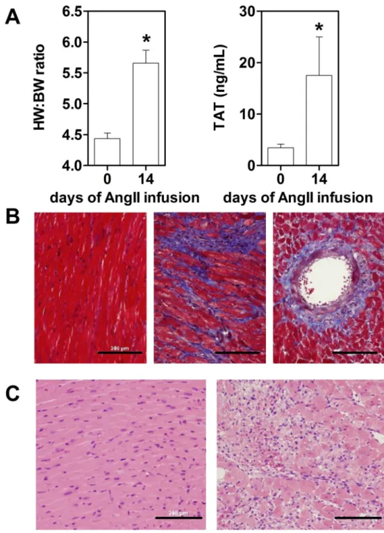
inflammation as well as activation of coagulation, WT mice were infused for up to 14 days with Ang II (600ng/kg/min). As expected, AT1 receptor stimulation in WT mice led to
significant increase in heart weight: body weight (HW:BW) ratios 14 days of Ang II infusion (Figure 1A). Importantly, we also observe a systemic activation of coagulation measured by plasma TATc levels at day 14 after pump installation (Figure 1A). The increased heart hypertrophy and procoagulant state were associated with interstitial and perivascular fibrosis within the heart muscle (Figure 1B) and increased cellular infiltrations (Figure 1C) after 7 days of Ang II infusion. Our observation suggests that chronic activation of the AT1 receptor leads to activation of coagulation which was associated with pathologic
cardiac fibrosis, inflammation and remodeling.
3.2. PAR-1 deficiency has no effect on blood pressure before and after Ang II infusion
To determine if PAR-1 activation contribute to the basal and Ang II dependent BP, BP in WT and PAR-1 deficient mice were measured before, 7 and 28 days after constant Ang II infusion. Consistently with previously published data, PAR-1 deficiency had no significant
A
uthor Man
uscr
ipt
A
uthor Man
uscr
ipt
A
uthor Man
uscr
ipt
A
uthor Man
uscr
effect on the baseline diastolic and systolic BP (Supplement 1) [24, 25]. Interestingly, infusion of Ang II at the concentration of 600ng/kg/min caused only modest but not
statistically significant increase of BP in both WT and PAR-1−/− mice measured at day 7 and 28 after initiation of infusion (Supplement 1). In addition, we did not find any differences between the genotypes with regards to the BP at day 7 and 28. This suggests that PAR-1 does not play a major role in baseline BP regulation and that the all observations in our study were due to BP-independent AT1- and PAR-1-mediated signaling events in the setting
resembling pre-HTN conditions [39].
3.3. PAR-1 deficiency attenuates Ang II-induced remodeling of the aorta
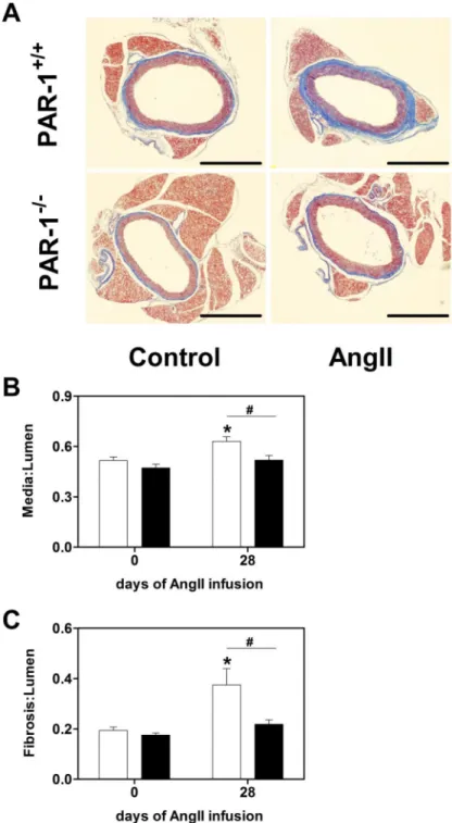
Ang II infusion into mice is known to lead to vascular remodeling in the aorta associated with an increased media thickness and enhanced fibrosis [6]. To determine if PAR-1 affects these parameters, media to lumen and fibrosis to lumen ratios of aortas were analyzed before and 28 days after Ang II infusion in WT and PAR-1 deficient mice. There were no
significant differences with regard to aorta media thickness and the amount of fibrotic tissue around the aorta in mice of both genotypes before Ang II infusion (Figure 2). As expected, chronic AT1 stimulation for 28 days led to increase in aorta media thickness and increased
collagen deposition visualized by Masson’s trichrome staining around the aorta in WT mice. Importantly both parameters were significantly attenuated in Ang II treated PAR-1−/− mice (Figure 2). These data suggest that PAR-1 dependent signaling pathways contribute to the vascular remodeling induced by chronic Ang II infusion.
3.4. Reduced expression of inflammatory and pro-fibrotic markers in the aortas of PAR-1 deficient mice
To determine if changes in vascular remodeling induced by Ang II are associated with changes in the expression profile of inflammatory and pro-fibrotic genes in the aorta, real-time PCR was performed on aorta samples of WT and PAR-1 deficient mice before, 7 and 28 days after Ang II infusion. Importantly, there were no significant differences between the genotypes in the baseline mRNA expression levels in the aorta of the inflammatory
mediators IL-1β, IL-6, TNF-α, MCP-1, CXCL1, and CXCL2 as well as pro-fibrotic mediators TGFβ1, TGFβ3, CTGF, and extracellular matrix components collagen Ia1, collagen Ia2 and collagen III (Figure 3 and data not shown). Except for CTGF, Ang II infusion led to a significant increase in the mRNA expression of all measured genes in WT mice at 7 days. Later at day 28, the aortic expression levels of the analyzed genes in WT mice returned to baseline with the exception of CTGF (Figure 3F). Importantly, PAR-1 deficiency was associated with less pro-inflammatory and pro-fibrotic gene expression compared at day 7 as well as CTGF at day 28 (Figure 3). These data indicate that PAR-1 is involved in regulating a transient pro-inflammatory and pro-fibrotic phenotype leading to vascular inflammation and remodeling in the aorta during chronic Ang II infusion.
3.5. Ang II-induced perivascular fibrosis of coronary arteries is reduced in Ang II treated PAR-1 deficient mice
Besides its effect on the aorta, Ang II leads to perivascular fibrosis of coronary vessels and heart hypertrophy [6]. To analyze Ang II-dependent changes in perivascular fibrosis and cardiac remodeling, hearts of WT and PAR-1 deficient mice were analyzed before, 7 and 28
A
uthor Man
uscr
ipt
A
uthor Man
uscr
ipt
A
uthor Man
uscr
ipt
A
uthor Man
uscr
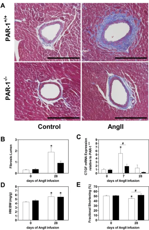
days after Ang II infusion. We did not observe any differences in the level of perivascular fibrosis around the cardiac arteries before Ang II infusion measured by fibrosis area to lumen area (Figure 4A). Chronic AT1 stimulation led to an increase of perivascular fibrosis
around coronary arteries in both WT and PAR-1 deficient mice visualized by Masson’s trichrome staining of heart sections (Figure 4A). However, this parameter was significantly increased only in WT but not PAR-1−/− mice group at day 28 (Figure 4B). Next, we
analyzed expression of CTGF in the heart which was shown to be associated with pathologic cardiac fibrosis and remodeling [38]. CTGF expression was significantly increased in the heart of WT mice 7 days after Ang II infusion and returned to the baseline (Figure 4 C) at day 28. Importantly, 7 days after Ang II infusion, CTGF mRNA expression was significantly lower in the heart of PAR-1 deficient mice compared to WT controls (Figure 4C).
3.6. PAR-1 deficient mice are protected against Ang II-induced heart dysfunction
Chronic AT1 stimulation is known to induce heart hypertrophy leading to heart dysfunction
[40]. To analyze changes in heart hypertrophy, heart weight to body weight ratios were calculated in WT and PAR-1 deficient mice before and 28 days after Ang II infusion. PAR-1 deficiency did not affect this parameter at baseline (Figure 4D). Heart weight to body weight ratios were significantly increased 28 days after Ang II infusion. However, PAR-1 deficiency did not affect the heart hypertrophy. Despite that, PAR-1 deficient mice had better preserved heart function compared to the WT mice 28 days after Ang II infusion (Figure 4E). This observation suggests that lack of PAR-1 dependent signaling protects against Ang II-induced heart dysfunction independently of heart hypertrophy.
4. Discussion
In this study, we demonstrated that PAR-1 deficiency attenuates pathologic Ang II-induced remodeling of the cardiovascular system. PAR-1 deficiency was associated with reduced mRNA expression of pro-inflammatory and pro-fibrotic markers which correlated with an attenuation of fibrosis and remodeling in the aortas of Ang II treated mice. In addition, we also observed reduced perivascular fibrosis of coronary vessels in hearts of PAR-1 deficient mice after of Ang II treatment. The reduced cardiac fibrosis in PAR-1 deficient mice led to a preserved heart function compared to Ang II treated WT mice. Interestingly, our observation was independent of a change in BP as well as heart hypertrophy since PAR-1 deficiency had no effect on these parameters. Our data indicate that PAR-1 plays a significant role in cardiovascular remodeling mediated by the BP-independent action of Ang II.
In our study, we were not able to detect increased BPs in anesthetized mice after 600 ng/kg/min Ang II infusion. This was surprising since even a lower Ang II dose (490 ng/kg/ min) was shown to increase BP in telemetric observed animals [41]. However, the BP discrepancy might be due to the measurement used. Anesthesia has cardio-depressive effects leading to hypotension which might mask the real BP elevation in our experimental mice [42]. Thus, our study mimicked a murine pre-HTN model with BP below levels defined as HTN. Pre-HTN is described as BP between 120 and 139 mmHg systolic or 80 and 89 mm Hg diastolic [39]. It is thought that pre-HTN status adds a moderate-to-high risk to the total cardiovascular risk [39]. Importantly, the Framingham Heart study showed that patients with
A
uthor Man
uscr
ipt
A
uthor Man
uscr
ipt
A
uthor Man
uscr
ipt
A
uthor Man
uscr
pre-HTN have an increased risk to develop HTN [43]. Furthermore, the ATTICA study reported that there was an association between pre-HTN and increased inflammation, which was linked to the atherosclerotic process [39]. It was concluded that increased inflammation might be a mechanism in the initiation and/or progression of pre-HTN [44, 45].
The ongoing inflammation might be mediated by both the innate and adaptive immune processes. The innate immune pathway not only responds to exogenous pathogens but can be also activated by damage-associated molecular patterns (DAMPs) released endogenous by stressed, damaged or necrotic cells [46]. DAMPs are present in cardiovascular diseases during HTN [47]. AT1 activation was shown to induce p53-dependent apoptosis of cultured
rat cardiomyocytes, which might lead the release of DAMPs [48]. Interestingly, Singh et al. reported that Ang II mediated cardiac hypertrophy and pro-inflammatory gene expression was mainly caused due to TRIF pathway activation [49]. The authors further showed that the expression of the TRIF-dependent cytokine CXCL10 was increased in MyD88−/− mice hearts but reduced in mice lacking TRIF signaling [49]. We found that PAR-1 stimulation enhanced toll-like receptor (TLR)-3/TRIF-dependent CXCL10 expression in cardiac fibroblasts and immune cells [34, 37]. This suggests that PAR-1 possibly enhanced pathologic TRIF signaling during Ang II infusion. Beside its effect on TLR signaling, PAR-1 activation induces the classical hallmarks of inflammation, including enhanced vascular permeability, upregulation of pro-inflammatory mediators and adhesion molecules [21]. In our study, we observed a PAR-1-dependent pro-inflammatory state in mice after Ang II infusion measured by increased IL-1β, IL-6, TNF-α, MCP-1, CXCL1 and CXCL2 expression in the aorta. Consistent with this observation, a study showed that increased plasma levels of IL-1β, a cytokine involved in monocyte activation, precedes changes in BP in HTN [50]. Furthermore, IL-6, MCP-1 and especially TNF-α were shown to be essential for the pathologic effects of Ang II on the vasculature by increasing oxidative stress, activating fibroblasts and attracting T cells [47, 51].
Expression of pro-inflammatory cytokines is further linked to an induction of a pro-fibrotic phenotype. In addition, a mild-to-moderate increase in BP was reported to stimulate fibroblasts and increase collagen formation leading to a fibrotic remodeling of the
myocardium with normal LV mass [52]. TGFβ1 mRNA and protein expression are increased in HTN patients [53]. CTGF has a role as a downstream mediator of the chronic fibrotic effects of TGFβ. Activated TGFβ induces CTGF expression in fibroblasts and
cardiomyocytes [54]. During this process fibroblasts differentiate into myofibroblasts, the major cell that synthesizes collagen in cardiac remodeling [55, 56]. Activation of PAR-1 on fibroblasts induced cell proliferation and expression of pro-fibrotic genes [32, 57]. Here, we found increased CTGF and TGFβ as well as collagen expressions in Ang II infused WT but not in PAR-1−/− mice. This PAR-1-dependent pro-fibrotic phenotype resulted in increased vascular and perivascular remodeling associated with fibrosis and possible reduced elasticity of the aorta and coronary arteries. This is consistent with the finding of Celik and colleagues that pre-HTN was associated with an impairment of the aortic elasticity and inflammation when compared to healthy controls [44, 58]. These changes may lead to reduced coronary flow very early in pre-HTN before hypertrophy is apparent and thus may cause subsequently ischemia and fibrosis [59, 60]. Structural abnormalities are already seen in pre-HTN
patients, although in a milder manner compared to newly diagnosed HTN patients which
A
uthor Man
uscr
ipt
A
uthor Man
uscr
ipt
A
uthor Man
uscr
ipt
A
uthor Man
uscr
might lead to cardiac remodeling [33, 61]. In our study, we did not observe any differences in the cardiac hypertrophy between the two genotypes. This was surprising since we and others have shown that PAR-1 contributes to cardiomyocyte hypertrophy and cardiac remodeling [23, 30]. However, the significant differences in fibrosis independent of hypertrophy might be due to the AT receptor distribution on cardiac fibroblasts and cardiomyocytes. Cardiomyocytes express comparable amounts of AT1 and AT2 receptors
whereas fibroblasts express predominantly AT1 [62]. In addition, AT2 was shown to
counteract AT1 activation [62]. Importantly, increased vascular inflammation as well as
arterial stiffness was shown to be a predictive for cardiovascular events [63, 64].
The remaining question for further studies is the physiologic activator of PAR-1 in the setting of chronic Ang II infusion, AT1 stimulation, and HTN. The most likely candidate is
thrombin. In our study, we observed increased TAT levels after chronic Ang II infusion. Consistent with our findings, it was shown that AT1 receptor stimulation and HTN was
linked to increased activation of coagulation due to increased TF expression and thrombin generation [11–13, 16, 17], which led to a pro-thrombotic phenotype [14]. Another possible activator is the non-canonical activation of PAR-1 by matrix-metalloproteinases (MMPs) 1 or 13 [7]. Recently, it was shown that MMP-1 enhanced Ang II induced vasoconstriction in endothelium-intact omental arteries in a PAR-1 dependent way ex vivo [65]. Furthermore, MMP-13 is expressed and active during Ang II induced HTN [66]. We showed that the MMP-13/PAR-1 pathways is active in cardiovascular diseases models [7, 18, 33, 34].
Importantly, besides its proven pathologic role, TF expressed by extravascular cells in the brain, lung and heart such as astrocytes, epithelial cells, smooth muscle cells, fibroblasts and cardiomyocytes maintains organ hemostasis [10, 11, 23, 67–71]. Reduced TF-dependent initiation of coagulation can result in hemorrhages, increased tissue fibrosis and reduced overall survival under healthy as well as pathologic conditions [10, 68, 71]. This dual role makes is difficult to use the TF blockage as viable therapy option [70]. Based on our data, further studies are warranted to investigate the effect of the PAR-1 inhibitor vorapaxar in Ang II-induced cardiovascular remodeling.
5. Conclusion
Taken together our data suggest that PAR-1 signaling pathway contributes to Ang II-induced cardiac fibrosis and heart dysfunction.
Supplementary Material
Refer to Web version on PubMed Central for supplementary material.
Acknowledgments
We thank Ying Zhang and Wyeth Alexander for excellent technical assistance, and Drs. Michael F. Bode and Tracy Stokol for for helpful comments.
The study was supported by grants from the American Heart Association to S.A. (14BGIA20380134) and to R.P. (09BGIA2150078), and the National Institute of Health to R.P. (RO1 HL096679). There are no financial interests.
A
uthor Man
uscr
ipt
A
uthor Man
uscr
ipt
A
uthor Man
uscr
ipt
A
uthor Man
uscr
References
1. Mehta PK, Griendling KK. Angiotensin II cell signaling: physiological and pathological effects in the cardiovascular system. Am J Physiol Cell Physiol. 2007; 292:C82–C97. [PubMed: 16870827] 2. Marchesi C, Paradis P, Schiffrin EL. Role of the renin-angiotensin system in vascular inflammation.
Trends Pharmacol Sci. 2008; 29:367–374. [PubMed: 18579222]
3. Wolny A, Clozel JP, Rein J, Mory P, Vogt P, Turino M, Kiowski W, Fischli W. Functional and biochemical analysis of angiotensin II-forming pathways in the human heart. Circ Res. 1997; 80:219–227. [PubMed: 9012744]
4. Sasamura H, Hein L, Krieger JE, Pratt RE, Kobilka BK, Dzau VJ. Cloning, characterization, and expression of two angiotensin receptor (AT-1) isoforms from the mouse genome. Biochem Biophys Res Commun. 1992; 185:253–259. [PubMed: 1599461]
5. Kato H, Ishida J, Matsusaka T, Ishimaru T, Tanimoto K, Sugiyama F, Yagami K, Nangaku M, Fukamizu A. Erythropoiesis and Blood Pressure Are Regulated via AT1 Receptor by Distinctive Pathways. PLoS One. 2015; 10:e0129484. [PubMed: 26107632]
6. Takayanagi T, Kawai T, Forrester SJ, Obama T, Tsuji T, Fukuda Y, Elliott KJ, Tilley DG, Davisson RL, Park JY, Eguchi S. Role of Epidermal Growth Factor Receptor and Endoplasmic Reticulum Stress in Vascular Remodeling Induced by Angiotensin II. Hypertension. 2015; 65:1349–1355. [PubMed: 25916723]
7. Antoniak S, Sparkenbaugh E, Pawlinski R. Tissue factor, protease activated receptors and pathologic heart remodelling. Thromb Haemost. 2014; 112:893–900. [PubMed: 25104210]
8. Pawlinski R, Pedersen B, Erlich J, Mackman N. Role of tissue factor in haemostasis, thrombosis, angiogenesis and inflammation: lessons from low tissue factor mice. Thromb Haemost. 2004; 92:444–450. [PubMed: 15351839]
9. Mackman N, Tilley RE, Key NS. Role of the extrinsic pathway of blood coagulation in hemostasis and thrombosis. Arterioscler Thromb Vasc Biol. 2007; 27:1687–1693. [PubMed: 17556654] 10. Pawlinski R, Tencati M, Holscher T, Pedersen B, Voet T, Tilley RE, Marynen P, Mackman N. Role
of cardiac myocyte tissue factor in heart hemostasis. J Thromb Haemost. 2007; 5:1693–1700. [PubMed: 17663739]
11. Muller DN, Mervaala EM, Dechend R, Fiebeler A, Park JK, Schmidt F, Theuer J, Breu V, Mackman N, Luther T, Schneider W, Gulba D, Ganten D, Haller H, Luft FC. Angiotensin II (AT(1)) receptor blockade reduces vascular tissue factor in angiotensin II-induced cardiac vasculopathy. Am J Pathol. 2000; 157:111–122. [PubMed: 10880382]
12. Koh KK, Chung WJ, Ahn JY, Han SH, Kang WC, Seo YH, Ahn TH, Choi IS, Shin EK. Angiotensin II type 1 receptor blockers reduce tissue factor activity and plasminogen activator inhibitor type-1 antigen in hypertensive patients: a randomized, double-blind, placebo-controlled study. Atherosclerosis. 2004; 177:155–160. [PubMed: 15488878]
13. Steffel J, Luscher TF, Tanner FC. Tissue factor in cardiovascular diseases: molecular mechanisms and clinical implications. Circulation. 2006; 113:722–731. [PubMed: 16461845]
14. Senchenkova EY, Russell J, Esmon CT, Granger DN. Roles of Coagulation and fibrinolysis in angiotensin II-enhanced microvascular thrombosis. Microcirculation. 2014; 21:401–407. [PubMed: 24495184]
15. Coughlin SR. Thrombin signalling and protease-activated receptors. Nature. 2000; 407:258–264. [PubMed: 11001069]
16. Ekholm M, Wallen NH, Johnsson H, Eliasson K, Kahan T. Long-term angiotensin-converting enzyme inhibition with ramipril reduces thrombin generation in human hypertension. Clin Sci (Lond). 2002; 103:151–155. [PubMed: 12149106]
17. Sawada K, Naiki M, Yago H, Matsushita K, Ohtsuki T, Kitagawa K, Matsumoto M, Hori M. Hypertension associated with reduced plasma thrombomodulin levels and a hypercoagulable state in rats. Clin Exp Hypertens. 2003; 25:73–84. [PubMed: 12611420]
18. Antoniak S, Mackman N. Coagulation, protease-activated receptors, and viral myocarditis. J Cardiovasc Transl Res. 2014; 7:203–211. [PubMed: 24203054]
19. Antoniak S, Pawlinski R, Mackman N. Protease-activated receptors and myocardial infarction. IUBMB Life. 2011; 63:383–389. [PubMed: 21438116]
A
uthor Man
uscr
ipt
A
uthor Man
uscr
ipt
A
uthor Man
uscr
ipt
A
uthor Man
uscr
20. Leger AJ, Covic L, Kuliopulos A. Protease-activated receptors in cardiovascular diseases. Circulation. 2006; 114:1070–1077. [PubMed: 16952995]
21. Major CD, Santulli RJ, Derian CK, Andrade-Gordon P. Extracellular mediators in atherosclerosis and thrombosis: lessons from thrombin receptor knockout mice. Arterioscler Thromb Vasc Biol. 2003; 23:931–939. [PubMed: 12676802]
22. Steinberg SF. The cardiovascular actions of protease-activated receptors. Mol Pharmacol. 2005; 67:2–11. [PubMed: 15371558]
23. Pawlinski R, Tencati M, Hampton CR, Shishido T, Bullard TA, Casey LM, Andrade-Gordon P, Kotzsch M, Spring D, Luther T, Abe J, Pohlman TH, Verrier ED, Blaxall BC, Mackman N. Protease-activated receptor-1 contributes to cardiac remodeling and hypertrophy. Circulation. 2007; 116:2298–2306. [PubMed: 17967980]
24. Cheung WM, Andrade-Gordon P, Derian CK, Damiano BP. Receptor-activating peptides distinguish thrombin receptor (PAR-1) and protease activated receptor 2 (PAR-2) mediated hemodynamic responses in vivo. Can J Physiol Pharmacol. 1998; 76:16–25. [PubMed: 9564545] 25. Darrow AL, Fung-Leung WP, Ye RD, Santulli RJ, Cheung WM, Derian CK, Burns CL, Damiano BP, Zhou L, Keenan CM, Peterson PA, Andrade-Gordon P. Biological consequences of thrombin receptor deficiency in mice. Thromb Haemost. 1996; 76:860–866. [PubMed: 8972001]
26. Cheung WM, D'Andrea MR, Andrade-Gordon P, Damiano BP. Altered vascular injury responses in mice deficient in protease-activated receptor-1. Arterioscler Thromb Vasc Biol. 1999; 19:3014– 3024. [PubMed: 10591683]
27. Wilcox JN, Rodriguez J, Subramanian R, Ollerenshaw J, Zhong C, Hayzer DJ, Horaist C, Hanson SR, Lumsden A, Salam TA, et al. Characterization of thrombin receptor expression during vascular lesion formation. Circ Res. 1994; 75:1029–1038. [PubMed: 7955141]
28. Takada M, Tanaka H, Yamada T, Ito O, Kogushi M, Yanagimachi M, Kawamura T, Musha T, Yoshida F, Ito M, Kobayashi H, Yoshitake S, Saito I. Antibody to thrombin receptor inhibits neointimal smooth muscle cell accumulation without causing inhibition of platelet aggregation or altering hemostatic parameters after angioplasty in rat. Circ Res. 1998; 82:980–987. [PubMed: 9598595]
29. Andrade-Gordon P, Derian CK, Maryanoff BE, Zhang HC, Addo MF, Cheung W, Damiano BP, D'Andrea MR, Darrow AL, de Garavilla L, Eckardt AJ, Giardino EC, Haertlein BJ, McComsey DF. Administration of a potent antagonist of protease-activated receptor-1 (PAR-1) attenuates vascular restenosis following balloon angioplasty in rats. J Pharmacol Exp Ther. 2001; 298:34–42. [PubMed: 11408522]
30. Sabri A, Muske G, Zhang H, Pak E, Darrow A, Andrade-Gordon P, Steinberg SF. Signaling properties and functions of two distinct cardiomyocyte protease-activated receptors. Circ Res. 2000; 86:1054–1061. [PubMed: 10827135]
31. Glembotski CC, Irons CE, Krown KA, Murray SF, Sprenkle AB, Sei CA. Myocardial alpha-thrombin receptor activation induces hypertrophy and increases atrial natriuretic factor gene expression. J Biol Chem. 1993; 268:20646–20652. [PubMed: 8397212]
32. Sabri A, Short J, Guo J, Steinberg SF. Protease-activated receptor-1-mediated DNA synthesis in cardiac fibroblast is via epidermal growth factor receptor transactivation: distinct PAR-1 signaling pathways in cardiac fibroblasts and cardiomyocytes. Circ Res. 2002; 91:532–539. [PubMed: 12242272]
33. Jaffre F, Friedman AE, Hu Z, Mackman N, Blaxall BC. Beta-adrenergic receptor stimulation transactivates protease-activated receptor 1 via matrix metalloproteinase 13 in cardiac cells. Circulation. 2012; 125:2993–3003. [PubMed: 22610965]
34. Antoniak S, Owens AP 3rd, Baunacke M, Williams JC, Lee RD, Weithauser A, Sheridan PA, Malz R, Luyendyk JP, Esserman DA, Trejo J, Kirchhofer D, Blaxall BC, Pawlinski R, Beck MA, Rauch U, Mackman N. PAR-1 contributes to the innate immune response during viral infection. J Clin Invest. 2013; 123:1310–1322. [PubMed: 23391721]
35. Antoniak S, Rojas M, Spring D, Bullard TA, Verrier ED, Blaxall BC, Mackman N, Pawlinski R. Protease-activated receptor 2 deficiency reduces cardiac ischemia/reperfusion injury. Arterioscler Thromb Vasc Biol. 2010; 30:2136–2142. [PubMed: 20724699]
A
uthor Man
uscr
ipt
A
uthor Man
uscr
ipt
A
uthor Man
uscr
ipt
A
uthor Man
uscr
36. Hathaway CK, Grant R, Hagaman JR, Hiller S, Li F, Xu L, Chang AS, Madden VJ, Bagnell CR, Rojas M, Kim HS, Wu B, Zhou B, Smithies O, Kakoki M. Endothelin-1 critically influences cardiac function via superoxide-MMP9 cascade. Proc Natl Acad Sci U S A. 2015; 112:5141–5146. [PubMed: 25848038]
37. Antoniak S, Tatsumi K, Bode M, Vanja S, Williams JC, Mackman N. Protease-activated receptor-1 enhances poly I:C induction of the anti-viral response in macrophages and mice. J Innate Immun. 2016
38. Antoniak S, Sparkenbaugh EM, Tencati M, Rojas M, Mackman N, Pawlinski R. Protease activated receptor-2 contributes to heart failure. PLoS One. 2013; 8:e81733. [PubMed: 24312345]
39. Chrysohoou C, Pitsavos C, Panagiotakos DB, Skoumas J, Stefanadis C. Association between prehypertension status and inflammatory markers related to atherosclerotic disease: The ATTICA Study. Am J Hypertens. 2004; 17:568–573. [PubMed: 15233975]
40. Hein L. Genetic deletion and overexpression of angiotensin II receptors. J Mol Med (Berl). 1998; 76:756–763. [PubMed: 9826120]
41. Mikolajczyk TP, Nosalski R, Szczepaniak P, Budzyn K, Osmenda G, Skiba D, Sagan A, Wu J, Vinh A, Marvar PJ, Guzik B, Podolec J, Drummond G, Lob HE, Harrison DG, Guzik TJ. Role of chemokine RANTES in the regulation of perivascular inflammation, T-cell accumulation, and vascular dysfunction in hypertension. FASEB J. 2016; 30:1987–1999. [PubMed: 26873938] 42. Constantinides C, Mean R, Janssen BJ. Effects of isoflurane anesthesia on the cardiovascular
function of the C57BL/6 mouse. ILAR J. 2011; 52:e21–e31. [PubMed: 21677360]
43. Vasan RS, Larson MG, Leip EP, Kannel WB, Levy D. Assessment of frequency of progression to hypertension in non-hypertensive participants in the Framingham Heart Study: a cohort study. Lancet. 2001; 358:1682–1686. [PubMed: 11728544]
44. Celik T, Yuksel UC, Demirkol S, Bugan B, Iyisoy A, Kabul HK, Kilic S, Fici F, Yaman H. Relationship between increased systemic inflammation and impaired aortic elasticity in young patients with prehypertension. Blood Press Monit. 2011; 16:55–61. [PubMed: 21415815] 45. Ferreira AS. Immunity, Inflammation, and Prehypertension: In What Order? J Clin Hypertens
(Greenwich). 2015; 17:775–776. [PubMed: 26146860]
46. Takeuchi O, Akira S. Pattern recognition receptors and inflammation. Cell. 2010; 140:805–820. [PubMed: 20303872]
47. De Miguel C, Rudemiller NP, Abais JM, Mattson DL. Inflammation and hypertension: new understandings and potential therapeutic targets. Curr Hypertens Rep. 2015; 17:507. [PubMed: 25432899]
48. Kajstura J, Cigola E, Malhotra A, Li P, Cheng W, Meggs LG, Anversa P. Angiotensin II induces apoptosis of adult ventricular myocytes in vitro. J Mol Cell Cardiol. 1997; 29:859–870. [PubMed: 9152847]
49. Singh MV, Cicha MZ, Meyerholz DK, Chapleau MW, Abboud FM. Dual Activation of TRIF and MyD88 Adaptor Proteins by Angiotensin II Evokes Opposing Effects on Pressure, Cardiac Hypertrophy, and Inflammatory Gene Expression. Hypertension. 2015; 66:647–656. [PubMed: 26195481]
50. Mauno V, Hannu K, Esko K. Proinflammation and hypertension: a population-based study. Mediators Inflamm. 2008; 2008:619704. [PubMed: 19125204]
51. Sriramula S, Francis J. Tumor Necrosis Factor - Alpha Is Essential for Angiotensin II-Induced Ventricular Remodeling: Role for Oxidative Stress. PLoS One. 2015; 10:e0138372. [PubMed: 26378790]
52. Muller-Brunotte R, Kahan T, Lopez B, Edner M, Gonzalez A, Diez J, Malmqvist K. Myocardial fibrosis and diastolic dysfunction in patients with hypertension: results from the Swedish Irbesartan Left Ventricular Hypertrophy Investigation versus Atenolol (SILVHIA). J Hypertens. 2007; 25:1958–1966. [PubMed: 17762662]
53. Lijnen PJ, Petrov VV, Fagard RH. Association between transforming growth factor-beta and hypertension. Am J Hypertens. 2003; 16:604–611. [PubMed: 12850397]
54. Rosin NL, Falkenham A, Sopel MJ, Lee TD, Legare JF. Regulation and role of connective tissue growth factor in AngII-induced myocardial fibrosis. Am J Pathol. 2013; 182:714–726. [PubMed: 23287510]
A
uthor Man
uscr
ipt
A
uthor Man
uscr
ipt
A
uthor Man
uscr
ipt
A
uthor Man
uscr
55. Chen MM, Lam A, Abraham JA, Schreiner GF, Joly AH. CTGF expression is induced by TGF- beta in cardiac fibroblasts and cardiac myocytes: a potential role in heart fibrosis. J Mol Cell Cardiol. 2000; 32:1805–1819. [PubMed: 11013125]
56. Villarreal FJ, Dillmann WH. Cardiac hypertrophy-induced changes in mRNA levels for TGF-beta 1, fibronectin, and collagen. Am J Physiol. 1992; 262:H1861–H1866. [PubMed: 1535758] 57. Chambers RC, Leoni P, Blanc-Brude OP, Wembridge DE, Laurent GJ. Thrombin is a potent
inducer of connective tissue growth factor production via proteolytic activation of protease-activated receptor-1. J Biol Chem. 2000; 275:35584–35591. [PubMed: 10952976]
58. Celik T, Iyisoy A, Kursaklioglu H, Turhan H, Cagdas Yuksel U, Kilic S, Kutsi Kabul H, Genc C. Impaired aortic elastic properties in young patients with prehypertension. Blood Press Monit. 2006; 11:251–255. [PubMed: 16932034]
59. Laine H, Raitakari OT, Niinikoski H, Pitkanen OP, Iida H, Viikari J, Nuutila P, Knuuti J. Early impairment of coronary flow reserve in young men with borderline hypertension. J Am Coll Cardiol. 1998; 32:147–153. [PubMed: 9669263]
60. Erdogan D, Yildirim I, Ciftci O, Ozer I, Caliskan M, Gullu H, Muderrisoglu H. Effects of normal blood pressure, prehypertension, and hypertension on coronary microvascular function.
Circulation. 2007; 115:593–599. [PubMed: 17283278]
61. Di Bello V, Talini E, Dell'Omo G, Giannini C, Delle Donne MG, Canale ML, Nardi C, Palagi C, Dini FL, Penno G, Del Prato S, Marzilli M, Pedrinelli R. Early left ventricular mechanics abnormalities in prehypertension: a two-dimensional strain echocardiography study. Am J Hypertens. 2010; 23:405–412. [PubMed: 20044741]
62. de Gasparo M, Catt KJ, Inagami T, Wright JW, Unger T. International union of pharmacology. XXIII. The angiotensin II receptors. Pharmacol Rev. 2000; 52:415–472. [PubMed: 10977869] 63. Chae CU, Lee RT, Rifai N, Ridker PM. Blood pressure and inflammation in apparently healthy
men. Hypertension. 2001; 38:399–403. [PubMed: 11566912]
64. Weber T, Auer J, O'Rourke MF, Kvas E, Lassnig E, Berent R, Eber B. Arterial stiffness, wave reflections, and the risk of coronary artery disease. Circulation. 2004; 109:184–189. [PubMed: 14662706]
65. Nugent WH, Mishra N, Strauss JF 3rd, Walsh SW. Matrix Metalloproteinase 1 Causes Vasoconstriction and Enhances Vessel Reactivity to Angiotensin II via Protease-Activated Receptor 1. Reprod Sci. 2015
66. Cheng C, Tempel D, van Haperen R, van Damme L, Algur M, Krams R, de Crom R. Activation of MMP8 and MMP13 by angiotensin II correlates to severe intra-plaque hemorrhages and collagen breakdown in atherosclerotic lesions with a vulnerable phenotype. Atherosclerosis. 2009; 204:26– 33. [PubMed: 19233360]
67. Wang S, Reeves B, Sparkenbaugh EM, Russell J, Soltys Z, Zhang H, Faber JE, Key NS, Kirchhofer D, Granger DN, Mackman N, Pawlinski R. Protective and detrimental effects of neuroectodermal cell-derived tissue factor in mouse models of stroke. JCI Insight. 2016; 1 68. Pawlinski R, Fernandes A, Kehrle B, Pedersen B, Parry G, Erlich J, Pyo R, Gutstein D, Zhang J,
Castellino F, Melis E, Carmeliet P, Baretton G, Luther T, Taubman M, Rosen E, Mackman N. Tissue factor deficiency causes cardiac fibrosis and left ventricular dysfunction. Proc Natl Acad Sci U S A. 2002; 99:15333–15338. [PubMed: 12426405]
69. Erlich JH, Boyle EM, Labriola J, Kovacich JC, Santucci RA, Fearns C, Morgan EN, Yun W, Luther T, Kojikawa O, Martin TR, Pohlman TH, Verrier ED, Mackman N. Inhibition of the tissue factor-thrombin pathway limits infarct size after myocardial ischemia-reperfusion injury by reducing inflammation. Am J Pathol. 2000; 157:1849–1862. [PubMed: 11106558]
70. Bode MF, Mackman N. Protective and pathological roles of tissue factor in the heart. Hamostaseologie. 2015; 35:37–46. [PubMed: 25434707]
71. Antoniak S, Tatsumi K, Hisada Y, Milner JJ, Neidich SD, Shaver CM, Pawlinski R, Beck MA, Bastarache JA, Mackman N. Tissue factor deficiency increases alveolar hemorrhage and death in influenza A virus-infected mice. J Thromb Haemost. 2016; 14:1238–1248. [PubMed: 26947929]
A
uthor Man
uscr
ipt
A
uthor Man
uscr
ipt
A
uthor Man
uscr
ipt
A
uthor Man
uscr
Figure 1. Infusion of Ang II leads to cardiac hypertrophy, systemic activation of coagulation associated with cardiac fibrosis and inflammation
(A) Increase in heart weight to body weight ratios (HW:BW) and plasma levels of thrombin-antithrombin complexes (TAT) in Ang II treated wild-type mice. (B) Masson’s trichrome staining of representative heart sections from control (left panel) and mice treated with Ang II for 7 days (middle and right panel). (C) Inflammatory cells are present in H&E stained heart sections from Ang II treated (right panel) but not from control mice (left panel). Bar = 200 µm. Data (mean ± SEM; n = 3 for each time point) were analyzed by Student’s t-test. Statistical significances is shown as * P<0.05.
A
uthor Man
uscr
ipt
A
uthor Man
uscr
ipt
A
uthor Man
uscr
ipt
A
uthor Man
uscr
Figure 2. PAR-1 deficiency attenuates Ang II-induced remodeling of aorta
(A) Representative cross sections of thoracic parts of aortas stained with Masson’s trichome. (B) Quantification of the media area and (C) fibrotic area to lumen area from PAR-1+/+ (white bars) and PAR-1−/− (black bars) mice aortas before and 28 days after constant Ang II infusion. Bar = 1 mm. Data (mean ± SEM; n = 5 for day 0, n =10 for day 28) were analyzed by 2-way ANOVA. Statistical significances is shown as # P<0.05 and * P<0.05 versus day 0 of the respective genotype.
A
uthor Man
uscr
ipt
A
uthor Man
uscr
ipt
A
uthor Man
uscr
ipt
A
uthor Man
uscr
Figure 3. Reduced expression of inflammatory and pro-fibrotic markers in the aortas of PAR-1 deficient mice
Real-time PCR analysis of mRNA expression of inflammatory mediators (A) IL-1β, (B) CXCL1 (KC) and (C) CXCL2 (inflammatory protein 2-alpha, MIP-2α), of pro-fibrotic markers (D) transforming growth factor (TGF) β1, (E) TGFβ3 and (F) connective tissue growth factor (CTGF) as well as extracellular matrix proteins (G) collagen (Coll) Ia1, (H) Coll Ia2 and (I) Coll III in the aortas of PAR-1+/+ (white bars) and PAR-1−/− (black bars) mice before, 7 and 28 days of constant Ang II infusion. Data (mean ± SEM; n = 6–10) were analyzed by 2-way ANOVA. Statistical significances is shown as # P<0.05 and * P<0.05 versus day 0 of the respective genotype.
A
uthor Man
uscr
ipt
A
uthor Man
uscr
ipt
A
uthor Man
uscr
ipt
A
uthor Man
uscr
Figure 4. PAR-1 deficient mice are protected against Ang II-induced cardiac fibrosis and heart dysfunction
(A) Representative sections of heart coronary vessels from PAR-1+/+ and PAR-1−/− mice after 28 days of constant Ang II infusion stained with Masson’s trichrome. (B)
Quantification of the fibrotic area to lumen area of heart coronary vessels from PAR-1+/+ (white bars) and PAR-1−/− (black bars) mice before and after 28 days of constant Ang II infusion. (C) Real-time PCR analysis of connective tissue growth factor (CTGF) mRNA expression in the heart of PAR-1+/+ and PAR-1−/− mice before, 7 and 28 days of continuous Ang II infusion. (D) Heart weight to body weight ratios (HW:BW) and (E) fractional
A
uthor Man
uscr
ipt
A
uthor Man
uscr
ipt
A
uthor Man
uscr
ipt
A
uthor Man
uscr
shortening measured by echocardiography before and 28 days of Ang II infusion. Bar = 200 µm. Data (mean ± SEM; n = 6–10) were analyzed by 2-way ANOVA. Statistical
significances is shown as # P<0.05 and * P<0.05 versus day 0 of the respective genotype.