The Role of Cytokinin in Female Gametophyte Development in Arabidopsis
Chia-Yi Cheng
A dissertation submitted to the faculty of the University of North Carolina at Chapel Hill in partial fulfillment of the requirements for the degree of Doctor of Philosophy in the
Department of Biology
Chapel Hill 2013
Approved by:
Joseph J. Kieber (Advisor)
Sarah R. Grant
Corbin D. Jones
Jason W. Reed
ii ABSTRACT
CHIA-YI CHENG: The Role of Cytokinin in Female Gametophyte Development in Arabidopsis
(Under the direction of Joseph J. Kieber)
Plants, unlike animals, have distinct haploid and diploid phases of their life cycle.
The haploid female gametophyte develops within the diploid maternal sporophytic tissue.
The signaling events regulating the development of the female gametophyte are just
beginning to be understood. The plant hormone auxin plays a role in the development of
the female gametophyte, but the contribution of other phytohormones has not been
examined in depth.
Here we explore and demonstrate a role for the plant hormone cytokinin in female
gametophyte development. We show that cytokinin function is enriched at one end of the
sporophytic tissue that supports the development of the female gametophyte. This
localized cytokinin signal in the maternal tissue is required for the initial specification of
iii
Acknowledgements
This dissertation had a life of its own and I merely followed. For all the assistance
I received along the way I am deeply grateful. I want to thank my advisor, Joe Kieber, for
his patient guidance and inspiring enthusiasm that have sustained and encouraged me
throughout the years. Advice given by my committee was very helpful and gave
refreshing perspective on my career.
I would like to thank Smadar Harpaz-Saad for her loving support and endless
encouragement. I thank Wenjing Zhang for the stimulating discussion and intellectual
input. I am grateful for my friends Ken-Yen Liu and Shiang-Kwei Wang for their solid
friendship.
I would also like to express my deepest gratitude to my parents, Kuo-Chuan
Cheng and Jang-Suen Wu, and my grandmother, Chia-Pao Shu for their unconditional
love. I should thank Sophia Shih and Louise White for their companionship and spirit.
In everything I give thanks to God, who allows me to experience the beauty of
iv
TABLE OF CONTENTS
ACKNOWLEDGEMENTS ... iii
TABLE OF CONTENTS ... iv
LIST OF TABLES ... ix
LIST OF FIGURES ... x
LIST OF ABBREVIATIONS ... xii
CHAPTER 1: Cytokinin Signaling ... 1
Introduction ... 1
1. Two-component elements are involved in cytokinin signaling ... 4
1.1 Phosphotransfer chemistry ... 4
1.2 His-Asp phosphorylation pathway in Arabidopsis thaliana... 5
1.3 Cytokinin two-component elements ... 6
2. Cytokinin receptors are histidine kinases ... 8
2.1 Discovery ... 8
2.2 Cytokinin receptors differ in biochemical nature ... 9
2.3 Functions ... 11
2.4 Cytokinin receptors have overlapping and specific expression pattern ... 12
2.5 The majority of cytokinin receptors localize in ER ... 13
v
2.7 Are AHKs the only cytokinin receptors? ... 16
3. The phosphotransfer proteins in Arabidopsis ... 18
3.1 AHPs shuttle phosphate between AHKs and ARRs ... 18
3.2 AHPs act downstream of AHKs and CKI1 ... 18
3.3 The subcellular localization of AHPs suggests a function in a bidirectional phosphorelay ... 19
3.4 AHP6 is a negative element in cytokinin two-component signaling ... 20
3.5 Nitric oxide regulates phosphotransfer proteins through S-nitrosylation ... 21
4. Response regulators in Arabidopsis ... 23
5. Type-A response regulators ... 25
5.1 Type-A ARRs are primary response genes in cytokinin signaling ... 25
5.2 Type-A response regulators negatively regulates cytokinin signaling ... 25
5.3 Phosphorylation of receiver domain is essential for type-A ARR function ... 26
5.4 Other signals cross-talk with cytokinin signaling via regulating type-A ARRs . 27 6. Type-B response regulators ... 28
6.1 Type-B ARRs are positive regulators in cytokinin signaling ... 28
6.2 The receiver domains of type-B ARRs have inhibitory effect on DNA-binding domains ... 29
6.3 Type-B ARRs function as transcription factors ... 30
6.4 The direct targets of type-B ARRs... 31
7. Cytokinin response factors ... 33
vi
CHAPTER 2: Cytokinin-Dependent Specification of the Functional Megaspore in the
Arabidopsis Female Gametophyte ... 36
Preface ... 36
Summary ... 36
Introduction ... 38
Results ... 42
Loss of cytokinin receptors results in female gametophytic lethality ... 42
Impaired cytokinin signaling results in incomplete megasporogenesis ... 45
Cytokinin receptors and activity are enriched in the chalaza of the ovule ... 50
Chalazal cytokinin signaling is required for the selection of functional megaspore 53 Discussion ... 55
Two-component signaling in female gametophyte development ... 55
Cytokinin is essential for functional megaspore specification ... 56
Signaling between the sporophyte and the gametophyte involves cytokinin ... 58
Origin of chalaza-localized cytokinin ... 59
Conclusion ... 60
Experimental Procedures ... 61
Acknowledgments ... 63
Supporting Information ... 64
vii
Preface ... 67
Summary ... 67
Cell Fate Determination in Ovule ... 69
Sporophytically Acting Genes in Regulating Integument Growth and Development .. 71
Perspectives ... 74
CHAPTER 4: Characterization of Phospho-Specific Type-A ARR Interacting Targets . 75 Preface ... 75
Introduction ... 75
Results and Discussion ... 79
Identification of phospho-specific type-A ARR interacting proteins ... 79
Establishment of the protoplast system to validate the phospho-specific interactions in vivo ... 80
Phylogenetic analysis of Arabidopsis ARID-containing proteins ... 81
Overexpressing ARID-GFP transgene results in retarded shoot development ... 83
BPC6 belongs to a GAGA-binding transcription factor family ... 85
Experimental procedures ... 87
Chapter 5: Discussion ... 90
Sporophytic signals in specifying gametophytic cell fate ... 90
viii
ix
LIST OF TABLES
Table 2.1. Segregation of single AHK in double ahk background ... 50
x
LIST OF FIGURES
Figure 1.1. Schematic diagram of two-component (TCS) system ... 2
Figure 1.2. Cytokinin signal transduction in Arabidopsis ... 3
Figure 1.3. Phylogenetic relationship of the Arabidopsis response regulators (ARR) ... 24
Figure 2.1. Two-component elements and female gametophyte development ... 39
Figure 2.2. Phenotypic analysis of different ahk triple mutants ... 43
Figure 2.3. Expression of AHK genes in ahk2-7 ahk3-3 cre1-12 ... 45
Figure 2.4. Characterization of the ovule phenotypes in the ahk2-7 ahk3-3 cre1-12 mutant. ... 46
Figure 2.5. Both ahk- and cki1-like phenotype are present in high order ahp and type-B arr mutants ... 49
Figure 2.6. Expression of cytokinin signaling elements in ovules ... 52
Figure 2.7. Cytokinin signaling is required for the functional megaspore selection ... 54
Figure 2.8. Two-component signaling in female gametophyte development ... 55
Figure 2.9. Phenotypic difference between spl-1 and ahk2-7 ahk3-3 cre1-12 ovules ... 58
Figure S2.1. Phenotypic analysis of female gametophyte in ahk2-1 ahk3-1 ahk4-1 ... 64
Figure S2.2. Phenotypic analysis of cki1-5 female gametophytes at FG7 ... 65
Figure S2.3. Expression of IPT1 during megasporogenesis ... 66
Figure 3.1. Cytokinin signaling regulates mitotic activity in the sporophytic tissues of ovule ... 73
Figure 4.1. Bimolecular fluorescence complementation validates phospho-specific interaction between ARID1 and ARR4 ... 81
Figure 4.2. . Phylogenetic relationship of ARID-containing proteins in Arabidopsis ... 82
xi
xii
LIST OF ABBREVIATIONS
AHK Arabidopsis histidine kinase
ARR Arabidopsis response regulator
BA N6-Benzyladenine
CHASE Cyclase/histidine kinase-associated sensing extracellular
Che Chemotaxis
CKI Cytokinin independent
CKX Cytokinin oxidase
CRF Cytokinin response factor
DM Degenerating megaspore
FG Female gametophyte
FM Functional megaspore
GARP DNA binding domain found in GOLDEN2 in maize, the ARRs, and the Psr1protein from Chlamydomonas
GUS ß-glucuronidase
HK Histidine kinase
HP Histidine phosphotransfer protein
Hpt Histidine-containing phosphotransfer proteins
IPT Isopentenyl transferase
MMC Megaspore mother cell
RR Response regulator
T-DNA Transferred-DNA from agrobacterium
CHAPTER 1: Cytokinin Signaling Introduction
In animals and plants, hormones generally act pleiotropically to regulate growth
and development. The phytohormone cytokinin has been linked to a wide array of
developmental processes since its identification in the 1950s as a factor which, in concert
with auxin, induced cell division in tobacco tissue1, 2. These processes include organ
initiation, meristem maintenance, chloroplast development, vascular differentiation, and
leaf senescence. Further, cytokinin also plays an important role in the responses to biotic
factors, such as pathogen defense and rhizobial symbiosis, and abiotic factors such as
cold, drought, and salt stress3, 4. How a signal mediates such diverse biological outputs
and how these responses are intertwined with other signaling pathways remain
fundamental questions in plant biology.
Molecular genetic studies in Arabidopsis have revealed that cytokinin perception
in this dicot model system is similar to bacterial two-component phosphotransfer signal
transduction systems. Two-component elements are also present in monocots5, 6, as well
as in lower plants such as the moss Physcomitrella patens7. The prototypical
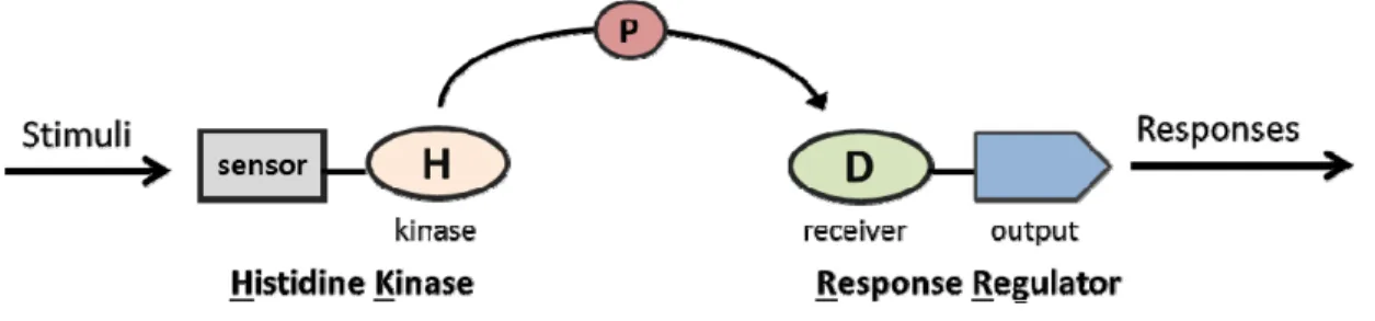
two-component system consists of two conserved proteins: a sensor kinase (histidine kinase,
HK) and a response regulator (RR) (Figure 1.1). In response to the environmental stimuli,
the HK autophosphorylates on a conserved His residue using the -phosphate from ATP
2
domain of the RR8, 9. In addition to the conserved N-terminal receiver domain, RRs also
have a variable C-terminal output domain that confers diversity in regulatory strategies.
The phosphorylation state of the RRs changes the function of the output domain, which
can participate in DNA binding and transcriptional control, perform enzymatic activities,
or mediate protein-protein interactions. Thus, RRs function as phospho-mediated
switches that couple environmental cues to cellular responses in a simple, direct
manner10, 11.
Cytokinin signaling, as well as all other known eukaryotic two-component-like
signaling, involves a more elaborate version of the two-component system known as a
multi-component phosphorelay (Figure 1.2). This involves hybrid kinases containing
both histidine kinase (HK) and receiver domains in a single protein, a His-containing
phosphotransfer proteins (HP), and response regulators (RR)12. The phosphotransfer
scheme occurs via a His-Asp-His-Asp phosphorelay that provides more targets for
modulation. The multiple-step phosphorelay systems are present in both prokaryotes and
eukaryotes.
The elucidation of the mechanism of cytokinin signaling has been hampered by
the genetic redundancy of the two-component genes in plants. Nevertheless, molecular
Figure 1.1. Schematic diagram of two-component (TCS) system. The prototypical TCS features a
3
Figure 1.2. Cytokinin signal transduction in Arabidopsis. Cytokinin binds to the membrane-bound AHK receptors, which initiates a phosphorelay through the AHPs and ultimately results in the
phosphorylation of type-B and type-A ARRs. The activated type-B ARRs induce the transcription of the type-A ARRs, which in turn act to negatively feedback the pathway.
genetic studies, primarily in the model species Arabidopsis thaliana, have revealed the
various two-component elements involved in cytoknin signaling, and the analysis of high
order loss-of-function mutants has shed light on the overlapping and distinct biological
roles of these two-component proteins13-18. However, the understanding of the underlying
mechanisms by which cytokinin achieves signaling specificity in its myriad roles
throughout plant growth and development, and how it integrates with other signaling
pathways is only beginning to be understood.
4
1. Two-component elements are involved in cytokinin signaling
1.1 Phosphotransfer chemistry
Two-component signaling acts as a ubiquitous mechanism by which bacteria
sense and respond to environmental cues. Most bacteria possess numerous
two-component systems to respond to a variety of environmental changes, such as
temperature (thermotaxis), light (phototaxis), salinity (osmotaxis), oxygen (aerotaxis),
and chemicals (chemotaxis). To date, two-component signal transduction has been found
in all domains: eubacteria, archaea, and eukarya; However, while these systems are
common in eubacteria and archaea, they are relatively rare in eukarya, and have not as
yet been found in animals, which instead rely on Ser/Thr/Tyr phosphorylation for much
of their signaling needs9. It is important to note that the chemistry of the phosphorelay
differs substantially from the phosphoesters involved in Ser/Thr/Try phosphorylation9.
The chemistry of the basic two-component system involves three phosphotransfer
reactions8, 9:
1. Autophophorylation: HK-His + ATP ↔ HK-His~P + ADP
2. Phosphotransfer: HK-His~P + RR-Asp ↔ HK-His + RR-Asp~P
3. Dephosphorylation: RR-Asp~P + H2O ↔ RR-Asp + Pi
The phosphorylation at His and Asp residues is thermodynamically distinct from the
phosphorylation at Ser, Thr, and Tyr residues, which are highly exothermic and thus
essentially irreversible. In contrast, the free energy associated with the phosphotransfer
between His and Asp residues is close to zero, which allows bidirectional flow of the
5
RRs is able to flow back to the HKs, effectively terminating the response and allowing
rapid, adaptive responses to environmental conditions.
1.2 His-Asp phosphorylation pathway in Arabidopsis thaliana
In 1993, the identification of a candidate ethylene receptor ETR1 with putative
His kinase and receiver domains in Arabidopsis thaliana was the first indication that
eukaryotes in general, and specifically plants harbored two-component signaling
systems19. In 1996, the second His-kinase, CKI1, was discovered as a potential mediator
of cytokinin signaling20, and shortly thereafter response regulators (RRs) were identified
as cytokinin primary response genes. Similarly, two-component elements were identified
in a number of fungal species. When the entire genome sequence of Arabidopsis was
elucidated in 2000, the complete two-component element repertoire was revealed: the
Arabidopsis genome encodes 11 genes predicted to be functional HKs, 5 HPs, and 23
RRs. Among the eleven HKs in Arabidopsis, five belong to the ethylene receptor family
members, though three of these are missing residues essential for histidine kinase
activity21. Further, the output of the ethylene receptor HKs occurs largely through a
Raf-like Ser/Thr kinases CTR1, which does not involve a His-Asp phosphorelay22. Among
the six other non-ethylene receptor HKs, AHK2, AHK3, and AHK4 are cytokinin
receptors; CKI1 functions upstream of the AHPs, both in vegetative and reproductive
stages23; AHK1/AtHK1 is a positive regulator of drought and salt stress responses24; and
6 1.3 Cytokinin two-component elements
In Arabidopsis, multiple two-component elements have been shown to act in
cytokinin signaling. The three cytokinin receptors (AHK2, AHK3, and
AHK4/CRE1/WOL) contain a conserved CHASE (Cyclase/Histidine kinase-associated
sensing extracellular) domain, which confers the ability to bind cytokinin with high
affinity. They also contain a histidine kinase domain, and both an authentic and a pseudo
receiver domain13, 18, 26. AHKs act partially redundantly as positive elements in cytokinin
signaling. The direct target of the AHKs are the five His-containing phosphotransfer
proteins (AHPs), which also act redundantly as positive elements in the primary
cytokinin signaling pathway15, 27, 28. There are 23 response regulators (ARRs) in
Arabidopsis that fall into two major classes based on phylogenetic analysis and domain
structure: type-A ARRs and type-B ARRs. The eleven type-B ARRs have a conserved
myb-like GARP DNA-binding domain following the N-terminal receiver domain and act
as partially redundant, positive elements in cytokinin signaling29-32. In contrast, the ten
type-A ARRs are comprised of essentially only a receiver domain and they act as
negative elements in cytokinin signaling17, 33.
Cytokinin signal transduction initiates when cytokinin binds to the CHASE
domain of the receptors AHKs to initiate autophosphorylation on a conserved His residue
within the histidine kinase domain (Figure 1.2). This phosphoryl group is then transferred
to an Asp residue within the C-terminal receiver domain of the AHKs. The AHPs then
shuttle the phosphoryl group from the AHKs to the type-B and type-A ARRs15, 27. The
phosphorylation activates the type-B ARRs, which regulate the expression of the primary
7
as negative feedback regulators of the primary signaling pathway35. Overall, the
phosphorelay in cytokinin signal transduction involves four sequential phosphorylation
events in the order His-Asp-His-Asp. This more elaborate architecture of the
phosphorelay provides additional opportunities for crosstalk with other signaling
pathways and provides a robust mechanism for shuttling the cytokinin signal to multiple
8 2. Cytokinin receptors are histidine kinases
2.1 Discovery
A histidine kinase (HK), CKI1 (CYTOKININ INSENSITIVE 1), was isolated
from an activation T-DNA tagging screening as a gene that when overexpressed
conferred shoot initiation in the absence of exogenous cytokinin. The CKI1 protein
consists of a histidine kinase domain, a single transmembrane domain, and a receiver
domain. Overexpression of CKI1 induces typical cytokinin responses to cultured cells,
including rapid proliferation, greening, shoot formation and inhibition of root formation.
The activation of cytokinin responses by CKI1 implicates it as a cytokinin signaling
element, but the gain-of-function nature of the allele complicates this conclusion.
Subsequent studies identified cre1 (cytokinin response 1) as a mutant that showed
reduced cytokinin sensitivity. CRE1 also encodes a histidine kinase. However, in contrast
to CKI1, CRE1 was demonstrated to bind cytokinin with high affinity and specificity,
and could complement yeast and bacterial HK mutants in a cytokinin-dependent manner.
This Arabidopsis Histidine Kinase (AHK4/CRE1/WOL) and its paralogues (AHK2 and
AHK3) contain a CHASE domain that binds cytokinin. How binding of cytokinin to the
CHASE domain transduces the signal across the membrane is not known. CKI1 and other
hybrid HKs that do not include the CHASE domain do not bind cytokinin; nevertheless,
they could feed into cytokinin signaling by phosphorylation of the AHPs in response to
the sensing of other signals beside cytokinin, and/or by forming heterodimers with the
9
2.2 Cytokinin receptors differ in biochemical nature
Naturally occurring cytokinins are adenine derivatives with a N6-side chain and
are classified as isoprenoid or aromatic depending on the nature of the side chain 36. The
binding preferences for the different AHKs have been studied in Escherichia coli. Strains
of E. coli expressing AHK3 or AHK4 have different sensitivity in recognizing various
cytokinin compounds 37. Both are most sensitive to the isoprenoid-type trans-zeatin (tZ)
and isopentenyladenine (iP) but differ significantly in the recognition of other cytokinin
compounds. Interestingly, all maize HKs recognize cis-zeatin (cZ) with high affinity 38.
Results from in planta experiments confirm the in vitro data, and further reveal different
sensitivity of each AHK towards tZ and iP; AHK2 and AHK4 show a comparable
activity by tZ and iP, while AHK3 shows the higher sensitivity to tZ compared with iP. A
higher functional similarity between AHK2 and AHK4 is supported by the
promoter-swap experiment, in which AHK4 expressed under the control of the AHK2 promoter
(but not the AHK3 promoter) is sufficient to complement the ahk2 ahk3 loss-of-function
phenotype 39. A chimeric protein that includes the CHASE-TM (transmembrane) of
AHK3 and the cytoplasmic domain of AHK4 could partially complement ahk2 ahk3
phenotype 39. The partial complementation suggests the property of the CHASE-TM
domain is critical but not the sole feature required for proper AHK3 function.
Disrupting AHK4 via T-DNA insertion or nonsense mutation does not cause a
substantial effect on plant growth and development. However, certain point mutations
within the AHK4 coding region produce the wooden leg (wol) alleles of CRE1, which
have fewer numbers of vascular initials during embryogenesis, and as a result, cause
10
T278I in the CHASE domain of AHK4 blocks its cytokinin binding activity in vitro. The
recently resolved crystal structure of the CHASE domain of AHK4 has rationalized that
the T278I mutation likely restricts the overall size of the binding pocket and thus affects
the binding capacity 41. The dominant-negative nature of wolT278I may result from the
phosphatase activity of unbound AHK4 42, which is observed in some prokaryotic
histidine kinases that possess both kinase and phosphatase activity9. Biochemical analysis
show that CRE1 can dephosphorylate multiple AHPs and this phosphatase activity
requires the conserved Asp residue in its receiver domain42. These results suggest that
AHK4 acts as both a kinase and a phosphatase in a bidirectional phosphorelay. Thus, in
the absence of cytokinin, phosphate from the ARRs could be removed via flow to AHK4,
through the AHPs, reflecting the reversibility of the various phosphorylation events in
this pathway.
A distinct wol-like allele of AHK4 has been isolated 43. This mutation results in a
wol-like root phenotype. Interestingly, the wol-2L506F displays intragenic
complementation of wol-1T278I, implying that the signal transduction involves
dimerization or higher order oligomerization. In the trans-hetergozygous plants
(wol-1/wol-2), the root vascular phenotype is wild-type, though they remain insensitive to
exogenous cytokinin. It is puzzling how these recessive mutations cause
dominant-negative effects on procambial cell proliferation, and display intragenic complementation
exclusively in vascular development. One explanation is that AHK4 represses, perhaps
via its phosphatase activity, cambium morphogenesis as a monomer 44. Binding of
cytokinin triggers the dimerization (or higher order oligomerization) and subsequently
11
ability either to bind cytokinin (wol-1) or to dimerize (wol-2) and thus repress procambial
development. In this model, the trans-heterozygotes wol-1/wol-2 would only have
compromised receptors including one copy of WOL-2 that can bind cytokinin but not
dimerize and one copy of WOL-1 that can dimerize but not bind cytokinin, which is
sufficient to release the repressing effect on vascular initiation but not for the response to
elevated levels of exogenous cytokinin.
2.3 Functions
Since their discovery, cytokinins have been shown to positively regulate shoot
growth and negatively regulate root elongation. Much of the work, however, has been
based on experiments using overexpressors and exogenous cytokinin treatment.
Mutations in the genes encoding various cytokinin two-component elements provide
novel ways to explore the functions of cytokinin in plant growth and development.
Primary root elongation and lateral root formation are inhibited in the ahk2 ahk3
ahk4 mutant, which is associated with cell cycle arrest as the transition from G2 → M
phase is delayed18. Moreover, the wol mutant displays a lack of the periclinal cell
divisions that occurs during vascular morphogenesis 40. However, single mutation in
AHK3 and multiple mutations in isopentenyltransferases (IPTs), which are essential to
cytokinin biosynthesis, results in longer primary root and a larger root meristem 45. These
results led to a model in which the cytokinin response curve, at least in the root, is
bell-shaped rather than linear 44. If this were the case, a minor reduction of cytokinin signaling
would induce root growth, while reduction beyond the threshold would abolish growth.
This is similar to the bell-shape response curve observed for cytokinin in shoot initiation
12
Disruption of the cytokinin receptors also perturbs shoot and floral development.
The shoot meristem size and leaf cell number are smaller in ahk2 ahk3 ahk4 mutants,
consistent with a role for AHKs as positive regulator of cell division 13, 14, 18. The ahk2
ahk3 ahk4 mutants only occasionally form an inflorescence stem and produce few sterile
flowers. These results suggest the transition from vegetative to inflorescence meristem is
defective, and that the floral meristem activity is depleted in ahk2 ahk3 ahk4 mutants.
The cytokinin receptors AHKs are also required in gametophyte development as the
strong triple mutant combinations result in complete male and female sterility 13, 46.
Interestingly, the weak ahk triple mutants produce a few flowers with reduced fertility,
but are capable of producing a few seeds. These findings indicate that cytokinin is
essential in floral development, however, the dosage of cytokinin signaling required
differs for different developmental stages47.
2.4 Cytokinin receptors have overlapping and specific expression pattern
The expression of the AHK genes overlap but display different levels in specific
tissues. In roots, AHK4 is expressed at a higher level as compared to AHK2 and AHK3; in
rosette leaves, AHK2 and AHK3 display the predominant expression and AHK4 is barely
detectable. Consistent with the expression patterns, ahk2 ahk3 mutants have smaller
rosettes while ahk3 ahk4 and ahk2 ahk4 are similar in size to the wild type; single ahk4
mutants display reduced cytokinin sensitivity in the root while the ahk2 and ahk3 mutants
exhibit normal sensitivity. The cytokinin-dependent induction of ARR15 and ARR16 are
compromised in the roots of the ahk4 mutants, but not in leaves. This suggests the
transcriptional induction of a subset of type-A ARRs in root by exogenous cytokinin
13
level in leaves, a specific role in regulating senescence is exclusively mediated by AHK3.
A gain-of-function mutation in AHK3 causes delayed leaf senescence whereas a loss of
function ahk3 mutant (but not ahk2 nor ahk4 single mutant) confers reduced cytokinin
sensitivity in leaf senescence. Chlorophyll retention is impaired in the ahk3 mutant, and
addition of a ahk2 mutation further magnifies this effect. The involvement of AHK4 only
becomes significant in the triple mutant, consistent with its weak expression level in
rosette leaves.
2.5 The majority of cytokinin receptors localize in ER
In addition to the redundant roles of the receptors in some contexts, genetic
studies have also reported specific roles for individual AHKs14, 46, 48. These unique
receptor functions could be the result of differences in expression patterns39, 46, 48, ligand
binding37, 39, protein structures41, 49, interacting targets50, or subcellular localizations51, 52.
Hydrophobicity analysis suggests putative transmembrane segments in the N-terminus of
AHK2, AHK3, and AHK4. Bioinformatic analysis using the PSORT (Prediction of
Protein Sorting Signals and Localization Sites) program suggested that they are localized
in the plasma membrane (PM). However, more recent data from biochemical and cell
biological assays show that, in Arabidopsis and maize, the majority of cytokinin receptors
are localized in the endoplasmic reticulum (ER) and expose the cytokinin-binding
CHASE domain to the ER lumen51-53. Consistent with this, endomembranes have higher
cytokinin binding affinity than the PM. Transiently expressed fluorescent fusion proteins
support the predominant localization to the ER for all three AHKs. Nevertheless, there is
a minor but functional relevant fraction of the receptors at the PM52. The canonical model
14
finding that their location is predominantly in the ER suggests that active cytokinins must
cross the plasma membrane and ER membrane in order to bind to the lumen localized
CHASE domain. Purine permeases (PUP) have been suggested, in Arabidopsis cell
culture and in yeast, to transport cytokinin and adenine into the cytosol54, 55. Genetic
evidence for the role of PUPs in cytokinin in planta is still lacking due to the large
number of PUP genes present in the Arabidopsis genome. Therefore, more studies are
needed to understand the mechanisms by which cytokinin enters the cell and is
transported into the ER lumen.
2.6 Non-receptor kinases also feed into two-component signal transduction
2.6.1 CKI1
As noted above, overexpression of CKI1 induced cytokinin-independent callus
formation in cultured Arabidopsis cells20. However, the lack of a CHASE domain in CKI
strongly suggests that this HK is not a cytokinin receptor per se. Recent studies have
shown that CKI1 can feed into downstream two-component signal transduction via the
phosphoproteins AHPs and the type-B ARRs23. The receiver domains of CKI1 can
interact with AHP2, AHP3, and AHP5 in yeast and plant protoplasts56. The phenotype
induced by overexpression of CKI1 is eliminated in the ahp1,2,3,4,5 mutant, suggesting
that AHPs act epistatically to CKI123. Further, phenotypic analysis has shown that in high
order ahp and type-B arr mutants, a subset of female gametophytes have similar
phenotypes as those observed in cki1 loss-of-function alleles, which supports a role for
15
type-B ARRs, under the control of CKI1 promoter is able to partially rescue the cki1
phenotype, further suggesting ARR1 is epistatic to CKI1.
Despite the lack of cytokinin-binding CHASE domain, overexpression of CKI1
was found to partially rescue multiple phenotypes in a wol mutant, including the
shortened primary root, defects in xylem development, and cytokinin insensitivity in
shoot regeneration assay23. In addition, ectopic expression of cytokinin biosynthetic
isopentenyltransferase IPT8 under the control of the CKI1 promoter is able to partially
rescue cki1 phenotype. The mechanisms by which elevated cytokinin levels complement
the cki1 phenotype is not clear, though likely involve increased activity of the
downstream phosphorelay.
2.6.2 Other non-receptor histidine kinases
The role of AHK1 in plant growth and development is only obvious in a triple
ahk1 ahk2 ahk3 mutant background. Addition of ahk1 to an ahk2 ahk3 mutant
significantly reduces the plant size and retards growth24. In addition, the ahk1 mutant is
more sensitive to drought stress, while ahk2 and ahk3 are more tolerant24.This suggests
that cytokinin receptors act in stress responses in a manner opposite to that of AHK1.
Comprehensive coexpression analysis reveals that AHK1 is coexpressed with a set of
type-A ARRs (ARR4, ARR5, ARR6, ARR8, ARR9) under abiotic stress conditions and
cytokinin treatment57. These results suggest a potential interaction between AHK1 and
cytokinin signaling in abiotic responses, though further studies are needed to confirm this.
CKI2/AHK5 is a histidine kinase that lacks a transmembrane domain. The genetic
role of AHK5 in the cytokinin two-component phosphorelay is not clear. AHK5 interacts
16
significance of these interactions remains an open question. AHK5 is an active histidine
kinase, but this activity is not dependent on cytokinin. Disruption of AHK5 results in a
normal shoot, and the roots displayed wild-type sensitivity to cytokinin, but were
hypersensitive to abscisic acid and ethylene, suggesting a potential role as a negative
regulator of these signaling pathways59.
2.7 Are AHKs the only cytokinin receptors?
Cytokinins were long thought to be essential for plant growth and development as
they regulated essential processes such as cell division and organogenesis. Surprisingly,
three independent ahk triple mutants harboring non-overlapping T-DNA alleles are
seedling viable, albeit quite stunted. One allelic combination has marginally reduced
fertility while the other two mutants are completely male and female sterile47. Recent
studies have revealed that even in the strongest ahk triple mutant, there is residual
full-length AHK3 transcript (~0.02% compared to wild type), indicating that none of these
three triple mutants represent null alleles47. This raises the question as to whether or not
cytokinin is essential for plant growth and development. One possibility is that it is not,
notwithstanding the residual AHK3 transcript. A second possibility to explain the
viability of the ahk2 ahk3 ahk4 mutants is that the residual AHK3 transcript, although
insufficient for male and female gametophyte development, is sufficient to support some
vegetative development. A final possibility is that there are additional cytokinin receptors
beyond AHK2, AHK3 and AHK4. Several cytokinin-binding proteins have been isolated
from various plant species including barley, maize, oat, and tobacco. However, the
evidence linking these to a physiological function in cytokinin signaling is lacking.
17
encodes a protein containing a CHASE domain at the N-terminus followed by a
serine/threonine kinase domain60. The CHASE domain of CHARK is 49% to 67%
identical to the cytokinin HK receptors in rice, maize, and Arabidopsis60. CHARK may be
a cytokinin-binding element unique to rice or monocots, although the cytokinin-binding
activity of the encoded product CHARK has not been verified. Further genetic analysis is
18 3. The phosphotransfer proteins in Arabidopsis
3.1 AHPs shuttle phosphate between AHKs and ARRs
Examination of the Arabidopsis genome reveals five genes encoding predicted
proteins with high sequence similarity to histidine-containing phosphotransfer (Hpts)
proteins from E. coli and yeast28. These Arabidopsis Hpts (AHPs) were shown to
complements a loss-of-function mutation in YPD1 in yeast, which is a Hpt protein
involved in osmosensing28. The AHPs were shown to rapidly transfer phosphoryl groups
from their own conserved His residue to the Asp residue of two response regulators61. In
addition to these functional AHPs, there is a sixth gene that lacks the conserved His
residue that is the site of phosphorylation called AHP6 (see below)15, 49. The AHPs
directly interact with both the upstream cytokinin receptors AHKs and the downstream
type-A and type-B ARRs. The AHPs act to shuttle a phosphoryl group from the Asp
residue of the receiver domains present in the cytokinin AHK receptors to the Asp
residue present in the receiver domains of the ARRs, thus transducing the signal from the
site of perception (the ER membrane) to the nucleus to regulate gene expression, and to
the type-A ARRs, some of which are in the nucleus and some of which are cytoplasmic.
3.2 AHPs act downstream of AHKs and CKI1
Reverse genetic experiments provided direct evidence for the role of five AHPs as
positive regulators in cytokinin signaling. Various combinations of T-DNA insertion
alleles in the five AHP loci were analyzed. AHP1, AHP2, AHP3 and AHP5 were found
to have overlapping roles as positive elements in cytokinin signaling using multiple
19
The quintuple ahp1ahp2-1 ahp3 ahp4 ahp5 mutant displayed phenotypes similar to ahk2
ahk3 ahk4 triple receptor mutants, including inhibition of primary root growth and loss of
metaxylem development. Interestingly, shoot development in the ahp1ahp2-1 ahp3 ahp4
ahp5 mutant was not as severely affected compared to the ahk2 ahk3 ahk4 mutant, which
is likely due to the residual full-length AHP2 transcript from the ahp2-1 allele used. A
quintuple ahp1ahp2-2 ahp3 ahp4 ahp5 mutant that incorporates a null ahp2-2 allele is
severely delayed in true leaf formation and dies at the seedling stage, which has not been
reported even in the strongest ahk2-7 ahk3-3 cre1-12 triple receptor mutant. As noted
above, the discrepancy in the phenotypic strength of these lines as compared to the null
AHP mutant is likely due to the fact that none of the ahk triple mutant combinations
represent complete receptor nulls (see 2.8). Alternatively, as the AHKs are likely not the
sole upstream regulators of the AHPs (see 2.6.1), the stronger phenotype of the
ahp1ahp2-1 ahp3 ahp4 ahp5 mutant may reflect disruption of the AHK- and/or
CKI1-dependent signaling pathways.
3.3 The subcellular localization of AHPs suggests a function in a bidirectional phosphorelay
Phospho-His and phospho-Asp residues are high energy molecules. As noted
above (see 1.1), the free energy associated with the various phosphorylation reactions that
occur in the phosphorelay is close to zero, which allows HP domains to act both as
phosphodonors and phosphoreceivers, and so to shuttle a phosphoryl group between two
or more receiver domains. That is, the high energy cytosolic phospho-AHP is capable of
donating the phosphoryl group to both the type-A and type-B response regulators, or to
20
nucleus in response to cytokinin treatment, but a more quantitative analysis demonstrated
that the AHPs are persistently nucleo-cytosolic and non-responsive to cytokinin or
phosphorylation62. Further, the size of the AHP-GFP fusion proteins exceeds the
exclusion limit of the nuclear pore, suggesting they are actively transported into and out
of the nucleus62. Together, this suggests that the AHPs can mediate the phosphorelay
from the membrane bound receptors to the mainly-nuclear localized response regulators.
In addition, the flow of phosphate can proceed from the receptors to the ARRs in
response to cytokinin activation of the AHK receptors, or from the ARRs back to the
AHKs upon cessation of the cytokinin signal.
3.4 AHP6 is a negative element in cytokinin two-component signaling
AHP6 was isolated in a genetic screen as a suppressor of the determinate root
phenotype of wol49. AHP6 lacks the conserved His residue that is the site of
phosphorylation, and thus is not a functional Hpt protein and does not participate in
phosphotransfer. AHP6 was shown to inhibit the phosphorelay in vitro from the His
kinase domain of SLN1 to its fused receiver domain. SLN1 is a hybrid HK involved in
the Saccharomyces cerevisiae osmosensing pathway. AHP6 also inhibits the
phosphotransfer from phosphorylated AHP1 to ARR1 in vitro, which suggests that AHP6
acts as an inhibitor of phosphotransfer, likely through a dominant negative mechanism49.
The role of AHP6 as a negative regulator of cytokinin signaling is also supported by
functional analysis in vivo. The ahp6-1 loss-of-function mutant has elevated basal
expression of the cytokinin primary response gene ARR15 and is hypersensitive to the
21
differentiation. Interestingly, cytokinin also negatively regulates AHP6 expression,
forming a mutual-regulatory circuit in regulating root development49.
3.5 Nitric oxide regulates phosphotransfer proteins through S-nitrosylation
In cells, nitric oxide (NO) can directly modify the cysteine thiol of a protein as a
redox-based posttranslational modification mechanism, which is known as S-nitrosylation.
It has been suggested that NO can regulate cytokinin signaling63. The expression of the
cytokinin reporter TCS-GFP as well as multiple cytokinin primary response genes are
reduced in the nox1 and gsnor1-3 mutants, which have elevated levels of endogenous
NO64. These NO-overexpressors are less sensitive to cytokinin in root and hypocotyl
elongation, root apical meristem size, and shoot regeneration assays, as well as in the
induction of cytokinin primary response genes, suggesting that NO negatively regulates
cytokinin signaling. This is likely the result of the S-nitrosylation of the AHP proteins on
the conserved cysteine residue, which reduces their phosphorylation level and hence
functionality. AHP1C115S, a non-nitrosylatable mutant protein, was resistant to the
inhibitory effect of NO donors and was able to complement the cytokinin-insensitivity of
high order ahp mutants. In contrast, an AHP1C115W mutant protein that mimics the
S-nitrosylation modification displayed reduced phosphorylation even in the absence of an
NO donor and did not complement ahp mutants. In vitro S-nitrosylation of AHP1
repressed its phosphotransfer activity to ARR1, a type-B ARR, demonstrating that
S-nitrosylation reduced the activity of the AHP. This represents a novel mechanism by
which environmental stimuli intertwine with endogenous signal transduction pathways.
S-22
nitrosylated cysteine residues are also present in AHKs and ARRs, although experimental
23 4. Response regulators in Arabidopsis
Response regulators were first implicated in cytokinin signaling when they were
identified as early cytokinin response genes in Arabidopsis and maize65, 66. Response
regulators contain a conserved receiver domain with a conserved Asp residue that is the
site of phosphorylation, which generally regulates their output activities. The Arabidopsis
response regulators (ARRs) fall into four major classes based on their domain structure
and the similarity of the amino acid sequences of the receiver domains67: type-A, type-B,
type-C ARRs, and the Arabidopsis pseudoresponse regulators (APRRs) (Figure 1.3). The
ten type-A ARRs are primary transcriptional targets of cytokinin signaling and contain
short C-terminal extensions following the conserved receiver domain. The eleven type-B
ARRs contain a receiver domain followed by an output domain that has DNA-binding
activity. The two type-C ARRs are structurally similar to type-A ARRs as they contain
only the receiver domains, however, they are not transcriptionally induced by cytokinin.
The role of type-C ARRs in cytokinin signaling remains unclear, although overexpression
of one of them confers reduced cytokinin sensitivity68. The APRRs contain complete
receiver domains but lack the conserved Asp residue for phosphorylation, although many
appear to be phosphorylated. A subset of the APRRs play a role in modulating circadian
rhythms and their phosphorylation status oscillates throughout the day.
Biochemical and genetic analyses have demonstrated that bacterial response
regulators (RRs) function as phosphorylation-mediated switches10, 11. Phosphorylation of
the highly conserved Asp residue in the receiver domain inactivates the protein in some
RRs and activates it in others. In agreement with a regulatory role of phosphorylation,
24
function. A subset of type-A ARR proteins is stabilized by cytokinin via phosphorylation
of the Asp residue33 (see 5.2). Similarly, mutation of the phospho-receiving Asp to
phospho-insensitive Asn in type-B ARRs abolished its activity to transactivate type-A
ARR669 (see 6.2). Together, these data suggest that phosphorylation is a common strategy
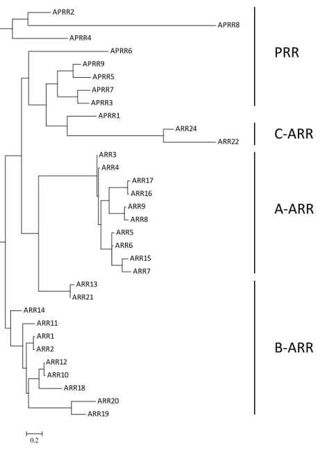
utilized by the ARRs to modulate their output response. APRR2 APRR8 APRR4 APRR6 APRR9 APRR5 APRR7 APRR3 APRR1 ARR24 ARR22 ARR3 ARR4 ARR17 ARR16 ARR9 ARR8 ARR5 ARR6 ARR15 ARR7 ARR13 ARR21 ARR14 ARR11 ARR1 ARR2 ARR12 ARR10 ARR18 ARR20 ARR19 0.2
PRR
C
‐
ARR
A
‐
ARR
B
‐
ARR
Figure 1.3. Phylogenetic relationship of the Arabidopsis response regulators (ARR). The amino
25 5. Type-A response regulators
5.1 Type-A ARRs are primary response genes in cytokinin signaling
The Arabidopsis type-A ARRs are a family of ten genes that fall into five distinct
pairs which, based on the analysis of the locations of the genes within the genome, likely
arose from the most recent genome duplication event in the evolution of Arabidopsis70.
The amino acid sequences of type-A ARRs are somewhat similar to that of the bacterial
single-domain response regulator CheY, which is comprised of only a receiver domain71.
The transcripts of type-A ARRs are rapidly and specifically induced by cytokinin, and
this induction is insensitive to inhibition of protein synthesis, and they are thus primary
response genes65. The cytokinin receptor AHKs, the AHPs, and type-B ARRs are
required for the rapid induction of type-A ARRs, indicating that their induction requires
an intact phosphorelay.
5.2 Type-A response regulators negatively regulates cytokinin signaling
Overexpression of type-A ARRs results in repression of ARR6-LUC activity,
leading to the hypothesis that they are negative regulators of cytokinin response72.
Consistent with this hypothesis, analysis of loss-of-function type-A arr mutants shows
that at least eight of the ten type-A ARRs are negative regulators of cytokinin signaling in
multiple cytokinin response assays35, 73. Single loss-of-function type-A arr mutants show
no significant difference from the wild type in the response to exogenous cytokinin
treatment in root elongation assays, while double and higher order multiple mutations in
type-A ARRs show increasing hypersensitivity to cytokinin35. In monocots and dicots,
26
meristem, presumably through increased cytokinin signaling output 74, 75. Consistently,
overexpression of the rice type-A response regulator OsRR6 leads to repression of shoot
regeneration in tissue culture and an aberrant dwarf phenotype in transgenic plants76.
Despite the well-described role of type-A ARRs as negative regulators in
cytokinin signaling, mutation of eight out of ten type-A ARRs does not cause dramatic
morphological phenotypes. It is possible that additional negative feedback loops might
compensate for the cytokinin hypersensitivity in this octuple mutant by decreasing
cytokinin levels, as several cytokinin degrading oxidases (CKX) are induced upon
cytokinin treatment77. The alternative, not mutually exclusive possibility is that type-A
ARRs might functionally overlap with other signaling elements to regulate plant growth
and development. Thus, the octuple mutations in type-A ARRs attenuate but not abolish
the output responses.
5.3 Phosphorylation of receiver domain is essential for type-A ARR function
The type-A ARR proteins exhibit in vitro activity typical of bacterial receivers as
they can be phosphorylated on the conserved Asp residue using a phospho-HP domain as
a phosphodonor71. Further, a subset of type-A ARR proteins is stabilized by cytokinin via
phosphorylation of the Asp residue33. Type-A ARR5D87A, a non-phosphorylatable mutant
protein, fails to complement the cytokinin hypersensitivity of a arr3 arr4 arr5 arr6
mutant, indicating that phosphorylation of the conserved Asp residue is required for its
proper function. In contrast, ARR5D87E, a phosphomimic version of the protein, can
partially rescue the cytokinin hypersensitivity of arr3 arr4 arr5 arr6 mutant. As this
ARR5D87E protein cannot be phosphorylated in the phosphorelay, it suggests that the
27
feedback cytokinin signaling. Such phospho-dependent interactions are a common
strategy used by bacterial single domain response regulators that lack a distinguishable
output domain. At least two independent yeast two-hybrid screenings have revealed that
type-A ARRs interact with candidate proteins in addition to the two-component
elements50, 78, which potentially may include such target proteins.
5.4 Other signals cross-talk with cytokinin signaling via regulating type-A ARRs
In addition to cytokinin, type-A ARRs are regulated by other inputs, presumably as a
means to modulate cytokinin signaling. For example, WUSCHEL (WUS), a
homeodomain transcription factor, specifically represses the expression of ARR5, ARR6,
ARR7, and ARR15 in shoot apical meristem to reduce their negative attenuation on
cytokinin signaling, thus maintaining stem cell fate79. Overexpression of the
phosphomimic form of ARR7 (35S::ARR7D85E) results in arrested shoot apical meristem
that is similar to the phenotype observed in a wus mutant79. Similarly, auxin, another
plant hormone, also cross talks with cytokinin signaling by regulating two type-A ARRs,
ARR7 and ARR15. Whereas cytokinin induces ARR7 and ARR15 in shoot apical meristem,
auxin represses the expression of these two type-A ARRs via AUXIN RESPONSE
FACTOR 5 (ARF5)80. Mutations of the ARF-binding site in the promoter region of
ARR15 result in ectopic expression of ARR15. Further, mutations of ARR7 and ARR15 in
arf5 background could partially rescue arf5 phenotype. These experiments provide
examples in which endogenous signals cross talk with cytokinin signaling via
28 6. Type-B response regulators
The type-B ARR family is comprised of eleven transcription factors that belong to
three sub-families based on the amino acid sequences of their receiver domains81, 82. The
subfamily I type-B ARRs have been the most thoroughly studied as they seem to take the
predominant role in cytokinin-mediated outputs. The type-B ARRs contain a receiver
domain at the N-terminus followed by a conserved plant-specific myb-related GARP
DNA-binding domain.
6.1 Type-B ARRs are positive regulators in cytokinin signaling
Type-B ARRs play overlapping roles in cytokinin signaling and plant
development16, 83. Analysis of loss-of-function subfamily I type-B arr mutants has enable
the elucidation of the endogenous functions of these genes. Single type-B arr mutants
show only slight cytokinin insensitivity in seedlings, while multiple mutations in
subfamily I ARR1, ARR10, ARR12 confer additive cytokinin insensitivity in primary root
growth, hypocotyl elongation, and shoot formation in tissue culture. These results suggest
that subfamily I type-B ARRs acts as redundant positive elements in cytokinin signaling.
Mutations in type-B ARRs also compromise the induction of cytokinin primary response
genes16, 32, indicating that they mediate the immediate transcriptional response to
cytokinin. The role of subfamily II and III type-B ARRs is not clear as our understanding
has been based on overexpression experiments81, which may not reflect their endogenous
functions. Subsequent analysis of loss-of-function mutations in subfamily II and III ARR
showed that there is no obvious phenotype in flower development, silique development,
29
Subfamily I type-B ARRs are broadly detectable by RT-PCR in vegetative and
reproductive tissues, with particular high expression in regions where phenotypes have
been reported in loss-of-function mutants, including young developing leaves and
meristems82. On the contrary, subfamily II and III type-B ARRs are barely detected by
RT-PCR, but their expression can be seen using promoter fusion to GUS reporter
protein82. This may reflect the rapid RNA turnover of type II and III type-B ARRs or very
specific expression domains. The restricted expression profile suggests that subfamily II
and III type-B ARRs may function in specific developmental processes. Consistent with
this, a recent report shows that ARR20, which belongs to subfamily III type-B ARR, is a
positive regulator of cytokinin signaling in regulating pavement cell morphogenesis.
Similar to the cytokinin double receptor mutant ahk3-3 cre1-12, the single arr20 mutant
displays a modest enhancement pavement cell interdigitation. This phenotype is not
observed even in arr1-3 arr10-5 arr12-1 mutant, which shows almost complete
insensitivity to exogenous cytokinin in multiple other assays16.
6.2 The receiver domains of type-B ARRs have inhibitory effect on DNA-binding domains
Cytokinin signaling influx activates type-B ARRs via phosphorylation at the
conserved Asp residue in the receiver domain. The N-terminal receiver domains of
type-B ARRs interact with the AHP phosphotransfer proteins as shown by in vitro and in vivo
experiments50, 61. Mutation of phospho-receiving Asp in ARR2 to phospho-insensitive
Asn abolishes the ability of this protein to be phosphorylated via phosphotransfer
30
promoter in response to cytokinin69, indicating that phosphorylation of the receiver
domain is required for type-B ARR function.
Transgenic plants overexpressing full-length type-B ARRs display wild-type
morphology; however, deletion of the receiver domain leads to constitutive activation of
this transcription factor31. Overexpressing several N-terminal truncated type-B ARRs
(ARR2, ARR11, ARR18, ARR19, ARR20, and ARR21), which contain only the
C-terminal DNA-binding domain, results in their constitutive activation and pleiotropic
phenotypes31, 81, 85, 86. These results indicate potential novel roles of type-B ARRs in plant
growth and development, although the ectopic and overexpression of these activated
type-B ARRs may not faithfully reflect their endogenous functions.
One simple model for the autoinhibitory effect of the type B N-terminal domains
is that the receiver domain blocks, through steric hindrance or direct interaction, the
activity of the DNA-binding domain. This is similar to the mechanism in many bacterial
RRs. Phosphorylation of the receiver domain triggers conformational change that
de-represses the inhibitory effect on the DNA-binding domain. However, the precise
mechanism underlying the inhibitory regulation has not been experimentally validated.
6.3 Type-B ARRs function as transcription factors
In vitro assays have shown that the GARP motifs of at least four type-B ARRs
bind to DNA in a sequence-specific manner; the sequences that ARR1/ARR2, ARR10,
and ARR11 preferentially bind are GAT(T/C), AGATT, and GGATT respectively30, 87, 88.
The core sequences are too short to specify direct targets because they appear too
frequently in DNA genomes. A study analyzing the cis-elements of the target genes
31
GAT(C/T)TT, and AAGAT(C/T)TT, which were found to be tandemly enriched in the
target promoters89. A meta-analysis of cytokinin regulated genes further showed that the
ARR1 consensus binding site AAGAT(C/T)TT was substantially overrepresented in the
regulatory regions of robustly cytokinin-responsive genes90. Single mutations of the
ARR1 binding sites AGATT to ACATT in the promoter region of type-A ARR15 was
sufficient to eliminate its cytokinin responsiveness in planta91, demonstrating that this
regulatory element is required for cytokinin-responsiveness.
Despite sharing a core binding sequence (GAT), ARR11, unlike other type-B
ARRs, does not bind to the cytokinin-responsive element AGATT88, suggesting
non-overlapping targets and hence function of type-B ARRs. Indeed, seven of the type-B
ARRs (ARR11, ARR14, and ARR18 of subfamily I; ARR13 of subfamily II; ARR19 and
ARR20 of subfamily III) under the control of the ARR1 native promoter cannot
complement the root growth phenotype of arr1 arr12, suggesting these proteins may
have distinct functions.
6.4 The direct targets of type-B ARRs
Type-B ARRs-regulated genes have been identified by microarray analyses of
wild type and multiple type-B arr mutants in response to cytokinin treatment16, 77, 92.
These studies indicate that type-B ARRs are essential for nearly all cytokinin-regulated
gene expression. Further, the endogenous levels of many genes not identified as
cytokinin-responsive are differentially expressed in the type-B arr mutants compared
with wild-type seedlings16. The different experimental conditions and combinations of
high order mutants used in these experiments make it difficult to decipher the target
32
a glucocorticoid (DEX) inducible chimeric transcription factor fused to ARR1 lacking its
receiver domain, have been used to identify the genes rapidly activated by ARR1ΔDDK
upon DEX treatment. These genes potentially represent direct targets of ARR1 and
included cytokinin oxidase, cytokinin hydroxylase, putative disease resistance response
proteins, and IAA3/SHY289. Chromatin immunoprecipitation and gel mobility shift
analyses confirmed that ARR1 directly associates with the promoter region of
33 7. Cytokinin response factors
Microarray results from different labs have shown that CYTOKININ RESPONSE
FACTORS (CRFs), a subset of AP2/ERF superfamily of transcription factors, are
transcriptionally up-regulated by cytokinin77, 89, 94. CRF includes a group of six core
members that contain a AP2/ERF domain and a CRF motif and represents a large clade
of AP2/ERF genes in land plant species95. In Arabidopsis seedlings, CRF2 and CRF5
show rapid (<30 minutes) induction upon cytokinin treatment while CRF6 induction does
not peak until later (>8 hours)96, indicating different kinetics in their response to
cytokinin. Despite the name, not every CRF has been found to be cytokinin-responsive.
Similarly, cytokinin up-regulates only a subset of the tomato CRFs, which also display
distinct kinetics in response to cytokinin 97.
The rapid induction of CRFs by cytokinin is compromised in type-B arr mutants,
leading to the hypothesis that CRFs regulate part of the transcription network
downstream of type-B ARRs96. Indeed, multiple cytokinin-regulated genes exhibit
reduced responsiveness in the loss-of-function crf mutants, suggesting CRFs are
responsible for a subset of cytokinin responses96. The induction of type-A ARRs,
however, is not dependent on CRFs as they retain the wild-type level of induction in crf
mutants. Overall, CRFs are hypothesized to form a side branch of the cytokinin response
34 Future directions
Twelve years after the initial discovery of a cytokinin receptor, remarkable
progress has been made in our understanding of cytokinin signaling. The two-component
elements modulate, via sequential phosphorelay events, cytokinin signal transduction.
Loss-of-function and gain-of-function mutants have, at least in part, overcome the gene
redundancy in this system and revealed the roles of cytokinin signaling in plant growth
and development. Meanwhile, the elucidation of the pathway has raised many
outstanding questions from perception of cytokinins to determine the output responses.
For example, the subcellular localization of cytokinin receptors in ER raises the question
as to how cytokinins are transported across plasma membrane and ER membrane to reach
the receptors. How is specificity in outputs achieved in the pathway, and how is this
pathway integrated with other signals to achieve appropriate growth and development?
How does the non-receptor histidine kinase CKI1 sense stimuli and feed into cytokinin
signaling? How does elevated cytokinin level partially complement cki1 phenotype?
The phosphotransfer proteins that shuttle the phosphoryl group between the
receptors and the response regulators provide another layer of regulation in this system.
At the cellular level, studies are needed understand the mechanism underlying the
transport of AHPs in and out of the nucleus, which allows the phospho-AHPs to maintain
the appropriate phosphorylation status of the system elements. As for the ARRs, the
mechanism by which type-A ARRs negatively feedback on cytokinin signaling is not yet
clear. Likewise, how phosphorylation de-represses the inhibitory effect of the receiver
35
Disruption of cytokinin signal transduction, at multiple branch points, implicates
the requirement of cytokinin in many aspects of plant growth and development. The next
step is to illuminate the mechanisms by which cytokinin signaling elicits the biological
CHAPTER 2: Cytokinin-Dependent Specification of the Functional Megaspore in the Arabidopsis Female Gametophyte
Preface
The following chapter was published in The Plant Journal volume 73 and my
contribution to the publication was that I designed the experiments and wrote the
manuscript. This work was the first example that specified the role of cytokinin in female
gametophyte development.
Summary
The life cycle of higher plants alternates between the diploid sporophytic and the
haploid gametophytic phases. In angiosperms, male and female gametophytes develop
within the sporophyte. During the female gametophyte (FG) development, a single
archesporial cell enlarges and differentiates into a megaspore mother cell, which then
undergoes meiosis to give rise to four megaspores. In most species of higher plants,
including Arabidopsis thaliana, the megaspore closest to the chalaza develops into the
functional megaspore (FM) and the remaining three megaspores degenerate. Here, we
examined the role of cytokinin signaling in FG development. We characterized the FG
phenotype in three triple mutants harboring non-overlapping T-DNA insertions in
cytokinin AHK receptors. We demonstrate that even the strongest line is not a complete
null for the cytokinin receptors. Only the strongest line displayed a near fully penetrant
37
phenotype. This suggests that cytokinin signaling is essential for FG development, but
that only a low threshold of signaling activity is required for this function. Further, we
demonstrate that there is elevated cytokinin signaling localized in the chalaza of the
ovule, which is contributed by the asymmetric localization of cytokinin biosynthetic
machinery and receptors. We show that FM-specific marker is absent in the multiple ahk
ovules, suggesting that disruption of cytokinin signaling elements in Arabidopsis blocks
the FM specification. Together, this study reveals a chalazal-localized, sporophytic
38 Introduction
In angiosperms, both male and female gametophytes are embedded in the
sporophytic tissues and the haploid and diploid generations coexist in a single organ. The
surrounding sporophyte supports meiosis and subsequent gametophyte development in
anthers and ovules, the male and female organs. The female gametophyte (embryo sac),
unlike pollen, remains physically connected to the maternal tissues before and after
fertilization. Ovules arise from the placenta within the carpels in flowering plants and are
the site of gametogenesis, which can be divided into megasporogenesis and
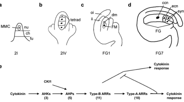
megagametogenesis98-101. Ovules have three structural domains along the proximal-distal
axis: the funiculus, the chalaza, and the nucellus (Figure 2.1a). During megasporogenesis,
a hypodermal archesporial cell enlarges and differentiates into megaspore mother cell
(MMC) that undergoes meiosis and gives rise to a tetrad of four megaspores (Figure
2.1b). In higher plants with Polygonum-type embryo sac development, including
Arabidopsis, rice, and maize, the megaspore closest to the chalaza develops into the
functional megaspore (FM), while the other three degenerate (Figure 2.1c). During the
subsequent megagametogenesis, the FM undergoes three rounds of mitosis, resulting in
an eight nuclei syncytium that partitions into four cell types after cellularization: two
synergid cells that are important for pollen tube guidance; the egg and central cells that
receive sperm cells for double fertilization; and three antipodal cells which degenerate by
39
Figure 2.1. Two-component elements and female gametophyte development. (a-d) Schematic depiction
of wild-type female gametophyte development in Arabidopsis. Abbreviations: MMC, megaspore mother cell; nu, nucellus; ch, chalaza; fu, funiculus; oi, outer integuments, ii, inner integuments; FM, functional megaspore; dm, degenerating megaspores; ccn, central cell nucleus; ecn, egg cell nucleus; syn, synergid cell nuclei. (e) Model of cytokinin response pathway in Arabidopsis. Cytokinin binds to the AHK
receptors, which initiate a phosphorelay through AHPs and ultimately result in the phosphorylation of type-B ARRs. The activated type-type-B ARRs elevate the transcription of the type-A ARRs, which in turn act to negatively regulate the pathway. CKI1, which acts gametophytically to regulate FG development after FG4, also acts through the AHPs. The number of genes in each family in Arabidopsis is shown in parentheses below each element.
Given the proximal connection between the sporophytic tissues and gametophytic
cells, it is anticipated that cellular communication is important during the developmental
processes103, 104. A sporophytic siRNA pathway involving ARGONAUTE9 (AGO9) is
crucial to specify cell fate in the Arabidopsis ovule105. In ago9 plants, more than one
sub-epidermal cell enlarges and contains a conspicuous nucleus in the ovule105. Recent
studies have begun to reveal the interactive role of hormone signaling between two
generations103, 106-108. The cell fate specification during syncytial development depends on
40
cytokinin receptors disrupt female gametogenesis through a sporophytic effect13, 106, yet
the underlying mechanisms remain to be elucidated.
Cytokinin is important in a wide array of developmental processes. Cytokinin
perception in plants is similar to bacterial two-component phosphorelay signal
transduction systems (TCS). In Arabidopsis, there are three cytokinin receptors
(Arabidopsis Histidine Kinases 2, [AHK2], AHK3, and AHK4/CRE1/WOL) that
autophosphorylate in response to the binding of cytokinin18, 26. AHKs then relay this
phosphoryl group to the Arabidopsis histidine phosphotransfer proteins (AHPs), which in
turn transfer the phosphoryl group to the Arabidopsis response regulators (ARRs) (Figure
2.1e)15, 28. CKI1, which encodes a histidine kinase that lacks a cytokinin-binding domain,
can also feed into downstream TCS signaling20, 72 and acts in the female gametophyte to
regulates its development23, 109, 110. The ARRs fall into four classes based on phylogenetic
analysis and domain structure: type-A ARRs, type-B ARRs, type-C ARRs, and the
Arabidopsis pseudoresponse regulators (APRRs). The eleven type-B ARRs are positive
elements in the primary cytokinin signal transduction network. The ten type-A ARRs are
rapidly transcriptionally up-regulated in response to cytokinin via direct activation by the
type-B ARRs and act to negatively regulate cytokinin signaling31, 34, 35, 65, 71, 72.
In Arabidopsis and rice, mutants defective in biosynthesis and perception of
cytokinin displayed reduced female fertility, suggesting that cytokinin has a conserved
role in regulating ovule development13, 18, 106, 111, 112. However, a triple receptor mutant
that showed cytokinin deficiency in multiple bioassays was able to form seeds14, raising
the possibility that the female-sterile phenotype was conditional. Here, we examined the