Associations of Blood Pressure Dipping Patterns With Left Ventricular
Mass and Left Ventricular Hypertrophy in Blacks: The Jackson Heart
Study
Marwah Abdalla, MD, MPH;* Melissa C. Caughey, MPH, PhD;* Rikki M. Tanner, MPH, PhD; John N. Booth III, MS; Keith M. Diaz, PhD; D. Edmund Anstey, MD; Mario Sims, PhD, MS; Joseph Ravenell, MD, MS; Paul Muntner, PhD; Anthony J. Viera, MD, MPH;
Daichi Shimbo, MD
Background-—Abnormal diurnal blood pressure (BP), including nondipping patterns, assessed using ambulatory BP monitoring, have been associated with increased cardiovascular risk among white and Asian adults. We examined the associations of BP dipping patterns (dipping, nondipping, and reverse dipping) with cardiovascular target organ damage (left ventricular mass index and left ventricular hypertrophy), among participants from the Jackson Heart Study, an exclusively black population–based cohort.
Methods and Results-—Analyses included 1015 participants who completed ambulatory BP monitoring and had echocardiography data from the baseline visit. Participants were categorized based on the nighttime to daytime systolic BP ratio into 3 patterns: dipping pattern (≤0.90), nondipping pattern (>0.90 to ≤1.00), and reverse dipping pattern (>1.00). The prevalence of dipping, nondipping, and reverse dipping patterns was 33.6%, 48.2%, and 18.2%, respectively. In a fully adjusted model, which included antihypertensive medication use and clinic and daytime systolic BP, the mean differences in left ventricular mass index between reverse dipping pattern versus dipping pattern was 8.32.1 g/m2 (P<0.001) and between nondipping pattern versus dipping pattern was 1.01.6 g/m2(P=0.536). Compared with participants with a dipping pattern, the prevalence ratio for having left ventricular hypertrophy was 1.65 (95% CI, 1.05–2.58) and 0.96 (95% CI, 0.63–1.97) for those with a reverse dipping pattern and nondipping pattern, respectively.
Conclusions-—In this population-based study of blacks, a reverse dipping pattern was associated with increased left ventricular mass index and a higher prevalence of left ventricular hypertrophy. Identification of a reverse dipping pattern on ambulatory BP monitoring may help identify black at increased risk for cardiovascular target organ damage. (J Am Heart Assoc. 2017;6:e004847. DOI: 10. 1161/JAHA.116.004847.)
Key Words: ambulatory blood pressure monitoring•black•dipping•diurnal variation•left ventricular hypertrophy•left
ventricular mass
I
n healthy adults, blood pressure (BP) is characterized by a circadian pattern during a 24-hour period with levels that are normally highest while awake and fall during sleep.1 Ambulatory BP monitoring (ABPM) can be used to assess thecircadian pattern of BP.1,2Based on the nighttime to daytime systolic BP (SBP) ratio, individuals can be categorized by dipping status into nondipping BP status (SBP ratio>0.90) or dipping BP status (SBP ratio ≤0.90).3 However, in some
From the Department of Medicine, Columbia University Medical Center, New York, NY (M.A., K.M.D., D.E.A., D.S.); Department of Medicine, University of North Carolina at Chapel Hill, NC (M.C.C.); Department of Epidemiology, University of Alabama at Birmingham, AL (R.M.T., J.N.B., P.M.); Department of Medicine, University of Mississippi Medical Center, Jackson, MS (M.S.); Department of Population Health, Center for Healthful Behavior Change, New York University Medical Center, New York, NY (J.R.); Hypertension Research Program and Department of Family Medicine, University of North Carolina at Chapel Hill, NC (A.J.V.).
Accompanying Data S1 and Tables S1 through S10 are available at http://jaha.ahajournals.org/content/6/4/e004847/DC1/embed/inline-supplementary-mate rial-1.pdf
*Dr Abdalla and Dr Caughey are co-first authors.
Correspondence to:Marwah Abdalla, MD, MPH, Center for Behavioral Cardiovascular Health, 622 West 168th Street, PH 9-321, Columbia University Medical
Center, New York, NY 10032. E-mail: [email protected]
Received January 10, 2017; accepted February 17, 2017.
ª2017 The Authors. Published on behalf of the American Heart Association, Inc., by Wiley. This is an open access article under the terms of the Creative Commons Attribution-NonCommercial-NoDerivs License, which permits use and distribution in any medium, provided the original work is properly cited, the use is non-commercial and no modifications or adaptations are made.
individuals there may be an excess rise or fall in nighttime BP relative to daytime BP.4,5Some investigators6have proposed that dipping status should be further categorized into 4 dipping patterns4: extreme dipping pattern (SBP ratio≤0.80), dipping pattern (0.80< SBP ratio≤0.90), nondipping pattern (0.90< SBP ratio ≤1.00), and reverse dipping pattern (SBP ratio>1.00), as these patterns may provide more prognostic information than dipping dichotomized as nondipping BP or dipping BP status.
Prior studies have demonstrated that nondipping and reverse dipping patterns are associated with increased mortality and cardiovascular events when compared with a dipping pattern.5,7–12Also, an association between nondipping BP status and subclinical target organ damage, including left ventricular mass index (LVMI) and left ventricular hypertrophy (LVH), has been previously demonstrated.13–16However, less is known about the association between reverse dipping pattern with LVMI or LVH. The prior studies17,18 that have examined the association between reverse dipping patterns and LVMI have provided conflicting results. These studies were conducted mostly among adults who had untreated hyper-tension. Neither study17,18included blacks, who are known to have a high prevalence of hypertension and hypertension-related cardiovascular target organ damage,19–21 including increased LVMI and a higher prevalence of LVH compared with whites.22 In addition, blacks are also more likely to have nondipping BP and higher nighttime BP compared with whites.23,24
In the current study, we examined the association of BP dipping patterns with LVMI and LVH among participants in the Jackson Heart Study (JHS), a large population-based cohort study comprised exclusively of blacks. We examined these associations for the overall population and for participants taking and not taking antihypertensive medication, separately. In secondary analyses, we also examined the associations of nighttime BP with LVMI and LVH.
Methods
Study Population
The JHS enrolled 5306 noninstitutionalized blacks, 21 years and older, between 2000 and 2004 from the Atherosclerosis Risk in the Community (ARIC) site in Jackson, MS, and a representative sample of urban and rural Jackson, MS, metropolitan tri-county (Hinds, Madison, and Rankin counties) residents, volunteers, randomly selected individuals, and secondary family members of enrolled participants.25 The current analyses were restricted to 1148 participants who underwent 24-hour ABPM following the baseline examination in 2000–2004. Participants who did not meet the Interna-tional Database of Ambulatory Blood Pressure in Relation to
Cardiovascular Outcome26 (IDACO) criteria for complete ABPM (n=102; described below), did not have complete echocardiography data on LVMI (n=27), or were missing clinic BP values at the baseline visit (n=4) were excluded, leaving a final sample size of 1015 participants.
The JHS was approved by the institutional review boards of the University of Mississippi Medical Center, Jackson State University, and Tougaloo College. The institutional review boards at Columbia University and University of Alabama at Birmingham approved the use of JHS data for the current analysis. All participants provided written informed consent.
Data Collection and Clinical Covariates
A detailed description of the data collection, methodology, and specimen collection and processing from examination 1 has been previously described and is available in Data S1.27,28 Data were collected during an in-home interview, clinic examination, and a 24-hour ABPM period. During the in-home interview, trained staff administered questionnaires to collect self-reported information on sociodemographics, selected health behaviors (eg, alcohol consumption, current smoking, and physical activity), and prior diagnosed comorbid condi-tions. During the clinic examination, trained technicians measured height, weight, and BP, and collected blood and urine samples. Antihypertensive medication use was defined by self-report. After the clinic examination, participants were given the opportunity to complete ABPM.
Clinic BP Measurement
Clinic BP measurements were taken using a Hawksley random zero sphygmomanometer (Hawksley and Sons Ltd, Lancing, United Kingdom) and an appropriately sized BP cuff. Cuff size was determined by measuring the upper-arm circumference. At each visit, participants rested for at least 5 minutes in a seated, upright position with their back and arms supported, feet flat on the floor, and legs uncrossed prior to having their BP measured. Trained staff conducted 2 BP measurements in the right arm. One minute elapsed between the 2 measurements. The JHS Coordinating Center conducted quality control by semiannual training and retrain-ing of staff, monitorretrain-ing digit preference for each technician, and comparing mean BP measurements within and between trained technicians. The 2 clinic-measured BP measurements were averaged for analysis. As previously described, BP measurements were calibrated using robust regression to an oscillometric device (Omron HEM-907XL, Omron Healthcare Inc., Lake Forest, IL).29 Prevalent clinic hypertension was defined as a mean clinic SBP ≥140 mm Hg, mean clinic diastolic BP (DBP) ≥90 mm Hg, or antihypertensive medica-tion use.
Blood Pressure Dipping and Left Ventricular Mass Abdalla et al
ORIGI
N
AL
RESE
Ambulatory BP Monitoring
Participants were fitted with an ABPM device (Spacelabs 90207, Spacelabs, Redmond, WA) on their nondominant arm following the baseline examination. Ambulatory BP was recorded every 20 minutes. After 24 hours, the device was removed and data were downloaded onto a computer and processed with Medifacts International’s Medicom software (Rockville, MD). IDACO criteria were used to define whether the ABPM period was complete. Specifically, participants were considered to have a complete ABPM if they had ≥10 daytime (10 AM–8 PM)
and ≥5 nighttime (12 AM–6 AM) SBP and DBP
measure-ments.26Mean daytime SBP and DBP and mean nighttime SBP and DBP were calculated by averaging the readings during the daytime and nighttime periods, respectively. Mean 24-hour BP was defined by averaging all available BP measurements from ABPM.
The nighttime to daytime SBP ratio was defined as mean nighttime SBP divided by mean daytime SBP.6 Dipping was categorized into 3 patterns based on the nighttime to daytime SBP ratio: dipping pattern (≤0.90), nondipping pattern (>0.90 to ≤1.00), and reverse dipping pattern (>1.00). Because of the small sample size (n=38) of participants with an extreme dipping pattern (≤0.80), these participants were included in the dipping pattern category (≤0.90).
Echocardiography
Certified sonographers performed 2-dimensional transtho-racic echocardiography (Sonos-4500, Philips Medical
Sys-tems, Amsterdam, Netherlands) using standardized
protocols.27 Echocardiograms were reviewed for clinical interpretation and analytical measurements by experienced cardiologists on networked image workstations (Vericis; Camtronics Medical Systems, Hartland, WI).27Left ventricular dimensions including left ventricular internal diameter in diastole (mm), interventricular septal thickness in diastole (IVSd, mm), and posterior wall thickness in diastole (PWTd, mm), were assessed according to American Society of Echocardiography (ASE) recommendations.30
Calculation of LVMI and LVH
Left ventricular mass, LVMI, and LVH were derived according to ASE and European Society of Cardiovascular Imaging recom-mendations.30Left ventricular mass was calculated using the ASE formula: 0.89(1.049((IVSD+LVEDD+PWTD)3 (L-VEDD)3))+0.6. LVMI was calculated as left ventricular mass/ body surface area.30 LVH was defined as increased LVMI ≥96 g/m2in women and≥116 g/m2in men).30
Statistical Analyses
Participant characteristics were calculated for the overall analytical sample and stratified into 3 dipping patterns (dipping, nondipping, and reverse dipping). Values were expressed as meanSD or percentages. Using ANOVA, mean differencesstandard error (SE) in LVMI between participants with dipping (referent), nondipping, and reverse dipping patterns were determined. In addition to an unadjusted model, 4 adjusted models were conducted using ANCOVA. Model 1 included adjustment for age, sex, and body mass index (BMI). Model 2 included the variables in model 1 plus diabetes mellitus, education level achieved, alcohol consumption, smoking status, physical activity, reduced estimated glomerular filtration rate (<60 mL/min per 1.73 m2), and antihypertensive medication use. Model 3 included adjustment for the variables in model 2 plus mean clinic SBP. Model 4 included adjustment for the
variables in model 3 plus mean daytime SBP. The
prevalence of LVH between participants with dipping, nondipping, and reverse dipping patterns was also calcu-lated. Prevalence ratios (PRs) and 95% CIs for having LVH were determined using Poisson regression models with sandwich estimators before and after adjustment for covariates as described for models 1 to 4 above, with the dipping pattern serving as the reference group. In secondary analyses, the differences in LVMI and preva-lence of LVH between participants with dipping, nondip-ping, and reverse dipping patterns were calculated by substituting mean daytime SBP with mean 24-hour SBP in model 4. In this model, the association of mean 24-hour SBP with LVMI and LVH was also examined.
Mean differences in LVMI by quartiles of nighttime SBP and quartiles of nighttime DBP, separately, were also calculated using ANCOVA and Poisson regression. For these analyses, the lowest quartile of nighttime SBP and DBP were the referent groups. In analyses for nighttime SBP, mean differences in LVMI were calculated before and after adjustment for covariates in models 1 to 4. In
the analyses for nighttime DBP, model 3 included
adjustment for mean daytime DBP instead of mean daytime SBP, and model 4 included adjustment for mean clinic DBP instead of mean clinic SBP. PRs and 95% CIs for having LVH associated with quartiles of nighttime SBP and DBP were determined using Poisson regression
models with sandwich estimators before and after
adjustment for covariates as described for models 1 to 4 above.
Subgroup analyses were conducted by repeating the
analyses among participants taking and not taking
antihypertensive medication, separately. The tests for interaction between antihypertensive medication use and
Blood Pressure Dipping and Left Ventricular Mass Abdalla et al
ORIGI
N
AL
RESE
dipping BP as well as nighttime SBP and nighttime DBP on LVMI and LVH were calculated in models including the full population, main effect terms, and multiplicative interaction terms (eg, dipping pattern9antihypertensive medication use). P<0.05 was considered to be statistically significant. Analyses were conducted using SAS version 9.4 (SAS Institute, Cary, NC).
Results
Baseline Characteristics
Among the participants included in this analysis, the meanSD age was 59.110.9 years, 67.9% were women, 63.0% had prevalent clinic hypertension, and 56.8% were taking antihypertensive medication (Table 1). The prevalence
Table 1. Characteristics of Jackson Heart Study Participants Included in the Current Analysis, Overall, and Stratified by BP Dipping
Patterns
Overall (N=1015)
Dipping (n=341)
Nondipping (n=489)
Reverse Dipping (n=185)
P
Value*
Sociodemographic characteristics
Age, y 59.110.9 57.710.9 58.911.2 62.49.7 <0.001
Female sex, % 67.9 68.9 65.0 73.5 0.096
Education<high school, % 18.8 15.6 19.1 23.8 0.069
Clinical characteristics
Body mass index, kg/m2 31.16.5 30.06.0 31.56.7 32.16.4 <0.001
Diabetes mellitus, % 24.1 18.6 23.3 36.1 <0.001
Low-density lipoprotein cholesterol, mg/dL 125.836.0 128.536.5 124.833.9 123.640.0 0.251
High-density lipoprotein cholesterol, mg/dL 54.015.1 55.015.6 53.514.7 53.115.2 0.291
Estimated glomerular filtration rate<60 mL/min per 1.73 m2, % 10.9 7.7 10.3 18.1 0.001
Health behaviors
Alcohol use
Nondrinker, % 69.7 63.9 69.9 80.0 <0.001
Moderate drinker, % 28.1 32.6 28.2 19.5 0.006
Heavy drinker, % 2.2 3.5 1.8 0.5 0.064
Current smoking, % 10.0 11.7 8.4 10.8 0.260
Total physical activity score 8.32.6 8.52.5 8.32.6 7.92.6 0.063
High risk for sleep apnea, % 67.2 61.6 67.9 75.7 0.004
BP-related characteristics
Clinic BP
Mean systolic BP, mm Hg 127.715.9 126.315.6 127.815.7 130.316.9 0.028
Mean diastolic BP, mm Hg 74.58.5 74.38.8 74.58.3 74.98.7 0.766
Prevalent clinic hypertension†, % 63.0 58.4 60.3 78.8 <0.001
Antihypertensive medication use, % 56.8 52.1 53.2 74.9 <0.001
Ambulatory BP
Mean daytime systolic BP, mm Hg 129.413.5 130.913.3 128.613.4 128.914.1 0.039
Mean daytime diastolic BP, mm Hg 77.99.3 79.69.3 77.29.1 76.69.6 <0.001
Mean nighttime systolic BP, mm Hg 121.115.8 112.011.9 121.913.3 135.616.4 <0.001
Mean nighttime diastolic BP, mm Hg 68.49.8 63.68.6 69.08.7 75.69.7 <0.001
Mean 24-h systolic BP, mm Hg 126.313.7 123.512.6 126.113.4 132.115.0 <0.001
Mean 24-h diastolic BP, mm Hg 74.18.9 73.28.7 74.08.8 76.29.3 <0.001
Data are presented as meanSD or percentage. Dipping blood pressure (BP) (≤0.90), nondipping BP (>0.90 to≤1.00), and reverse BP dipping (>1.00). Nighttime to daytime systolic BP (SBP) ratio is defined as mean nighttime SBP divided by mean daytime SBP.
*Analysis of variancePvalue comparing differences between participants with dipping, nondipping, and reverse dipping patterns.
†Prevalent clinic hypertension is defined as a mean clinic SBP≥140 mm Hg or mean clinic diastolic BP≥90 mm Hg or self-report of current antihypertensive medication use.
Blood Pressure Dipping and Left Ventricular Mass Abdalla et al
ORIGI
N
AL
RESE
of dipping, nondipping, and reverse dipping patterns was 33.6%, 48.2%, and 18.2%, respectively. Among the 3 dipping patterns, reverse dipping was associated with the oldest age, highest body mass index and clinic SBP, and the highest proportion of women, diabetes mellitus, reduced estimated glomerularfiltration rate, non-alcohol use, high risk for sleep apnea, prevalent clinic hypertension, and antihypertensive medication use.
Associations of BP Dipping Patterns With LVMI
and LVH
In an unadjusted model, compared with participants with a dipping pattern, meanSE LVMI was 9.92.1 g/m2 higher for participants with a reverse dipping pattern (P<0.001) and 1.61.6 g/m2
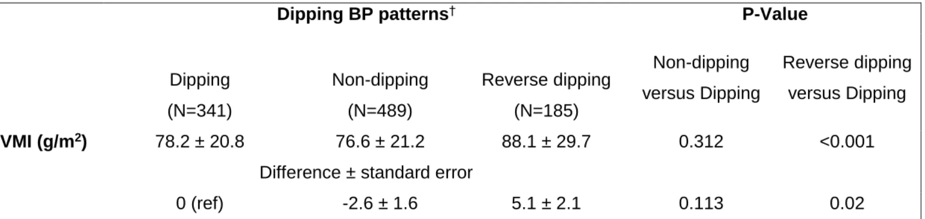
lower for participants with a nondipping pattern (P=0.312, Table 2). In a fully adjusted model including daytime SBP, compared with participants with a dipping pattern, meanSE LVMI was 8.32.1 g/m2 higher for participants with a reverse dipping pattern (P<0.001) and 1.01.6 g/m2 lower for participants with a nondipping pattern (P=0.536). In a fully adjusted model controlling for mean 24-hour SBP instead of mean daytime SBP (Table S1), compared with participants with a dipping pattern, meanSE LVMI was 5.12.1 g/m2 higher for participants with a reverse dipping pattern (P=0.02) and 2.61.6 g/m2 lower for participants with a nondipping pattern (P=0.113). In this model, higher mean 24-hour SBP was associated with higher LVMI (P<0.001, Table S2).
Among participants taking antihypertensive medication, the prevalence of a dipping pattern, nondipping pattern, and reverse dipping pattern was 30.8%, 45.4%, and 23.8%, respec-tively. Among participants not taking antihypertensive medi-cation, the prevalence of a dipping pattern, nondipping pattern,
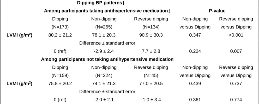
and reverse dipping pattern was 37.1%, 52.3%, and 10.5%, respectively. Among participants taking and not taking antihy-pertensive medication, those with reverse dipping pattern had 11.02.8 and 2.13.4 g/m2higher LVMI, respectively, com-pared with their counterparts with a dipping pattern in a fully adjusted model including mean daytime SBP (interaction P=0.068, Table 3). In a fully adjusted model including mean 24-hour SBP instead of mean daytime SBP, compared with participants with a dipping pattern, those with a reverse dipping pattern had 7.72.8 g/m2 higher and 1.03.4 g/m2 lower LVMI among those taking and not taking antihypertensive medication, respectively (interactionP=0.096, Table S3).
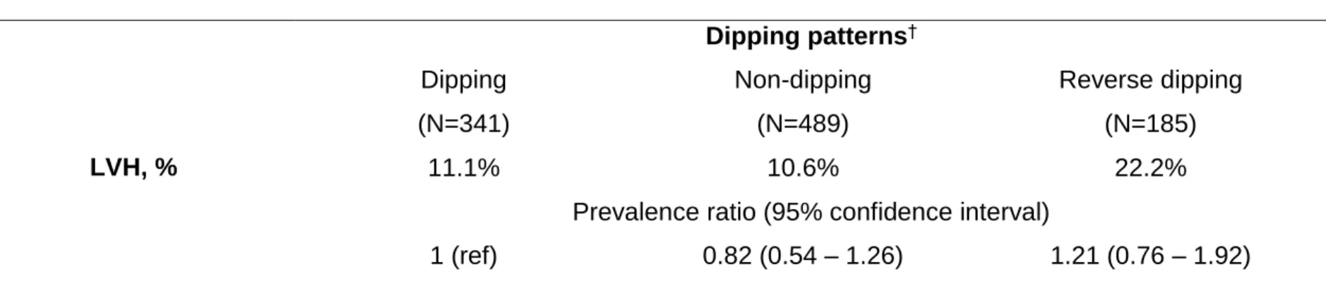
The prevalence of LVH for participants with a dipping pattern, nondipping pattern, and reverse dipping pattern was 11.1%, 10.6%, and 22.2%, respectively (Table 4). The PR for having LVH for reverse dipping pattern versus dipping pattern was 1.99 (95% CI, 1.33–2.98) in an unadjusted model. In a fully adjusted model including mean daytime SBP, the PR for having LVH for reverse dipping pattern versus dipping pattern was 1.65 (95% CI, 1.05–2.58). The PR for having LVH was 1.21 (95% CI, 0.76–1.92) in a fully adjusted model when adjusting for mean 24-hour SBP instead of mean daytime SBP (Table S4). In this latter model, higher mean 24-hour SBP was associated with an increased PR for having LVH (1.32 [95% CI, 1.15–1.51], Table S5).
In a fully adjusted model including mean daytime SBP, the PRs for having LVH among participants with a reverse dipping pattern versus a dipping pattern were 1.78 (95% CI, 1.07– 2.95) and 1.48 (95% CI, 0.52–4.16) among those taking and not taking antihypertensive medication, respectively (interac-tion P=0.904, Table 5). In a fully adjusted model including mean 24-hour SBP instead of mean daytime SBP, the PRs for having LVH among participants with a reverse dipping pattern versus dipping pattern were 1.39 (95% CI, 0.83–2.33) and
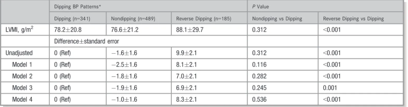
Table 2. Differences in LVMI Between Participants With Nondipping and Reverse Dipping Versus Dipping BP Patterns
Dipping BP Patterns* PValue
Dipping (n=341) Nondipping (n=489) Reverse Dipping (n=185) Nondipping vs Dipping Reverse Dipping vs Dipping
LVMI, g/m2 78.220.8 76.621.2 88.129.7 0.312 <0.001
Differencestandard error
Unadjusted 0 (Ref) 1.61.6 9.92.1 0.312 <0.001
Model 1 0 (Ref) 2.51.6 8.12.1 0.116 <0.001
Model 2 0 (Ref) 1.81.6 7.02.1 0.282 <0.001
Model 3 0 (Ref) 1.91.6 6.92.1 0.245 0.001
Model 4 0 (Ref) 1.01.6 8.32.1 0.536 <0.001
Data are presented as meanSD or unadjusted/adjusted mean difference compared with referent (Ref) groupstandard error. Nighttime to daytime systolic blood pressure (SBP) ratio is defined as mean nighttime SBP/mean daytime SBP. Model 1 includes adjustment for age, sex, and body mass index. Model 2 includes additional adjustment for diabetes mellitus, education level, alcohol consumption, smoking status, physical activity, estimated glomerularfiltration ratio<60 mL/min per 1.73 m2, and antihypertensive medication use. Model 3 includes additional adjustment for mean clinic SBP. Model 4 includes additional adjustment for mean daytime SBP. BP indicates blood pressure; LVMI, left ventricular mass index. *Dipping pattern is based on the nighttime to daytime SBP ratio and defined by 3 patterns: dipping (≤0.90), nondipping (>0.90 to≤1.00), and reverse dipping (>1.00). Blood Pressure Dipping and Left Ventricular Mass Abdalla et al
ORIGI
N
AL
RESE
0.92 (95% CI, 0.33–2.59) among those taking and not taking antihypertensive medication, respectively (interaction P=0.504, Table S6).
The PRs for having LVH among participants with a nondipping pattern versus their counterparts with a dipping pattern were not statistically significant in an unadjusted model (0.95 [95% CI, 0.64–1.42], Table 4) or a fully adjusted model including either mean daytime SBP (0.96 [95% CI, 0.63–1.47], Table 4) or mean 24-hour SBP (0.82 [95% CI, 0.54–1.26], Table S4). In a fully adjusted model including mean daytime SBP, the PRs for having LVH among partic-ipants with a nondipping pattern versus dipping pattern were 0.96 (95% CI, 0.58–1.60) and 1.04 (95% CI, 0.47–2.31) among those taking and not taking antihypertensive medica-tion, respectively (interactionP=0.777, Table 5).
Association of Nighttime BP With LVMI and LVH
Higher quartiles of nighttime SBP and DBP were each associated with higher LVMI in unadjusted and fully adjusted
models (Table 6). In an unadjusted model, the PRs for having LVH associated with quartiles 2, 3, and 4 versus quartile 1 of nighttime SBP were 1.21 (95% CI, 0.65–2.23), 2.13 (95% CI, 1.23–3.69), and 3.40 (95% CI, 2.04–5.69), respectively (Table 7). The unadjusted PRs for LVH associated with quartiles 2, 3, and 4 versus quartile 1 of nighttime DBP were 0.80 (95% CI, 0.48–1.34), 0.96 (95% CI, 0.59–1.57), and 1.79 (95% CI, 1.17–2.72), respectively. The associations of higher quartiles of nighttime SBP and DBP with LVH were not statistically significant in fully adjusted models.
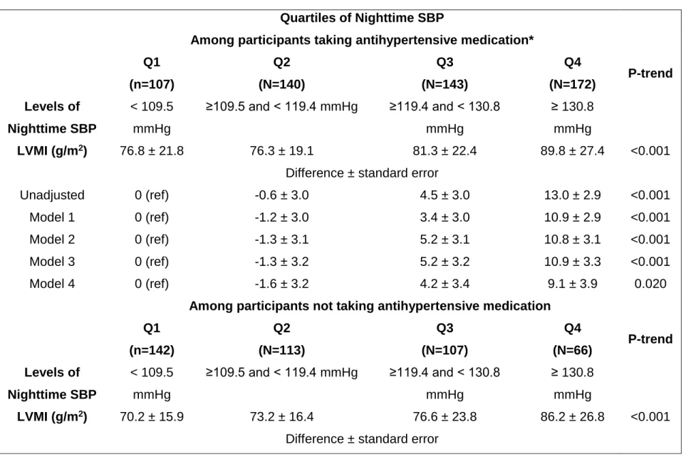
The association between higher quartiles of nighttime SBP and LVMI did not differ between participants taking versus not taking antihypertensive medication (interaction P=0.853, Table S7). In contrast, the association between higher quartiles of nighttime DBP and LVMI was stronger among participants taking versus not taking antihypertensive medication (interaction P<0.001, Table S8). Higher quartiles of nighttime SBP (Table S9) and nighttime DBP (Table S10) were not associated with a statistically significant increased prevalence of LVH among participants taking and not taking antihypertensive medication.
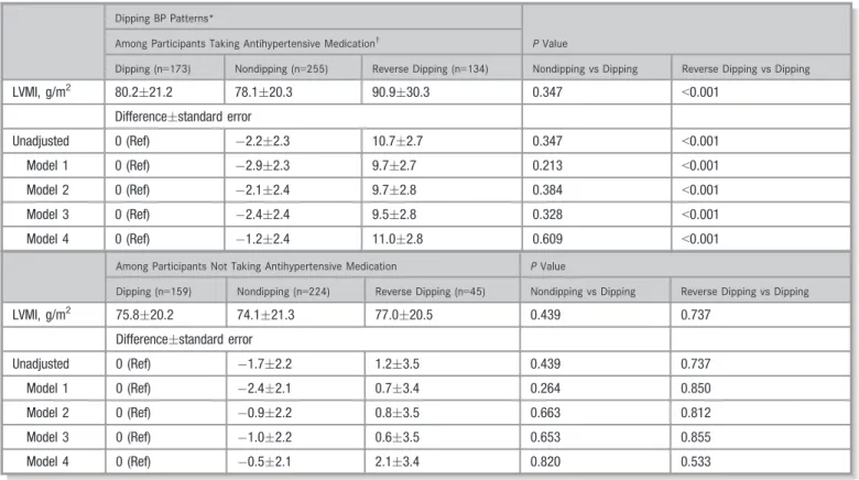
Table 3. Differences in LVMI Among Participants With Dipping, Nondipping, and Reverse Dipping BP Patterns Stratified by
Antihypertensive Medication Use
Dipping BP Patterns*
PValue Among Participants Taking Antihypertensive Medication†
Dipping (n=173) Nondipping (n=255) Reverse Dipping (n=134) Nondipping vs Dipping Reverse Dipping vs Dipping
LVMI, g/m2 80.221.2 78.120.3 90.930.3 0.347 <0.001
Differencestandard error
Unadjusted 0 (Ref) 2.22.3 10.72.7 0.347 <0.001
Model 1 0 (Ref) 2.92.3 9.72.7 0.213 <0.001
Model 2 0 (Ref) 2.12.4 9.72.8 0.384 <0.001
Model 3 0 (Ref) 2.42.4 9.52.8 0.328 <0.001
Model 4 0 (Ref) 1.22.4 11.02.8 0.609 <0.001
Among Participants Not Taking Antihypertensive Medication PValue
Dipping (n=159) Nondipping (n=224) Reverse Dipping (n=45) Nondipping vs Dipping Reverse Dipping vs Dipping
LVMI, g/m2 75.820.2 74.121.3 77.020.5 0.439 0.737
Differencestandard error
Unadjusted 0 (Ref) 1.72.2 1.23.5 0.439 0.737
Model 1 0 (Ref) 2.42.1 0.73.4 0.264 0.850
Model 2 0 (Ref) 0.92.2 0.83.5 0.663 0.812
Model 3 0 (Ref) 1.02.2 0.63.5 0.653 0.855
Model 4 0 (Ref) 0.52.1 2.13.4 0.820 0.533
Data are presented as meanSD or unadjusted/adjusted mean difference compared with referent (Ref) groupstandard error. Model 1 includes adjustment for age, sex, and body mass index. Model 2 includes additional adjustment for diabetes mellitus, education level, alcohol consumption, smoking status, physical activity, and estimated glomerularfiltration ratio
<60 mL/min per 1.73 m2. Model 3 includes additional adjustment for mean clinic systolic blood pressure (SBP). Model 4 includes additional adjustment for mean daytime SBP. BP indicates blood pressure; LVMI, left ventricular mass index.
*Dipping pattern is based on the nighttime to daytime SBP ratio and defined by 3 patterns: dipping (≤0.90), nondipping (>0.90 to≤1.00), and reverse dipping (>1.00). Nighttime to daytime SBP ratio is defined as mean nighttime SBP/mean daytime SBP.
†The overall test for interaction between antihypertensive medication use and dipping patterns on left ventricular mass index (LVMI) isP=0.086. The test for interaction between
antihypertensive medication use and nondipping pattern isP=0.707. The test for interaction between antihypertensive medication use and reverse dipping pattern isP=0.068. Blood Pressure Dipping and Left Ventricular Mass Abdalla et al
ORIGI
N
AL
RESE
Discussion
In the current population-based study of blacks, participants with a reverse dipping BP pattern had higher LVMI and a
higher prevalence of LVH when compared with their counter-parts with a dipping BP pattern. These associations were present after multivariable adjustment for demographics, cardiovascular risk factors, antihypertensive medication use,
Table 4. Prevalence and Prevalence Ratios for LVH Associated With Dipping Patterns
Dipping Patterns*
Dipping (n=341) Nondipping (n=489) Reverse Dipping (n=185)
LVH, % 11.1 10.6 22.2
Prevalence ratio (95% CI)
Unadjusted 1 (Ref) 0.95 (0.64–1.42) 1.99 (1.33–2.98)
Model 1 1 (Ref) 0.91 (0.62–1.34) 1.66 (1.11–2.49)
Model 2 1 (Ref) 0.89 (0.58–1.37) 1.48 (0.95–2.32)
Model 3 1 (Ref) 0.88 (0.57–1.35) 1.47 (0.94–2.29)
Model 4 1 (Ref) 0.96 (0.63–1.47) 1.65 (1.05–2.58)
Left ventricular hypertrophy (LVH) is defined as left ventricular mass index≥96 g/m2in women and≥116 g/m2in men according to the American Society of Echocardiography recommendations. Nighttime to daytime systolic blood pressure (SBP) ratio is defined as mean nighttime SBP/mean daytime SBP. Model 1 includes adjustment for age, sex, and body mass index. Model 2 includes additional adjustment for diabetes mellitus, education level, alcohol consumption, smoking status, physical activity, estimated glomerularfiltration ratio
<60 mL/min per 1.73 m2, and antihypertensive medication use. Model 3 includes additional adjustment for mean clinic SBP. Model 4 includes additional adjustment for mean daytime SBP. Ref indicates referent.
*Dipping pattern is based on the nighttime to daytime SBP ratio and defined by three 3: dipping (≤0.90), nondipping (>0.90 to≤1.00), and reverse dipping (>1.00).
Table 5. Prevalence Ratios for Having LVH Associated With Dipping Patterns Stratified by Antihypertensive Medication Use
Dipping Patterns*
Among Participants Taking Antihypertensive Medication†
Dipping (n=173) Nondipping (n=255) Reverse Dipping (n=134)
LVH, % 14.5 12.6 24.6
Prevalence ratio (95% CI)
Unadjusted 1 (Ref) 0.87 (0.53–1.41) 1.70 (1.07–2.72)
Model 1 1 (Ref) 0.87 (0.53–1.42) 1.64 (1.02–2.65)
Model 2 1 (Ref) 0.89 (0.53–1.50) 1.63 (0.98–2.70)
Model 3 1 (Ref) 0.88 (0.53–1.48) 1.61 (0.97–2.67)
Model 4 1 (Ref) 0.96 (0.58–1.60) 1.78 (1.07–2.95)
Among Participants Not Taking Antihypertensive Medication
Dipping (Ref) (n=159) Nondipping (n=224) Reverse Dipping (n=45)
LVH, % 7.6 6.7 11.1
Prevalence ratio (95% CI)
Unadjusted 1 (Ref) 0.89 (0.43–1.84) 1.47 (0.55–3.96)
Model 1 1 (Ref) 0.85 (0.42–1.72) 1.16 (0.43–3.15)
Model 2 1 (Ref) 0.98 (0.45–2.12) 1.25 (0.41–3.78)
Model 3 1 (Ref) 0.96 (0.44–2.10) 1.21 (0.40–3.61)
Model 4 1 (Ref) 1.04 (0.47–2.31) 1.48 (0.52–4.16)
Left ventricular hypertrophy (LVH) is defined as left ventricular mass index≥96 g/m2in women and
≥116 g/m2in men according to the American Society of Echocardiography recommendations. Model 1 includes adjustment for age, sex, and body mass index. Model 2 includes additional adjustment for diabetes mellitus, education level, alcohol consumption, smoking status, physical activity, estimated glomerularfiltration ratio<60 mL/min per 1.73 m2. Model 3 includes additional adjustment for mean clinic systolic blood pressure (SBP). Model 4 includes additional adjustment for mean daytime SBP. Ref indicates referent.
*Dipping pattern is based on the nighttime to daytime SBP ratio and defined by 3 patterns: dipping (≤0.90), nondipping (>0.90 to≤1.00), and reverse dipping (>1.00). Nighttime to daytime SBP ratio is defined as mean nighttime SBP/mean daytime SBP.
†The overall test for interaction between antihypertensive medication use and dipping patterns on LVH isP=0.919. The test for interaction between antihypertensive medication use and
nondipping pattern isP=0.777. The test for interaction between antihypertensive medication use and reverse dipping pattern isP=0.904. Blood Pressure Dipping and Left Ventricular Mass Abdalla et al
ORIGI
N
AL
RESE
clinic BP, and daytime BP. In contrast, participants with a nondipping versus a dipping pattern did not have higher LVMI or a higher prevalence of LVH. Further, higher nighttime BP was associated with higher LVMI but not the prevalence of LVH.
Although a reverse dipping pattern is associated with increased mortality and cardiovascular events when com-pared with a dipping pattern,5,8–12 it is unclear whether a reverse dipping pattern also confers an increased risk for subclinical cardiovascular target organ damage. Among 682 participants with hypertension, the majority of whom were not taking antihypertensive medications in the Korean Ambulatory Blood Pressure Study, there were no differences in LVMI between participants with a reverse dipping or nondipping pattern each versus a dipping pattern.18In contrast, in a study of 376 Serbian adults with untreated hypertension, reverse dipping pattern and nondipping BP patterns were each associated with higher LVMI when compared with a dipping BP pattern.17Additionally, 31% of participants with a reverse dipping pattern and 17% of participants with a nondipping pattern had LVH compared with only 9% of participants with a
dipping pattern. These studies17,18 were not population-based, included mostly individuals who had untreated hyper-tension, and did not include blacks. Several studies31–36have examined BP dipping in blacks or Caribbean blacks. These studies were typically small,31,34 were not population-based,31–34,36 did not examine reverse dipping31–34 or LVMI,31–33and did not consider antihypertensive medication use.31–35The results of our study are novel and extend the findings of these prior studies by demonstrating in a large population-based cohort of blacks that a reverse dipping pattern is associated with higher LVMI and LVH, independent of antihypertensive medication use.
Higher quartiles of nighttime SBP and nighttime DBP were also associated with higher LVMI, highlighting the important contribution of nighttime BP in cardiovascular disease risk. Nighttime BP may be a stronger predictor of cardiovascular events than daytime BP among individuals with treated hypertension37,38 and among population-based cohorts.39 In a population-based study of 1682 Italian participants, nighttime SBP was associated with higher left ventricular mass.40Additionally, Yi et al18demonstrated that the highest
Table 6. Differences in LVMI Associated With Quartiles (Qs) of Nighttime SBP (Upper Panel) and Nighttime DBP (Lower Panel)
Quartiles of Nighttime SBP
PTrend Q1 (n=253) Q2 (n=259) Q3 (n=258) Q4 (n=245)
Levels of nighttime SBP <109.5 mm Hg ≥109.5 and<119.4 mm Hg ≥119.4 and<130.8 mm Hg ≥130.8 mm Hg
LVMI, g/m2 73.018.8 74.717.9 80.023.5 89.728.0 <0.001
Differencestandard error
Unadjusted 0 (Ref) 1.72.0 7.02.0 16.72.0 <0.001
Model 1 0 (Ref) 0.42.0 4.92.0 13.02.1 <0.001
Model 2 0 (Ref) 0.32.0 4.42.0 10.42.2 <0.001
Model 3 0 (Ref) 0.42.0 4.32.1 10.22.3 <0.001
Model 4 0 (Ref) 1.42.1 2.02.3 6.32.9 0.026
Quartiles of Nighttime DBP
PTrend Q1 (n=255) Q2 (n=253) Q3 (n=256) Q4 (n=251)
Levels of nighttime DBP <61.7 mm Hg ≥61.7 and<67.5 mm Hg ≥67.5 and<74.4 mm Hg ≥74.4 mm Hg
LVMI, g/m2 76.422.9 74.919.0 78.720.0 87.028.2 <0.001
Differencestandard error
Unadjusted 0 (Ref) 1.52.0 2.32.0 10.62.0 <0.001
Model 1 0 (Ref) 1.22.0 2.32.0 9.62.1 <0.001
Model 2 0 (Ref) 2.12.0 1.32.1 7.02.1 <0.001
Model 3 0 (Ref) 1.82.1 1.82.1 7.82.2 <0.001
Model 4 0 (Ref) 2.42.1 0.42.3 5.32.7 0.013
Data are presented as meanSD or unadjusted/adjusted mean difference compared with referent (Ref) groupstandard error. Upper panel: Model 1 includes adjustment for age, sex, and body mass index. Model 2 includes additional adjustment for diabetes mellitus, education level, alcohol consumption, smoking status, physical activity, estimated glomerularfiltration ratio
<60 mL/min per 1.73 m2, and antihypertensive mediation use. Model 3 includes model 2 plus additional adjustment for mean clinic systolic blood pressure (SBP). Model 4 includes bodel 3 plus additional adjustment for mean daytime SBP. Lower panel: Model 1 includes adjustment for age, sex, and body mass index. Model 2 includes additional adjustment for diabetes mellitus, education level, alcohol consumption, smoking status, physical activity, estimated glomerularfiltration ratio<60 mL/min per 1.73 m2, and antihypertensive mediation use. Model 3 includes model 3 plus additional adjustment for mean clinic diastolic blood pressure (DBP). Model 4 includes model 2 plus additional adjustment for mean daytime DBP. Blood Pressure Dipping and Left Ventricular Mass Abdalla et al
ORIGI
N
AL
RESE
versus lowest quartile of nighttime SBP was associated with higher LVMI among individuals with untreated and treated hypertension. The results from the current study extend both these findings by demonstrating that higher nighttime BP was associated with increased LVMI, independent of clinic BP and daytime BP among a population-based sample of blacks.
In the current study, the associations of reverse dipping pattern with LVMI and LVH were smaller when adjusting for mean 24-hour SBP instead of mean daytime SBP. These results indicate that the associations of reverse dipping pattern with cardiovascular target organ damage may be explained by higher mean 24-hour SBP levels, and that reverse dipping may represent an ABPM phenotype for which daytime BP but not nighttime BP is controlled. Therefore, among blacks, BP control over the daytime and nighttime periods may be associated with a larger reduction in cardiovascular disease risk compared with BP control only during the daytime period. Some studies41–43have previously
demonstrated that nighttime dosing of antihypertensive medications may improve nighttime and 24-hour BP control and restores dipping BP among adults with nondipping BP. Given the results of the current study, future studies should examine whether nighttime dosing of antihypertensive med-ications lowers the risk of cardiovascular target organ damage or cardiovascular disease events among blacks with a reverse dipping pattern.
In the current study, 18.2% of participants had a reverse dipping pattern; 23.8% and 10.5% for those taking and not taking antihypertensive medication, respectively. The preva-lence of a reverse dipping pattern among participants taking antihypertensive medication was high, which is consistent with the findings of prior studies for which the prevalence ranged from 15.6% to 26.7% among individuals with treated hypertension.9,18,37,44–46Although the timing of antihyperten-sive medication was not recorded in the JHS, it is likely that most participants took their medications during the daytime period. Therefore, the higher prevalence of reverse dipping
Table 7. Prevalence and Prevalence Ratios for LVH Associated With Quartiles (Qs) of Nighttime SBP (Upper Panel) and Nighttime
DBP (Lower Panel)
Quartiles of Nighttime SBP
Q1 (n=253) Q2 (n=259) Q3 (n=258) Q4 (n=245)
Levels of nighttime SBP <109.5 mm Hg ≥109.5 and<119.4 mm Hg ≥119.4 and<130.8 mm Hg ≥130.8 mm Hg
LVH, % 6.7 8.1 14.3 22.9
Prevalence ratio (95% CI)
Unadjusted 1 (Ref) 1.21 (0.65–2.23) 2.13 (1.23–3.69) 3.40 (2.04–5.69)
Model 1 1 (Ref) 1.12 (0.61–2.06) 1.87 (1.07–3.28) 2.76 (1.59–4.80)
Model 2 1 (Ref) 1.03 (0.52–2.04) 1.85 (1.03–3.34) 2.40 (1.31–4.39)
Model 3 1 (Ref) 1.03 (0.52–2.05) 1.85 (1.29–4.47) 2.40 (1.29–4.47)
Model 4 1 (Ref) 0.93 (0.46–1.86) 1.43 (0.75–2.75) 1.51 (0.69–3.29)
Quartiles of Nighttime DBP
Q1 (n=255) Q2 (n=253) Q3 (n=256) Q4 (n=251)
Levels of nighttime DBP <61.7 mm Hg ≥61.7 and<67.5 mm Hg ≥67.5 and<74.4 mm Hg ≥74.4 mm Hg
LVH, % 11.4 9.1 10.9 20.3
Prevalence ratio (95% CI)
Unadjusted 1 (Ref) 0.80 (0.48–1.34) 0.96 (0.59–1.57) 1.79 (1.17–2.72)
Model 1 1 (Ref) 0.89 (0.53–1.48) 1.21 (0.74–1.97) 2.24 (1.45–3.46)
Model 2 1 (Ref) 0.77 (0.44–1.35) 1.04 (0.61–1.79) 1.86 (1.16–2.99)
Model 3 1 (Ref) 0.80 (0.45–1.41) 1.11 (0.64–1.92) 2.02 (1.24–3.29)
Model 4 1 (Ref) 0.74 (0.42–1.31) 0.94 (0.53–1.67) 1.49 (0.79–2.78)
Left ventricular hypertrophy (LVH) is defined as left ventricular mass index≥96 g/m2in women and≥116 g/m2in men according to the American Society of Echocardiography recommendations. Upper panel: Model 1 includes adjustment for age, sex, and body mass index. Model 2 includes additional adjustment for diabetes mellitus, education level, alcohol consumption, smoking status, physical activity, estimated glomerularfiltration ratio<60 mL/min per 1.73 m2, and antihypertensive mediation use. Model 3 includes model 2 plus additional adjustment for mean clinic SBP. Model 4 includes model 3 plus additional adjustment for mean daytime systolic blood pressure (SBP). Lower panel: Model 1 includes adjustment for age, sex, and body mass index. Model 2 includes additional adjustment for diabetes mellitus, education level, alcohol consumption, smoking status, physical activity, estimated glomerularfiltration ratio<60 mL/min per 1.73 m2, and antihypertensive mediation use. Model 3 includes model 3 plus additional adjustment for mean clinic diastolic blood pressure (DBP). Model 4 includes model 2 plus additional adjustment for mean daytime DBP. Ref indicates referent.
Blood Pressure Dipping and Left Ventricular Mass Abdalla et al
ORIGI
N
AL
RESE
pattern among those taking versus not taking antihyperten-sive medication may be partially explained by the daytime dosing of antihypertensive medication, leading to a reduction in daytime relative to nighttime BP. In the current study, the prevalence of having a high risk of sleep apnea was highest among participants with a reverse dipping pattern. Therefore, another explanation for the high prevalence of reverse dipping pattern is sleep apnea, which is common among individuals with treated hypertension and is associated with higher nighttime BP and a high prevalence of nondipping BP pattern.47–50
Finally, in the current study, the associations of reverse dipping with LVMI and LVH appeared to be evident only among participants taking antihypertensive medication. Although this finding suggests that reverse dipping may be more benign among those not taking antihypertensive medication, there was no evidence of an interaction between antihypertensive medication and dipping patterns on LVMI and LVH, indicating that the associations of reverse dipping with LVMI and LVH were not modified by antihypertensive medication use.
Study Strengths and Limitations
There are several strengths of the current study. We used data from a large and well-characterized population-based cohort of blacks. The JHS is one of the largest studies of ABPM conducted among blacks. Further, high-quality echocardiog-raphy was conducted among JHS participants using standard-ized procedures. There are also several possible limitations. Participants in JHS underwent ABPM during only one 24-hour period, and the short-term reproducibility of dipping patterns may be limited.51The study had only a few participants (n=38) with an extreme dipping pattern. Sleep diaries and informa-tion regarding reasons for nighttime awakening, which may impact nighttime BP, were not collected. Lastly, the current analysis was cross-sectional, and we cannot determine the direction of the association between dipping patterns and LVMI and LVH.
Conclusions
Approximately 1 in 5 blacks in the current study had a reverse dipping pattern. Participants with a reverse dipping pattern had higher LVMI and a higher prevalence of LVH compared with participants with a dipping pattern, Further, higher nighttime SBP and DBP values were associated with higher levels of LVMI. The data from the current study suggest that the identification of a reverse dipping pattern on ABPM may identify blacks at increased risk for cardiovascular target organ damage.
Acknowledgments
The authors would like to thank the Jackson Heart Study participants, investigators, and staff for their valuable contributions and long-term commitment to the study.
Sources of Funding
The Jackson Heart Study is supported and conducted in collaboration with Jackson State University (N01-HC-95170), University of Mississippi Medical Center (N01-HC-95171), and Touglaoo College (N01-HC-95172) and contracts HHSN
268201300046C, HHSN268201300047C, HHSN268201
300048C, HHSN268201300049C, and HHSN2682013000 50C from the National Heart, Lung, and Blood Institute (NHLBI) and the National Institute on Minority Health and Health Disparities (NIMHD) at the National Institutes of Health (NIH). This work was also supported by the NIH (HL047540,
HL117323, HL117323-02S2, K24-HL125704, 2T32HL00
7854-21) from the NHLBI, Bethesda, MD, and the American Heart Association (15SFRN2390002). The views expressed in this manuscript are those of the authors and do not necessarily represent the views of the NHLBI, the NIH, the U.S. Department of Health and Human Services, or the American Heart Association.
Disclosures
Dr Muntner received an institutional grant from Amgen Inc. Dr Shimbo is a consultant for Abbott Vascular and Novartis Pharmaceuticals Corporation. The remaining authors have no disclosures to report.
References
1. Pickering TG, Shimbo D, Haas D. Ambulatory blood-pressure monitoring.N Engl J Med. 2006;354:2368–2374.
2. Shimbo D, Abdalla M, Falzon L, Townsend RR, Muntner P. Role of ambulatory and home blood pressure monitoring in clinical practice: a narrative review. Ann Intern Med. 2015;163:691–700.
3. O’Brien E, Sheridan J, O’Malley K. Dippers and non-dippers. Lancet. 1988;2:397.
4. Kario K, Pickering TG, Matsuo T, Hoshide S, Schwartz JE, Shimada K. Stroke prognosis and abnormal nocturnal blood pressure falls in older hypertensives. Hypertension. 2001;38:852–857.
5. Fagard RH. Dipping pattern of nocturnal blood pressure in patients with hypertension.Expert Rev Cardiovasc Ther. 2009;7:599–605.
6. O’Brien E, Parati G, Stergiou G, Asmar R, Beilin L, Bilo G, Clement D, de la Sierra A, de Leeuw P, Dolan E, Fagard R, Graves J, Head GA, Imai Y, Kario K, Lurbe E, Mallion JM, Mancia G, Mengden T, Myers M, Ogedegbe G, Ohkubo T, Omboni S, Palatini P, Redon J, Ruilope LM, Shennan A, Staessen JA, vanMontfrans G, Verdecchia P, Waeber B, Wang J, Zanchetti A, Zhang Y; European Society of Hypertension Working Group on Blood Pressure Monitoring. European Society of Hypertension position paper on ambulatory blood pressure monitoring.J Hypertens. 2013;31:1731–1768.
7. Ben-Dov IZ, Kark JD, Ben-Ishay D, Mekler J, Ben-Arie L, Bursztyn M. Predictors of all-cause mortality in clinical ambulatory monitoring: unique aspects of blood pressure during sleep.Hypertension. 2007;49:1235–1241.
8. Fagard RH, Thijs L, Staessen JA, Clement DL, De Buyzere ML, De Bacquer DA. Night-day blood pressure ratio and dipping pattern as predictors of death and cardiovascular events in hypertension.J Hum Hypertens. 2009;23:645–653.
Blood Pressure Dipping and Left Ventricular Mass Abdalla et al
ORIGI
N
AL
RESE
9. Kim BK, Kim YM, Lee Y, Lim YH, Shin J. A reverse dipping pattern predicts cardiovascular mortality in a clinical cohort.J Korean Med Sci. 2013;28:1468– 1473.
10. Salles GF, Reboldi G, Fagard RH, Cardoso CR, Pierdomenico SD, Verdecchia P, Eguchi K, Kario K, Hoshide S, Polonia J, de la Sierra A, Hermida RC, Dolan E, O’Brien E, Roush GC. Prognostic effect of the nocturnal blood pressure fall in hypertensive patients: the Ambulatory Blood Pressure Collaboration in Patients With Hypertension (ABC-H) meta-analysis. Hypertension. 2016;67:693–700.
11. Kario K, Shimada K. Risers and extreme-dippers of nocturnal blood pressure in hypertension: antihypertensive strategy for nocturnal blood pressure.Clin Exp Hypertens. 2004;26:177–189.
12. Ohkubo T, Imai Y, Tsuji I, Nagai K, Watanabe N, Minami N, Kato J, Kikuchi N, Nishiyama A, Aihara A, Sekino M, Satoh H, Hisamichi S. Relation between nocturnal decline in blood pressure and mortality. The Ohasama Study.Am J Hypertens. 1997;10:1201–1207.
13. Pickering TG. The clinical significance of diurnal blood pressure variations. Dippers and nondippers.Circulation. 1990;81:700–702.
14. Verdecchia P, Porcellati C, Schillaci G, Borgioni C, Ciucci A, Battistelli M, Guerrieri M, Gatteschi C, Zampi I, Santucci A, Santucci C, Reboldi G. Ambulatory blood pressure. An independent predictor of prognosis in essential hypertension.Hypertension. 1994;24:793–801.
15. Verdecchia P, Schillaci G, Gatteschi C, Zampi I, Battistelli M, Bartoccini C, Porcellati C. Blunted nocturnal fall in blood pressure in hypertensive women with future cardiovascular morbid events.Circulation. 1993;88:986–992.
16. Verdecchia P, Schillaci G, Guerrieri M, Gatteschi C, Benemio G, Boldrini F, Porcellati C. Circadian blood pressure changes and left ventricular hypertrophy in essential hypertension.Circulation. 1990;81:528–536.
17. Ivanovic BA, Tadic MV, Celic VP. To dip or not to dip? The unique relationship between different blood pressure patterns and cardiac function and structure. J Hum Hypertens. 2013;27:62–70.
18. Yi JE, Shin J, Ihm SH, Kim JH, Park S, Kim KI, Kim WS, Pyun WB, Kim YM, Kim SK. Not nondipping but nocturnal blood pressure predicts left ventricular hypertrophy in the essential hypertensive patients: the Korean Ambulatory Blood Pressure multicenter observational study.J Hypertens. 2014;32:1999– 2004.
19. Cooper RS, Wolf-Maier K, Luke A, Adeyemo A, Banegas JR, Forrester T, Giampaoli S, Joffres M, Kastarinen M, Primatesta P, Stegmayr B, Thamm M. An international comparative study of blood pressure in populations of European vs. African descent.BMC Med. 2005;3:2.
20. Flack JM, Sica DA, Bakris G, Brown AL, Ferdinand KC, Grimm RH Jr, Hall WD, Jones WE, Kountz DS, Lea JP, Nasser S, Nesbitt SD, Saunders E, Scisney-Matlock M, Jamerson KA; International Society on Hypertension in Blacks. Management of high blood pressure in Blacks: an update of the International Society on Hypertension in Blacks consensus statement. Hypertension. 2010;56:780–800.
21. Kurian AK, Cardarelli KM. Racial and ethnic differences in cardiovascular disease risk factors: a systematic review.Ethn Dis. 2007;17:143–152.
22. Kizer JR, Arnett DK, Bella JN, Paranicas M, Rao DC, Province MA, Oberman A, Kitzman DW, Hopkins PN, Liu JE, Devereux RB. Differences in left ventricular structure between black and white hypertensive adults: the Hypertension Genetic Epidemiology Network study. Hypertension. 2004;43:1182–1188.
23. Profant J, Dimsdale JE. Race and diurnal blood pressure patterns. A review and meta-analysis.Hypertension. 1999;33:1099–1104.
24. Muntner P, Lewis CE, Diaz KM, Carson AP, Kim Y, Calhoun D, Yano Y, Viera AJ, Shimbo D. Racial differences in abnormal ambulatory blood pressure monitoring measures: results from the Coronary Artery Risk Development in Young Adults (CARDIA) study.Am J Hypertens. 2015;28:640–648.
25. Wilson JG, Rotimi CN, Ekunwe L, Royal CD, Crump ME, Wyatt SB, Steffes MW, Adeyemo A, Zhou J, Taylor HA Jr, Jaquish C. Study design for genetic analysis in the Jackson Heart Study.Ethn Dis. 2005;15:S6-30–S6-37.
26. Thijs L, Hansen TW, Kikuya M, Bjorklund-Bodegard K, Li Y, Dolan E, Tikhonoff V, Seidlerova J, Kuznetsova T, Stolarz K, Bianchi M, Richart T, Casiglia E, Malyutina S, Filipovsky J, Kawecka-Jaszcz K, Nikitin Y, Ohkubo T, Sandoya E, Wang J, Torp-Pedersen C, Lind L, Ibsen H, Imai Y, Staessen JA, O’Brien E; IDACO Investigators. The International Database of Ambulatory Blood Pressure in relation to Cardiovascular Outcome (IDACO): protocol and research perspectives.Blood Press Monit. 2007;12:255–262.
27. Carpenter MA, Crow R, Steffes M, Rock W, Heilbraun J, Evans G, Skelton T, Jensen R, Sarpong D. Laboratory, reading center, and coordinating center data management methods in the Jackson Heart Study. Am J Med Sci. 2004;328:131–144.
28. Taylor HA Jr, Wilson JG, Jones DW, Sarpong DF, Srinivasan A, Garrison RJ, Nelson C, Wyatt SB. Toward resolution of cardiovascular health disparities in
African Americans: design and methods of the Jackson Heart Study.Ethn Dis. 2005;15:S6-4–S6-17.
29. Abdalla M, Booth JN III, Seals SR, Spruill TM, Viera AJ, Diaz KM, Sims M, Muntner P, Shimbo D. Masked hypertension and incident clinic hyperten-sion among blacks in the Jackson Heart Study.Hypertension. 2016;68:220– 226.
30. Lang RM, Badano LP, Mor-Avi V, Afilalo J, Armstrong A, Ernande L, Flachskampf FA, Foster E, Goldstein SA, Kuznetsova T, Lancellotti P, Muraru D, Picard MH, Rietzschel ER, Rudski L, Spencer KT, Tsang W, Voigt JU. Recommendations for cardiac chamber quantification by echocardiography in adults: an update from the American Society of Echocardiography and the European Association of Cardiovascular Imaging.J Am Soc Echocardiogr. 2015;28:1–39.e14.
31. Fumo MT, Teeger S, Lang RM, Bednarz J, Sareli P, Murphy MB. Diurnal blood pressure variation and cardiac mass in American blacks and whites and South African blacks.Am J Hypertens. 1992;5:111–116.
32. McMullan CJ, Yano Y, Bakris GL, Kario K, Phillips RA, Forman JP. Racial impact of diurnal variations in blood pressure on cardiovascular events in chronic kidney disease.J Am Soc Hypertens. 2015;9:299–306.
33. Mellman TA, Brown TS, Kobayashi I, Abu-Bader SH, Lavela J, Altaee D, McLaughlin L, Randall OS. Blood pressure dipping and urban stressors in young adult African Americans.Ann Behav Med. 2015;49:622–627.
34. Mezue K, Isiguzo G, Madu C, Nwuruku G, Rangaswami J, Baugh D, Madu E. Nocturnal non-dipping blood pressure profile in black normotensives is associated with cardiac target organ damage.Ethn Dis. 2016;26:279–284.
35. Ogedegbe G, Spruill TM, Sarpong DF, Agyemang C, Chaplin W, Pastva A, Martins D, Ravenell J, Pickering TG. Correlates of isolated nocturnal hypertension and target organ damage in a population-based cohort of African Americans: the Jackson Heart Study.Am J Hypertens. 2013;26:1011– 1016.
36. Pogue V, Rahman M, Lipkowitz M, Toto R, Miller E, Faulkner M, Rostand S, Hiremath L, Sika M, Kendrick C, Hu B, Greene T, Appel L, Phillips RA; African American Study of Kidney Disease and Hypertension Collaborative Research Group. Disparate estimates of hypertension control from ambulatory and clinic blood pressure measurements in hypertensive kidney disease.Hypertension. 2009;53:20–27.
37. de la Sierra A, Banegas JR, Segura J, Gorostidi M, Ruilope LM; CARDIORISC Event Investigators. Ambulatory blood pressure monitoring and develop-ment of cardiovascular events in high-risk patients included in the Spanish ABPM registry: the CARDIORISC Event study. J Hypertens. 2012;30:713– 719.
38. Fagard RH, Celis H, Thijs L, Staessen JA, Clement DL, De Buyzere ML, De Bacquer DA. Daytime and nighttime blood pressure as predictors of death and cause-specific cardiovascular events in hypertension. Hypertension. 2008;51:55–61.
39. Hansen TW, Li Y, Boggia J, Thijs L, Richart T, Staessen JA. Predictive role of the nighttime blood pressure.Hypertension. 2011;57:3–10.
40. Cuspidi C, Facchetti R, Bombelli M, Sala C, Negri F, Grassi G, Mancia G. Nighttime blood pressure and new-onset left ventricular hypertrophy:findings from the Pamela population.Hypertension. 2013;62:78–84.
41. Hermida RC, Ayala DE, Fernandez JR, Calvo C. Chronotherapy improves blood pressure control and reverts the nondipper pattern in patients with resistant hypertension.Hypertension. 2008;51:69–76.
42. Hermida RC, Ayala DE, Mojon A, Fernandez JR. Influence of circadian time of hypertension treatment on cardiovascular risk: results of the MAPEC study. Chronobiol Int. 2010;27:1629–1651.
43. Hermida RC, Ayala DE, Mojon A, Fernandez JR. Decreasing sleep-time blood pressure determined by ambulatory monitoring reduces cardiovascular risk.J Am Coll Cardiol. 2011;58:1165–1173.
44. Dolan E, Stanton AV, Thom S, Caulfield M, Atkins N, McInnes G, Collier D, Dicker P, O’Brien E; ASCOT Investigators. Ambulatory blood pressure monitoring predicts cardiovascular events in treated hypertensive patients— an Anglo-Scandinavian cardiac outcomes trial substudy. J Hypertens. 2009;27:876–885.
45. Drawz PE, Pajewski NM, Bates JT, Bello NA, Cushman WC, Dwyer JP, Fine LJ, Goff DC Jr, Haley WE, Krousel-Wood M, McWilliams A, Rifkin DE, Slinin Y, Taylor A, Townsend R, Wall B, Wright JT, Rahman M. Effect of intensive versus standard clinic-based hypertension management on ambulatory blood pres-sure: results from the SPRINT (Systolic Blood Pressure Intervention Trial) ambulatory blood pressure study.Hypertension. 2017;69:42–50.
46. Muxfeldt ES, Cardoso CR, Salles GF. Prognostic value of nocturnal blood pressure reduction in resistant hypertension.Arch Intern Med. 2009;169:874– 880.
47. Nieto FJ, Young TB, Lind BK, Shahar E, Samet JM, Redline S, D’Agostino RB, Newman AB, Lebowitz MD, Pickering TG. Association of sleep-disordered
ORIGI
N
AL
RESE
ARCH
breathing, sleep apnea, and hypertension in a large community-based study. Sleep Heart Health Study.JAMA. 2000;283:1829–1836.
48. Onen SH, Lesourd B, Ouchchane L, Lin JS, Dubray C, Gooneratne NS, Onen F. Occult nighttime hypertension in daytime normotensive older patients with obstructive sleep apnea.J Am Med Dir Assoc. 2012;13:752–756. 49. Peppard PE, Young T, Palta M, Skatrud J. Prospective study of the association
between sleep-disordered breathing and hypertension. N Engl J Med. 2000;342:1378–1384.
50. Seif F, Patel SR, Walia HK, Rueschman M, Bhatt DL, Blumenthal RS, Quan SF, Gottlieb DJ, Lewis EF, Patil SP, Punjabi NM, Babineau DC, Redline S, Mehra R. Obstructive sleep apnea and diurnal nondipping hemodynamic indices in patients at increased cardiovascular risk.J Hypertens. 2014;32:267–275.
51. Cuspidi C, Meani S, Salerno M, Valerio C, Fusi V, Severgnini B, Lonati L, Magrini F, Zanchetti A. Reproducibility of nocturnal blood pressure fall in early phases of untreated essential hypertension: a prospective observational study. J Hum Hypertens. 2004;18:503–509.
ORIGI
N
AL
RESE
ARCH
Data S1.
Supplemental Methods
Data Collection & Clinical Covariates
During clinic examination, trained African American interviewers administered
standardized questionnaires to assess selected demographic and behavior
characteristics: age, sex,
education, marital status
, socioeconomic status, alcohol
consumption, current smoking, and physical activity.
1Education was measured as the
highest level of schooling completed and classified into two categories within this study
as “less than high school” or “greater than high school”. Physical activity over the past
12 months was assessed using the JHS
Physical Activity Cohort (JPAC) survey
, a
30-item validated questionnaire.
2, 3The JPAC has four index scores that correspond to four
physical activity domains (active living, work, sport, and home/life). The total physical
activity score was calculated as the sum of the four index scores, with work scores set
to 0 for participants who reported no paid or volunteer work during the past year.
Higher
scores represent more daily physical activity. Current smoking was defined by
affirmative responses to the questions “Have you smoked more than 400 cigarettes in
your lifetime?” and “Do you now smoke cigarettes?”
Daily alcohol consumption was
assessed from a validated food frequency questionnaire
4and in our study
defined as
“none”: 0 drinks/week, “moderate” consumption: 1-14 and 1-7 alcoholic drinks/week for
men and women respectively, and “heavy” consumption: >14 and >7 alcoholic
drinks/week for men and women respectively.
5Risk of obstructive sleep apnea was
assessed using the STOP-BANG questionnaire.
6, 7The STOP-BANG questionnaire has
during sleep, tiredness during the daytime, hypertension status, body mass index >35
kg/m
2, being aged >50 years, and having a neck circumference > 40 cm and being
male. Individuals with ≥3 attributes described above were categorized as high risk for
obstructive sleep apnea.
Participants were asked to bring any medications taken within 2 weeks prior to
the baseline examination to the clinic visit and were transcribed verbatim. Medication
coding was performed by a pharmacist using the Medispan dictionary and classified into
categories according to the Therapeutic Classification System. Antihypertensive
medication use was defined by self-report. Participants were asked to avoid caffeine,
eating, heavy physical activity, smoking, and alcohol intake for 12 hours prior to the
clinic examination.
During the clinic examination, weight and height were measured for
each participant.
B
ody mass index was calculated as the weight in kilograms divided by
height in meters squared (kg/m
2).
Fasting blood samples were collected according to
standardized procedures
8and processed at two central laboratories (University of
Mississippi Medical Center and the University of Minnesota).
8Total and high-density
lipoprotein (HDL) cholesterol was quantified by an oxidase method and low-density
lipoprotein (LDL) cholesterol was calculated using the Freidewald equation.
8Estimated
glomerular filtration rate (eGFR) was calculated using the Chronic Kidney Disease
Epidemiology Collaboration (CKD-EPI) equation.
9Reduced eGFR was defined as <60
ml/min/1.73 m
2. Diabetes was defined as a fasting (≥8 hours) serum glucose ≥126
mg/dL or hemoglobin A1c ≥6.5% or use of insulin or oral hypoglycemic medications
Table S1.
Differences in left ventricular mass index* between participants with non-dipping and reverse dipping versus
dipping blood pressure patterns.
Dipping BP patterns
†P-Value
Dipping
(N=341)
Non-dipping
(N=489)
Reverse dipping
(N=185)
Non-dipping
versus Dipping
Reverse dipping
versus Dipping
LVMI (g/m
2)
78.2 ± 20.8
76.6 ± 21.2
88.1 ± 29.7
0.312
<0.001
Difference ± standard error
0 (ref)
-2.6 ± 1.6
5.1 ± 2.1
0.113
0.02
Data presented as mean ± standard deviation or unadjusted/adjusted mean difference compared to referent group ± standard error.
BP=blood pressure
LVMI=left ventricular mass index
Ref=referent
SBP=systolic blood pressure
*Adjusted for age, sex, and body mass index, diabetes, education level, alcohol consumption, smoking status, physical activity,
estimated glomerular filtration ratio < 60 ml/min/1.73m
2,
antihypertensive medication use, mean clinic SBP, and mean 24-hour SBP.
†Dipping pattern is based on the nighttime-to-daytime SBP ratio and defined by three patterns: dipping (≤0.90), non-dipping (>0.90 to
Table S2.
Association between mean 24-hour SBP and left ventricular mass index.
B coefficient
±
standard error*
P-value
Mean 24-hour SBP, per 10 mmHg increase
3.1 ± 0.6
<0.001
B= beta
SBP=systolic blood pressure
*Adjusted for age, sex, and body mass index, diabetes, education level, alcohol consumption, smoking status, physical activity,
estimated glomerular filtration ratio < 60 ml/min/1.73m
2,
antihypertensive medication use, mean clinic SBP, and dipping BP patterns.
Table S3.
Differences in left ventricular mass index* among participants with dipping, non-dipping, and reverse dipping
blood pressure patterns stratified by antihypertensive medication use.
Dipping BP patterns
†
Among participants taking antihypertensive medication
‡
P-value
Dipping
(N=173)
Non-dipping
(N=255)
Reverse dipping
(N=134)
Non-dipping
versus Dipping
Reverse dipping
versus Dipping
LVMI (g/m
2)
80.2 ± 21.2
78.1 ± 20.3
90.9 ± 30.3
0.347
<0.001
Difference ± standard error
0 (ref)
-2.9 ± 2.4
7.7 ± 2.8
0.224
0.007
Among participants not taking antihypertensive medication
Dipping
(N=159)
Non-dipping
(N=224)
Reverse dipping
(N=45)
Non-dipping
versus Dipping
Reverse dipping
versus Dipping
LVMI (g/m
2)
75.8 ± 20.2
74.1 ± 21.3
77.0 ± 20.5
0.439
0.737
Difference ± standard error
0 (ref)
-2.0 ± 2.1
-1.0 ± 3.4
0.361
0.774
Data presented as mean ± standard deviation or unadjusted/adjusted mean difference compared to referent group ± standard error.
LVMI=left ventricular mass index
Ref=referent
SBP=systolic blood pressure
*Adjusted for age, sex, and body mass index, diabetes, education level, alcohol consumption, smoking status, physical activity,
estimated glomerular filtration ratio < 60 ml/min/1.73m
2,
antihypertensive medication use, mean clinic SBP, and mean 24-hour SBP
.
†Dipping pattern is based on the nighttime-to-daytime SBP ratio and defined by three patterns: dipping (≤0.90), non-dipping (>0.90 to
≤1.00) and reverse dipping (>1.00). Nighttime-to-daytime SBP ratio is defined as mean nighttime SBP/mean daytime SBP.
Table S4.
Prevalence and prevalence ratios* for left ventricular hypertrophy associated with dipping patterns.
Dipping patterns
†Dipping
(N=341)
Non-dipping
(N=489)
Reverse dipping
(N=185)
LVH, %
11.1%
10.6%
22.2%
Prevalence ratio (95% confidence interval)
1 (ref)
0.82 (0.54 – 1.26)
1.21 (0.76 – 1.92)
Left ventricular hypertrophy defined as LVMI ≥ 96 g/m
2in females and LVMI ≥ 116 g/m
2in males according to the American Society
of Echocardiography recommendations.
BP=blood pressure
LVH=left ventricular hypertrophy
LVMI=left ventricular mass index
Ref=referent
SBP=systolic blood pressure
*Adjusted for age, sex, and body mass index, diabetes, education level, alcohol consumption, smoking status, physical activity,
estimated glomerular filtration ratio < 60 ml/min/1.73m
2,
antihypertensive medication use, mean clinic SBP, and mean 24-hour SBP.
†Dipping pattern is based on the nighttime-to-daytime SBP ratio and defined by three patterns: dipping (≤0.90), non-dipping (>0.90 to
Table S5.
Association between mean 24-hour SBP and left ventricular hypertrophy.
Prevalence ratio (95% confidence interval)*
P-value
Mean 24-hour SBP, per 10 mmHg
increase
1.32 (1.15 – 1.51)
<0.001
SBP=systolic blood pressure
*Adjusted for age, sex, and body mass index, diabetes, education level, alcohol consumption, smoking status, physical activity,
estimated glomerular filtration ratio < 60 ml/min/1.73m
2Table S6.
Prevalence ratios* for having left ventricular hypertrophy associated with dipping patterns stratified by
antihypertensive medication use.
Dipping patterns
†
Among participants taking antihypertensive medication
‡
Dipping
(N=173)
Non-dipping
(N=255)
Reverse dipping
(N=134)
LVH, %
14.5%
12.6%
24.6%
Prevalence ratio (95% confidence interval)
1 (ref)
0.84 (0.51 – 1.40)
1.39 (0.83 – 2.33)
Among participants not taking antihypertensive medication
Dipping (ref)
(N=159)
Non-dipping
(N=224)
Reverse dipping
(N=45)
LVH, %
7.6%
6.7%
11.1%
Prevalence ratio (95% confidence interval)
1 (ref)
0.82 (0.36 – 1.88)
0.92 (0.33 – 2.59)
Left ventricular hypertrophy defined as LVMI ≥ 96 g/m
2in females and LVMI ≥ 116 g/m
2in males according to the
American Society of Echocardiography recommendations.
LVH=left ventricular hypertrophy
LVMI=left ventricular mass index
Ref=referent
SBP=systolic blood pressure
*Adjusted for age, sex, and body mass index, diabetes, education level, alcohol consumption, smoking status, physical activity,
estimated glomerular filtration ratio < 60 ml/min/1.73m
2,