OPTIMIZING THE PERFORMANCE OF PHASE-CHANGE CONTRAST AGENTS FOR MEDICAL ULTRASONOGRAPHY
Paul S. Sheeran
A dissertation submitted to the faculty of the University of North Carolina at Chapel Hill in partial fulfillment of the requirements for the degree of Doctor of Philosophy in the Department of Biomedical Engineering.
Chapel Hill 2014
c
2014
Abstract
PAUL S. SHEERAN: Optimizing the Performance of Phase-Change Contrast Agents for Medical Ultrasonography.
(Under the direction of Paul A. Dayton)
In the past two decades, perfluorocarbon (PFC) droplets have been investigated for biomedical applications in several imaging modalities. More recently, interest has in-creased in ‘phase-change’ PFC droplets (or ‘phase-change’ contrast agents (PCCAs)), which can convert from liquid to gas with an external energy input. In the field of ultrasound, phase-change droplets present an attractive alternative to traditional mi-crobubble agents for many diagnostic and therapeutic applications.
microbubble contrast agents. Finally, the benefits of these techniques bothin vitro and in vivo are demonstrated in applications such as ultrasound diagnostic and molecular imaging, ultrasound-mediated tissue ablation, and drug delivery across the blood-brain barrier.
My path to graduate school has been anything but straightforward, and so I’m deeply indebted to those who have pushed me in the right direction at each step. I would like
to thank my family for always encouraging me to pursue knowledge and deeper understanding. Without the encouragement of Denise Caballero McCann, I would probably never have seriously thought about returning for a doctorate. I would like to
thank Paul Dayton for inviting me to work in his lab - I was able to find one of the few fields perfect for me that combines medical science, engineering, and acoustics. Paul Dayton and Terry Matsunaga have been the best advisors I could have asked for
and handed me a project that turned out to be way more fun that any of us imagined. Much of the work I accomplished was possible because of a graduate fellowship from the National Science Foundation that allowed me to explore brand
new ideas. Without it, many of the portions of this work I found most interesting would never have been investigated. I would like to thank the collaborators I’ve had
in these studies, especially those in the Matsunaga lab at University of Arizona and the Konofagou lab at Columbia University. Though not listed as an author on most of the publications, Steven Feingold assisted in some way with virtually every chapter of this dissertation - either in calibrations and equipment setup, or in honest criticism that led to improvements. And finally, I am grateful for the labmates I’ve had in the last 5 years - especially the ‘originals’ (Lee, Jason, Ryan, and Steve). This has really
Acknowledgments
To Ansley, for being my biggest supporter and for giving me purpose To my family, for teaching me to always dig a little deeper
Table of Contents
Table of Contents . . . vii
List of Figures . . . xvii
List of Tables . . . xxii
List of Abbreviations . . . xxiii
1 Phase-Change Contrast Agents in Medical Ultrasonography . . . 1
1.1 Introduction . . . 1
1.2 History and Influencing Factors . . . 4
1.3 Applications . . . 11
1.3.1 Microscale Applications . . . 13
1.3.2 Nanoscale Applications . . . 17
1.4 Selection of PFCs . . . 23
1.5 In Vivo Studies . . . 25
1.6 Current Challenges . . . 29
1.7 Thesis Outline . . . 31
2 PCCAs Composed of Low Boiling Point Perfluorocarbons . . . . 35
2.1 Introduction . . . 35
2.2.1 Theory . . . 37
2.2.2 Preparation of Micron-Sized Perfluorocarbon Droplets . . . . 41
2.2.3 Experimental Apparatus . . . 44
2.2.4 Acoustics . . . 45
2.2.5 Sizing Droplets . . . 46
2.2.6 Analysis of Images . . . 46
2.2.7 Vaporization Thresholds of Droplets and Expansion . . . 47
2.3 Results . . . 48
2.3.1 Droplet Sizing and Optical Verification . . . 48
2.3.2 Vaporization Threshold for Individual Droplets . . . 48
2.3.3 Resulting Bubble Diameter . . . 49
2.4 Discussion and Conclusion . . . 51
3 Microbubble Condensation . . . 58
3.1 Introduction . . . 58
3.2 Materials & Methods . . . 60
3.2.1 Nanodroplet Formulation via Microbubble Condensation . . 60
3.2.2 Sample Sizing . . . 61
3.2.3 Experimental Apparatus . . . 61
3.2.4 Image Analysis . . . 61
3.2.5 Vaporization Thresholds and Precursor Size . . . 62
3.3 Results and Discussion . . . 63
3.3.1 Microbubble Size Prior to Condensation . . . 63
3.3.2 Sub-micrometer Particle Analysis of Nanodroplets . . . 63
3.3.3 Micrometer-Range Sizing of Droplets . . . 65
3.3.4 Vaporization of Microbubble Condensation Nanodroplets . . 69
3.3.6 Estimation of Originating Droplet Size . . . 74
3.3.7 Comparison of Methods . . . 77
3.4 Conclusion . . . 77
4 Modifying Sensitivity and Stability through PFC Mixing . . . . 79
4.1 Introduction . . . 79
4.2 Materials & Methods . . . 81
4.2.1 Microbubble Preparation . . . 81
4.2.2 Microbubble Condensation Procedure . . . 81
4.2.3 Sample Sizing . . . 82
4.2.4 Sample Stability . . . 82
4.2.5 Experimental Apparatus . . . 83
4.3 Results . . . 84
4.3.1 Microbubble and Droplet Size Distribution . . . 84
4.3.2 Droplet Size Distribution . . . 86
4.3.3 Droplet Emulsion Stability . . . 87
4.3.4 Microscale Droplet Thresholds . . . 88
4.4 Discussion . . . 90
4.5 Conclusion . . . 97
5 Measuring Activation of Nanoscale PCCAs . . . 98
5.1 Introduction . . . 98
5.2 Materials & Methods . . . 103
5.2.1 Decafluorobutane Nanodroplet Preparation . . . 103
5.2.2 Experimental Setup . . . 103
5.2.5 Distribution Tracking Theory and Processing . . . 107
5.3 Results . . . 111
5.3.1 Influence of Vaporization Pulse . . . 111
5.3.2 Bubble Distribution Tracking . . . 113
5.4 Discussion . . . 116
5.4.1 Vaporization Pulse . . . 116
5.4.2 Optimizing Activation via Distribution Tracking . . . 118
5.5 Summary . . . 125
6 Detecting Acoustic Signatures of PCCAs . . . 127
6.1 Introduction . . . 127
6.2 Materials & Methods . . . 129
6.2.1 Phase-Change Contrast Agent Preparation . . . 129
6.2.2 High-Speed Video Experimental Setup . . . 130
6.2.3 Acoustic Interrogation Experimental Setup . . . 131
6.3 Results and Discussion . . . 133
6.3.1 Ultra-High-Speed Video Experiments . . . 134
6.3.2 Acoustic Interrogation Results . . . 141
6.3.3 A Simple Spectral Approach to Droplet Detection . . . 148
6.3.4 Limitations and Future Directions . . . 153
6.4 Conclusion . . . 158
7 An In Vitro Proof-of-Principle of PCCA Molecular Imaging . . 159
7.1 Introduction . . . 159
7.1.1 Ultrasound Molecular Imaging . . . 159
7.1.2 Phase-Change Contrast Agents . . . 162
7.2 Materials & Methods . . . 164
7.2.1 Microbubble Preparation . . . 164
7.2.2 Microbubble Condensation . . . 165
7.2.3 Cell Culture . . . 165
7.2.4 Microscopy . . . 165
7.2.5 Shell Preservation . . . 166
7.2.6 Experimental Apparatus . . . 166
7.3 Results . . . 168
7.3.1 Sample Sizing . . . 168
7.3.2 Shell Preservation . . . 170
7.3.3 Ultrasonic Vaporization and Microscopy . . . 171
7.4 Discussion . . . 174
7.5 Conclusion . . . 178
8 Summary and Future Directions . . . 179
8.1 Introduction . . . 179
8.2 The Benefit of Microfluidics-Based Production . . . 180
8.2.1 Summary . . . 183
8.3 Tailoring Thermal Stability and Sensitivity to Ultrasound . . . 184
8.3.1 Use of Volatile PFCs to Reduce Vaporization Thresholds . . 184
8.3.2 Microbubble Condensation . . . 187
8.3.3 Fine Tuning of Properties by Perfluorocarbon Mixing . . . . 188
8.3.4 Altering Distribution with Microbubble Size-Selection . . . . 190
8.3.5 Summary . . . 192
8.4 Improving Methods of Measurement, Detection, and Activation . . 195
8.4.1 Measuring Activation by Changes in Bubble Distribution . . 195
8.4.3 Maximizing Bubbles and Minimizing Bioeffects . . . 200
8.4.4 Summary . . . 201
8.5 In Vitro and In Vivo Demonstrations . . . 202
8.5.1 In Vitro Demonstration of PCCA Molecular Imaging . . . . 202
8.5.2 In Vitro and In Vivo Demonstration of HIFU Ablation . . . 203
8.5.3 Blood-Brain Barrier Permeabilization with PCCAs . . . 207
8.5.4 In Vitro and In Vivo PCCA Imaging Sequences . . . 210
8.5.5 Summary . . . 215
8.6 A Final Note on the Significance of Mechanical Index . . . 215
8.7 Conclusions . . . 217
A Guide to the Appendix . . . 218
A.1 Overview . . . 218
A.1.1 Appendix B . . . 218
A.1.2 Appendix C . . . 218
A.1.3 Appendix D . . . 219
A.1.4 Appendix E . . . 219
A.1.5 Appendix F . . . 219
A.1.6 Appendix G . . . 220
B Precision Manufacture of PCCAs using Microfluidics . . . 221
B.1 Introduction . . . 221
B.1.1 Acoustic Droplet Vaporization Agents . . . 221
B.1.2 Acoustic Droplet Vaporization Applications . . . 222
B.1.3 Considerations of ADV Agent Size Distribution . . . 223
B.2 Materials & Methods . . . 224
B.2.2 Microfluidic Device Fabrication . . . 225
B.2.3 Microfluidic Experimental Apparatus . . . 226
B.2.4 Sample Production . . . 226
B.2.5 Stability Analysis . . . 228
B.3 Results . . . 228
B.3.1 Precision Size Control . . . 229
B.3.2 Production Rate . . . 230
B.3.3 Acoustic Responses . . . 230
B.3.4 Long-term Stability . . . 231
B.4 Discussion . . . 231
B.5 Conclusion . . . 233
C Clinical-Scale Microfluidic Generation of PCCAs . . . 235
C.1 Introduction . . . 235
C.2 Materials & Methods . . . 240
C.2.1 Device Design . . . 240
C.2.2 Device Operation: Droplet Formation Regime . . . 240
C.2.3 Microfabrication and Assembly . . . 241
C.2.4 Chemicals . . . 242
C.2.5 Droplet Stability . . . 243
C.2.6 Vaporization of Droplets in a Water Bath . . . 244
C.3 Results and Discussion . . . 245
C.3.1 Design Optimization . . . 245
C.3.2 Droplet Formation Regime . . . 245
C.3.3 Generation of Liquid Perfluoropentane Droplets . . . 247
C.3.4 Stability . . . 252
C.4 Conclusion . . . 255
D Microfluidic Generation of Acoustically Active Nanodroplets . . 257
D.1 Introduction . . . 257
D.2 Experimental Section . . . 258
D.2.1 Reagent Preparation . . . 258
D.2.2 Microfluidic Device Fabrication . . . 259
D.2.3 Optical Observation . . . 259
D.2.4 Pressure Control Flow System . . . 259
D.2.5 Optical Imaging and Sizing . . . 262
D.2.6 DLS Sizing . . . 263
D.2.7 TEM and Sizing . . . 263
D.2.8 Acoustic Droplet Vaporization . . . 264
D.3 Results and Discussion . . . 264
D.4 Conclusion . . . 266
E Focused Ultrasound-Induced BBB Opening Using PCCAs . . . . 268
E.1 Introduction . . . 268
E.2 Materials & Methods . . . 271
E.2.1 Contrast Agent Generation . . . 271
E.2.2 In Vitro Acoustic Nanodroplet Vaporization Setup . . . 272
E.2.3 Animal Preparation . . . 273
E.2.4 In Vivo BBB Opening Ultrasound Setup . . . 274
E.2.5 In Vivo BBB Opening Protocol . . . 275
E.2.6 Fluorescence Imaging Analysis . . . 276
E.2.7 Acoustic Emission Analysis . . . 277
E.3 Results . . . 278
E.3.1 Contrast Agent Generation . . . 278
E.3.2 In Vitro Acoustic Nanodroplet Vaporization . . . 280
E.3.3 In Vivo FUS-Induced BBB Opening . . . 283
E.3.4 Contrast Agent-Dependent BBB Opening Threshold . . . . 285
E.3.5 Safety . . . 289
E.4 Discussion . . . 291
E.5 Conclusion . . . 297
F PCCAs Enhance HIFU Thermal Delivery . . . 298
F.1 Introduction . . . 298
F.2 Materials & Methods . . . 301
F.2.1 Microbubble Preparation and Condensation . . . 301
F.2.2 Nanodroplet and Microbubble Sizing . . . 302
F.2.3 Tissue-Mimicking Phantoms . . . 302
F.2.4 Tumor-Mimicking Model for HIFU Ablation . . . 303
F.2.5 Focused Ultrasound . . . 304
F.2.6 Acoustic Imaging and Volume of the Vaporization Field . . . 305
F.2.7 Lesion Formation . . . 306
F.2.8 MR Thermometry . . . 307
F.2.9 Statistical Analysis . . . 308
F.3 Results . . . 309
F.3.1 Nanodroplet and Microbubble Size . . . 309
F.3.2 Onset of Vaporization . . . 313
F.3.3 Vaporization Field Volume . . . 313
F.3.4 Ablation Lesion Volume . . . 314
F.3.6 MR Thermometry . . . 318
F.4 Discussion . . . 319
F.5 Conclusion . . . 322
G Parameter Optimization for Spatial Control of HIFU Ablation . 324 G.1 Introduction . . . 324
G.2 Materials & Methods . . . 328
G.2.1 Preparation of the PSNEs . . . 328
G.2.2 Tissue-Mimicking Phantom . . . 329
G.2.3 Gel Model of Diseased Tissue . . . 331
G.2.4 Focused Ultrasound . . . 331
G.2.5 Vaporization Field Shape and Location . . . 334
G.2.6 Vaporization Field and Ablation Lesion Volumes . . . 335
G.2.7 Statistical Analysis . . . 336
G.3 Results . . . 336
G.3.1 Vaporization Field Shape and Location . . . 336
G.3.2 Vaporization Field Volume . . . 338
G.3.3 Ablation Lesion Size . . . 342
G.3.4 Optimizing Acoustic Parameters . . . 343
G.3.5 Comparing Vaporization Volumes with Ablation Lesions . . 343
G.4 Discussion . . . 345
G.5 Conclusion . . . 349
List of Figures
1.1 Droplet Vaporization Diagram . . . 2
1.2 ADV Tissue Occlusion . . . 12
1.3 ADV Aberration Correction . . . 15
1.4 ADV Tumor Treatment . . . 18
1.5 ADV HIFU Heat Delivery . . . 20
1.6 ADV Tumor Regression . . . 26
2.1 Perfluorocarbon Boiling Point Elevation . . . 40
2.2 Experimental Setup for Vaporization of PFC Droplets . . . 45
2.3 Perfluorocarbon Microdroplet Vaporization Thresholds . . . 50
2.4 Experimental Microdroplet Droplet Expansion . . . 51
2.5 Perfluorocarbon Microdroplet Vaporization Examples . . . 52
2.6 Droplet Theoretical Expansion Factor . . . 56
3.1 Microbubble Precursor Sizing . . . 63
3.2 Sub-micron Droplet Sizing . . . 64
3.3 Sub-micron Control Sizing . . . 65
3.4 Sub-micron Droplet Sizing in Micrometer Range . . . 67
3.5 Bubble Clouds Produced by Vaporized Droplets . . . 70
3.6 Size Distribution of Bubbles from Vaporization . . . 72
3.8 Vaporization of Sub-micron DFB Droplets . . . 75
3.9 Experimental Histogram of Originating Droplets . . . 76
4.1 Microbubble Condensation Diagram . . . 80
4.2 Perfluorocarbon Droplet Sizing: Pure PFC vs. Mixture . . . 86
4.3 Changes in Emulsion Distribution Over Time . . . 88
4.4 Changes in Emulsion Concentration Over Time . . . 89
4.5 OFP Droplet Vaporization Example . . . 90
4.6 Pure PFC Droplet Thresholds at 8 MHz . . . 91
4.7 Mixture PFC Droplet Thresholds at 8 MHz . . . 92
4.8 Droplet Distribution Produced by DFB and DDFP Mixtures . . . . 94
4.9 Droplet Stability of DFB and DDFP Mixtures . . . 95
4.10 Microbubbles Produced by OFP Droplet Vaporization . . . 96
5.1 Droplet Distribution . . . 104
5.2 Experimental Setup . . . 105
5.3 Microbubble Destruction by Ultrasound Pulse . . . 110
5.4 Microbubble Fusion by Ultrasound Pulse . . . 113
5.5 Ultrasound Vaporization Pulses . . . 114
5.6 Ultrasound Bubble Generation . . . 117
5.7 Sparse Bubbles Before Vaporization . . . 118
5.8 Changes in Distribution Mean with Increasing Pressure . . . 119
5.9 Changes in Distribution Mode with Increasing Pressure . . . 120
5.10 Changes in Distribution with Increasing Pressure . . . 121
5.11 Changes in Distribution Skew Statistics with Increasing Pressure . . 122
6.1 Droplet Oscillation in Brightfield . . . 135
6.3 Droplet Expansion Ratios . . . 139
6.4 Control Acoustic Reflections . . . 142
6.5 Bubble Acoustic Reflections . . . 144
6.6 Droplet Acoustic Reflections . . . 146
6.7 Droplet Acoustic Amplitude . . . 147
6.8 DFB Acoustic Detection Results . . . 152
6.9 OFP Acoustic Detection Results . . . 153
6.10 DFB Acoustic Detection dB Increases . . . 154
6.11 OFP Acoustic Detection dB Increases . . . 155
7.1 A Targeted Droplet Approach to Molecular Imaging . . . 161
7.2 Experimental Setup for Targeted Droplets . . . 166
7.3 Targeted Bubble and Droplet Sizing . . . 169
7.4 Preservation of Lipid Shell During Condensation . . . 170
7.5 Targeted Droplet B-Mode Overlay . . . 171
7.6 Targeted Droplets with HUVEC Cells . . . 172
7.7 Targeted Droplet B-Mode Overlay Volumetric Render . . . 173
7.8 Targeted Droplet Statistics . . . 175
7.9 Targeted Droplet Statistics: Subsets . . . 177
8.1 Droplets from Size-Selected Bubbles . . . 192
8.2 In Vitro Contrast Generation . . . 211
8.3 In Vivo Contrast Generation . . . 213
B.1 Droplet Production from a Microfluidic Device . . . 229
B.2 Vaporization Thresholds for Monodisperse Droplets . . . 230
B.3 Droplet Stability . . . 231
C.2 Microfluidic Device Diagram . . . 239
C.3 Droplet Formation Regimes . . . 244
C.4 Production Diameter as a Function of Flow Rate . . . 246
C.5 Production Frequency as a Function of Flow Rate . . . 247
C.6 Output in the Dripping Regime . . . 248
C.7 Production Frequency by Droplet Size . . . 249
C.8 Production Polydispersity by Droplet Size . . . 250
C.9 Production Diameter by Flow Ratio . . . 251
C.10 Production Rate by Flow Ratio . . . 252
C.11 Stability in Storage . . . 253
C.12 Thermal Vaporization . . . 254
D.1 Microfluidic Device . . . 260
D.2 Nanoparticle Sizing by DLS . . . 262
D.3 Nanoparticle Sizing by TEM . . . 263
E.1 In Vivo Experimental Setup . . . 274
E.2 Representative Size Distributions . . . 279
E.3 Droplet Activation in Optical Tests . . . 280
E.4 Droplet Activation in Optical Tests . . . 281
E.5 Fluorescence Enhancement In Vivo: Statistics . . . 284
E.6 Fluorescence Enhancement In Vivo: Microscopy . . . 285
E.7 Cavitation Detection In Vivo: Statistics . . . 286
E.8 Cavitation Detection In Vivo: Comparison . . . 287
E.9 Cavitation Detection In Vivo: Differential SCD . . . 288
E.10 Representative Histological Images . . . 290
F.1 Acrylamide Phantom Diagram . . . 304
F.2 HIFU MR Diagram . . . 307
F.3 B-Mode Assessment of Vaporization Volume . . . 310
F.4 Optical Assessment of Lesion Volume . . . 311
F.5 Optical Assessment of Lesion Characteristics . . . 312
F.6 MR Thermometry Data . . . 314
F.7 MR Thermometry Images . . . 315
F.8 MR Thermometry Traces . . . 317
G.1 Acrylamide Phantom Examples . . . 330
G.2 Representative Acoustic Images of Vaporization Fields . . . 332
G.3 Change in Vaporization Field Shape . . . 333
G.4 Changes in Shape and Location of Vaporization Fields . . . 337
G.5 Lesion Volume, Separation Time, and Insonation Time . . . 339
G.6 Volume of Vaporization Field by Duration of Insonation . . . 340
G.7 Ablation Lesion Size as a Function of Separation Time . . . 341
G.8 Ablation Lesion Size as a Function of Duration of Insonation . . . . 342
List of Tables
1.1 Perfluorocarbon Properties (Extended) . . . 5 1.2 Vaporization Influencing Factors . . . 10 1.3 In Vivo ADV Studies . . . 28
2.1 Perfluorocarbon Properties . . . 43
3.1 Bubble Sizes after Vaporization . . . 68 4.1 Bubble Sizes Before Condensation . . . 85
B.1 Polydispersity Data . . . 227 B.2 Production Rates . . . 234
D.1 Droplet Production Characteristics . . . 261
List of Abbreviations
AAP Albumin-Acrylamide Phantom
ADV Acoustic Droplet Vaporization
ANOVA Analysis of Variance
BBB Blood-Brain Barrier
CPS Cadence Pulse Sequence
CT Computed Tomography
DDFP Dodecafluoropentane (C5F12)
DFB Decafluorobutane (C4F10)
DLS Dynamic Light Scattering
DPPC Dipalmitoylphosphatidylcholine
DPPE-PEG2000 1,2-dipalmitoyl-sn-glycero- 3-phosphoethanolamine-N- [methoxy (polyethylene- glycol)-2000]
DSPC Distearoylphosphatidylcholine
DSPE-MAL-PEG2000 1,2-distearoyl-sn-glycero- 3- phosphoethanolamine-N- maleimide (polyethylene- glycol)-2000
EPR Enhanced Permeability and Retention
FUS Focused Ultrasound
FWHM Full-Width Half-Max
HEPES 4-(2-hydroxyethyl) piperazine-1- ethanesulfonic acid
HIFU High-Intensity Focused Ultrasound
HUVEC Human Umbilical Vein Endothelial Cell
ICD Inertial Cavitation Dose
LA Laser Ablation
LITT Laser-Induced Thermotherapy
MCA Microbubble Contrast Agent
MR Magnetic Resonance
MRI Magnetic Resonance Imaging
ND Nanodroplet
OFP Octafluoropropane (C3F8)
PBS Phosphate-Buffered Saline
PCCA Phase Change Contrast Agent
PDMS Polydimethylsiloxane
PFB Perfluorobutane (C4F10)
PFC Perfluorocarbon or Perfluorochemical
PFMP Perfluoro(2-methyl-3-pentanone) (C6F12O)
PFP Perfluoropentane (C5F12)
PNP Peak Negative Pressure
PPM Parts Per Million
PPP Peak Positive Pressure
PSNE Phase-Shift Nanoemulsion
RAD Cyclo-Arg-Ala-Asp-D-Tyr-Cys
RFA Radiofrequency Ablation
RGD Cyclo-Arg-Gly-Asp-D-Tyr-Cys
ROI Region of Interest
SCD Stable Cavitation Dose
SPECT Single Photon Emission Computed Tomography
TEM Transmission Electron Microscopy
UCA Ultrasound Contrast Agent
US Ultrasound
CHAPTER 1
Phase-Change Contrast Agents in Medical Ultrasonography
1.1 Introduction
The relatively recent concept of designing diagnostic and therapeutic phase-change contrast agents (PCCAs) based on an ultrasound triggered phase-transition has pro-vided medical researchers with a ‘middle-ground’ between the inert liquid emulsion and gas-based microbubble contrast agent (MCA) platforms. PCCAs can be generated in a wide range of useful sizes and, once activated, show high echogenicity (Figure 1.1). The ability to ultrasonically induce activation with temporal and spatial specificity has resulted in the emergence of a diverse set of techniques for in vivo and in vitro applications.
Perfluorochemicals and perfluorocarbons (referred to collectively here as PFCs), in both the liquid and gaseous state, have proven to be favorable compounds for inert liquid emulsions, MCAs, and PCCAs due to their unique properties. When adminis-tered in vivo, liquid emulsions of high boiling point PFCs are inert, non-toxic in small doses, and can have relative stability in circulation due to high molecular weight, im-miscibility in water, and low surface tension [1; 2]. Depending on the choice of PFC,
c
2012 Bentham Science Publishers (benthamscience.com) Portions reprinted, from PS Sheeran
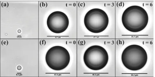
Figure 1.1: Vaporization of PFC droplets as a result of exposure to ultrasonic pulses. Shown here, a droplet of dodecafluoropentane encapsulated in a lipid shell is stable in vitro at 37◦C, but vaporizes upon exposure to a 5 MHz ultrasonic pulse. The phase-transition results in a bubble approximately 5 to 6 times larger.
imaging beyond ultrasound either through endogenous PFC properties or through the inclusion of other agents. PFCs, particularly those with higher boiling points, carry a high number of fluorine atoms that can be used for 19F MR spectroscopy [21; 22].
In-clusion of paramagnetic nanoparticles, fluorescent nanoparticles, and/or radioisotopes enable these PFC emulsions to be extended to 1H MR T1-weighted imaging,
SPECT-CT [4; 5; 23; 21; 22], and optical fluorescence imaging [9; 10]. Particular PFCs may also serve as radiopaque blood pool agents for CT due to the long intravascular half-life [1; 3; 8].
MCAs for diagnostic and therapeutic ultrasound have provided a promising plat-form for addressing a number of issues, including applications in echocardiography, microvascular perfusion imaging, thermal ablation enhancement, molecular imaging, drug and gene delivery, and thrombolysis [24; 25; 26; 27; 28; 29; 30; 31; 32; 33; 34; 35; 36; 37; 38; 39; 40; 41]. PFCs have been favored for the gaseous core for more than a decade because of low solubility compared to other gases [41; 42]. MCAs are typically produced with diameters in the 1 to 5 µm range, which is a compromise be-tween enabling free passage through capillary beds, and maximizing ultrasound imaging sensitivity. When excited by ultrasound energy, the highly compressible microbubbles exhibit nonlinear behavior. This has led to microbubble-specific imaging modes that enable contrast imaging with high sensitivity and low background signal from tissue [43; 44; 45; 46; 47; 48]. However, sizes optimal for contrast enhancement limit passage to within the vascular space [38], and microbubble persistencein vivo is typically only on the order of minutes [49].
[57], and methods of drug/gene delivery [58] have included discussions of PCCAs, and a few review articles have centered on the unique properties of these agents [59; 60; 61]. In this chapter, a brief survey of the history of phase-change contrast agents and the multiple environmental, design, and ultrasound-based factors that influence droplet vaporization is presented. The many proposed applications at both the microscale and nanoscale are discussed with a summary of the in vivo studies performed to date that show promising results for future use in humans. Issues involved with PCCA design as well as possibilities for future studies and applications are addressed.
1.2 History and Influencing Factors
Although the general physics of droplet vaporization has been well-described in the literature, phase-transition as a result of ultrasound energy and the range of di-agnostic and therapeutic possibilities for PCCAs have only begun to emerge in the past two decades. Phase-change colloids for ultrasonic imaging were first proposed by Quay in 1996 [62], resulting in development of the contrast agent EchoGenR, although
clinical trials were ultimately discontinued. EchoGenRwas fundamentally different
Perfluorocarbons and perfluorochemicals have been particularly attractive com-pounds in conjunction with PCCAs - not only due to the properties stated earlier, but also because many have boiling points near physiological temperatures, which allows for the design of droplets in or near a superheated state. Table 1.1 lists selected PFCs that have been used for inert liquid emulsions, MCAs, and PCCAs along with their physical properties, if available, gathered from several sources of literature [17; 19; 67]. DDFP, in particular, has been the most often-used compound because its boiling point of 29◦C allows for the possibility of droplet generation at room temperature, and expo-sure to physiological temperatures results in a superheated droplet that can be easily converted with ultrasound - although in practice, droplet size becomes a factor due to the effects of surface tension.
Surface tension plays a large role in both the threshold of vaporization for a droplet and the subsequent volumetric expansion. Beyond ambient pressure, a PFC droplet will experience an additional Laplace pressure as a result of surface tension effects over a defined radius [52]:
∆P =Pinside−Poutside = 2σ
r (1.1)
between the ambient pressure a compound experiences and the temperature required to induce phase-transition is approximated by the Antoine vapor-pressure equation, which is a derivation of the Clausius-Clapeyron relation. The Antoine vapor-pressure equation is defined as:
T = B
A−logP −C (1.2)
where T is the ambient temperature, P is the ambient pressure exerted on the droplet, andA,B, andC are experimentally derived constants observed for a particular temperature range. The relation shows that as the pressure is raised on the droplet core, the energy (in the form of temperature and/or ultrasound) required to induce phase transition increases. Because the Laplace pressure is an inverse function of radius, this effect becomes more pronounced for droplets in the nanometer range. By ideal gas laws with surface tension included, once a PFC droplet is vaporized, the resulting bubble will theoretically expand approximately 3 to 6 times in diameter, depending on initial droplet size, ambient pressure, and the density of the PFC selected [70]. Several studies have shownin vitro that bubbles may expand beyond ideal gas law predictions due to the influx of dissolved gases present in the surrounding media [71; 72]. In theory, this additional expansion is likely to be more pronounced for larger bubbles than smaller bubbles, as the internal pressure of the gas core due to surface tension is less.
studies have shown that increasing pulse duration at clinically relevant ultrasound fre-quencies results in no significant decrease in the vaporization pressure needed to achieve ADV, unless the duration is on the order of 1 millisecond or greater [73; 74; 75; 76]. For these longer pulse lengths, a decrease in the ultrasound pressure needed to induce va-porization has been observed. Although this may seem to be indicative of acoustically-induced heating, Lo and colleagues found that by using shorter periodic pulses unlikely to cause heating, but with an equivalent total ‘on-time’ as the longer pulses, a similar decrease in required ultrasound pressure resulted - indicating heating may not be the primary mechanism at work [74]. Other studies have provided convincing evidence that acoustic droplet vaporization may be initiated primarily by mechanical effects such as acoustic and hydrodynamic cavitation. Kripfgans and colleagues used high-speed op-tical microscopy to observe droplet deformations due to acoustic pressure, and showed that droplets with diameters in the micrometer range vaporized once a deformational threshold was reached, independent of original size [77]. More recent studies have shown vaporization can be induced independently of inertial acoustic cavitation, which has a number of implications for safetyin vivo [75; 76].
for droplets greater than 2 micrometers in diameter, but appears to be insignificant for sub-micrometer droplets.
The stability of circulating PCCAsin vivois not well characterized to date, although a few studies have suggested that certain PCCAs show greater circulation times than their counterpart perfluorocarbon-filled MCAs. Rapoport and colleagues have pub-lished several in vivo studies using polymer-coated, drug-loaded DDFP nanodroplets suggesting that a significant amount of nanodroplets were still in circulation as much as 4 to 5 hours after intravenous injection in mice [51; 52; 89]. When subjecting these droplets to ultrasonic energy, a substantial reduction in tumor growth was achieved over controls. Recent studies from this group using higher boiling-point perfluorocar-bons have suggested that the residence time may also be significantly influenced by the design of the encapsulating shell [53]. Zhang and colleagues recently published results showing that micron-sized droplets of DDFP were able to achieve an equivalent ef-fect when activated 30 minutes after intravenous injection in a canine model compared to those activated immediately after injection, although they did not test timepoints beyond 30 minutes [54]. In general, it is likely that longevity in circulation will be a function of PFC boiling point, design of the encapsulating shell, and droplet size, although many preclinical studies are needed to characterize these effects in vivo.
1.3 Applications
As tumors grow and recruit new vasculature, the rapidly-formed vessels often exhibit a degree of ‘loose’ organization. Inter-endothelial gaps that normally prevent passage of large molecules into interstitial space are characteristically wider depending on tumor type, and can allow for extravasation of particles well in to the hundreds of nanometers in size [90; 91]. PCCAs that can take advantage of the EPR effect have been proposed for applications such as ultrasound-mediated drug delivery, diagnostic imaging, and enhanced thermal ablation. In both size ranges, droplets can be administered intra-arterially, intravenously, or through direct intra-tumoral injection and then vaporized [50; 92; 93; 85]. In a few cases, the applications proposed for nanoscale droplets are similar to those for microscale droplets - such as drug delivery to solid tumors and enhancing the effects of HIFU therapy. Future in vivo work will be needed to develop an understanding of the tradeoffs inherent in choosing to pursue these techniques with microscale vs. nanoscale droplets. The specific applications are discussed below.
1.3.1 Microscale Applications
Vascular Occlusion Agents
Beginning with some of the earliest literature available on ADV, there has been significant interest in designing droplets in the micrometer size range for the purpose of vessel occlusion [50; 71]. Once vaporized, droplets near 5 µm in diameter will result in microbubbles that are on the order of 30µm - sufficient to occlude microvasculature. By using focused ultrasound transducers, vaporization can be induced in the feeder arteries of kidneys (and by extension, tumors), resulting in the ability to occlude with a high degree of spatial specificity (Figure 1.2) [92; 85]. This type of occlusion may be especially beneficial in enhancing thermal therapy of tumors, such as in radio-frequency ablation. Often the blood supply of the tumor can act as a heat sink - dissipating the heat and reducing the efficacy of the treatment. By occluding nearby vasculature, thermal delivery may be more successful. Reduced blood flow by occlusion may also be used to induce hypoxia in tumors, although future studies will be needed to determine whether the influence of PFC oxygen solubility reduces the degree of induced hypoxia in practice [50]. ADV-based occlusion appears to provide significant opportunity with regard to drug delivery. Residence time of drugs could be increased through reduced blood flow, allowing for enhanced diffusion into a targeted region. Drugs may be coinjected systemically or in the form of drug delivery vehicles such as liposomes, or alternately could be incorporated into the ADV occlusion agent through a dual-phase emulsion or multiple emulsions. Perhaps the most promising illustration of ADV-based occlusion was given by Fabiilli and colleagues [94]:
1. Through drug-incorporated emulsions, targeted occlusion can coincide with re-lease of a chemical embolic agent - thus sustaining embolization.
locally, enhancing efficacy of therapeutic delivery.
3. Hypoxia as a result of prolonged ischemia may be useful for activation of biore-ductive prodrugs - which potentially can be encapsulated in the emulsion process.
Although no in vivo validation is available to date, Fabiilli and colleagues have shown preliminaryin vitro proof-of-concept of encapsulation of thrombin and chloram-bucil in PCCA emulsions, followed by release with ultrasound-triggered vaporization [94; 95]. These studies also demonstrate the need for further optimization of formula-tion and drug loading as well as control over non-US induced drug release and droplet size prior to preclinical studies. How these emulsion techniques perform compared to alternative platforms, such as microscale PFC droplets co-injected with drug loaded liposomes or micelles, will also need to be characterized. Ultimately, the success of ADV-based occlusion techniques depends on the dynamics involved in the transport and lodging of microbubbles generated from PCCAs. Due to a number of influencing factors, these bubbles may become lodged in the microvasculature near the site of va-porization, may slide along the vascular space, or may interact in a complex manner with vessel bifurcations downstream. It is also important that the occlusive bubbles not be so large as to damage or rupture the vessel wall. Several physical models have been developed by the University of Michigan group to simulate these dynamics [79; 80; 81; 96; 97; 98]. Although some experimental studies are available on non-ADV microbubble embolization [99; 100], further experimental results from ADV-generated microbubbles are needed to validate these models as they extend to pulsatile blood flow in the microvasculature.
Aberration Correction
Figure 1.3: Ultrasound transcranial radiofrequency lines gathered usingex vivo human skulls. Misalignment of the peak amplitude of an ADV point beacon due to aberration (top) was corrected using time-reversal focusing (bottom). Reprinted from Ultrasound in Medicine and Biology, Vol. 34, Haworth KJ, Fowlkes JB, Carson PL, Kripfgans OD, “Towards aberration correction of transcranial ultrasound using acoustic droplet vaporization”, 435-45 (2008), with permission from Elsevier.
low-concentration droplets can be vaporized with a degree of spatial specificity, lodge in a vessel, and create a stationary point beacon for iterative methods [50; 71; 101]. Preliminary results using acoustic droplet vaporization have been promising for focusing on transmit (Figure 1.3), but have not yet been extended to image reconstruction [101].
Cavitation Nucleation Agents
formation in vitro as well as in vivo [54]. When droplets were introduced to tissue-mimicking phantoms, the exposure time needed to create similar lesions as controls was decreased by a factor of 2.5. When equal exposure times were used, the average lesion volume was 7-fold greater than phantoms without droplets. In vivo results proved even more promising, resulting in a 15- fold increase in volume for lesions formed in the canine liver at equal exposure time to controls without droplets.
Internal Markers for Intraoperative Guidance
One of the most recently-developed applications for PCCAs involves harnessing their sensitivity to heat and ultrasound pressure to create internal markers that may assist during therapeutic or surgical procedures. Huang and colleagues proposed a method where PCCAs designed to vaporized at a specific temperature are injected near the periphery of a tumor site and activated once the tissue reaches a lethal thermal dose - providing real-time feedback on intraoperative ablation margins [109]. They demonstrated this concept in vitro using micron-sized PLGA-encapsulated PCCAs. More recently, Couture and colleagues proposed ‘internal tattooing’, where PCCAs loaded with payloads of fluorescent markers are activated to release the fluorescent payload at the transducer focus [110]. This could be used to label areas of interest and delineate regions preoperatively with a high degree of spatial specificity that would then be visible intraoperatively through fluorescence imaging. They demonstrate proof-of-conceptin vivo by ultrasonically activating PCCAs and labeling tissues at different locations in a chicken embryo.
1.3.2 Nanoscale Applications
Intra-tumoral Diagnostics and Therapeutic Delivery
Figure 1.4: Schematic representation of passive drug targeting through the defective tumor microvasculature using an echogenic drug delivery system. The system com-prises micelles (small circles), PFC nanodroplets (stars), and PFC microbubbles (large circles). Lipophilic drugs can be localized in the micelle cores and in the walls of nanodroplets/microbubbles. Tumors are characterized by defective vasculature with large gaps between the endothelial cells, which allows extravasation of drug-loaded mi-celles and small nanodroplets into the tumor interstitium. Primary microbubbles are formed from the vaporization of nanodroplets due to hyperthermia or ultrasound, and larger microbubbles appear due to coalescence of the primary microbubbles. Rapoport N, Gao Z, Kennedy A, “Multifunctional nanoparticles for combining ultrasonic tumor imaging and targeted chemotherapy”, J Natl Cancer Inst, 2007, Vol. 99, 1095-106, by permission of Oxford University Press.
ultrasound combined with droplet vaporization enhances local drug delivery (Figure 1.4) [111]. The resulting microbubbles (as well as larger secondary bubbles formed by coalescence) can be used as real-time delivery confirmation. Over a wide range of studies, her group has demonstrated the promise of PCCAs for delivery of paclitaxel and doxorubicin in vitro and in vivo for various cancer models using DDFP droplets encapsulated in a polymer shell [51; 52; 89; 93; 112; 111; 113]. The group has recently begun to explore the use of much higher boiling-point PFCs for increased stability, reversible bubble formation, and co-registration with19F MRI, although they conclude that the temporary bubbles observed in the study may be due to gases dissolved in the PFC rather than actual vaporization of the PFC itself [53]. Matsuura and colleagues have provided promising preliminary evidence that incorporated nanoparticles such as quantum dots may act as additional cavitation nuclei within the core, and appear to decrease the ultrasound vaporization threshold significantly [87]. They suggest that PCCAs with incorporated nanoparticles may offer a means of spatially and temporally controlling nanoparticle deposition, and could produce a means to extend ADV-based agents to other therapeutic and imaging modalities.
Thermal Ablation Therapy and Therapeutic Bioeffects
Figure 1.5: Presence of bubbles formed by vaporized DDFP nanodroplets increased the thermal delivery by ultrasound to a tissue-mimicking phantom. Temperature elevations were measured during a 10-second HIFU exposure with and without an ADV pulse. The function generator was switched from continuous to pulse mode to fire the ADV pulse, and then back to continuous mode for heat deposition. The switching period led to the temperature drop during that period of time. The measured temperature reached a plateau in the presence of vaporized nanodroplets, most likely because of shielding effects. Reprinted from Ultrasound in Medicine & Biology, Vol 36, Zhang P, Porter T, “An in vitro study of a phase-shift nanoemulsion: a potential nucleation agent for bubble enhanced HIFU tumor ablation”, 1856-66 (2010), with permission from Elsevier.
Other Applications
Nanoscale PCCAs were used by Mohan and Rapoport to aid in the study of intra-cellular delivery of doxorubicin, including factors that influence penetration into the cell nucleus [116]. Their results suggest that ultrasound in the presence of microbub-bles, including those created by vaporized nanodroplets, transiently permeabilizes the cell nucleus and allows penetration of therapeutic drugs into the nucleus. Asami and colleagues have also proposed that nanoscale PCCAs could be used to characterize vis-coelastic properties of tumors by analyzing the waveforms received post-vaporization [117].
Many possible applications of the PCCA platform await additional exploration:
1. Targeting ligands could be incorporated in the encapsulating shell to provide a means of PCCA-based molecular imaging similar to that of microbubbles and inert liquid PFC emulsions [33; 118; 119; 120; 121]. The effect that incorporated ligands may have onin vivo aspects such as stability in circulation and clearance by the reticuloendothelial system will need to be carefully addressed. Once va-porized, PCCAs may be manipulated to enhance targeting through ultrasound phenomena such as acoustic radiation force [122; 123; 124].
3. Several of the previously mentioned sources have either suggested or given prelim-inary demonstration of the possibility of co-imaging with CT and MR techniques, and through incorporation of nanoparticles this may be extended to additional modalities. Strohm and colleagues recently showed that incorporation of PbS nanoparticles into micron-sized droplets of DDFP allowed the droplets to be va-porized by near-infrared laser irradiation due to heat generated by the particles [130]. This may be harnessed in the future to develop new techniques in photoa-coustic imaging.
4. PCCAs could be co-injected with other platforms to create novel imaging and treatment techniques. Lo and colleagues showed that co-injection with MCAs significantly decreased the vaporization threshold for PCCAs by enhancing the acoustic field in the vicinity of the droplets [74]. Therefore, microbubbles, which could also be targeted for angiogenesis, might be useful in reducing the vapor-ization threshold for extravasated nanodroplets and improving drug delivery ef-ficiency. A platform of drug-carrying liposomes/micelles and PCCAs could be developed such that the vaporized droplets confirm therapeutic delivery and cavitation-based effects from the resulting bubbles enhance release of drugs. Fi-nally, a coinjection of both nanoscale and microscale PCCAs could create a treat-ment where drugs are simultaneously released in the tumor interstitium by ex-travasated nanodroplets and in the vascular space by larger droplets that vaporize to form tissueoccluding microbubbles.
spectral information than MCAs [72; 132], which could lead to further contrast-specific imaging techniques.
1.4 Selection of PFCs
When designing phase-change contrast agents, the choice of which PFC comprises the core will create inherent trade-offs. As the energy needed to vaporize droplets of a certain size increases, the likelihood of inducing unwanted bioeffects for certain applications also increases. However, some applications may place a premium on high droplet stability over how easily the droplets can be vaporized. The optimal choice of PFC can largely be determined by considering:
1. The ideal size-regime of the droplets (nanoscale vs. microscale)
2. The ultrasound frequency used
3. Whether intended for diagnostic or therapeutic ultrasound machines
4. Whether ultrasound-induced bioeffects are acceptable
the droplets should be made from the PFC with the lowest possible boiling point that allows for stable circulation at physiological temperatures. For some therapeutic applications, the pressures used are already relatively high to create desired bioeffects, and droplets should remain stable until a desired activation pulse is delivered. For these applications, PFCs with a higher boiling point may be more suitable.
Most studies have shown that for applications such as vascular occlusion and aber-ration correction, microscale dodecafluoropentane- based droplets appear to be suffi-ciently stable and are able to be vaporizedin vitro at pressures within what diagnostic imaging machines typically provide [75; 76], although the pressures used to vaporizein vivo can be much higher [85]. DDFP and perfluorohexane (PFH) have been the most commonly-studied PFCs for use with nanoscale droplets. At these sizes, the optimal PFC choice is chiefly application-dependent. In a study with DDFP nanoemulsions for the purposes of enhancing thermal delivery to tumors and creating focal lesions, Zhang and Porter suggest that as the temperature rises in tissues, the nanodroplets just outside of the focal region may vaporize more easily and lead to unpredictable prefocal lesions [114]. A PFC with a higher boiling point, such as PFH, may prove better in this case, as it could still be vaporized and would lead to more predictable lesion formation. For co-imaging with19F MR, a PFC with a higher number of Fluorine
atoms per molecule may be desirable, although PFCs with more Fluorine atoms also have higher boiling points [53].
the focal region, and so lower thresholds may lead to a greater number of vaporized droplets and increased drug delivery and/or flow reduction. A number of the previously mentioned in vitro studies have shown that DDFP droplets have remarkable stability at body temperature, even though the bulk boiling point of DDFP is much lower than body temperature. A study by Giesecke and Hynynen demonstrated that vaporizing micron-sized albumin-coated DDFP droplets by heat alone required temperatures as much as 40◦C above the typical boiling point of DDFP as a bulk fluid [73], and the difference may increase as droplet size is reduced to the nanometer range. Therefore, methods of making PCCAs inherently more sensitive to ultrasound energy are necessary for expanding use toward diagnostic applications. This may be possible by including agents to facilitate nucleation within the droplet core (reducing the purity of the PFC), or by choosing alternative compounds that require less energy to vaporize.
One additional method of droplet design is to alter the amount of energy needed to vaporize a specific droplet by creating a mixture of PFCs with different boiling points. This technique was first proposed by Kawabata and colleagues, who demonstrated that mixing DDFP (b.p. 29◦C) and 2H,3H-DDFP (b.p. 53◦C) resulted in a droplet that required more energy to vaporized than if it contained only DDFP, but less than if it contained only 2H,3H-DDFP [86]. This technique could be useful in designing PCCAs for an optimal trade-off between stability in circulation and pressure required to induce vaporization.
1.5 In Vivo Studies
Figure 1.6: Demonstration of drug delivery by the interaction of ultrasound and drug-loaded nanodroplets/micelles. A nu/nu mouse bearing two ovarian carcinoma tumors immediately before (A)and 3 weeks after the treatment (B). The mouse was treated by four systemic injections of paclitaxel-loaded nanodroplets given twice weekly, while only the right tumor was sonicated by 1 MHz continuous-wave ultrasound 4 hours after the injection. The left tumor grew at the same rate as the controls, while the right tumor regressed, demonstrating paclitaxel release into the tumor volume. Reprinted from J Control Release, Vol. 138, Rapoport NY, Kennedy AM, Shea JE, Scaife CL, Nam KH, “Controlled and targeted tumor chemotherapy by ultrasound-activated na-noemulsions/microbubbles”, 268-76 (2009), with permission from Elsevier.
vaporized in vivo to produce desired effects such as reduced blood perfusion and ther-apeutic drug delivery (Figure 1.6). In a few instances, droplet-induced bioeffects were observed. An early occlusion-based study by Kripfgans and colleagues using a rabbit model showed that filtering droplet emulsions to transcapillary sizes (99.99%<6µm in diameter) and lowering doses to approximately 2×10⁷ droplets/kg eliminated instances
of pulmonary hyperinflation [92]. They suggest that because the rabbit model may be more susceptible to this bioeffect [133], it may be possible to use much higher doses for greater embolization in humans. Another occlusion-based study in a canine model by Zhang and colleagues noted instances of cardiac arrhythmia and one instance of animal death when droplets (99.9% < 10 µm in diameter) were administered in doses on the order of 1×10⁸ droplets/kg to 4×10⁸ droplets/kg through an intracardiac method [85].
hypothesized that these effects were a result of droplets occluding a coronary arteri-ole, inducing ischemia. Although no cardiac arrhythmia was observed for intravenous administration, doses approaching 2×10⁹ droplets/kg resulted in respiratory distress
and changes in blood chemistry. A dose of 3×10⁹ droplets/kg of droplets this size (a
total perfluorocarbon dose of 0.2 g/kg) was determined to be fatal to the canines. In a recent study by Zhang and colleagues aimed at assessing microscale DDFP droplets for thermal ablation, a dose of 1×10⁸ droplets/kg (99% < 8 µm in diameter) was administered intravenously in a canine model and no adverse bioeffects were reported [54]. All other studies noted no significant undesired bioeffects. Future studies are needed to optimize ultrasonic vaporizationin vivo.
1.6 Current Challenges
A thorough literature review of the current state of PCCAs reveals several areas to fundamentally improve the clinical potential of the PCCA platform:
1. Overcoming the Increase in Vaporization Energy at Nanoscale: In some
2. Increasing the Uniformity of Activation: Because the activation pressure of a droplet depends on the initial diameter, polydisperse distributions result in non-uniform activation. The techniques involved in preparing sub-micron emul-sions typically yield polydisperse distributions, but for applications involving mi-croscale droplets the uniformity of response (and therefore the effectiveness of the treatment) may be improved by creating monodisperse distributions through techniques such as microfluidic particle generation.
3. Tailoring Droplet Performance to Desired Application: Nearly all the
studies to date have investigated PCCAs composed of a single perfluorocarbon. Although not well explored in the literature, the ability to manipulate the in-terplay of vaporization energy and thermal stability (resistance to spontaneous vaporization) for a droplet are essential in order to maximize the performance for a given application. As the range of applications is wide and continues to grow yearly, designing an agent that is optimally stable and one that vaporizes with ideal pressures may require a careful balance of effects. Choosing single perflu-orocarbon species inherently limits the researcher to discrete points in the sen-sitivity/stability continuum, and so developing methods to more precisely ‘tune’ droplets will be essential in clinical translation.
4. Determining Appropriate Activation Thresholds for Polydisperse
Sub-micron Distributions: Most studies aimed at determining appropriate
nanoscale emulsionsin vivo.
5. Isolation of Signals Specific to PCCAs for in vivo Detection of
Activa-tion: The nonlinear oscillations produced by conventional microbubble contrast
agents while under the influence of an ultrasonic pulse can be easily detected and isolated from tissue through a variety of techniques (see Section 1.1). However, no such ‘signature’ exists for PCCAs that allows differentiation from tissue or standard microbubbles. If such a signature were to be discovered, it would enable new forms of ultrasound contrast imaging and would allow spatial and tempo-ral mapping of PCCA activation to correlate with therapeutic and/or diagnostic goals.
6. Demonstration of Utility for Diagnostic and Molecular Imaging
Pur-poses: A wealth of studies have shown the utility of PCCAs as therapeutic
agents. However, as a result of the relatively high vaporization thresholds for typical PCCA formulations, few studies have shown that they can be used as purely diagnostic agents (that is, their use as agents to generate imaging con-trast) or molecular imaging agents. If vaporization thresholds can be reduced, demonstrations of the diagnostic potential of PCCAs will greatly aid the push toward clinical use.
1.7 Thesis Outline
We first address the need to develop PCCAs inherently more sensitive to ultrasound energy by exploring the incorporation of more volatile compounds than had previously been considered in the literature. InChapter 2 we first treat the Laplace pressure in-crease theoretically to explore alternative perfluorocarbons to form the droplet core, and then provide proof-of-principle that droplets can be crafted from these compounds, re-sulting in emulsions with reduced vaporization thresholds compared to similar droplets of DDFP or PFH.
InChapter 3, we present a novel particle generation technique termed ‘microbubble
condensation’ that allows formation of nanoscale emulsions from volatile perfluorocar-bons (i.e. that exist as a gas at room temperature). By first generating a population of perfluorocarbon microbubbles ideal for ultrasound interaction, PCCAs can be formed by condensing the gaseous precursors to the liquid state through a combination of de-creased ambient temperature and inde-creased ambient pressure. We demonstrate that the resulting droplets have size distribution peaks on the order of 200-300 nm in diam-eter, and can be vaporized with ultrasound pressures within the limits of those set for clinical diagnostic imaging. Once vaporized, the particles expand to form bubbles of ideal size for ultrasound imaging and therapy.
In Chapter 4 we explore the inherent tradeoffs involved with forming PCCAs
from volatile compounds. Though gains are made with regard to reduced vaporiza-tion thresholds, in vitro thermal stability of the emulsions decrease as PFC volatility increases, which suggests reduced circulation profiles in vivo. We show that the sensi-tivity to ultrasound energy and the thermal stability of an emulsion can be modified by creating PFC mixtures during microbubble condensation - allowing fine-tuning of the droplet performance for the intended application.
(e.g. optical verification of individual droplet vaporization events) or phenomenolog-ical (e.g. measure of ultrasound backscatter produced after a vaporization pulse has been delivered). Both of these are limited for the typically broad distributions of nanoscale emulsions. In physical (optical) methods, the initial size of the droplets is below the wavelength of visible light, and therefore unresolvable. Phenomenological methods are often highly skewed by the presence of large outliers in the distribution, and so associating the measurement with a representative size is not straightforward.
InChapter 5we present an alternative method of assessingin vitro activation
thresh-olds for nanoscale emulsions based on optical measurements of the change in resulting bubble distribution with increasing ultrasound pressure. We also give preliminary ev-idence that once droplets are vaporized, they may be destroyed or made to fuse with nearby bubbles by the continuing vaporization pulse - highlighting the importance of optimized vaporization pulse sequences.
In Chapter 6 we demonstrate that the underlying physics of droplet vaporization
and expansion can, under certain conditions, produce new acoustic information that can be detected and differentiated from typical tissue and microbubble signals. When droplets formed from volatile compounds are activated with brief ultrasonic pulses, the momentum of expansion drives the bubbles to overexpand and oscillate to a final bubble diameter. These oscillations produce exponentially decaying sinusoids on the order of 1 MHz with size-dependent frequency and amplitude properties that can be captured with an ultrasonic transducer. A simple droplet-detection algorithm is imple-mented to characterize activation as a function of peak negative pressure and droplet concentration.
in the HUVEC cell line. We first generate targeted droplets by modifying the precursor bubbles to contain targeting ligands and then revert the particles to the liquid state. After a period of incubation with the cells, we are able to assess the level of angiogenic expression by activating the droplets with a clinical diagnostic ultrasound machine.
In Chapter 8we summarize our ongoingin vivo work to assess circulation profiles
and imaging contrast generated using custom imaging sequences from a diagnostic ultrasound probe, and frame the future of PCCA development in the context of the platform improvements demonstrated in this thesis.
Although not an integral portion of this thesis, we have included an appendix of 6 ad-ditional studies that stem from the concepts and improvements herein. InAppendices
B, C, and D we use the microfluidics platform to generate monodisperse microscale
CHAPTER 2
PCCAs Composed of Low Boiling Point Perfluorocarbons
2.1 Introduction
FDA-approved microbubble contrast agents are commonly produced with the ma-jority of the population between 1 and 5 µm in diameter to allow for safe passage through the circulatory system and provide significant contrast for imaging. Many tumor types exhibit characteristically permeable vasculature, with endothelial gaps typically between 200 to 600 nm and show poor lymphatic clearance, also known as the EPR effect [90; 91; 137]. A gas-filled contrast agent small enough to extravasate into tumor interstitium would ultimately be much less echogenic than commonly studied microscale MCAs and would provide limited US contrast [138]. Therefore, an agent capable of clearing inter-endothelial gaps and subsequently being transformed into a gas-filled MCA would have unique possibilities.
The application of acoustic droplet vaporization is a potential method of designing
c
2011 World Federation for Ultrasound in Medicine & Biology (wfumb.org) Portions reprinted,
from PS Sheeran, VP Wong, S Luois, RJ McFarland, WD Ross, S Feingold, TO Matsunaga, and PA Dayton. “Decafluorobutane as a Phase-Change Contrast Agent for Low-Energy Extravascular Ultrasonic Imaging” Ultrasound Med Biol, 2011 Sept; 37(9): 1518-1530.
c
2011 American Chemical Society (acs.org) Portions reprinted, from PS Sheeran, S Luois, PA
a contrast agent that can exploit the EPR effect and provide imaging contrast in tumor extravascular space. In brief, the vaporization of a liquid droplet depends primarily on the properties of the surrounding fluid (viscosity, ambient temperature and pressure), the droplet diameter, and the energy introduced into the system (heating, mechani-cal energy) (see Table 1.2). Often a lipid or polymer shell is used to both stabilize the droplet from coalescence and to alter the droplet surface tension. Most studies involving ADV-based liposomal nano/micro-emulsions have used either stabilized or superheated liquids in the perfluorocarbon (PFC) family, as many have boiling points near physiologic temperatures and are similar to commonly-used MCA perfluorocar-bons that have significant advantages in imaging applications with less toxicity at the small volumes used [1]. The most common PFCs used to date, dodecafluoropentane and perfluorohexane (PFH), are liquids at room temperature. When encased in lipid or polymer shells, nano/micro-emulsions of DDFP and PFH are able to stay in solution at body temperature and can be activated by additional energy input.
An agent with sufficient stability to extravasate into the extracellular space, yet labile enough to be vaporized at sufficiently low acoustic intensities so as to not induce unwanted bioeffects would be optimal. Most studies of phase-change contrast agents to date have shown that DDFP droplets near the desired size range vaporize with the least energy input compared with alternative compounds. The input pressure needed to vaporize them can be lowered even further by altering the duration of the excitation acoustic pulse [74]. By using ultrasound frequencies above 1 MHz and pulse lengths in the millisecond range, micron-sized droplets can be vaporized with pressures considered safe for diagnostic procedures [75]. However, data suggest that the vaporization of sub-micron droplets may require substantially more energy.
Choosing alternative lower boiling point PFCs could lower the vaporization threshold for sub-micron droplets, although they then have the potential to be relatively unstable compared with their higher boiling point counterparts. In some applications, such as thermal ablation enhancement, droplet stability through a range of temperatures above 37◦C may be a priority over low vaporization thresholds [114].
In this chapter, it is demonstrated that low boiling point perfluorocarbons can be manipulated to achieve the desired low vaporization threshold. Theoretical estimations of boiling point elevation are used to reveal that two previously unused compounds -decafluorobutane (DFB) and octafluoropropane (OFP) - may be able to be formed into metastable droplets that remain in the liquid state at body temperature. DFB, for example, has a boiling point of -1.7◦C, significantly lower than other PFCs commonly used in ADV, which may allow vaporization at much lower pressures. This could, in turn, significantly decrease the chance of unwanted bioeffects due to ultrasound exposure. To date, no study of phase-change contrast agents has explored the use of these compounds, which are used as the gas core component in several MCAs approved for clinical applications. The aim of this study is to develop a better understanding of the trade-offs inherent in choosing a lower boiling-point PFC. The results show the ability to produce stable lipid-encapsulated phase-change agents of DFB that can vaporize at lower pressures than similarly sized emulsions of higher boiling-point PFCs.
2.2 Materials & Methods
2.2.1 Theory
1.2. This equation uses experimental results to develop a basic relationship between temperature and pressure as a droplet of a particular substance vaporizes. Following reasoning outlined by Rapoportet al., a droplet will experience an additional pressure due to interfacial surface tension effects, defined as the Laplace pressure (eqn. 1.1) [112]. PFCs typically have fairly low surface tension values against air (on the order of 10 mN/m at room temperature), but high surface tension against aqueous solutions (on the order of 60 mN/m). Because the Laplace pressure is an inverse function of radius, smaller droplets will experience greater pressure. Encapsulating the droplets in a lipid or polymer shell stabilizes the droplets from coalescence and alters the interfacial surface tension. Depending on the properties of the encapsulating shell, a larger re-sulting surface tension may cause an increase in the pressure exerted, which essentially increases the vaporization temperature of the droplet. In designing agents for human medical imaging purposes, the ambient pressure may be defined as
Pamb =Patm+Pbody (2.1)
where Patm = 101.325 kPa and Pbody is a representative pressure inside the human body (vascular or other). Although intravascular pressure is inherently pulsatile, for the purposes of these calculations, an average value of Pbody = 12.67 kPa was used. With a total pressure exerted on the droplet of:
P =Pamb+ ∆P =Patm+Pbody +
2σ
r (2.2)
The resulting modified Antoine vapor-pressure equation is:
T = B
A−log (Patm+Pbody+ 2rσ)
Published surface tensions often vary between 25 mN/m and 50-60 mN/m, depend-ing on surfactant properties [139; 140]. Although the exact surface tension of lipid solutions used in this study were not known, a value near 51 mN/m was sufficient for the purposes of these initial calculations in that it provided a Laplace pressure near the upper limit of what can be expected. The constants A, B and C were gathered from the National Institute of Standards and Technology (NIST) Chemistry WebBook [19] for the nearest available temperature range. Figure 2.1 shows the relationship be-tween droplet diameter and predicted vaporization temperature for octafluoropropane, decafluorobutane, dodecafluoropentane, and perfluorohexane (natural boiling points of -37.6 ◦C, -1.7 ◦C, 29◦C and 56.6◦C, respectively). While the constants used are not expected to predict the vaporization relationship completely accurately in the desired temperature range, the calculation shows that DFB droplets appear to have the poten-tial to remain stable in the 200−600 nm diameter range at temperatures just above body temperature. This suggests that they may require a small amount of additional energy (such as US) to induce vaporization compared with other PFCs, if droplets can be generated stably. Although the temperature required to induce vaporization increases substantially for droplets near 200−300 nm in diameter, others have shown successful vaporization of droplets through ultrasonic energy at temperatures as much as 40◦C below their boiling point, as in the case of perfluorohexane droplets vaporizing at room and body temperature [73; 75]. According to the estimations, octafluoro-propane droplets have the potential to remain stable at sizes near 200 nm, although the -37.6 ◦C boiling point presents significant production challenges. With a boiling point of -1.7◦C, DFB droplet generation can be explored under more practical experi-mental conditions.
Figure 2.1: Predicted vaporization temperature of lipid encapsulated perfluorocarbons based on Antoine vapor pressure equation. As droplet diameter decreases, the temper-ature required to vaporize increases exponentially.
the expansion factor when a liquid undergoes a phase conversion to the gaseous state. Because perfluorocarbons are immiscible in the liquid state and have low diffusivity in the gaseous state, it is assumed that the number of moles is constant from the liquid phase to the gaseous phase (nl =ng). The moles of PFC in the spherical droplet can be computed as
nl =
4πr3
lρl
3M (2.4)
where rl is the radius of the liquid droplet, ρl is the liquid density and M is the molar mass. Substituting this into the ideal gas law and simplifying as a ratio of the gas-phase radius to liquid-phase radius gives:
rg
rl
= 3 r
ρlRT
Expanding with eqn 2.2 gives:
rg
rl
= 3
s
ρlRT
M(Patm+Pbody+ 2rgσ)
(2.6)
As rg approaches very large values, the surface tension component becomes negli-gible.
Decafluorobutane has a molar mass of M = 0.238 kg/mol and at 37◦C (310 K)
ρl = 1500 kg/m3. Evaluating eqn 2.6 with in vivo (Pbody = 12.67 kPa) and in vitro (Pbody = 0 kPa) conditions and neglecting surface tension effects reveals that, based on the assumptions given, a droplet of DFB can be predicted to expand to an approximate upper limit of 5.2 to 5.4 times its original diameter once vaporized (neglecting any deviations from ideal gas laws). Rearranging eqn 2.6 such that it is solved for liquid droplet radius becomes:
rl=
3
s
M r2
g[rg(Patm+Pbody) + 2σ]
ρlRT
(2.7)
This allows one, based on ideal gas laws and surface tension effects, to estimate the size of the droplet that vaporized to become a bubble of a known size. Evans et al. show that eqn 2.7 can also be solved for rg, providing a numerically equivalent, though much more complex, solution [70].
2.2.2 Preparation of Micron-Sized Perfluorocarbon Droplets
were stored in vacuo overnight.
The lipid films were rehydrated with approximately 1 mL of (4-(2-hydroxyethyl) piperazine-1-ethanesulfonic acid) (HEPES) buffer (pH = 7.4) (Sigma-Aldrich, Co., St. Louis, MO, USA) and sonicated for 10 min in a water bath sonicator (Branson 1510; Branson Ultrasonic Corporation, Danbury, CT, USA) at 50 to 60◦C. The rehydrated films were then subjected to 10 freeze-thaw cycles. The solution was then stirred for 10 min at 50 to 60◦C. The resulting concentration of the lipid solution was approximately 20 mg/mL.