Sca-1 Positive Pancreatic Progenitor Cells: A Replacement for Transplanted Islets
Lisa Lynn Samuelson
A thesis submitted to the faculty of the University of North Carolina at Chapel Hill in partial fulfillment of the requirements for the degree of Master of Science in the Department of Pathology and Laboratory Medicine.
Chapel Hill 2011
©2011
ABSTRACT
Lisa Lynn Samuelson: Sca-1 Positive Pancreatic Progenitor Cells: A Replacement for Transplanted Islets
(Under the direction of David A. Gerber)
ACKNOWLEDGEMENTS
First and foremost I would like to thank my advisor, Dr. David Gerber for being not only a boss but a friend over the past 9 years. It is rare to find a superior that looks out for you and has your best interests in mind. I have never doubted that this is exactly how you feel. Thank you for giving me the opportunity to pursue my degree. Without your continued support and guidance my Masters would never have been possible. I appreciate everything you have taught me, about science and about life. I marvel everyday at your ability to multi-task and balance your many roles in life. I hope that I have served you well during my time at UNC and that I will continue to do so. To my committee members, Dr. William Coleman and Dr. Monte Willis I say thank you for your time and willingness to guide and help me through my graduate career. Dr. Coleman, I say a special thank you for creating this program and giving research technicians like myself the chance to further our education. I also say a special thank you to Dr. Tim Sadiq. You provided me with my introduction to science and lab life. It is the foundation and practices that you taught me that have allowed me to be successful.
ever I see that family is the most important thing we can have and I am grateful everyday that all of you are part of mine.
TABLE OF CONTENTS
ABSTRACT.. ... .iii
ACKNOWLEDGEMENTS ... iv
TABLE OF CONTENTS ... vi
LIST OF TABLES ... ix
LIST OF FIGURES ...x
LIST OF ABBREVIATIONS ... xii
INTRODUCTION...1
DIABETES PATHOGENESIS ...1
PANCREAS ANATOMY AND FUNCTION ...3
COMPLICATIONS AND SIDE EFFECTS OF BLOOD SUGAR IRREGULARITIES ...4
TREATMENT OPTIONS FOR DIABETICS ...5
STEM-CELLS AND DIABETES ...6
EMBRYONIC STEM CELLS ...7
ADULT DERIVED STEM CELLS ...8
INDUCED PLURIPOTENT STEM CELLS ...9
TRANSDIFFERENTIATIONS OF STEM CELLS ...9
BIOREACTORS FOR CELL CULTURE AND TISSUE ENGINEERING ...10
MICE ………..……….………..…...13
ISOLATION OF PANCREATIC PROGENITOR CELLS ...13
CELL ENRICHMENT AND CULTURE ...14
FLUORESCENT IMMUNOPHENOTYPING AND FLOW CYTOMETRY ...15
HISTOLOGY AND IMMUNOHISTOCHEMISTRY ...15
RT-PCR...16
WESTERN BLOT ANALYSIS...16
INSULIN ELISA ...18
CELLULAR PROLIFERATION ASSAY ...19
MICROCARRIER PREPARATION ...19
BIOREACTOR CULTURE ...20
Results ...21
ISOLATION OF A PANCREATIC PROGENITOR CELLS ...21
Cellular Localization Within Murine Pancreas ... 21
Enrichment and Purification of Sca-1+ PPCs ... 22
Two Dimensional Culture of Sca-1+ Pancreatic Progenitor Cells ... 24
CHARACTERIZATION OF SCA-1+ PANCREATIC PROGENITOR CELLS ...25
Immunohistochemical Analysis of Sca-1+ Colonies In-Vitro ... 25
PPC Expansion and Capacity to Proliferate ... 25
Interim Conclusions ... 27
TRANSCRIPTION AND TRANSLATION OF SCA-1+ PPCS ...30
PPCs Express Transcription Factors Indicative of β-Cell Lineage as well as all Other Cell Fates of Adult Pancreas ... 30
Interim Conclusions ... 35
FUNCTIONAL ANALYSIS OF SCA-1+ PPC’S ...37
PPCs Release Basal Amounts of Insulin In Vitro ... 37
Interim Conclusions ... 39
THREE DIMENSIONAL CULTURE ...41
Bioreactor Culture of Sca-1+ PPCs ... 41
Insulin Secretion of Sca-1+ PPCs in Bioreactors ... 42
Bioreactor Culture of β-TC6 Cell Line ... 45
Comparison of 2-D and 3-D Transcription ... 48
Comparison of 2-D and 3-D Translation ... 48
Improved Insulin Secretion in Bioreactor ... 52
Interim Conclusions ... 52
Discussion...56
SUMMARY ...56
ISOLATION OF A NOVEL SCA-1+ PANCREATIC PROGENITOR CELL ...57
BIOREACTOR CULTURE FOR IMPROVED PANCREATIC PROGENITOR CELL FUNCTION ...58
FUTURE DIRECTIONS ...59
LIST OF TABLES
LIST OF FIGURES
Figure 1. Sca-1 expression in normal murine pancreas tissue. ... 22
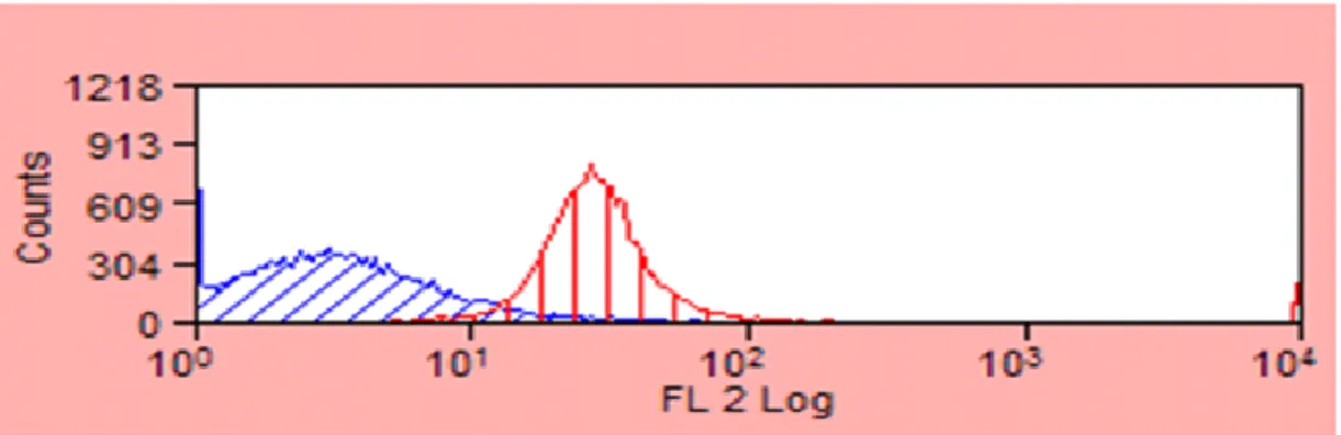
Figure 2. Flow cytometry analysis of immunomagnetically separated Sca-1+ PPCs. ... 24
Figure 3. Changes in Sca-1+ colony morphology throughout culture. ... 26
Figure 4. Sca-1 immunohistochemistry in culture. ... 28
Figure 5. Proliferative ability of Sca-1+ PPCs. ... 29
Figure 6. Transcription factors of interest in pancreatic progenitor development. ... 31
Figure 7. Semi quantitative PCR analysis of Sca-1+ PPCs ... 33
Figure 8. PDX-1 expression of Sca-1+ colonies ... 34
Figure 9. Protein expression of Sca-1+ PPCs. ... 36
Figure 10. Functional ability of Sca-1+ PPCs to produce insulin. ... 38
Figure 11. Rapid response of Sca-1+ PPCs to high glucose conditions. ... 40
Figure 13. Microcarriers and culture bags for bioreactor
experimentation. ... 43
Figure 14. Attachment of Sca-1+ PPCs to microcarrier culture
beads. ... 44
Figure 15. Sca-1+ PPCs respond to increased glucose in 3-D
bioreactor cultures. ... 46
Figure 16. Morphology of β-TC6 cell when grown in
monolayer versus bioreactor culture. ... 47
Figure 17. Cell clusters formed by β-TC6 cells in bioreactors
resemble normal islets in architecture. ... 49
Figure 18. Semi quantitative PCR results for β-TC6 cell line in
monolayer and bioreactor culture. ... 50
Figure 19. Comparison of protein expression from β-TC6 cells
contained in culture dishes and 3-D bioreactors. ... 51
Figure 20. Bioreactors allow for increased secretion of insulin
LIST OF ABBREVIATIONS ADA AU BrdU BSA Ca2+ Ck19 CO2 DMEM EDTA ELISA ES cell FBS FEP G GDM GFP g/L Hes1 HRP iPS cell Klf4
American Diabetes Association arbitrary units
Bromodeoxyuridine Bovine serum albumin Calcium
cytokeratin 19 Carbon dioxide
Dulbecco’s Modified Eagle’s medium Ethylenediaminetetraacetic acid
enzyme linked immunoabsorbant assay embryonic stem cell
fetal bovine serum
transparent fluoro ethylene propylene gauge
gestational diabetes mellitus green fluorescent protein grams per liter
hairy and enhancer of split 1 horseradish peroxidase induced pluripotent stem cell
Mg2+ mg/dL mL mm mM NASA ng/mL Oct3/4 PBS PCR PDX1 PE PP PPC rpm RWV Sca-1 Sox2 T1D T2D WHO WT α magnesium
milligrams per deciliter milliliter
millimeter millimolar
National Aeronautics Space Administration Nanograms per milliliter
octamer 3 / 4
Dulbecco’s phosphate buffered saline polymerase chain reaction
pancreas and duodenal homeobox gene 1 Phycoerythrin
pancreatic polypeptide pancreatic progenitor cell revolutions per minute rotating wall vessel stem cell antigen 1
SRY (sex determining region Y)-box 2 Type 1 diabetes
Type 2 diabetes
World Health Organization Wildtype
β δ µg µg/ml
beta delta microgram
INTRODUCTION
DIABETES PATHOGENESIS
Diabetes is a disease characterized by the body’s inability to produce insulin or an inability to respond to insulin’s actions. Insulin is a hormone produced by the pancreas which regulates the amount of glucose circulating in the blood. It acts by stimulating cells to absorb circulating glucose and use it for energy. When cells are unable to absorb circulating glucose, it accumulates in the blood, a state known as hyperglycemia and fat is burned as an alternate source of energy. The breakdown of fat for fuel causes toxic acids, known as ketones, to be left behind in urine and blood. The opposite can also occur with insufficient amounts of glucose circulating in the blood leaving cells unable to function due to lack of an energy source. This state is known as hypoglycemia, or low blood sugar. Normal blood glucose levels are expected to be in the range of 80-140 mg/dl. Diabetics have fragile regulatory mechanisms with respect to glycemic control and therefore are unable to prevent hyper- and hypo-glycemic events. It is not uncommon for diabetic patients to experience blood sugar levels ranging from 30-600 mg/dl.
The cause of the autoimmune attack on pancreatic β-cells in T1D is unknown. There is some evidence for a genetic factor in the disease as it is seen in families. However, environment and random developmental factors seem to be key players1. T1D is the main focus of this research.
Type 2 diabetes (T2D), or non-insulin-dependent diabetes, is due to a decrease in the body’s sensitivity to respond to insulin or secondary to an insufficiency in insulin production. This form of the disease does not result from an autoimmune attack on the islets of the pancreas as seen in T1D. While not fully understood, this form of diabetes is strongly associated with obesity and sedentary lifestyles and currently accounts for over 90% of the total cases of the disease2. It is also associated with advancing age, with increased risk occurring after age 45. T2D, can progress to the point of islet exhaustion. This results from the normally functioning islets over-compensating for the loss of function or loss of sensitivity to insulin and their eventual hypertrophy and cessation of activity. When this occurs T2D effectively becomes T1D.
PANCREAS ANATOMY AND FUNCTION
The pancreas is an organ that has both exocrine and endocrine function. Embryonic pancreatic development in vertebrate animals occurs from the dorsal and ventral protrusions of the primitive gut epithelium, which grow, branch, and eventually fuse to form the whole gland around the seventh week of gestation6. The human pancreas is located in the abdomen posterior to the stomach, attached to the small intestine at the duodenum and extending to the hilum of the spleen. The human pancreas is roughly 20 centimeters in length and weighs 85-90 grams7.
The exocrine portion of the pancreas functions in digestion. The cells that compose this portion include acinar cells that secrete numerous enzymes from the pancreas into the duodenum aiding in digestion and breakdown of food as it passes through the digestive tract. These pancreatic enzymes include trypsin, lipase, amylase, and carboxypeptidase. The exocrine portion of the pancreas accounts for 80-85% of the organ’s makeup and is thus a major component of pancreas anatomy7.
COMPLICATIONS AND SIDE EFFECTS OF BLOOD SUGAR IRREGULARITIES
All diabetic patients are at risk for serious life threatening complications secondary to poor glucose regulation. The Diabetes Control and Complications Research Trial demonstrated that maintaining blood sugar levels within defined ranges and keeping a close monitor of the disease can greatly decrease the risk of developing life-threatening complications and aide in slowing diease progression9.
Several acute complications of the disease can affect diabetics on a daily basis. These include bouts of hypoglycemia leading to confusion, headache, fatigue, nausea, numbness, irritability, slurred speech, ataxia, and impaired judgment. Untreated hypoglycemia can ultimately lead to loss of consciousness, diabetic coma, and possible death. Conversely, if blood sugar levels rise above normal, patients can experience polyuria, dizziness, confusion, cardiac arrhythmia, blurred vision, lethargy, and diabetic ketoacidosis as a result of ketones spilling into the blood and urine.
vasculature, as seen in diabetic retinopathy7. Intracellular hyperglycemia with disturbances in polyol pathways leads to oxidative stress which causes damage to all cellular components and affects organs like the kidneys, vasculature, nerves, and eyes.7.
TREATMENT OPTIONS FOR DIABETICS
Treatment for diabetes, especially T1D, is limited. T2D and GDM can often be treated successfully with oral medication, diet, and lifestyle changes. However, T1D treatment is more invasive as deficiency of insulin production requires the injection of exogenous insulin. Insulin was discovered in the 1920’s and proven effective at lowering blood glucose levels in test animals and experimental patients. This form of insulin was generated via extraction from animal sources and its use was limited. As a result, treatment remained largely centered around diet and exercise and the disease continued to lead to premature death. It was not until the 1950’s that the amino-acid structure of insulin was elucidated and then the 1970’s before the first synthetic insulin was produced. With its production came the commercial sale of the medication in the 1980’s. Insulin has since been the standard of care for type 1 diabetics with many patients receiving multiple injections daily in order to maintain glucose levels as close to normal as possible. Insulin can be administered via hypodermic injection or given mechanically through an external insulin pump that maintains a steady release of drug subcutaneously.
major problem is the fact that it takes more than one organ donor to yield enough islets for a successful transplant. The average donor generates approximately 500,000 viable islets but the typical recipient requires roughly 1 million healthy islets to be successfully weaned from exogenous insulin. Another drawback to the procedure is the requirement for immunosuppressive treatment after transplant to prevent rejection of the transplanted allogeneic cells. Longstanding immunosuppression is complicated by an increased risk of systemic infections and malignancies in the recipient. A final limitation to islet transplantation is the autoimmune nature of the disease that destroyed the patient’s original islets. Over time this same pathologic process attacks the transplanted islets, ultimately leaving the patient insulin-dependent again. The Collaborative Islet Transplant Registry reported that 13% of islet recipients experience total loss of transplant function by 6 months and this increases to 42% by 3 years post-transplant11. Other studies are more pessimistic with reports that by 2 years post-transplant 76% of patients require exogenous insulin due to failure of the transplanted cells12.
STEM-CELLS AND DIABETES
highly proliferative, easily accessible, and unlimited, all qualities of a stem/progenitor cell population.
EMBRYONIC STEM CELLS
Embryonic stem cells (ES cells), were first cultivated in 1981 from mouse blastocysts14. Later experiments yielded ES cells obtained and cultivated from human tissues15. Embryonic stem cells are a potential candidate for islet replacement because they are pluripotent, capable of self-renewal and have the potential to differentiate into any cell type within the body. Subsequently, focus has been aimed at committing ES cells to a pancreatic β-cell fate. Early experiments with ES cells used the formation of embryoid bodies, or three dimensional aggregates, which were differentiated into insulin-producing cells16, 17. These approaches generate low numbers of insulin-producing cells and led researchers to experiment with over-expressing transcription factors that were known to force β-cell formation in an effort to yield robust insulin-producing populations18
. Recent studies examined ES cells in conjunction with a myriad of signaling factors, growth factors, feeder cells, and/or culture conditions19, 20. Groups have also independently reported differentiating human ES cells into pancreatic lineage cells by recapitulating endocrine development. These cells were differentiated via a protocol generating definitive endoderm, posterior foregut, pancreatic endoderm and finally hormone expressing endocrine cells19, 21, 22.
ethical, political, and moral concerns as ES cells are derived from embryos and many people hold strong religious and moral beliefs about obtaining them.
ADULT DERIVED STEM CELLS
INDUCED PLURIPOTENT STEM CELLS
Induced pluripotent stem cell (iPS cell) technology uses a somatic cell taken from the body and driven backwards in development using select embryologic factors. This brings the cell(s) to a phenotypic state similar to ES cells where they are capable of differentiation into any cell type of the body. This technology was initially reported by Takahashi and Yamanaka in 2006 when they were able to retro-virally induce Oct3/4, Sox2, Klf4, and c-Myc into mouse fibroblast cells and generate cells with properties similar to ES cells29. In 2008 human skin fibroblast derived iPS cells were differentiated into insulin-secreting islet-like clusters30. Although this technology creates a highly proliferative, functional cell source it does have its drawbacks. First and perhaps most importantly, these cells, because they are induced into ES-like cells with unlimited proliferative ability, give rise to teratomas when transplanted into animals. Because of their unlimited and unregulated proliferative potential there is no way to inhibit the formation of cancerous growths. Also, in order to get the transcription factors needed into the cells, retroviruses or lentiviruses are used. The implications of these technologies remain unknown but there is concern that after placing these cells back into humans the viral vectors could activate unwanted gene expression.
TRANSDIFFERENTIATIONS OF STEM CELLS
these tissues may be able to transform into a pancreatic cell type if given the correct signals and growth factors which are conducive to pancreatic differentiation. The theory of transdifferentiation states that tissues derived from the same region of a developing embryo share transcription factors and only differ in the expression of a few factors which determine their ultimate fate32. There has been limited success with transdifferentiation of liver cell populations to a pancreatic phenotype via over-expression of pancreas and duodenal homeobox gene 1 (PDX1)33, 34. In addition, groups are beginning to see a relationship between the biliary system of the liver and the pancreas35. It has been shown that Hes1-null mice display conversion of their common bile duct into pancreas tissue expressing the full array of pancreatic cell types36.
BIOREACTORS FOR CELL CULTURE AND TISSUE ENGINEERING
EARLY ATTEMPTS WITH BIOREACTOR DEVICES
Bioreactor devices have proven to be effective in creating an optimal 3-D environment for cell culture. Early bioreactor experiments were pioneered by the National Aeronautics Space Administration (NASA). They recognized that space flight offers the potential for zero gravity culture conditions which closely mimic the developmental process during embryogenesis. In 1984 human embryonic kidney cells were attached to microcarriers in the microgravity of space and found to have the potential for enhanced attachment and proliferation compared to cultures grown on Earth38. The success of the NASA space flight experiments led to the conceptual development of Earth-bound microgravity bioreactor systems.
RATIONALE AND GOALS FOR PROJECT
Clinical advances in cell transplant techniques have made islet transplant a successful means of temporarily relieving diabetic patients of the need for administration of exogenous insulin. However, because of the limited success and the need for multiple cell infusions, islet transplant is reserved for only select cases. It is with this in mind that we have set out to identify an adult cell source which can be used to replace islets currently used for transplant.
We have isolated an adult derived murine progenitor cell from pancreas tissue purified for stem cell antigen 1 (Sca-1). We theorize that this pancreatic progenitor cell (PPC) population can be expanded in culture and is capable of differentiation into a cell with β-cell characteristics. We propose that we can grow a Sca-1 positive PPC population in 2-D culture and maintain them over long periods of time while identifying their potential for differentiation and their potential to produce insulin in response to glucose increases. Because of this cell’s proliferative capability and pancreatic genotype and phenotype we postulate that it could be a suitable alternative cell type for transplant therapies for diabetes treatment.
Materials and Methods
MICE
All mice were obtained from The Jackson Laboratory (Bar Harbor, ME), including the standard C57BL/6 strain (stock # 000664; for our purposes termed Wild Type or WT), and mice from a C57 background with an enhanced EGFP cDNA constitutively expressed using the β-actin promoter (stock # 003291, termed GFP+). Animals were maintained on standard rodent chow under a constant 12 hour day/12 hour night cycle. C57BL/6 and GFP positive mice were used for isolation of PPCs. Care and use of animals was approved by the Institutional Animal Care and Use Committee at the University of North Carolina at Chapel Hill in accordance with the principles and procedures outlined in the National Institutes of Health Guide for the Care and Use of Laboratory Animals.
ISOLATION OF PANCREATIC PROGENITOR CELLS
dissociated tissue was filtered through a 70µm filter to yield a single cell suspension, centrifuged for 5 minutes at 1100 rpm, and then resuspended in calcium and magnesium chloride free Dulbecco’s Phosphate Buffered Saline (PBS; Sigma; Saint Louis, MO) containing 0.5% BSA and 2mM EDTA prior to sorting by MACS®.
CELL ENRICHMENT AND CULTURE
FLUORESCENT IMMUNOPHENOTYPING AND FLOW CYTOMETRY
Cells were stained for immunofluorescense using Sca-1 antibody conjugated to Phycoerythrin (PE) (BD Biosciences; San Jose, CA). Cells stained with an IgG2a antibody (BD Biosciences) were used as isotype controls and unstained cells used as a negative control. Cells were suspended at 1 X 10⁶ cells per ml in phosphate buffered saline (PBS) containing 2% FBS at room temperature. Cells were incubated with antibody for 20 minutes, centrifuged and resuspended in cold buffer twice to wash, before being analyzed on a Beckman-Coulter CyAn ADP flow cytometer provided by the UNC flow cytometry core facility.
HISTOLOGY AND IMMUNOHISTOCHEMISTRY
For fluorescent immunohistochemistry, indirectly labeled antibodies to Sca-1 (rat anti mouse Ly-6A/E; BD Biosciences; San Jose, CA), and rabbit anti-PDX-1 (Millipore; Temecula, CA) were used. Secondary antibodies used include goat anti-rat Texas Red (Molecular Probes; Carlsbad, CA), and goat-anti rabbit AF594 (Invitrogen; Carlsbad, CA) respectively. DAPI was used to counter stain nuclei (Sigma Aldrich; St Louis, MO).
RT-PCR
Polymerase chain reaction (PCR) analysis was performed on total RNA extracted from freshly isolated and cultured Sca-1+ cells. Islet RNA was used as a control. A total of 2µg of total RNA was used as a template to create complementary DNA using RETROscript® kit (Ambion; Austin, TX) per the manufacturer’s protocols. The following genes of pancreatic progenitors were selected; Nestin, Hnf6, Ptf1alpha, PDX-1; markers of endocrine progenitors; Pax6, Nkx2.2, PDX-1, Ptf1alpha, Ngn3, Neuro D; and differentiated cell markers; insulin 1, insulin 2, and glut 2. The presence of CK19, a ductal cell marker, was also assayed. Primer sequences are listed in Table 1. Basic conditions included a 94 degree denaturation following by 35 cycles of denaturation at 94 degrees, annealing at specified melting points, and elongation at 72 degrees, all followed by a final elongation at 72 degrees and a final hold of 4 degrees.
WESTERN BLOT ANALYSIS
Table 1: Semi-quantitative PCR primers.
Primer
5’-3’
Annealing
Temp
Size
Ptf1alpha
catagagaacgaaccaccctttgag
cttgagacaggtcctttgaggcacg
60
294
Nkx2.2
aaaggtatggaggtgacgcct
tcatgttgcgggtcatgtcga
54
190
Ngn3
aagagcgagttggcactgagc
gcgtatcgcctggtgtcgaa
56
223
Insulin 1
tagtgaccagctataatcagagac
agccaaggtctgaaggtc
70
288
Insulin 2
ccctgctggccctgctctt
aggtctgaaggtcacctgct
60
213
Nestin
ggagagtcgcttagaggtgc
gaagagaaccgaaaggactg
58
327
Glut 2
ggataaattcgcctggatga
tcaaggtctttggtttcctt
53
298
NeuroD
cttggccaagaactacatctgg
ccgttgaagagaaagtttgtgc
53
430
CK19
ctgcagatgacttcagaacc
ggccatgatctcatactgac
62
299
Pax 6
tcacagcggagtgaatcag
cccaagcaaagatggaag
58
332
Hnf6
gcaatggaagtaattcagggcag
cgtgacagtcgttgaagaagtac
60
471
PDX-1
ctttcccgtggatgaaatcc
gtcaagttcaacatcactgcc
1mL phosphatase inhibitor for every 100mL. Lysates were purified by centrifugation at 14,000g for 2 minutes and stored at -20ºC. Protein concentrations were determined by using a standard Bradford assay on a BioTek microplate reader. Total cell proteins (50µg/lane) were separated by SDS-PAGE, transferred to nitrocellulose membrane, incubated with blocking buffer (5% nonfat dry milk in Tris Buffered Saline with 0.05% Tween 20, pH 7.5), and probed with primary antibodies overnight. Antibodies were directed against Glucagon (1:1000), Amylase (1:2000), Insulin Receptor alpha (1:1000), Ngn3 (1:1000) (Santa Cruz Biotechnology; Santa Cruz, CA), Pdx1 (1:5000), and Glut-2 (1:1000) (Milipore; Billerica, MA). After 1 hour incubation with appropriate HRP secondary antibodies (Dako; Carpinteria, CA), peroxidase activity was detected by enhanced chemiluminescence. Densitometric signals from western blots were analyzed with NIH-ImageJ software (http://rsb.info.nih.gov/ij/). Protein levels were calculated in arbitrary units (AU) normalized with β-actin protein levels.
INSULIN ELISA
final set of experiments were performed in an attempt to generate a large difference in insulin produced by using low glucose (0mM) and high glucose (100mM) conditions over 1 hour of culture. All samples were analyzed for insulin concentration according to manufacturer’s protocol for the Mercodia Mouse Insulin ELISA (Alpco Diagnostics; Salem, NH). Assays were performed by the UNC Center for Gastrointestinal Biology and Disease (CGIBD) immunotechnology core facility. Values were expressed in ng/mL.
CELLULAR PROLIFERATION ASSAY
Cell growth and proliferation were measured using the CyQUANT® NF Cell Proliferation Assay Kit (Invitrogen Molecular Probes™; Eugene, OR) which measures DNA content. Cells were allowed to proliferate in 96 well culture dishes for set time points of 0, 1, 4, 7, 14, and 21 days. On test day growth medium was removed and 50µl of 1X binding dye from kit added. Cells were further incubated for 30 minutes and assayed on a BioTek Microplate reader at emission wavelengths of 485 and 530nm. Absorbance values obtained were correlated to cell numbers using a standard curve (created previously with a rapidly proliferating cell line).
MICROCARRIER PREPARATION
50ml/g. The next day the solution was removed and the beads washed three times with sterile PBS. Before use with PPCs beads were suspended in 2mL of 2µg/mL fibronectin solution, washed with 2mL PBS, then resuspended in 2mL of 1µg/ml concavalin A followed by washing with PBS. Cell lines were mixed with beads immediately after third PBS wash.
BIOREACTOR CULTURE
Results
ISOLATION OF A PANCREATIC PROGENITOR CELL
Cellular Localization Within Murine Pancreas
Enrichment and Purification of Sca-1+ PPCs
Fluorescence-activated cell sorting analysis was utilized to quantitatively assess the number of the total Sca-1+ cells in the murine pancreas. Results show that 1.13 ± 1.28% and 2.3 ± 0.21% of neonatal and adult pancreatic cells are Sca-1+ respectively. Adult mice generated a higher percentage of Sca-1+ cells but fewer cells overall (690 compared to 16,950 from neonates). For this reason, neonatal mice, ~2 weeks of age were used in all experiments because they gave a higher yield of Sca-1+ cells. Adult mice were used in selected comparison studies.
Two Dimensional Culture of Sca-1+ Pancreatic Progenitor Cells
Figure 3 demonstrates cell colony morphology. These images are representative of the colonies demonstrated when Sca-1+ PPCs are grown using modified cell propagation medium conditions51. Our modified conditions include low glucose DMEM with 10% FBS, 1.4 units ESGRO, 1µl/mL BMP4, and B27 supplement. Using the immunomagnetic separation technique, MACS®, approximately 1.5 million Sca-1+ cells are recovered from a single murine pancreas. Upon initially plating the cells they are seen as individual, single cells. Very early in culture (day 1-3), small colonies form (colony diameter ranging from 50-100µm) (Figure 3a). The colonies are comprised of small and tightly packed cells with a high nuclear to cytoplasm ratio. The cells comprising these colonies rapidly proliferate and expand to form larger colonies several hundred microns in diameter. The morphology of the cells changes while they proliferate. Within two weeks the cells exhibit increasing cytoplasm and their morphologic features are consistent with an epithelial appearance while the colony rapidly expands across the tissue culture dish (Figure 3b). By day 21 of culture, the colonies approach confluence and require passage to maintain viability. After the first passage, cells acquire a fibroblastic phenotype and lose their colony-forming capacity. While small colonies are no longer seen, cells continue to rapidly proliferate and can be maintained through 35+ passages (Figure 3C).
CHARACTERIZATION OF SCA-1+ PANCREATIC PROGENITOR CELL
Immunohistochemical Analysis of Sca-1+ Colonies In-Vitro
progenitor colonies by fluoro-immunohistochemistry for Sca-1 expression and confirmed that the colonies are composed of Sca-1+ cells (Figure 4).
PPC Expansion and Capacity to Proliferate
The CyQuant proliferation assay was utilized to assess PPC proliferation. The assay is based on fluorescent dye binding to DNA of viable cells. As evidenced by the graph in Figure 5, twenty-four hours after isolating and plating the cells there is a decrease in viable cell number to 60% of the starting cell population. This decrease likely reflects the fact that the isolation procedure leads to cellular necrosis due to the fragile nature of this primary cell population. Mechanical and enzymatic stress as well as removing cells from their natural environment contributes to cell death. Additional cells are lost from the culture as some cells do not attach to the dishes after plating. Cell numbers continue to decline through day 4 and at that point our cell population represents approximately 36% of cells compared with the starting population. After day 4 the viable cells undergo a period of recovery and notable proliferation. By day 7 there is an increase to 102% of cells present compared with Day 4. Cell numbers continue to increase and by day 14 there is an 8.7-fold increase compared with the numbers seen at day 4. By day 21, cell numbers plateau and remain constant at 11x the numbers seen on day 4. This coincides with visible confluence of cells on the dish and likely cell-cell contact inhibition secondary to a depletion of space for the cells to further proliferate.
Interim Conclusions
28
29
ducts of the pancreas. There is no Sca-1 expression seen within the islets or within the acinar cells of the organ. We are able to consistently isolate and purify this cell population using immunomagnetic cell separation technology specific for Sca-1 and obtain a population with 94% purity. Sca-1+ cells form rapidly proliferating colonies of tightly packed small cells when placed in culture. Initially these colonies are compact and composed of cells that have a high nuclear to cytoplasm ratio. However, as the cells continue to proliferate their morphology changes and they develop an epithelioid appearance with larger cytoplasm. Once cells are confluent they are capable of passage. When passaged morphology changes again and cells appear fibroblastic in nature for the remainder of culture. Cells positive for Sca-1 maintain their expression in culture after forming stem cell colonies. Sca-1+ PPCs are a highly proliferative cell population. Upon initial plating cells undergo a period of cell death. After 4 days in culture cells recover and a period of rapid proliferation ensues with ultimate cell numbers being 11 times higher than their lowest numbers on day 4 of culture.
TRANSCRIPTION AND TRANSLATION OF SCA-1+PPC’S
PPCs Express Transcription Factors Indicative of β-Cell Lineage as well as all Other Cell Fates of Adult Pancreas
developmental markers Hnf6, Ptf1α, and PDX-1 (Figure 7). During cell culture (passage 2) the markers Hnf6 and PDX-1 are down-regulated. Hnf6 is subsequently up-regulated by P4. Expression of markers leading to an endocrine progenitor fate Nkx2.2, Ptf1alpha, Ngn3, and NeuroD were analyzed. Nestin, which has been shown to be necessary to differentiate embryonic stem cells toward a pancreatic cell fate was also analyzed53. These genes persist throughout passage, while Pax6 and PDX-1 disappear as passage number increases. Markers of endocrine cells, Insulin 1 and 2 are present at P0 and P2, but decrease by P4. Another differentiated cell marker, Glut 2 is very weakly expressed at P2 and P4. Because prior histological analysis showed Sca-1+ cells correlating with ductal areas we subsequently evaluated expression levels of CK19, which was present at all time points in culture and passage.
PDX-1 expression is required for differentiation of pancreatic stem cells toward a pancreatic β-cell54
. Based on this information and the fact that we see transcription of the gene we analyzed the Sca-1+ progenitor population for co-expression of PDX-1. PDX-1 expression in the Sca-1+ colonies was analyzed by fluoro-immunohistochemistry. Figure 8 shows that Sca-1+ progenitor colonies do exhibit PDX-1 in their cytoplasm. PDX-1 is normally found located in the nucleus of cells. Expression in the cytoplasm of PPCs would suggest transcription but not translation of the protein into an active form. As seen by other investigators, in rat and human islets PDX-1 is found in an inactive form in the cytoplasm of the islet when glucose concentrations are low. PDX-1 is then activated by phosphorylation when glucose concentrations increase and the active form is translocated to the cell nucleus
55-58
34
Differentiation of PPCs into Multiple Pancreatic Cell Fates
Western blot analysis was employed to analyze cell cultures to determine differentiation toward the endocrine and exocrine features of the pancreatic population. Cells were assayed at passage 0, 2, 4, and 7. Ngn3 and Pdx1 were chosen as products of early cell differentiation associated with β cell fate; insulin receptor alpha, a marker of fully developed Beta cells; glucagon a marker of alpha cells; and amylase a product of the acinar cells. Final analysis shows cellular expression of PDX-1 and Ngn3 at low levels after isolation and throughout the time points assayed. Insulin receptor alpha expression was higher at all time points but there was no significant variation throughout culture. Amylase was likewise observed at low levels throughout the culture period. Results are summarized in Figure 9.
Interim Conclusions
36
Figure 9. Protein expression of Sca-1+ PPCs. Western blot analyses performed at passage numbers 0, 2, 4, and 7 for pancreatic proteins
does not provide a 100% pure population of Sca-1+ cells. In addition, maintaining the cells in a 2-D environment limits their ability to develop normally and forces the cells to differentiate based on constraints of the culture vessel as well as causing cell death due to limited space. PDX-1 is a transcription factor that is present not only in early development in the pancreas but is also essential for the differentiation of mature β-cells. For this reason its presence would be essential in any cell population with potential of becoming an islet-like cell. PPCs do in fact express PDX-1 throughout culture at the transcriptional level. We also analyzed cells in culture for the presence of PDX-1 after colony formation and found that the colonies do express PDX-1 in their cytoplasm.
Protein analysis of the Sca-1+ PPC population was done via western blot. Analysis shows that cells express the developmental markers Ngn3 and PDX-1 throughout culture and passage. The cells also express mature endocrine proteins Glucagon and Insulin Receptor α. Likewise, mature exocrine cell marker Amylase is expressed throughout culture. The expression levels of each protein vary throughout the culture duration showing the ability for endocrine and exocrine function, but a significant trend is in a particular direction is not observed.
FUNCTIONAL ANALYSIS OF SCA-1+PPC’S
PPCs Release Basal Amounts of Insulin In Vitro
relatively constant amount of insulin across time points and glucose concentrations, neonatal cells spike in insulin production at P2 and then return to immediate post isolation (P0) levels by P4. There is no statistically significant increase in insulin secretion from low to high glucose conditions at any passage for either age group.
Initial ELISA data was collected allowing 48 hours for the cells to be glucose stimulated. As insulin secretion is typically measured by analyzing the rapid spike in response to acute changes in glucose concentration this physiologic cellular response could be missed over longer collection times like those delineated in Figure 10. To test the cells’ acute phase response an experiment was performed using a 4 hour assessment with sample collections at 30 minutes, 1 hour, 2 hours, and 4 hours after glucose challenge (Figure 11). Islets were used as a control to demonstrate normal rapid response to changes in glucose conditions. Sca-1+ cells produced basal amounts of insulin throughout the process, without a significant increase in production as a response to increased glucose concentrations in the media. PPCs in low glucose conditions produced slightly more insulin than their high glucose counterpart.
Interim Conclusions
released more insulin than their high glucose counterparts. This was in stark contrast to islets which showed a drastic increase in insulin secretion at high concentrations and only a minor increase in low glucose conditions.
THREE DIMENSIONAL CULTURE
Bioreactor Culture of Sca-1+ PPCs
Bioreactor culture devices were designed and created with assistance from Dr Robert Dennis of the department of Biomedical Engineering at UNC (Figure 12). The base design is a rotating wall vessel which circles around a fixed point creating conditions of zero gravity and allowing cells to remain suspended throughout the duration of culture. The device measures 8 X 8 X 6 inches and is maintained inside a carbon dioxide infused incubator.
Insulin Secretion of Sca-1+ PPCs in Bioreactors
Cultures were kept in bioreactors until PPCs proliferated to the point where they covered the microcarrier beads. At this point they were placed in high glucose conditions and analyzed for their ability to secrete insulin over 4 hours. Insulin levels increased over a 2 hour time period but levels then declined by 4 hours of increased glucose stimulation (Figure 15). This pattern, which is similar to that seen in fully functioning islets, was a new observation. All previous attempts at glucose challenge of the PPCs resulted in a steady release of insulin over the entire experimental time and no spike in secretion in response to increased glucose. By simply removing PPCs from a monolayer culture and placing them into a 3-D environment they demonstrate a physiologically ability to function in a more normal manner.
Bioreactor Culture of β-TC6 Cell Line
up to 200 µm. As evidenced in Figure 17, the 3-D architecture of these cell clusters resembles that of viable islets and is strikingly different than the monolayer dish architecture.
Comparison of 2-D and 3-D Transcription
Semi-quantitative PCR shows that over the course of 12 days transcription of pancreatic markers is up-regulated in bioreactor cultures compared to monolayer dish cultures. At day 5 the amounts of mRNA expressed are similar. However, by day 12 there is a trend towards increased mRNA expression in bioreactor cultures across all transcription factors studied (PDX1, NeuroD, Insulin1, Insulin2, Isl1, Pax4, Pax6, GATA4, GATA6). Also of interest is the observation that developmental markers Nestin and Ngn3, and exocrine markers Amylase and Ptf1α are expressed by day 12 in bioreactors, but not expressed in 2-D static dish cultures. Figure 18 graphically illustrates these results.
Comparison of 2-D and 3-D Translation
50
Figure 18. Semi quantitative PCR results for β-TC6 cell line in monolayer and bioreactor culture. Cells were cultured in either dish or
Figure 19. Comparison of protein expression from β-TC6 cells contained in culture dishes
and 3-D bioreactors. Expression of both PDX-1 and Glut2 is increased in bioreactor cultures
bioreactors 1.61 AU. Although expression differences were different between bioreactor and dish cultures the values did not reach statistical significance.
Improved Insulin Secretion in Bioreactor
β-TC6 cell lines were cultured in standard monolayer dish cultures and 3-D bioreactor cultures. We then measured the ability of the cells to secrete insulin in response to glucose stimulation. Figure 20 shows that over the course of an hour there was only a minor response by β-TC6 cells on dishes to an increased glucose concentration; 22.1ng/ml at 30 minutes, 25.3ng/ml at 1 hour. This was surprising as cells cultured in the absence of glucose (0nM) produced higher amounts of insulin 26.9ng/ml at 30 minutes, 45.7 ng/ml at 1 hour than cells in high glucose conditions (100mM). Alternately, cells cultured in bioreactors showed a measurable increase in insulin production in high glucose conditions with 51.6ng/ml at 30 minutes, 182.5ng.ml at 1 hour. All low glucose conditions in bioreactors resulted in lower readings of insulin production 55.8ng/ml at 30 minutes, 78.4ng/ml at 1 hour.
Interim Conclusions
When assayed for their ability to release insulin PPCs in bioreactors demonstrate function not seen in dish culture. Over the course of 2 hours the amount of insulin released steadily increased by 6 fold in response to continuing culture in high glucose surroundings.
The β-TC6 cell line was used for the majority of bioreactor experiments because of its high proliferative ability and similarities to our endocrine progenitor cell type, notably its ability to produce insulin. When placed in 3-D bioreactors the β-TC6 cells form aggregates on microcarrier beads. These aggregates then break away from the beads and form free standing structures that remain suspended in the culture medium. Aggregates are very similar to islets in their architecture and morphology. In contrast, the same cells on culture dishes form flattened layers of cells that look endothelial in nature.
Discussion
SUMMARY
Diabetes is currently the seventh leading cause of death in the US, the leading cause of kidney failure, lower limb amputations, and blindness, as well as a major contributor to heart disease and stroke61. Over 220 million people worldwide currently have diabetes62. Twenty-six million people (or 8.3% of the population) have diabetes in the United States alone61. It is predicted that by the year 2025, 333 million people worldwide will suffer from the disease63.
Current treatment options for Type 1 diabetics do not offer strict enough control of blood sugar levels to prevent the development of the previously mentioned complications. Islet cell transplant is a clinically proven method to treat Type I diabetes but it is challenged by severe limitations in donor availability, poor long term success rate, and requirement for immunosuppression. For these reasons an alternative cell source that could provide physiologic glucose responsiveness while replacing islet transplantation could offer a cure to the approximately 2 million newly diagnosed T1D patients each year in the US alone61.
ISOLATION OF ANOVEL SCA-1+PANCREATIC PROGENITOR CELL
There is much debate in the field of stem cell biology whether an adult derived progenitor cell exists in pancreatic tissue. Initial studies showed that new β-cells arise as a result of division of pre-existing β-cells throughout life and after pancreas injury, thus casting doubt on the idea of stem cell contributions64. Subsequent reports aimed to confirm this phenomenon verify that cells are maintained by replication of already differentiated β-cells65-67. Alternately, others have shown that a stem cell does in fact exist in the pancreas. Seaberg et al. reported in 2004 that pancreatic precursors could be isolated from adult mouse pancreas23. They were able to demonstrate that the cells are positive for many β-cell markers and capable of releasing insulin in response to glucose stimulation. However, the amount of insulin produced by these cells was much lower than what is produced by a normal β-cell. Recent reports have shown that endocrine precursors can be derived from surgically resected portions of human pancreas and that islets themselves contain a population of mesenchymal stem cells68, 69.
BIOREACTOR CULTURE FOR IMPROVED PANCREATIC PROGENITOR CELL FUNCTION
Three dimensional culture offers unlimited potential in cultivation of cell populations. Unlike static dish cultures, 3-D systems allow for more robust growth of cell populations while enhancing cell to cell interaction and cell migration. Cells have expanded surface area to differentiate and divide without the predefined basement membrane of a culture dish. Normal cell division occurs through a series of events that relies on spatial and temporal organization as well as mechanical cues, communication between cells and their matrix, and communication between individual cells, all of which are enhanced in a 3-D environment74.
Previous research has shown that pancreatic cell lines from rat exocrine tissue are highly proliferative when placed in a rotating 3-D culture and that they are able to maintain their cell markers and potential for differentiation43. Porcine pancreatic tissue has also been cultured in 3-D and demonstrated the ability to form islet-like structures and secrete inslin75. Similarly, islets have been placed in 3-D culture devices along with co-cultures of sertoli rat cells and show preservation of physiologic function76.
Using our bioreactor a murine endocrine cell line, β-TC6, forms islet-like structures not seen in static monolayer culture. We have also demonstrated up-regulation of transcription and translation of progenitor and differentiated pancreatic markers as well as improved insulin secretion ability of this cell line when compared to dish cultures. Initial experiments with our Sca-1+ PPC population in a 3-D environment demonstrate cell stability and proliferation in culture while the cells gain the ability to respond to changes in glucose in their environment. We have shown a physiologic change in the ability to produce insulin for the β-TC6 cell line when moving it from a static 2-D culture to a dynamic 3-D bioreactor.
FUTURE DIRECTIONS
One major obstacle to overcome with the Sca-1+ PPC population is the fact that there is no ortholog for Sca-1 in humans. The Sca-1+ cells are co-localized in the peri-ductal region of the pancreas and we postulate that they may also be positive for CK19, a ductal marker in the pancreas. Recent reports have identified progenitor cells along the ductal epithelia of murine pancreas78-80. Further investigation into this staining may offer a key to finding and isolating these cells in other mammals.
REFERENCES
1. Todd JA, Wicker LS. Genetic protection from the inflammatory disease type 1 diabetes in humans and animal models. Immunity. Sep 2001;15(3):387-395.
2. Zimmet P, Alberti KG, Shaw J. Global and societal implications of the diabetes epidemic. Nature. Dec 13 2001;414(6865):782-787.
3. Serlin DC, Lash RW. Diagnosis and management of gestational diabetes mellitus. Am Fam Physician. Jul 1 2009;80(1):57-62.
4. Lambrinoudaki I, Vlachou SA, Creatsas G. Genetics in gestational diabetes mellitus: association with incidence, severity, pregnancy outcome and response to treatment.
Curr Diabetes Rev. Nov;6(6):393-399.
5. Association AD. Gestational diabetes mellitus. Diabetes Care. Jan 2004;27 Suppl 1:S88-90.
6. Edlund H. Pancreatic organogenesis--developmental mechanisms and implications for therapy. Nat Rev Genet. Jul 2002;3(7):524-532.
7. Kumar A, Fausto. Robbins and Cotran Pathologic Basis of Disease. 7 ed: Elsevier Saunders; 2005.
8. Cabrera O, Berman DM, Kenyon NS, Ricordi C, Berggren PO, Caicedo A. The unique cytoarchitecture of human pancreatic islets has implications for islet cell function. Proc Natl Acad Sci U S A. Feb 14 2006;103(7):2334-2339.
9. Diabetes Control and Complications Trial Research Group. Effect of intensive diabetes treatment on the development and progression of long-term complications in adolescents with insulin-dependent diabetes mellitus: Diabetes Control and Complications Trial. J Pediatr. Aug 1994;125(2):177-188.
10. Shapiro JAM, Lakey JRT, Ryan EA, et al. Islet transplantation in seven patients with type 1 diabetes mellitus using a glucocorticoid-free immunosuppressive regimen.
New England Journal of Medicine. 2000;343(4):230-238.
13. Brennand K, Melton D. Slow and steady is the key to beta-cell replication. J Cell Mol Med. Mar 2009;13(3):472-487.
14. Evans MJ, Kaufman MH. Establishment in culture of pluripotential cells from mouse embryos. Nature. Jul 9 1981;292(5819):154-156.
15. Thomson JA, Itskovitz-Eldor J, Shapiro SS, et al. Embryonic stem cell lines derived from human blastocysts. Science. 1998;282(5391):1145-1147.
16. Soria B, Roche E, Berna G, Leon-Quinto T, Reig JA, Martin F. Insulin-secreting cells derived from embryonic stem cells normalize glycemia in streptozotocin-induced mice. Diabetes. 2000;49:157-162.
17. Soria B. In-vitro differentiation of pancreatic beta-cells. Differentiation. Oct 2001;68(4-5):205-219.
18. Lavon N, Yanuka O, Benvenisty N. The effect of overexpression of Pdx1 and Foxa2 on the differentiation of human embryonic stem cells into pancreatic cells. Stem Cells.
Aug 2006;24(8):1923-1930.
19. D'Amour KA, Bang AG, Eliazer S, et al. Production of pancreatic hormone-expressing endocrine cells from human embryonic stem cells. Nat Biotechnol. Oct 19 2006.
20. Kroon E, Martinson LA, Kadoya K, et al. Pancreatic endoderm derived from human embryonic stem cells generates glucose-responsive insulin-secreting cells in vivo. Nat Biotech. 2008;26(4):443-452.
21. Phillips BW, Hentze H, Rust WL, et al. Directed differentiation of human embryonic stem cells into the pancreatic endocrine lineage. Stem Cells Dev. Aug 2007;16(4):561-578.
22. Jiang J, Au M, Lu K, et al. Generation of Insulin-Producing Islet-Like Clusters from Human Embryonic Stem Cells. Stem Cells. August 1, 2007 2007;25(8):1940-1953.
23. Seaberg RM, Smukler SR, Kieffer TJ, et al. Clonal identification of multipotent precursors from adult mouse pancreas that generate neural and pancreatic lineages.
Nat Biotechnol. Sep 2004;22(9):1115-1124.
25. Baeyens L, De Breuck S, Lardon J, Mfopou JK, Rooman I, Bouwens L. In vitro generation of insulin-producing beta cells from adult exocrine pancreatic cells.
Diabetologia. Jan 2005;48(1):49-57.
26. Minami K, Okuno M, Miyawaki K, et al. Lineage tracing and characterization of insulin-secreting cells generated from adult pancreatic acinar cells. Proc Natl Acad Sci U S A. Oct 18 2005;102(42):15116-15121.
27. Means AL, Meszoely IM, Suzuki K, et al. Pancreatic epithelial plasticity mediated by acinar cell transdifferentiation and generation of nestin-positive intermediates.
Development. Aug 2005;132(16):3767-3776.
28. Desai BM, Oliver-Krasinski J, De Leon DD, et al. Preexisting pancreatic acinar cells contribute to acinar cell, but not islet beta cell, regeneration. J Clin Invest. Apr 2007;117(4):971-977.
29. Takahashi K, Yamanaka S. Induction of pluripotent stem cells from mouse embryonic and adult fibroblast cultures by defined factors. Cell. Aug 25 2006;126(4):663-676.
30. Tateishi K, He J, Taranova O, Liang G, D'Alessio AC, Zhang Y. Generation of Insulin-secreting Islet-like Clusters from Human Skin Fibroblasts. J. Biol. Chem.
November 14, 2008 2008;283(46):31601-31607.
31. Wells JM, Melton DA. Early mouse endoderm is patterned by soluble factors from adjacent germ layers. Development. Apr 2000;127(8):1563-1572.
32. Sahu S, Tosh D, Hardikar AA. New sources of beta-cells for treating diabetes. J Endocrinol. Jul 2009;202(1):13-16.
33. Ferber S, Halkin A, Cohen H, et al. Pancreatic and duodenal homeobox gene 1 induces expression of insulin genes in liver and ameliorates streptozotocin-induced hyperglycemia [see comments]. Nature Medicine. 2000;6(5):568-572.
34. Horb ME, Shen CN, Tosh D, Slack JM. Experimental conversion of liver to pancreas.
Curr Biol. Jan 21 2003;13(2):105-115.
37. Hammond TG, Hammond JM. Optimized suspension culture: the rotating-wall vessel. Am J Physiol Renal Physiol. July 1, 2001 2001;281(1):F12-25.
38. Tschopp A, Cogoli A, Lewis ML, Morrison DR. Bioprocessing in space: human cells attach to beads in microgravity. J Biotechnol. 1984;1:287-293.
39. Croughan MS, Hamel JF, Wang DI. Hydrodynamic effects on animal cells grown in microcarrier cultures. 1987. Biotechnol Bioeng. Oct 5 2006;95(2):295-305.
40. Croughan MS, Sayre ES, Wang DI. Viscous reduction of turbulent damage in animal cell culture. Biotechnol Bioeng. Feb 20 1989;33(7):862-872.
41. Schwarz RP, Goodwin TJ, Wolf DA. Cell culture for three-dimensional modeling in rotating-wall vessels: an application of simulated microgravity. J Tissue Cult Methods. 1992;14(2):51-57.
42. Rutzky LP, Bilinski S, Kloc M, et al. Microgravity culture condition reduces immunogenicity and improves function of pancreatic islets1. Transplantation. Jul 15 2002;74(1):13-21.
43. Serra M, Brito C, Leite SB, et al. Stirred bioreactors for the expansion of adult pancreatic stem cells. Ann Anat. Jan 2009;191(1):104-115.
44. Gerlach JC, Hout M, Edsbagge J, et al. Dynamic 3D culture promotes spontaneous embryonic stem cell differentiation in vitro. Tissue Eng Part C Methods. Apr 21 2009.
45. Wang X, Ye K. Three-dimensional differentiation of embryonic stem cells into islet-like insulin-producing clusters. Tissue Eng Part A. Aug 2009;15(8):1941-1952.
46. Wright N, Samuelson L, Walkup MH, Chandrasekaran P, Gerber DA. Enrichment of a bipotent hepatic progenitor cell from naive adult liver tissue. Biochem Biophys Res Commun. Feb 8 2008;366(2):367-372.
47. Holmes C, Stanford WL. Stem Cell Antigen-1: Expression, Function, and Enigma.
Stem Cells. Mar 22 2007.
49. Chandra V, G. S, Phadnis S, Nair PD, Bhonde RR. Generation of Pancreatic Hormone-Expressing Islet-Like Cell Aggregates from Murine Adipose Tissue-Derived Stem Cells. Stem Cells. 2009;27(8):1941-1953.
50. Dekel B, Zangi L, Shezen E, et al. Isolation and characterization of nontubular sca-1+lin- multipotent stem/progenitor cells from adult mouse kidney. J Am Soc Nephrol.
Dec 2006;17(12):3300-3314.
51. Ta M, Choi Y, Atouf F, Park CH, Lumelsky N. The defined combination of growth factors controls generation of long-term-replicating islet progenitor-like cells from cultures of adult mouse pancreas. Stem Cells. Aug 2006;24(7):1738-1749.
52. Zaret KS, Grompe M. Generation and Regeneration of Cells of the Liver and Pancreas. Science. December 5, 2008 2008;322(5907):1490-1494.
53. Lumelsky N, Blondel O, Laeng P, Velasco I, Ravin R, McKay R. Differentiation of embryonic stem cells to insulin-secreting structures similar to pancreatic islets.
Science. 2001.
54. Hui H, Perfetti R. Pancreas duodenum homeobox-1 regulates pancreas development during embryogenesis and islet cell function in adulthood. Eur J Endocrinol.
2002;146(2):129-141.
55. Macfarlane WM, McKinnon CM, Felton-Edkins ZA, Cragg H, James RF, Docherty K. Glucose stimulates translocation of the homeodomain transcription factor PDX1 from the cytoplasm to the nucleus in pancreatic beta-cells. J Biol Chem. Jan 8 1999;274(2):1011-1016.
56. Andrali SS, Sampley ML, Vanderford NL, Ozcan S. Glucose regulation of insulin gene expression in pancreatic beta-cells. Biochem J. Oct 1 2008;415(1):1-10.
57. Guillemain G, Da Silva Xavier G, Rafiq I, Leturque A, Rutter GA. Importin beta1 mediates the glucose-stimulated nuclear import of pancreatic and duodenal homeobox-1 in pancreatic islet beta-cells (MIN6). Biochem J. Feb 15 2004;378(Pt 1):219-227.
59. Poitout V, Stout LE, Armstrong MB, Walseth TF, Sorenson RL, Robertson RP. Morphological and functional characterization of beta TC-6 cells--an insulin-secreting cell line derived from transgenic mice. Diabetes. Mar 1995;44(3):306-313.
60. Poitout V, Olson LK, Robertson RP. Insulin-secreting cell lines: classification, characteristics and potential applications. Diabetes Metab. Feb 1996;22(1):7-14.
61. Prevention CfDCa. National Diabetes Fact Sheet: national estimates and general information on diabetes and prediabetes in the United States. Atlanta, GA; 2011.
62. WHO WHO. 2011:Diabetes Fact Sheet.
63. Narayan KMV, Zhang P, Williams D, et al. How should developing countries manage diabetes? Canadian Medical Association Journal. September 26, 2006 2006;175(7):733-.
64. Dor Y, Brown J, Martinez OI, Melton DA. Adult pancreatic beta-cells are formed by self-duplication rather than stem-cell differentiation. Nature. May 6 2004;429(6987):41-46.
65. Nir T, Melton DA, Dor Y. Recovery from diabetes in mice by beta cell regeneration.
J Clin Invest. Sep 2007;117(9):2553-2561.
66. Brennand K, Huangfu D, Melton D. All beta cells contribute equally to islet growth and maintenance. PLoS Biol. Jul 2007;5(7):e163.
67. Teta M, Rankin MM, Long SY, Stein GM, Kushner JA. Growth and regeneration of adult beta cells does not involve specialized progenitors. Dev Cell. May 2007;12(5):817-826.
68. Shyu JF, Wang HS, Shyr YM, et al. Alleviation of hyperglycemia in diabetic rats by intraportal injection of insulin-producing cells generated from surgically resected human pancreatic tissue. J Endocrinol. Mar 2011;208(3):233-244.
69. Carlotti F, Zaldumbide A, Loomans CJ, et al. Isolated human islets contain a distinct population of mesenchymal stem cells. Islets. May-Jun;2(3):164-173.
71. Oh H, Bradfute SB, Gallardo TD, et al. Cardiac progenitor cells from adult myocardium: homing, differentiation, and fusion after infarction. Proc Natl Acad Sci U S A. Oct 14 2003;100(21):12313-12318.
72. Asakura A, Seale P, Girgis-Gabardo A, Rudnicki MA. Myogenic specification of side population cells in skeletal muscle. J Cell Biol. Oct 14 2002;159(1):123-134.
73. Welm BE, Tepera SB, Venezia T, Graubert TA, Rosen JM, Goodell MA. Sca-1(pos) cells in the mouse mammary gland represent an enriched progenitor cell population.
Dev Biol. May 1 2002;245(1):42-56.
74. Haycock JW. 3D cell culture: a review of current approaches and techniques.
Methods Mol Biol.695:1-15.
75. Chawla M, Bodnar CA, Sen A, Kallos MS, Behie LA. Production of islet-like structures from neonatal porcine pancreatic tissue in suspension bioreactors.
Biotechnol Prog. Mar-Apr 2006;22(2):561-567.
76. Cameron DF, Hushen JJ, Nazian SJ. Formation of insulin-secreting, Sertoli-enriched tissue constructs by microgravity coculture of isolated pig islets and rat Sertoli cells.
In Vitro Cell Dev Biol Anim. Sep 2001;37(8):490-498.
77. Eibl R, Kaiser S, Lombriser R, Eibl D. Disposable bioreactors: the current state-of-the-art and recommended applications in biotechnology. Appl Microbiol Biotechnol.
Mar;86(1):41-49.
78. Li W-C, Rukstalis JM, Nishimura W, et al. Activation of pancreatic-duct-derived progenitor cells during pancreas regeneration in adult rats. J Cell Sci. August 15, 2010 2010;123(16):2792-2802.
79. Rovira M, Scott SG, Liss AS, Jensen J, Thayer SP, Leach SD. Isolation and characterization of centroacinar/terminal ductal progenitor cells in adult mouse pancreas. Proc Natl Acad Sci U S A. Jan 5;107(1):75-80.