Novel AAV-mediated Therapeutic Strategies for Epilepsy
Stacey Beth Foti
A dissertation submitted to the faculty of the University of North Carolina at Chapel Hill in partial fulfillment of the requirements for the degree of Doctor of Philosophy in the
curriculum of Neurobiology
Chapel Hill 2008
iii ABSTRACT
Stacey Beth Foti: Novel AAV-mediated Therapeutic Strategies for Epilepsy (Under the direction of Thomas McCown and Richard Jude Samulski)
Epilepsy afflicts 2.1 million people in the United States, and many patients develop drug resistant seizures. For these patients, gene therapy may be an attractive treatment option. Recent studies have shown that viral vector-mediated expression of neuropeptides such as galanin and neuropeptide Y (NPY) can attenuate seizure sensitivity and prevent seizure-induced cell death in the brain. NPY13-36 is a C-terminal peptide fragment of NPY that activates the NPY-Y2 receptor, thought to mediate the anti-seizure activity. Therefore we investigated if recombinant adeno-associated virus (AAV)-mediated expression and constitutive secretion of NPY or NPY13-36 could alter limbic seizure sensitivity. We found that AAV-mediated delivery of both NPY and NPY13-36 attenuates limbic seizures, and provides a vector technology platform for delivering therapeutic peptide fragments with increased receptor selectivity.
sequence was sufficient to cause secretion of both proteins, 2) the proteins were cleaved from one another regardless of position relative to the cleavage sequence, 3) cleavage efficiency of secreted proteins was 100%, 4) if the cleavage sequence was absent, uncleaved fusion protein was secreted into the medium.
v
ACKNOWLEDGEMENTS
TABLE OF CONTENTS
List of Tables ...x
List of Figures ... xi
Chapter 1 Introduction...1
I.A. Epilepsy ...1
I.B. Gene Therapy...4
I.C. AAV Biology...6
I.D. AAV Vectors ...10
I.E. Potential Gene Therapy Targets for Epilepsy ...13
I.F. Ex Vivo Gene Therapy...17
I.G. In Vivo Viral Vector-mediated Alterations in Receptor Function ...19
I.H. In Vivo Viral Vector-mediated Expression and Secretion of Neuropeptides and Peptide Fragments...22
I.I. Combination Therapy...25
Chapter 2 Methods...31
II.A. rAAV Vectors: Cloning and Construction ...31
II.A.1. Cloning Plasmids for rAAV-CB-FIB-NPY and rAAV-CB-FIB-NPY13-36...31
II.A.2. Cloning Plasmids for Multiple Gene Product Delivery Vectors ...32
vii
II.B. In Vitro Methods...35
II.B.1. Cell Culture and Transfection...35
II.B.2. Immunoprecipitation and Western Blotting ...35
II.C. rAAV Production, Purification, and Characterization...36
II.D. In Vivo Methods ...44
II.D.1. Experimental Animals...44
II.D.2. rAAV Vector Microinjection ...44
II.D.3. In Vivo Detection of AAV-Derived NPY or NPY13-36...46
II.D.4. In Vivo Detection of AAV-Derived GFP ...47
II.D.5. Kainic Acid Treatment ...47
Chapter 3 Results...48
III.A. rAAV-Mediated Expression and Constitutive Secretion of NPY or NPY13-36 Suppresses Seizure Activity In Vivo...48
III.B. A Strategic Approach for Delivering Multiple Gene Products Using a Single AAV Vector ...51
III.B.1. In Vitro Characterization of Dual Reporter Gene Product Delivery Vectors...53
III.B.2. In Vitro Characterization of Double FIB Multiple Gene Product Delivery Vectors ...59
III.B.3. In Vivo Characterization of Double FIB Multiple Gene Delivery Vectors...62
III.B.4. In Vitro Characterization of Single FIB Multiple Gene Product Delivery Vectors ...65
III.B.5. In Vivo Functional Test of Single FIB Multiple Gene Product Delivery Vectors ...68
IV.A. rAAV-Mediated Expression and Constitutive Secretion
of NPY or NPY13-36 Suppresses Seizure Activity In Vivo...73
IV.B. A Strategic Approach for Delivering Multiple Gene Products Using a Single AAV Vector ...78
Chapter 5 The Future of AAV-mediated Gene Therapy in Brain: Obstacles and Opportunities...83
V.A. Vector Delivery...83
V.B. Vector Safety, Tolerability, and Efficacy...85
V.B.1. Vector Targeting...85
V.B.1.a. Capsid-mediated Vector Targeting...86
V.B.1.b. Cell Type-specific Promoter-mediated Vector Targeting ...90
V.B.1.c. Inducible and Conditional Promoter-mediated Vector Targeting ...92
V.B.2. Cis-acting Regulatory Elements...93
V.B.3. Immune Response ...95
V.C. Conclusions and Progress of Current Clinical Trials in Brain ...100
Appendices...104
Appendix A. Plasmids Generated and Characterized ...104
Appendix B. Multiple Gene Product Delivery Vectors ...107
Appendix C. Nerve Growth Factor (NGF) and Glial Cell Derived Neurotrophic Factor (GDNF)...107
Appendix D. Publications ...112
Appendix D.1. Adeno-associated Virus-Mediated Expression and Constitutive Secretion of NPY or NPY13-36 Suppresses Seizure Activity In Vivo...113
ix
LIST OF TABLES
Table 1-1 Current AAV-mediated Gene Therapy Clinical Trials for
Neurological Diseases...7
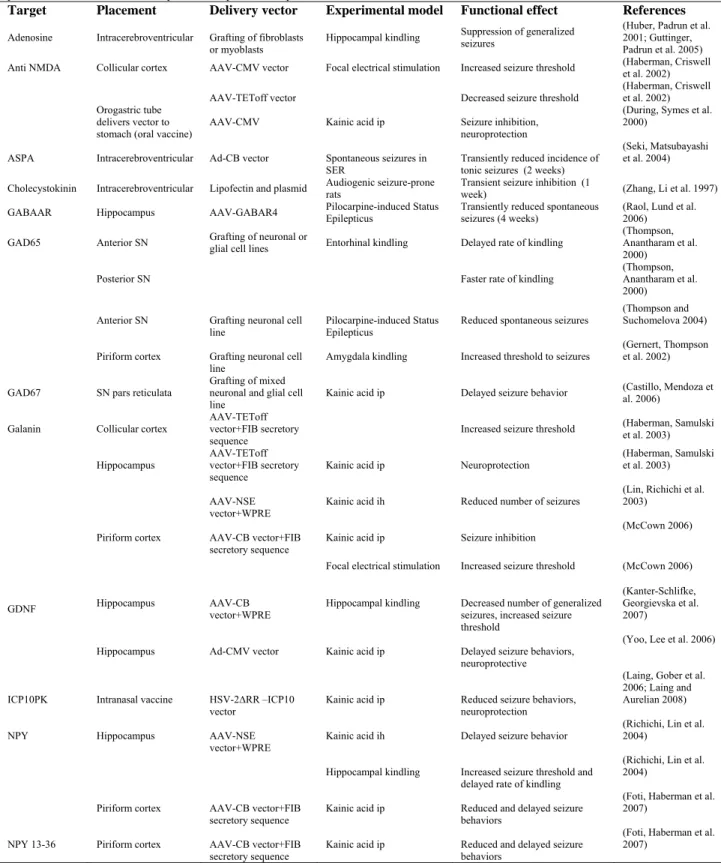
Table 1-2. Potential Therapeutic Targets that Influence Seizure Behavior ...16
Table 2-1. PCR Primers for Multiple Gene Product Delivery Vectors ...34
Table 5-1. Current Open Gene Therapy Trials for Neurological Diseases...101
xi
LIST OF FIGURES
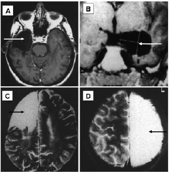
Figure 1-1. Different Types of Epilepsy Surgery ...3
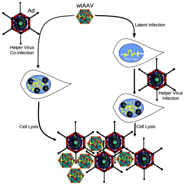
Figure 1-2 AAV Latent and Lytic Life Cycles ...9
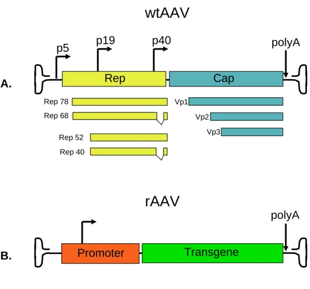
Figure 1-3. Comparing wtAAV to rAAV ...11
Figure 1-4. Ex Vivo versus In Vivo Viral Gene Delivery...18
Figure 1-5. Strategies to Co-express Gene Products ...26
Figure 2-1. Dot Blot, Standard Curve, and Titer Calculations for AAV2-CB-FIB-NPY and AAV2-CB-FIB-NPY13-36...37
Figure 2-2. Dot Blot, Standard Curve, and Titer Calculations for AAV2-CB-FIB-GAL-RKRRKR-FIB-EGFP, AAV2-CB-FIB-EGFP, and AAV2-CB-EGFP ...38
Figure 2-3. Dot Blot, Standard Curve, and Titer Calculations for Multiple Gene Product Delivery Vectors ...39
Figure 2-4. Infectious Center Assay of AAV2-CB-FIB-GAL- RKRRKR-EGFP ...40
Figure 2-5. Infectious Center Assay of AAV2-CB-FIB-EGFP- RKRRKR-GAL...41
Figure 2-6. Infectious Center Assay of AAV2-CB-FIB-GAL-EGFP ...42
Figure2-7. Infectious Center Assay of AAV2-CB-FIB-GAL- RKRRKR-NPY13-36 ...43
Figure 2-8. Coronal Section of the Rat Brain Depicting the Location of Vector Microinjection...45
Figure 3-1. AAV Vectors that Express and Constitutively Secrete NPY or NPY13-36...49
Figure 3-2. The Effects of AAV-FIB-NPY and AAV-FIB-NPY13-36 Vectors on the Expression of Limbic Seizure Behaviors ...50
Figure 3-4. AAV Vectors Characterized in Initial Multiple Gene
Product Delivery Studies ...54 Figure 3-5. Luciferase Assay on Concentrated Medium ...56
Figure 3-6. Luciferase Antibody Protein Analysis of Luciferase-GFP
Multiple Gene Product Delivery Constructs...57 Figure 3-7. GFP Antibody Protein Analysis of Luciferase-GFP Multiple
Gene Product Delivery Constructs...58 Figure 3-8. Double FIB Multiple Gene Product Delivery Vectors...60 Figure 3-9. In Vitro Protein Analysis of Double FIB Multiple Gene Product
Delivery Constructs ...61 Figure 3-10. In Vivo Fluorescence Pattern in Rat Cortex after Infusion of
AAV2-CB-FIB-GAL-RKRRKR-FIB-EGFP,
AAV2-CB-FIB-EGFP, or AAV2-CB-EGFP...63 Figure 3-11. The Effects of AAV2-CB-FIB-GAL-RKRRKR-FIB-EGFP on
the Expression of Limbic Seizure behaviors ...64 Figure 3-12. AAV Vectors Characterized in Final Multiple Gene Product
Delivery Studies...66 Figure 3-13. In Vitro Characterization of Fluorescence Patterns, Cleavage,
and Secretion of Proteins Derived from Multiple Gene
Product Delivery Vectors...67 Figure 3-14. Immunofluorescence of Neurons in the Piriform Cortex That
Have Been Transduced with GFP-containing Vectors ...69 Figure 3-15. The In Vivo Presence of FIB-GAL-RKRRKR-NPY13-36
mRNA 1 Week After Vector Infusion into the Piriform Cortex ...70 Figure 3-16. The Effects of Multiple Gene Product Delivery Vectors on
Limbic Seizure Behavior ...71 Figure 4-1. Regulated versus Constitutive Secretion in Neurons ...76 Figure 6-1. Immunoprecipitation Followed by SDS-PAGE and Coomassie
Staining of Multiple Gene Product Delivery Vectors for
xiii
Chapter 1 Introduction
I.A. Epilepsy
About 2.1 million people in the United States are living with epilepsy (Hirtz,
Thurman et al. 2007), with a world wide prevalence of 1 percent (Hauser, Hesdorffer et
al. 1990), making epilepsy one of the most common neurological diseases. Epilepsy
syndromes can be classified into two major categories based on the origin of seizure
activity. In generalized epilepsy the seizure originates bilaterally from the cerebral
hemispheres and typically involves the whole brain. For partial or focal epilepsy, the
area of seizure genesis is localized to one or more defined areas termed foci, although it
may spread to involve the entire brain.
Temporal lobe epilepsy (TLE), is a partial epilepsy where seizure genesis occurs
in temporal lobe structures such as the hippocampus and amygdala, and is the most
frequent and severe form of adult focal epilepsy (Cohen, Navarro et al. 2002). Patients
with TLE can have seizures that cause them to lose consciousness. If the seizures go
untreated, they can lead to severe brain damage. Having epilepsy is not only debilitating,
it can also be a significant economic burden. The estimated annual economic cost of
epilepsy in the United States including the direct cost of treatment and the indirect costs
such as lost productivity and wages is 12.5 billion dollars (Shafer and Begley 2000).
treatment method for epilepsy is to administer anti-epileptic drugs (AEDs) to arrest the
seizures or at least reduce their frequency and intensity. If seizures are left untreated,
they can increase in duration and subsequently cause more activity-induced pathology in
the brain. The AEDs attempt to prevent seizure activity by four major mechanisms: (1)
increasing gamma-aminobutyric acid (GABA)-ergic actions which are predominantly
inhibitory; (2) decreasing excitatory glutamatergic transmission; (3) decreasing
voltage-dependant calcium release; and (4) decreasing voltage-voltage-dependant sodium conductance
(Ure and Perassolo 2000).
Since seizures seem to result from hyperexcitable networks of neurons, the AEDs
are designed to enhance inhibitory neurotransmission and hyperpolarize neurons.
Unfortunately, even with new AEDs available, the number of drug resistant epilepsies
has not decreased (Loscher and Leppik 2002). Patients who continue to exhibit
intractable seizures or those who develop intolerable side effects while on AEDs are
considered to have medically refractory epilepsy. Unfortunately, 30–40% of all patients
with epilepsy have medically intractable seizures, and only half of these patients are
candidates for surgery (Shafer, Hauser et al. 1988; Engel, Levesque et al. 1992; Sander
1993; Siegel 2004), which is currently one of the few treatment options for medically
refractory patients. Additionally, 20% of the patients who do undergo temporal lobe
surgery will still have seizures post-operatively (Spencer, Spencer et al. 1984).
Even though surgical techniques have become more refined, several factors still
limit success. The highest surgical success rate is achieved if the epileptogenic tissue can
be directly removed using procedures like focus surgery, anterior temporal lobectomy,
and total lobe resection (See Figure 1-1). Lower success is achieved when the area of
Figure 1-1. Different Types of Epilepsy Surgery (modified from Siegel 2003).
A. Temporal lobectomy, B. Selective amygdalo-hippocampectomy, C. Frontal
lobectomy, and D. Anatomical hemispherectomy. Arrows show where brain has been
4
seizure genesis overlaps with tissues that serve important sensory or motor functions. For these patients, preventing seizure spread by surgically interrupting the hyperexcitable circuits is accomplished through procedures like amygdalo-hippocampectomy and corpuscollosectomy. Surgical complications such as visual field deficits, transient or persistent hemiparesis, infections, epidural hematoma, dysphasia, global memory deficits, and transient psychosis or depression may also limit success (Siegel 2004). Although current surgical procedures can be beneficial to patients, more effective and potentially less invasive treatment strategies need to be designed.
Theoretically, a gene therapy approach to seizure suppression is an attractive alternative treatment option for focal epilepsy. Focal epilepsy arises from a circumscribed and hyperexcitable network of neurons whose synchronous electrical discharge creates seizure activity. Since the area of seizure genesis is confined, the localized nature of in vivo gene therapy should prove capable of influencing a site that
initiates seizure activity, therefore preventing the cascade leading to seizure spread. One significant advantage of localized gene delivery is the limited exposure of the rest of the brain and body to the therapeutic compounds, thereby minimizing the potential for unwanted side effects. Furthermore, gene therapy allows for localized de novo synthesis
of the therapeutic compounds in the brain, surmounting the difficulty of delivering therapeutic compounds with short half-lives across the otherwise impermeable blood brain barrier.
I.B. Gene Therapy
offers the possibility that chronic disease progression (which occurs with medically intractable seizures) can be halted and the symptoms can be controlled, substantially increasing the quality of life for the patient. Using a virus as a means to deliver therapeutic genes circumvents many of the problems that non-viral vector-based gene therapy is subject to, such as inefficient gene transfer and transient gene expression (Huang, Hung et al. 1999). In order to complete their lifecycle, viruses must efficiently deliver their genes into the nucleus of a host cell. Thus, viruses can be exploited as excellent gene delivery tools by substituting therapeutic genes in place of viral genes.
6
Lentiviral vectors (HIV based) are effective in transducing neurons, do not seem to cause cytotoxicity upon central nervous system (CNS) infection, and are capable of long term transgene expression (Blomer, Naldini et al. 1997; Bosch, Perret et al. 2000). However, since lentiviral vectors randomly integrate into the host cell’s genome, they can cause insertional mutagenesis. These random insertions may disrupt or activate cellular genes including oncogenes. Recently, class 1 non-integrative lentiviral vectors derived from HIV type 1 were developed and characterized in brain tissue (Philippe, Sarkis et al. 2006; Yanez-Munoz, Balaggan et al. 2006). These vectors can be further modified to be self-inactivating (Miyoshi, Blomer et al. 1998), a combination that substantially reduces the risks of insertional mutagenesis and replication-competence, thereby generating safer lentiviral vectors for gene therapy in the brain.
AAV vectors which are based on a non-pathogenic and replication deficient virus offer the safest method of viral gene delivery in the brain. In addition, AAV vectors are capable of long term stable gene expression and do not elicit an immune response upon infection, making it the viral vector of choice in clinical trials for neurological diseases (see Table 1-1 and http://www.wiley.co.uk/wileychi/genmed/clinical/).
I.C. AAV Biology
Table 1-1. Current AAV-mediated Gene Therapy Clinical Trials for Neurological Diseases. This data is current to July 2007. For more details on these clinical trials please see: http://www.wiley.co.uk/wileychi/genmed/clinical/
Trial ID: Title: Gene Disease
US-469 Subthalamic GAD Gene Transfer in Parkinson’s Disease Patients Who Are Candidates for Deep Brain Stimulation.
Glutamic acid decarboxylase 65-67 (GAD 65-65-67)
Parkinson's Disease
US-593 A Phase I Open-Label Safety Study of Intrastriatal Infusion of Adeno-Associated Virus Encoding Human Aromatic L-amino Acid Decarboxylase (AAV-hAADC-2) in Subjects with Advanced Parkinson's Disease
Aromatic L-amino Acid Decarboxylase (AADC)
Parkinson's Disease
US-623 A Phase I/II, Dose-Escalating, Randomized and Controlled Study to Assess the Safety, Tolerability, and Efficacy of CERE-110 [Adeno-Associated Virus (AAV)-based, Vector-Mediated Delivery of beta-Nerve Growth Factor (NGF)] in Subjects with Mild to Moderate Alzheimer's Disease
Nerve growth factor
(NGF) Alzheimer's Disease
US-669 Hippocampal NPY Gene Transfer in Subjects with Intractable Temporal Lobe Epilepsy
Neuropeptide Y
(NPY) Epilepsy
US-689 A Phase I, Open-Label Study of CERE-120 (Adeno-Associated Virus Serotype 2 [AAV2]-Neurturin [NTN]) to Assess the Safety and Tolerability of Intrastriatal Delivery to Subjects with Idiopathic Parkinson's Disease
Neurturin (NTN) Parkinson's Disease
US-788 Multicenter, Randomized, Double-Blind, Sham Surgery-Controlled Study of CERE-120 (Adeno-Associated Virus Serotype 2 [AAV2]-Neurturin [NTN]) to Assess the Efficacy and Safety of Bilateral Intraputamenal (IPu) Delivery in Subjects With Idiopathic Parkinson's Disease
8
unless facilitated by a helper virus such as Ad or Herpes Simplex virus (HSV) (Buller, Janik et al. 1981; Bauer and Monreal 1986). When helper viruses are absent and the host cell is not subjected to DNA damaging agents or metabolic inhibitors, AAV will establish a latent infection within the cell (Yalkinoglu, Heilbronn et al. 1988). This latent infection will occur by site-specific integration of AAV into the host genome at human chromosome 19q13.3qter AAVS1 site (Samulski, Zhu et al. 1991; Muzyczka 1992), by AAV persistence as an episome within the nucleus (Yue and Duan 2003; McCarty, Young et al. 2004), or by random integration at chromosomal double-strand breaks (Miller, Petek et al. 2004). AAV is stable in this latent state and can persist for years until rescued by helper virus co-infection, which reinitiates the lytic cycle (Cheung, Hoggan et al. 1980) (see Figure 1-2).
AAV virions are comprised of a linear single-stranded DNA genome of 4.7 kb, which is encapsidated by a non-enveloped icosahedral shell, measuring approximately 22 nm in diameter. The elegant simplicity of the genome is astounding, with only two genes
rep (replication) and cap (capsid) encoding nonstructural and structural proteins
respectively. From a single open reading frame (ORF) the rep gene encodes four
replication proteins (Rep78, Rep 68, Rep 52, and Rep 40), which collectively are responsible for site-specific integration, DNA nicking, and helicase activity. Rep proteins also respond to intracellular cues (like the presence of helper virus) to regulate the AAV promoters that drive expression of its genome (Pereira, McCarty et al. 1997). The cap gene encodes three overlapping proteins (VP1, VP2, and VP3) which differ from
Figure 1-2 AAV Latent and Lytic Life Cycles.
In the absence of helper virus, AAV latently integrates into host chromosomes, with 70% of the genomes being targeted to the human chromosome 19q13.3qter AAVS1 site. If the cells are then superinfected with a helper virus, AAV enters its lytic phase and initiates virus replication. AAV can also immediately enter its lytic phase upon helper virus co-infection, resulting in the replication of both AAV and the helper virus.
wtAAV
19q13.3qter Ad
Helper Virus Co-infection
Cell Lysis
Helper Virus Infection
Cell Lysis Latent Infection
wtAAV
19q13.3qter 19q13.3qter 19q13.3qter Ad
Helper Virus Co-infection
Cell Lysis
Helper Virus Infection
10
approximately a 1:1:20 ratio and T=1 icosahedral symmetry (Rose, Maizel et al. 1971; Muzyczka 1992). The rep and cap genes are flanked by the AAV inverted terminal
repeats (TR), which are the only cis-acting elements required for genome replication and
packaging (Samulski, Chang et al. 1987; Samulski, Chang et al. 1989). I.D. AAV Vectors
Since the AAV TR is the only required cis-element for replication and
encapsidation, the two viral genes (rep and cap) can be removed and replaced by a
transgene expression cassette (see Figure 1-3). As long as rep and cap are provided in trans, AAV vectors can be produced and subsequently purified. Building on experiments
by Ferrari et al., (1996) which demonstrated that Ad helper function could be conferred
using noninfectious Ad DNA, pioneering work was done by Xiao, Li, and Samulski, allowing production of high titer AAV vectors in the absence of contaminating helper viruses (Ferrari, Samulski et al. 1996; Ferrari, Xiao et al. 1997; Xiao, Li et al. 1998). Consequently these vectors are safer for clinical trial applications. In addition, there are approximately 300 nucleotides of viral DNA present in the AAV vector expression cassette (the TR), and this sequence is not translated into viral proteins. Perhaps this lack of translated viral proteins reduces the risk of AAV vector genomes eliciting a host cell immune response, and may contribute to the longevity of transgene expression (longer than 3 years in humans) (Duan, Sharma et al. 1998; Jiang, Pierce et al. 2006).
Figure 1-3. Comparing wtAAV to rAAV.
A. The wtAAV genome encodes two viral genes, the rep and cap, and is flanked by
terminal repeats (TR). The four Rep proteins: Rep78, Rep68, Rep52 and Rep40 are responsible for site-specific integration, DNA nicking, and helicase activity. All four proteins are transcribed from the p5 and p19 promoters and by alternative splicing. The three viral structural proteins VP1, VP2, and VP3 share the same C-terminus and form trimers and pentamers that ultimately come together and encapsidate the single stranded genome. They are transcribed from the p40 promoter and by alternative splicing. B. Since the AAV terminal repeats are the only cis-elements required for viral vector
(a.k.a. recombinant AAV or rAAV) production, rep and cap can be removed and
replaced by a transgene expression cassette. As long as rep and cap are provided in trans, AAV vectors can be produced and subsequently purified.
wtAAV
A.
p19
p40
polyA
Rep
Cap
Vp2 Vp1
Vp3 Rep 78
Rep 52 Rep 40 Rep 68
p5
Promoter
Transgene
rAAV
B.
12
clinical trials in brain. Several AAV serotypes have been tested in the brain, although not every serotype was tested at the same time in the same model. While there is some variability, some generalizations can be made: capsids from AAV9, AAV8, AAV7, and AAV5 cause transduction in neurons (and a few glia) farthest from the site of injection (Klein, Dayton et al. 2006; Taymans, Vandenberghe et al. 2007; Klein, Dayton et al. 2008), followed by AAV1 (Burger, Gorbatyuk et al. 2004) which still transduces neurons farther away than AAV2, which seems to have the most localized effects. AAV4 appears to transduce ependymal cells exclusively, severely limiting its use as a vector for neurological diseases (Davidson, Stein et al. 2000). Furthermore, with the discovery of novel serotypes and the ability to engineer new capsid mutations by rational design and directed evolution approaches, it is now possible to enhance viral transduction and targeting (see Chapter 5 for detailed discussion).
Another concern that potentially limits AAV vector efficiency is the ability and speed with which the infected cells orchestrate viral genome second strand synthesis. Since traditional AAV vectors are single stranded, the DNA must be converted to a double stranded form before gene expression can be initiated, and the process is dependent upon the host cell-mediated DNA synthesis (Ferrari, Samulski et al. 1996; Fisher, Gao et al. 1996). One way to overcome this obstacle was pioneered by McCarty
et al, and involves generating a self-complimentary AAV vector (scAAV) that upon
uncoating could fold back on itself through intramolecular base pairing, creating a duplexed form (McCarty, Monahan et al. 2001; McCarty, Fu et al. 2003; Wang, Ma et al. 2003). Once duplexed, the efficiency with which transcriptionally active double stranded molecules are formed is greatly enhanced. Not only does this scAAV vector allow gene expression to occur much faster, it also has been reported to increase the level of transgene expression (McCarty, Monahan et al. 2001; Yang, Schmidt et al. 2002). While these vectors have shown great promise, they are even further limited in their packaging capacity to around 2.3kb (Wu, Zhao et al. 2007), which may restrict the range of therapeutic applications.
I.E. Potential Gene Therapy Targets for Epilepsy
14
primarily achieved through GABAergic neurotransmission, and loss of this inhibition has been implicated in epilepsy, making GABA a good therapeutic target. This is evident by the fact that many of clinically effective AEDs enhance GABA action either by increasing the time GABA remains in the synapse (by blocking its intracellular catabolism or by blocking its reuptake transporter, GAT1), or by acting at GABA receptors (to increase chloride ion flux into the neuron which hyperpolarizes it and prevents the firing of new action potentials).
calcium and cyclic GMP (Ure and Perassolo 2000). Even channels for ions such as sodium, potassium, and calcium regulate neuronal excitability, and their manipulation could provide in vivo seizure control. However, a liability with many of these targets in a
gene therapy context is that success will depend on the pattern of neuronal transduction, i.e. the type of neuron and its position within a functional circuit will influence seizure outcome. For example, reducing excitatory tone of a primary output neuron will reduce overall excitability, but reducing excitatory tone of an inhibitory interneuron may actually increase seizure severity.
16
Table 1-2. Potential Therapeutic Targets that Influence Seizure Behavior.
Abbreviations are: AAV: adeno-associated virus; ASPA: aspartoacylase; Ad: adenovirus; CB: cytomegalovirus enhancer, chicken beta-actin promoter; CMV: cytomegalovirus promoter; GAD: glutamic acid decarboxylase; GDNF: glial-derived neurotrophic factor; ICP10PK: ICP 10 protein kinase; ih: intrahippocampal; ip: intraperitoneal; NMDA: N-methyl-D-aspartate; NPY: Neuropeptide Y; NSE: neuron- specific enolase; SER: spontaneously epileptic rats; SN: substantia nigra; TET off: tetracycline-off regulatable promoter; WPRE: woodchuck hepatitis virus post-transcriptional element.
Target Placement Delivery vector Experimental model Functional effect References
Adenosine Intracerebroventricular Grafting of fibroblasts
or myoblasts Hippocampal kindling
Suppression of generalized seizures
(Huber, Padrun et al. 2001; Guttinger, Padrun et al. 2005) Anti NMDA Collicular cortex AAV-CMV vector Focal electrical stimulation Increased seizure threshold (Haberman, Criswell et al. 2002)
AAV-TEToff vector Decreased seizure threshold (Haberman, Criswell et al. 2002) Orogastric tube
delivers vector to stomach (oral vaccine)
AAV-CMV Kainic acid ip Seizure inhibition, neuroprotection
(During, Symes et al. 2000)
ASPA Intracerebroventricular Ad-CB vector Spontaneous seizures in
SER Transiently reduced incidence of tonic seizures (2 weeks)
(Seki, Matsubayashi et al. 2004)
Cholecystokinin Intracerebroventricular Lipofectin and plasmid Audiogenic seizure-prone rats Transient seizure inhibition (1 week) (Zhang, Li et al. 1997)
GABAAR Hippocampus AAV-GABAR4 Pilocarpine-induced Status Epilepticus Transiently reduced spontaneous seizures (4 weeks) (Raol, Lund et al. 2006)
GAD65 Anterior SN Grafting of neuronal or glial cell lines Entorhinal kindling Delayed rate of kindling (Thompson, Anantharam et al. 2000)
Posterior SN Faster rate of kindling (Thompson, Anantharam et al. 2000)
Anterior SN Grafting neuronal cell
line Pilocarpine-induced Status Epilepticus Reduced spontaneous seizures
(Thompson and Suchomelova 2004)
Piriform cortex Grafting neuronal cell
line Amygdala kindling Increased threshold to seizures
(Gernert, Thompson et al. 2002)
GAD67 SN pars reticulata Grafting of mixed neuronal and glial cell
line Kainic acid ip Delayed seizure behavior
(Castillo, Mendoza et al. 2006)
Galanin Collicular cortex AAV-TEToff vector+FIB secretory sequence
Increased seizure threshold (Haberman, Samulski et al. 2003)
Hippocampus AAV-TEToff vector+FIB secretory
sequence Kainic acid ip Neuroprotection
(Haberman, Samulski et al. 2003)
AAV-NSE
vector+WPRE Kainic acid ih Reduced number of seizures
(Lin, Richichi et al. 2003)
Piriform cortex AAV-CB vector+FIB
secretory sequence Kainic acid ip Seizure inhibition
(McCown 2006)
Focal electrical stimulation Increased seizure threshold (McCown 2006)
GDNF Hippocampus AAV-CB vector+WPRE Hippocampal kindling Decreased number of generalized seizures, increased seizure threshold
(Kanter-Schlifke, Georgievska et al. 2007)
Hippocampus Ad-CMV vector Kainic acid ip Delayed seizure behaviors, neuroprotective
(Yoo, Lee et al. 2006)
ICP10PK Intranasal vaccine HSV-2∆RR –ICP10
vector Kainic acid ip Reduced seizure behaviors, neuroprotection
(Laing, Gober et al. 2006; Laing and Aurelian 2008)
NPY Hippocampus AAV-NSE
vector+WPRE Kainic acid ih Delayed seizure behavior
(Richichi, Lin et al. 2004)
Hippocampal kindling Increased seizure threshold and delayed rate of kindling
(Richichi, Lin et al. 2004)
Piriform cortex AAV-CB vector+FIB
secretory sequence Kainic acid ip Reduced and delayed seizure behaviors
(Foti, Haberman et al. 2007)
NPY 13-36 Piriform cortex AAV-CB vector+FIB
secretory sequence Kainic acid ip Reduced and delayed seizure behaviors
I.F. Ex Vivo Gene Therapy
There are two basic modalities for gene therapy: ex vivo gene therapy which relies
on genetically modifying cells in vitro then implanting them into the target tissue, or in vivo gene therapy where the transfer of genetic material occurs directly within the host
via viral or non-viral mediated gene delivery (see Figure 1-4). To date, ex vivo gene
therapy experiments in brain have been carried out using fibroblasts (Huber, Padrun et al. 2001), myoblasts (Lisovoski, Wahrmann et al. 1997; Guttinger, Padrun et al. 2005), CNS progenitor cells (Martinez-Serrano and Bjorklund 1997), immortalized neurons (Gernert, Thompson et al. 2002; Longhi, Watson et al. 2004), and astrocytes (Lundberg, Horellou et al. 1996; Ericson, Wictorin et al. 2002). In order for this ex vivo approach to be
successful in treating epilepsy, the implanted cells must be engineered to secrete a product that will enhance neuronal inhibition. In the Boison lab, genetically engineered fibroblasts (Huber, Padrun et al. 2001) and myoblasts (Guttinger, Padrun et al. 2005) were made to secrete the inhibitory neuromodulator adenosine, and then grafted into the lateral ventricles or hippocampus of rats. While the rate of limbic kindling was significantly attenuated during the first week post-implantation, by the fourth week there was a drastic reduction of seizure suppression. The authors determined that the loss of therapeutic efficacy was due to low viability of the grafted cells. These results point to a universal problem with ex vivo gene therapy: the loss of graft viability over time. With
18
Figure 1-4. Ex Vivo versus In Vivo Viral Gene Delivery.
2. Culture Cells 4. Inject Genetically Modified
Cells into Rodent
3. Infect Cells with Virus Encoding Therapeutic Gene
1. Harvest Cells from Rodent A. Ex VivoViral Gene Therapy
2. Culture Cells 4. Inject Genetically Modified
Cells into Rodent
3. Infect Cells with Virus Encoding Therapeutic Gene
1. Harvest Cells from Rodent A. Ex VivoViral Gene Therapy
4. Inject Genetically Modified Cells into Rodent
3. Infect Cells with Virus Encoding Therapeutic Gene
1. Harvest Cells from Rodent A. Ex VivoViral Gene Therapy
3. Infect Cells with Virus Encoding Therapeutic Gene
1. Harvest Cells from Rodent A. Ex VivoViral Gene Therapy
1. Harvest Cells from Rodent A. Ex VivoViral Gene Therapy
1. Harvest Cells from Rodent A. Ex VivoViral Gene Therapy
B.In VivoViral Gene Therapy
1. Inject Virus Encoding Therapeutic Gene Directly into Rodent
B.In VivoViral Gene Therapy
More recently the Boison lab has used genetically engineered human mesenchymal stem cells (hMSC) where lentiviral RNAi mediates knockdown of adenosine kinase (ADK), the gene responsible for adenosine metabolism and influx from the extracellular space back into cells (Ren, Li et al. 2007). The authors demonstrate reduced seizure duration and neuronal loss after graft implantation in the hippocampus followed by intra-amygdaloid injection of kainic acid (Ren, Li et al. 2007). This type of approach would be compatible with autologous cell grafting in patients, and thus represents a potential clinical therapy if the grafts of mesenchymal stem cells can remain viable in the brain over time.
The Boison lab is also currently exploring implantation of genetically modified embryonic stem cell-derived neural precursors with biallelic genetic disruption of ADK (Li, Steinbeck et al. 2007). Implanting these grafts into the hippocampus caused increased levels of extracellular adenosine and suppressed kindling epileptogenesis (Li, Steinbeck et al. 2007). While the authors only conducted these experiments for 26 days, they see integration of the grafted cells and neuronal maturation markers expressed in about half of their grafted cells; leading the authors to speculate that graft viability may extend beyond the limited time course analyzed.
I.G. In Vivo Viral Vector-Mediated Alterations in Receptor Function
As mentioned before, enhancing inhibitory receptor function may be an effective target for antiepileptic gene therapy. Thus, an obvious approach would be to increase GABA-mediated fast neuronal inhibition, which is achieved through GABAA receptors
20
GABRα4 and poor in GABRα1 have been found in both humans with temporal lobe epilepsy and in rodent models of epilepsy, and show reduced GABA-mediated inhibition in the presence of zinc (Buhl, Otis et al. 1996; Gibbs, Shumate et al. 1997; Brooks-Kayal, Shumate et al. 1998; Brooks-Kayal, Shumate et al. 1999). In addition, after pilocarpine-induced status epilepticus, GABRα4 expression increases while GABRα1 expression decreases in the dentate gyrus of adult rats (Brooks-Kayal, Shumate et al. 1998). While these adult rats go on to develop spontaneous seizures, postnatal day 10 rats that are subjected to the same paradigm fail to develop spontaneous seizures (Zhang, Raol et al. 2004). Furthermore, the postnatal day 10 rats exhibit an increase in GABRα1 expression in dentate gyrus neurons (Zhang, Raol et al. 2004), suggesting that overexpression of GABRα1 may confer seizure protection if overexpressed in the adults. To investigate this hypothesis, Raol et al. used an AAV5 vector to express GABRα1 from the GABRα4
Another strategy to limit seizure activity would be to reduce or block excitatory amino-acid receptors, which may help restore inhibitory tone and prevent excitotoxicity. Since activation of NMDA receptors has been shown to increase seizure sensitivity (McCown, Givens et al. 1987), it is logical to try to limit their activation. In fact, NMDA channels are a good target for a gene therapy approach because although they are comprised of multiple subunits that are coded for by several genes, each channel must have the required NR1 subunit to be fully functional (Monyer, Sprengel et al. 1992; Hollmann and Heinemann 1994; Yamakura and Shimoji 1999). Therefore, knockdown of NMDAR1 protein should result in fewer functional NMDA channels. Haberman et al.
tested this hypothesis using an NMDAR1 antisense construct delivered by an AAV2 vector (Haberman, Criswell et al. 2002). While in vitro NMDAR1 receptor function and in vivo NMDAR1 protein levels were significantly reduced, the seizure behavior outcome
22
important liability when modulating receptors, ion channels, or even neurotransmitters and transporters: results may be dependant upon the pattern of transduction.
I.H. In Vivo Viral Vector-Mediated Expression of Neuropeptides and Peptide Fragments
Neuropeptides and their receptors are found in areas often associated with seizure generation, and their physiological effects are predominantly inhibitory suggesting convulsant action. In particular, both galanin and NPY (and their analogs) have anti-convulsant effects in several models of experimentally induced seizures (Mazarati, Liu et al. 1998; Mazarati, Hohmann et al. 2000; Mazarati and Wasterlain 2002; Haberman, Samulski et al. 2003; Mazarati 2004; McCown 2006; Foti, Haberman et al. 2007). Experimental evidence suggests that prolonged overstimulation, such as recurring seizures, depletes galanergic innervation (Mazarati, Liu et al. 1998) and destroys a subset of GABA-ergic NPY containing interneurons in the hippocampus (Sloviter 1991; Sperk, Marksteiner et al. 1992; Schwarzer, Williamson et al. 1995). This finding was also validated in patients with mesial temporal lobe sclerosis (de Lanerolle, Kim et al. 1989). Taken together, these data suggest that overexpressing galanin and NPY in the ictogenic brain regions may not only suppress seizures and prevent them from spreading, but might also compensate for depleted neuropeptides, conferring some neuroprotection.
The use of AAV vectors to express galanin and NPY in the hippocampus and piriform cortex has been explored with very good results. Haberman et al. first
preceded by the secretory signal sequence of the laminar protein, fibronectin. Thus, the active peptide would be constitutively secreted from the transduced cell. The elegance of this approach hinges on the fact that the newly synthesized neuropeptide will be packaged in vesicles and continuously released, independent of neuronal activity, essentially bypassing the traditional neuropeptide regulatory pathways. These studies showed that the fibronectin secretion signal sequence (FIB) did cause constitutive secretion of the gene product from transfected cells in vitro and, upon transduction in
vivo, significantly attenuated focal seizure sensitivity and prevented seizure-induced hippocampal cell damage (Haberman, Samulski et al. 2003).
In a subsequent publication, Lin et al. found that AAV mediated expression of a
24
and severity of kainic acid-induced seizure behaviors. Thus, the piriform cortex is a good place to deliver and test potential therapeutic targets for anti-seizure efficacy.
As previously discussed, there is evidence to suggest that NPY can exhibit anti-seizure activity in vivo (Woldbye, Larsen et al. 1997; Mazarati and Wasterlain 2002;
peptide fragments, which have demonstrated receptor selectivity. Thus, we infused AAV2 vectors that mediate expression and constitutive secretion of NPY or the peptide fragment NPY13-36 into the piriform cortex, and evaluated their ability to suppress kainic acid-induced limbic seizure behaviors (see Chapter 3).
I.I. Combination Therapy
Delivering individual inhibitory neuropeptides to the brain to prevent seizures has been well studied (see above), but to date no one has tried to deliver multiple neuropeptides simultaneously from an AAV vector. In the last 20 years, many different virus-based strategies were explored to co-express two genes from vectors: 1. internal promoters, 2. internal initiation (IRES), 3. self-processing peptides (CHYSEL), 4. fusion proteins, 5. proteolytic processing, and finally 6. a combination of separate vectors where each carries one transgene (see Figure 1-5). Internal promoter vectors (sometimes referred to as bicistronic vectors) have two transcriptional units, each with its own open reading frame that results in the production of two proteins. While these vectors are quite popular, problems arise because of the uncoupled transcription of both genes. For example, it has been reported that transcriptional interference and promoter silencing can result in the transcription of only one gene (Hippenmeyer and Krivi 1991; Nakajima, Ikenaka et al. 1993; Zaboikin and Schuening 1998).
26
Figure 1-5. Strategies to Co-express Gene Products.
A) Internal promoter vectors have two transcriptional units, each with its own open reading frame that results in the production of two proteins. B) Internal initiation vectors have a single transcript with two open reading frames and two sites of translation. The first open reading frame is cap-dependent, while translation of the second open reading frame depends upon an internal sequence called an internal ribosomal entry site (IRES). C) Self-processing peptide or cis-acting
hydrolase element (CHYSEL) vectors have a single transcriptional unit with a single open reading frame and one site of translation. The CHYSEL sequence is inserted in frame between the two protein sequences, and upon translation, the CHYSEL produces a disruption in translation. D) Proteolytic processing vectors also have a single transcriptional unit with a single open reading frame and one site of translation. The proteolytic cleavage sequence is cloned in frame as a linker, and a chimeric fusion protein is generated that is capable of undergoing post-translational cleavage in the presence of trans proteases. E) Fusion protein vectors have a single
transcriptional unit with a single open reading frame and one site of translation. A single chimeric fused protein is generated. (Arrows depict promoters, star depicts cleavage site)
IP
Gene 2
polyA
polyA
Gene 1
AAA
AAA
Protein 1 Protein 2
A
Gene 1
IRES
Gene 2
polyA
AAA
Protein 1 Protein 2
B
polyA
AAA
Protein 1 Protein 2 Post-translational Cleavage and Removal of Sequence
Protein 1 Protein 2
*
Gene 1
SequenceCleavageGene 2
D
Gene 2
polyA
Gene 1
AAA
Protein 1 Protein 2
E
Gene 2
polyA
CHYSEL
AAA
Protein 1 CHYSE
Gene 1
C
Protein 2
L
IP
Gene 2
polyA
polyA
Gene 1
AAA
AAA
Protein 1 Protein 2
A
IP
IP
Gene 2
Gene 2
polyA
polyA
polyA
polyA
Gene 1
Gene 1
AAA
AAA
AAA
AAA
Protein 1
Protein 1 Protein 2Protein 2
A
Gene 1
IRES
Gene 2
polyA
AAA
Protein 1 Protein 2
B
Gene 1
IRES
Gene 2
polyA
AAA
Protein 1 Protein 2
Gene 1
IRES
Gene 2
polyA
AAA
Gene 1
IRES
Gene 2
polyA
Gene 1
Gene 1
IRES
IRES
Gene 2
Gene 2
polyA
polyA
AAA
AAA
Protein 1
Protein 1 Protein 2Protein 2
B
polyA
AAA
Protein 1 Protein 2 Post-translational Cleavage and Removal of Sequence
Protein 1 Protein 2
*
Gene 1
SequenceCleavageGene 2
D
polyA
polyA
AAA
Protein 1 Protein 2 Post-translational Cleavage and Removal of Sequence
Protein 1 Protein 2
*
AAA
AAA
Protein 1
Protein 1 Protein 2Protein 2 Post-translational Cleavage and Removal of Sequence
Protein 1
Protein 1 Protein 2Protein 2
*
Gene 1
Gene 1
SequenceCleavageGene 2
Gene 2
Cleavage Sequence
D
Gene 2
polyA
Gene 1
AAA
Protein 1 Protein 2
E
Gene 2
polyA
Gene 1
AAA
Protein 1 Protein 2
Gene 2
Gene 2
polyA
polyA
Gene 1
Gene 1
AAA
AAA
Protein 1 Protein 2 Protein 1
Protein 1 Protein 2Protein 2
E
Gene 2
polyA
CHYSEL
AAA
Protein 1 CHYSE
Gene 1
C
Protein 2
L
Gene 2
Gene 2
polyA
polyA
CHYSEL CHYSEL
AAA
AAA
Protein 1 CHYSE
Protein 1
Protein 1 CHYSECHYSE
Gene 1
Gene 1
C
Protein 2
L Protein 2Protein 2 L
the expression of the upstream gene) (Mizuguchi, Xu et al. 2000). Perhaps the biggest drawback of both vector strategies discussed so far (internal promoters and IRES) is that they contain large DNA sequences (often >0.5kb), which compete with the transgene for a limited amount of space in an AAV vector (around 4.7kb).
Self-processing peptide or cis-acting hydrolase element (CHYSEL) vectors have a
single transcriptional unit and one site of translation with a single open reading frame. The CHYSEL sequence is inserted in frame between the two protein sequences, and upon translation, the CHYSEL produces a disruption in translation. The ribosome then releases the first protein and continues to translate the second protein, resulting in the production of two proteins (Donnelly, Luke et al. 2001). While it has been demonstrated that higher levels of the second protein are achieved when using CHYSEL in contrast to IRES (Furler, Paterna et al. 2001), a potential problem is that after processing, the CHYSEL sequence remains fused to the C-terminus of the upstream protein, and an extra proline residue will be added to the N-terminus of the second protein (de Felipe 2002). This extra sequence may interfere with protein bioactivity, especially with relatively small proteins such as neuropeptides that need to bind to receptors to initiate signaling.
28
and intracellular or membrane bound proteins, further limiting the applicability of this approach.
There are also major drawbacks with using a co-infection of two separate vectors where each carries one transgene. Three populations of transduced neurons will result based on a Poisson distribution: some neurons will be transduced only by the first vector, some neurons will be transduced only by the second vector, and some neurons will be transduced by both. This kind of transduction pattern can complicate the analysis of transgene interactions, because the observed therapeutic effects will not be attributable solely to the co-transduced neurons. For these reasons we chose to pursue a proteolytic processing vector strategy where a fusion protein gene product is processed into separate bioactive proteins. This method will ensure that each transduced neuron expresses both gene products, and utilizes only a small amount of the AAV vector genome, since proteolytic cleavage consensus sequences can be as small as two amino acids (Seidah and Chretien 1999). In addition, we can use this same strategy in the future to express more than two therapeutic peptides simultaneously, and to manipulate the ratios of expressed peptides by inserting multiple copies of the same peptide. Furthermore, by using a single vector we can reduce viral particle number (and viral capsid proteins), which would likely increase clinical safety.
endotoxins (Nakayama 1997). Furin cleaves precursors into bioactive proteins by recognizing and cleaving strings of basic amino acids such as arginines and lysines. In addition, furin is ubiquitously expressed in neurons and glia throughout the brain (Day, Schafer et al. 1993), making it an ideal pro-protein convertase to target with our constitutively secreted neuropeptides platform. The furin pathway has already been used in a gene therapy context where a furin cleavage site was engineered into the rat preproinsulin-I gene, which was then packaged into a recombinant retroviral vector and delivered to the liver (Muzzin, Eisensmith et al. 1997). It was shown that correctly processed and functional insulin could be secreted from the ectopic organ (Muzzin, Eisensmith et al. 1997). In a subsequent study, Margaritis et al. used a furin cleavage
consensus sequence in an AAV vector cassette between Factor VIIa subunits, then delivered the vector into the spleens of hemophiliac mice (Margaritis, Arruda et al. 2004). Phenotypic correction was achieved using this strategy (Margaritis, Arruda et al. 2004).
While both of these studies demonstrate that engineering a furin cleavage consensus sequence into a transgene can be successful in a gene therapy context, only one functional protein was delivered. In a study by Gaken et al., a furin cleavage
30
Previous experiments suggest that it is possible to use a proteolytic strategy to deliver multiple peptides from AAV vectors, but to date no one has tried. We are in a unique position to conduct these experiments and combine them with our constitutively secreted neuropeptides platform. We expect improved cleavage efficiencies over previous attempts because we are using a FIB sequence that will direct our chimeric protein to the constitutive secretory pathway where furin is most active (Van de Ven, Creemers et al. 1991). We first proved the concept that the therapeutic gene galanin and the reporter gene GFP could be correctly cleaved from one another and secreted into the medium of transfected cells in vitro. Then, we evaluated the in vivo function of each
Chapter 2 Methods
II.A. rAAV Vectors: Cloning and Construction
II.A.1. Cloning Plasmids for rAAV-CB-FIB-NPY and rAAV-CB-FIB-NPY13-36
The expression cassette backbone is the same for both NPY and NPY13-36
plasmids, where gene expression is driven by the hybrid chicken beta-actin promoter, the
mature coding sequence of the transgene is followed by the SV-40 polyadenylation
sequence (polyA), and the cassette is flanked by AAV2 TRs. The full length NPY active
peptide sequence was amplified by RT-PCR from rat brain RNA using primers directed
to the mature peptide sequence, such that melting and reannealing of two separate PCR
products resulted in 5’ AgeI and 3’ NotI overhangs (Zeng 1998). The 3’ primer included
a stop codon to properly terminate translation. The sequence of the primers used is as
follows: (NPY 5’ long) CCG GTA ATG TAC CCC TCC AAG CCG; (NPY 5’ short) TA
ATG TAC CCC TCC AAG CCG; (NPY 3’short) GC TCA ATA TCT CTG TCT GGT
G; (NPY 3’ long) GGC CGC TCA ATA TCT CTG TCT GGT G. The PCR product was
ligated into the AgeI-NotI sites of the AAV2 backbone plasmid, resulting in the plasmid
pTR-CB-NPY. Next, the fibronectin signal sequence (nucleotides 208–303) was derived
from the rat fibronectin mRNA sequence (GenBank Accession No. X15906) and
oligonucleotides corresponding to both strands were generated (Midland Certified
32
could be ligated in front of NPY, resulting in the plasmid pTR-CB-FIB-NPY. For NPY 13-36, the same cloning strategy was used, except the plasmid pTR-CB-FIB-NPY served as the PCR template and cloning backbone. The primers used to generate NPY13-36 were as follows: (NPY13-36 5’ long) 5’-CCG GTA ATG CCA GCA GAG GAC ATG-3’, (NPY13-36 5’ short) 5’-TA ATG CCA GCA GAG GAC ATG GC-ATG-3’, (NPY13-36 3’ short) 5’-GC TCA ATA TCT CTG TCT GGT G-3’, (NPY13-36 3’ long) 5’-GGC CGC TCA ATA TCT CTG TCT GGT G-3’. Again, the PCR product was ligated into the AgeI-NotI site of pTR-CB-FIB-NPY, replacing the “FIB-NPY”. Then the annealed FIB oligonucleotides were ligated into the AgeI site resulting in the plasmid pTR-CB-FIB-NPY13-36. All plasmids were sequenced to verify accuracy.
II.A.2. Cloning Plasmids for Multiple Gene Product Delivery Vectors
sequence of the primers used is as follows: (Gal 5’ long) 5’-CCG GTA ATG GGC TGG ACC CTG AAC-3’, (Gal 5’ short) 5’-TA ATG GGC TGG ACC CTG AAC-3’, (Gal 3’ short) 5’-GC TCA TGT GAG GCC ATG CTT G-3’, (Gal 3’ long) 5’-GGC CGC TCA TGT GAG GCC ATG CTT G-3’. The PCR product was ligated into the AgeI-NotI sites of the AAV2 plasmid backbone, resulting in the plasmid pTR-CB-GAL. Then the annealed FIB oligonucleotides were ligated into the AgeI site resulting in the plasmid pTR-CB-FIB-GAL. Using pTR-CB-FIB-GAL as a backbone, the other plasmids were generated by PCR so the furin cleavage consensus sequence arginine-lysine-arginine-arginine-lysine-arginine (RKRRKR) was cloned in frame between two genes. Briefly, two pairs of oligonucleotides were designed for each construct, such that a forward and reverse primer corresponded to each gene plus 3 of the six amino acids that make up the furin consensus sequence (see Table 2-1). The oligonucleotides included the restriction sites NheI and NotI that would be used for cloning. Two fragments each containing the gene of interest plus half of the furin consensus sequence were generated by PCR, then the products were digested with NheI and NotI, and ligated into the plasmid backbone using a sticky-blunt-sticky ligation. All plasmids were sequenced to verify accuracy.
II.A.3. DNA Sequencing
34
Table 2-1 PCR Primers for Multiple Gene Product Delivery Vectors
Construct Name Primers Used Melting
Temperature
TR-CB-FIB-GAL-RKRRKR-EGFP F1= GTACGGAAGTGTTACTTCTGCTC R1= 5’PTCTCTTTCTTGTGAGGCCATGCTT
F2= 5’PAGAAAGAGAATGGTGAGCAAGGGCGAGGAGC R2= CTTATCATGTCTGGATCCCCGCGGCC
55.0°C 56.0°C 66.0°C 64.0°C
TR-CB-FIB-EGFP-RKRRKR-GAL F1= GTACGGAAGTGTTACTTCTGCTC R1= 5’PTCTCTTTCTCTTGTACAGCTCGTC F2= 5’PAGAAAGAGAGGCTGGACCTGAACAGCG R2= CTTATCATGTCTGGATCCCCGCGGCC
55.0°C 56.0°C 64.0°C 64.0°C
TR-CB-FIB-GAL-EGFP
F1= GTACGGAAGTGTTACTTCTGCTC R1= 5’PTGTGAGGCCATGCTTGTCGCT F2= 5’PATGGTGAGCAAGGGCGAGGAGCTG R2= CTTATCATGTCTGGATCCCCGCGGCC
55.0°C 56.0°C 63.0°C 64.0°C
TR-CB-FIB-GAL-RKRRKR-NPY13-36
F1= GTACGGAAGTGTTACTTCTGCTC R1= 5’PTCTCTTTCTTGTGAGGCCATGCTT F2= 5’PAGAAAGAGACCAGCAGAGGACATGGCC R2= CTTATCATGTCTGGATCCCCGCGGCC
II.B. In Vitro Methods
II.B.1. Cell Culture and Transfection
293 cells were cells were maintained at 37°C in a 5% CO2 atmosphere in
Dulbecco’s modified Eagle’s medium (DMEM), supplemented with 10% fetal bovine serum (FBS) and penicillin-streptomycin (PS). Cells were plated at a density of 5.5x10⁵ cells per mL medium in 60mm dishes the day prior to transfection. Cells were transfected using the polyethyleneimine (PEI) technique (Grieger, Choi et al. 2006). Forty-eight hours after transfection, cells were imaged then medium and lysates were harvested. Briefly, medium was cleared by centrifugation at 2500 RPM at 4°C for 5 minutes to remove any cellular debris. Cells were scraped and rinsed with 2mL Dulbecco phosphate buffered saline (DPBS) then lysed using 1mL lysis buffer (50mM Tris pH8.0, 150mM NaCl, 50mM NaF, 1% NP-40) per 60mm plate. Lysates were cleared by spinning in centrifuge at maximum speed at 4°C for 5 minutes, supernatant was transferred to a clean tube.
II.B.2 Immunoprecipitation and Western Blotting
removed by vacuum and beads were washed 3 times using 1mL DPBS. Load dye was added to beads along with water and betamercaptoethanol, then samples were boiled 5 minutes and cooled to room temperature before loading on 10% Bis-Tris gel (NuPage) Samples were transferred by western blot to a nitrocellulose membrane and blocked using 10% nonfat milk in tris buffered saline + 0.1% Tween (TBST). Blot was incubated at 4°C overnight in 1:1000 dilution of primary antibody Living Colors A.V. Monoclonal antibody JL-8 (BD). Blot was rinsed 3 times 5 minutes in TBST then incubated in a 1:3000 dilution of goat-anti-mouse HRP (Pierce) Signal was detected using the West femto-chemiluminescence kit (Pierce) according to the manufacturer’s instructions. II.C. rAAV Production, Purification, and Characterization
Recombinant AAV2 was produced and purified as previously described (Rabinowitz, Rolling et al. 2002) with the following modifications: 293 cells (10 15cm plates/prep)were transfected with 60μg transgene plasmid, 120μg XX-680, and 100μg PXR2 via the polyethyleneimine (PEI) technique. Nuclei were isolated and lysed using sonication. AAV particles were purified by cesium chloride density gradient. Peak fractions were determined via dot blot hybridization, and extensively dialyzed against DPBS + 10% (wt/vol) D-sorbitol. Final titer was determined by dot blot hybridization (modified southern blot) using a probe against the CB promoter sequence so all viruses could be titered on the same blot (see Figures 2-1 through 2-3) Infectious center assays (ICA) were performed using C12 cells (293 cells with Rep stably integrated) as previously described (Grieger, Choi et al. 2006) (see Figures 2-4 through 2-7). AAV particles were analyzed to determine purity and ratio of empty to full particles by staining
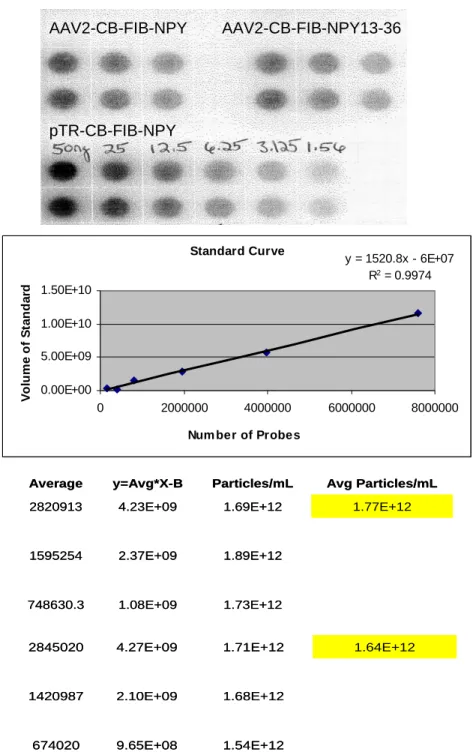
Figure 2-1. Dot Blot, Standard Curve, and Titer Calculations for AAV2-CB-FIB-NPY and AAV2-CB-FIB-NPY13-36. The top panel shows three dilutions (across) of the viruses performed in duplicate (down). Dilutions of known amounts (nanograms) of one of the plasmids used to make the rAAV vectors were included (pTR-CB-FIB-NPY). Upon hybridization with a radiolabeled probe that was complementary to a fragment of the CB promoter, a standard curve was generated (middle panel). Then a linear regression analysis was done, and the derived equation (displayed in the upper left corner of the standard curve graph) was used to calculate the number of rAAV particles present in each dilution (bottom panel), then the average particles present per mL.
AAV2-CB-FIB-NPY AAV2-CB-FIB-NPY13-36 pTR-CB-FIB-NPY AAV2-CB-FIB-NPY AAV2-CB-FIB-NPY13-36 pTR-CB-FIB-NPY 1.73E+12 1.08E+09 748630.3 1.89E+12 2.37E+09 1595254 1.77E+12 1.69E+12 4.23E+09 2820913 Avg Particles/mL Particles/mL y=Avg*X-B Average 1.73E+12 1.08E+09 748630.3 1.89E+12 2.37E+09 1595254 1.77E+12 1.69E+12 4.23E+09 2820913 Avg Particles/mL Particles/mL y=Avg*X-B Average 1.54E+12 9.65E+08 674020 1.68E+12 2.10E+09 1420987 1.64E+12 1.71E+12 4.27E+09 2845020 1.54E+12 9.65E+08 674020 1.68E+12 2.10E+09 1420987 1.64E+12 1.71E+12 4.27E+09 2845020 Standard Curve
y = 1520.8x - 6E+07 R2 = 0.9974
0.00E+00 5.00E+09 1.00E+10 1.50E+10
0 2000000 4000000 6000000 8000000
Num ber of Probes
38
Figure 2-2. Dot Blot, Standard Curve, and Titer Calculations for AAV2-CB-FIB-GAL-RKRRKR-FIB-EGFP, AAV2-CB-AAV2-CB-FIB-GAL-RKRRKR-FIB-EGFP, and AAV2-CB-EGFP. The left panel shows three dilutions (across) of the viruses performed in duplicate (down). Dilutions of known amounts (nanograms) of one of the plasmids used to make the rAAV vectors were included (pTR-CB-FIB-GAL-RKRRKR-FIB-EGFP). Upon hybridization with a radiolabeled probe that was complementary to a fragment of the CB promoter, a standard curve was generated (bottom panel). Then a linear regression analysis was done, and the derived equation (displayed in the upper left corner of the standard curve graph) was used to calculate the number of rAAV particles present in each dilution (right panel), then the average particles present per mL.
AAV2-CB-FIB-GAL-RKRRKR-FIB-EGFP side and peak fractions
AAV2-CB-FIB-EGFP AAV2-CB-EGFP
50 25 12.5 6.25 3.125
AAV2-CB-FIB-GAL-RKRRKR-FIB-EGFP side and peak fractions
AAV2-CB-FIB-EGFP AAV2-CB-EGFP
50 25 12.5 6.25 3.125
5.76271E+11 7.20E+08 38473.47 5.80512E+11 7.26E+08 38756.62 5.47932E+11 4.87012E+11 1.22E+09 65028.63 Avg particles/mL Particles/mL
# * X+B Average 5.76271E+11 7.20E+08 38473.47 5.80512E+11 7.26E+08 38756.62 5.47932E+11 4.87012E+11 1.22E+09 65028.63 Avg particles/mL Particles/mL
# * X+B Average 2.16418E+12 2.71E+09 144486.61 2.27565E+12 2.84E+09 151928.995 2.1967E+12 2.15028E+12 5.38E+09 287117.055 2.16418E+12 2.71E+09 144486.61 2.27565E+12 2.84E+09 151928.995 2.1967E+12 2.15028E+12 5.38E+09 287117.055 2.51044E+12 3.14E+09 167603.77 2.42044E+12 3.03E+09 161595.12 2.41071E+12 2.30127E+12 5.75E+09 307278.77 2.51044E+12 3.14E+09 167603.77 2.42044E+12 3.03E+09 161595.12 2.41071E+12 2.30127E+12 5.75E+09 307278.77 1.88995E+12 2.36E+09 126178.295 1.93021E+12 2.41E+09 128866.455 1.87706E+12 1.81101E+12 4.53E+09 241815.59 1.88995E+12 2.36E+09 126178.295 1.93021E+12 2.41E+09 128866.455 1.87706E+12 1.81101E+12 4.53E+09 241815.59
Standard Curve y = 18225x + 1E+09 R2 = 0.9899
0 5000000000 10000000000 15000000000 20000000000 25000000000
0 200000 400000 600000 800000 1000000 1200000
Number of Probes
Figure 2-3. Dot Blot, Standard Curve, and Titer Calculations for Multiple Gene Product Delivery Vectors. The left panel shows three dilutions (across) of the viruses performed in duplicate (down). The virus order is as follows 1) upper left, FIB-GAL-RKRRKR-EGFP, 2) upper right, FIB-EGFP-RKRRKR-GAL, 3) lower left, FIB-GAL-EGFP, and 4) lower right, AAV2-CB-FIB-GAL-RKRRKR-NPY13-36. Dilutions of known amounts (nanograms) of one of the plasmids used to make the rAAV vectors were included (pTR-CB-FIB-GAL-EGFP). Upon hybridization with a radiolabeled probe that was complementary to a fragment of the CB promoter, a standard curve was generated (bottom panel). Then a linear regression analysis was done, and the derived equation (displayed in the upper left corner of the standard curve graph) was used to calculate the number of rAAV particles present in each dilution (right panel), then the average particles present per mL.
6.44569E+11 4.03E+08 503472.1 1.03451E+12 1.29E+09 909902 1.09097E+12 1.14743E+12 2.87E+09 1629115 Avg Particles/mL Particles/mL
Avg * x - B Average 6.44569E+11 4.03E+08 503472.1 1.03451E+12 1.29E+09 909902 1.09097E+12 1.14743E+12 2.87E+09 1629115 Avg Particles/mL Particles/mL
Avg * x - B Average 5.3877E+11 3.37E+08 473285.3 1.10069E+12 1.38E+09 947668.1 1.16655E+12 1.2324E+12 3.08E+09 1726094 5.3877E+11 3.37E+08 473285.3 1.10069E+12 1.38E+09 947668.1 1.16655E+12 1.2324E+12 3.08E+09 1726094 2.4716E+12 1.54E+09 1024766 2.88779E+12 3.61E+09 1967467 2.84914E+12 2.81049E+12 7.03E+09 3527150 2.4716E+12 1.54E+09 1024766 2.88779E+12 3.61E+09 1967467 2.84914E+12 2.81049E+12 7.03E+09 3527150 6.71364E+12 4.20E+09 2235117 6.56656E+12 8.21E+09 4066745 6.00733E+12 5.4481E+12 1.36E+10 6537437 6.71364E+12 4.20E+09 2235117 6.56656E+12 8.21E+09 4066745 6.00733E+12 5.4481E+12 1.36E+10 6537437 AAV2-CB-FG-RKRRKR-E AAV2-CB-FE-RKRRKR-G AAV2-CB-F-G-E AAV2-CB-FG-RKRRKR-NPY13-36 pTR-CB-FIB-GAL-EGFP AAV2-CB-FG-RKRRKR-E AAV2-CB-FE-RKRRKR-G AAV2-CB-F-G-E AAV2-CB-FG-RKRRKR-NPY13-36 pTR-CB-FIB-GAL-EGFP
Standard Curve y = 2190.5x - 7E+08 R2 = 0.991
-2000000000 0 2000000000 4000000000 6000000000 8000000000 10000000000 12000000000 14000000000
0 1000000 2000000 3000000 4000000 5000000 6000000
Num ber of probe s
40
Figure 2-4. Infectious Center Assay of AAV2-CB-FIB-GAL-RKRRKR-EGFP. A) 500 particles, B) 1000 particles, and C) 5000 particles of AAV2-CB-FIB-GAL-RKRRKR-EGFP were co-infected with Ad in C12 cells, blotted on a membrane, and hybridized with radiolabeled probe that was complementary to a fragment of the CB promoter to determine the number of infectious units per mL (IU/mL). This virus is 3.6x10⁹ IU/mL.
AAV2-CB-FIB-GAL-RKRRKR-EGFP
A
B
C
AAV2-CB-FIB-GAL-RKRRKR-EGFP
A
B
Figure 2-5. Infectious Center Assay of AAV2-CB-FIB-EGFP-RKRRKR-GAL. A) 500 particles, B) 1000 particles, and C) 5000 particles of AAV2-CB-FIB-EGFP-RKRRKR-GAL were co-infected with Ad in C12 cells, blotted on a membrane, and hybridized with radiolabeled probe that was complementary to a fragment of the CB promoter to determine the IU/mL. This virus is 1.0x10⁹ IU/mL.
AAV2-CB-FIB-EGFP-RKRRKR-GAL
A
C
B
AAV2-CB-FIB-EGFP-RKRRKR-GAL
A
C
42
Figure 2-6. Infectious Center Assay of AAV2-CB-FIB-GAL-EGFP.
A) 500 particles, B) 1000 particles, and C) 5000 particles of AAV2-CB-FIB-GAL-EGFP were co-infected with Ad in C12 cells, blotted on a membrane, and hybridized with radiolabeled probe that was complementary to a fragment of the CB promoter to determine the IU/mL. This virus is 2.0x10⁹ IU/mL.
AAV2-CB-FIB-GAL-EGFP
A
B
C
AAV2-CB-FIB-GAL-EGFP
A
B
Figure 2-7. Infectious Center Assay of AAV2-CB-FIB-GAL-RKRRKR-NPY13-36. A) 500 particles, B) 1000 particles, and C) 5000 particles of AAV2-CB-FIB-GAL-RKRRKR-NPY13-36 were co-infected with Ad in C12 cells, blotted on a membrane, and hybridized with radiolabeled probe that was complementary to a fragment of the CB promoter to determine the IU/mL. This virus is 9.0x10⁹ IU/mL.
AAV2-CB-FIB-GAL-RKRRKR-NPY13-36
A
B
C
AAV2-CB-FIB-GAL-RKRRKR-NPY13-36
44
with 1% aqueous uranyl acetate and visualizing with transmission electron microscopy (LEO-EM910, accelerating voltage = 80 KV).
II.D. In Vivo Methods
II.D.1. Experimental Animals
All of the animals were pathogen-free male Sprague–Dawley rats obtained from Charles Rivers. The animals were maintained in a 12 hour light–dark cycle and had free access to food and water. All care and procedures were in accordance with the Guide for the Care and Use of Laboratory Animals (DHHS Publication No. [NIH]85-23), and all procedures received prior approval by the University of North Carolina Institutional Animal Care and Usage Committee.
II.D.2. rAAV Vector Microinjection
For AAV infusions, rats were first were anesthetized with 50 milligrams per kilogram, intraperitoneal (mg/kg, ip) pentobarbital and placed into a stereotaxic frame. Using a 32 gauge stainless steel injector and a Sage infusion pump, the rats received 2 or 3µL of virus (depending on virus titer) over 20 minutes into the piriform cortex (inter-aural line (IAL) 6.7 millimeters (mm), lateral 6.0mm, vertical 8.4mm, according to the
Figure 2-8. Coronal Section of the Rat Brain Depicting the Location of Vector Microinjection. rAAV2 vector is stereotaxically injected bilaterally into the piriform cortex, denoted by arrows. The coordinates are inter-aural line (IAL) 6.7 millimeters (mm), lateral 6.0mm, vertical 8.4mm, according to the atlas of Paxinos and Watson (1998).
46
II.D.3. In Vivo Detection of AAV-Derived NPY or NPY 13-36
As previously described (Haberman, Samulski et al. 2003; Foti, Haberman et al. 2007) the vector-derived NPY could not be visualized in vivo by immunohistochemistry,
likely due to the fact that the secreted NPY or NPY 13-36 would be rapidly degraded, especially during the perfusion procedure. Thus, in vivo activity of the vectors
AAV-FIB-NPY or AAV-FIB-NPY 13-36 was validated by demonstrating the presence of vector-derived mRNA. Two rats received AAV-FIB-NPY and two received AAV-FIB-NPY 13-36 vector infusions into the piriform cortex as described above. Then, 1 week later, the animals received an overdose of pentobarbital (100mg/kg, ip) and were subsequently decapitated. The brain was removed, and the piriform cortex was dissected out. The tissue was stored in RNAlater (Ambion, Austin, TX, USA) at -80°C. Subsequently, the RNA was extracted from the tissue (Promega SV-40 total RNA isolation kit; Madison, WI, USA) and reverse transcribed using AMV reverse transcriptase and oligo(dT) primers. The subsequent PCR used primers that were designed to span the FIB-NPY (and thus the FIB-NPY 13-36) sequence, which can only be derived from the rAAV vector: (FIB, 5’) 5’-CTA GCA GTC CTG TGC CTG-3’, (NPY and NPY13-36, 3’) 5’ –GCT CAA TAT CTC TGT CTG GTG-3’.